Seed development and inheritance studies in
apomictic maize-Tripsacum hybrids reveal
barriers for the transfer of apomixis into sexual crops
OLIVIER LEBLANC*
,1, DANIEL GRIMANELLI
1, MARTHA HERNANDEZ-RODRIGUEZ
2, PABLO A. GALINDO
2,
ANA M. SORIANO-MARTINEZ
2,#and ENRICO PEROTTI
2,31Laboratoire Génome et Développement des Plantes (LGDP), UMR 5096 IRD-CNRS-Université de Perpignan, Montpellier, France, 2International Maize and Wheat Improvement Center, Texcoco 56130, Mexico and
3Plant Cell Biology Group, Australian National University, Canberra ACT, Australia
ABSTRACT Apomixis in plants covers a variety of cloning systems through seeds of great potential for plant breeding. Among long-standing approaches for crop improvement is the attempt to exploit wild relatives as natural, vast reservoirs for novel genetic variation. With regard to apomixis, maize possesses an apomictic wild relative, Tripsacum, which we used to produce advanced maize-Tripsacum hybrid generations. However, introgression of apomixis in maize has failed so far. In order to understand the how’s and why’s, we undertook characterization of seed development and inheritance studies in these materials. We show that apomictic seeds suffer from epigenetic loads. Both seed tissues, the endosperm and the embryo, displayed developmen-tal defects resulting from imbalanced parendevelopmen-tal genomic contributions and aberrant methylation patterns, respectively. Progeny characterization of several maize-Tripsacum hybrid generations allowed significant progress toward the unraveling of the genetics of apomixis. First, chromo-some deletion mapping showed that expression of apomixis requires one single Tripsacum chromosome. However, inheritance studies revealed that female gametes inheriting this segment were unequivalent carriers depending on their origin: unreduced gametes transmit a functional segment, whereas progeny derived from reduced ones reproduced sexually. Finally, chromo-somal or genomic dosage variation barely affected the apomictic phenotype suggesting no dependency for ploidy in these materials. We conclude that epigenetic information imposes constraints for apomictic seed development and seems pivotal for transgenerational propagation of apomixis. The nature of the triggering mechanisms remains unknown as-yet, but it certainly explains the modest success relative to the development of apomictic maize thus far.
KEY WORDS:
apomixis, seed development, maize, epigenetics, interspecific hybrid
Introduction
The Tripsacum genus comprises wild relatives of maize (Zea mays L.) widely distributed across the American continent and highly variable in many aspects (Berthaud et al., 1997, Randolph, 1970). Efforts towards allele mining out of this diverse, genetic reservoir have been limited so far (for instance, resistance to Puccinia sorghi; Bergquist, 1981). One notable exception con-cerns apomixis or the formation of maternal embryos within seeds
BIOLOGY
www.intjdevbiol.com
*Address correspondence to: Olivier Leblanc. Institut de Recherche pour le Développement, 911 Avenue Agropolis, BP 64 501, 34394 Montpellier Cedex
05, France. Fax: +33-04-6741-6181. e-mail: [email protected] Lab web address: http://lgdp.univ-perp.fr
#Present address: Unidad de Biología Tecnología y Prototipos (UBIPRO), Universidad Nacional Autónoma de México, 54090 Tlalnepantla, Estado de México,
México
Accepted: 12 September 2008. Published online: 18 December 2008.
(Koltunow, 1993, Nogler, 1984), a reproductive behavior of great potential for agriculture and plant breeding (Spillane et al., 2004). Apomixis in higher plants is achieved through a number of routes, most of which still lack detailed characterization. Apomic-tic developments have been categorized into adventitious embryony and gametophytic apomixis depending on the cell type
ISSN: Online 1696-3547, Print 0214-6282
© 2008 UBC Press Printed in Spain
that gives rise to maternal embryos, i.e. somatic cells within the ovule and parthenogenetical egg cells within unreduced megaga-metophytes, respectively (Gustafsson, 1946, Koltunow, 1993, Nogler, 1984). Recent works in species reproducing through distinct pathways, i.e. Hieracium subgenus pilosella, Poa, and Tripsacum, suggest that gametophytic apomixis (referred to as apomixis herein) relies upon either spatial or temporal
misexpression of genes acting during female sexual reproduction (Albertini et al., 2004, Grimanelli et al., 2003, Tucker et al., 2003). However, although candidate genes showing differences in spa-tial and temporal expression patterns between apomicts and their sexual counterparts have been identified, both their involvement in the control of apomixis and function remain largely speculative (Ozias-Akins, 2006). Inheritance studies have provided a rela-tively simple view for the genetics of apomixis, which was reported to depend on a few dominant genes in most species investigated (Asker and Jerling, 1992, Ozias-Akins and van Dijk, 2007). Challenging this, further molecular mapping and cytogenetic characterization of the chromosomal region(s) linked to the trait in several species have unveiled attributes frequently associated with complex loci, such as lack of recombination, trans-acting mechanism for gamete elimination, heterochromatinization and DNA rearrangements (Ozias-Akins and van Dijk, 2007). To date, the genetic control of apomixis remains confuse, as no apomictic locus has been molecularly resolved. Alike, whether the complex nature of apomictic loci or polyploidy, a usual feature of apomictic genomes, take part in the control of the trait is unknown. Fueling the debate, functional roles for polyploidy have been hypoth-esized, including epigenetic gene deregulation (Koltunow and Grossniklaus, 2003), ploidy-dependent gene expression (Cervigni et al., 2008, Quarin et al., 2001), or ectopic gene expression resulting from genome asynchrony (Carman, 1997). On the other hand, these complexities were proposed to have evolved once apomictic reproduction established, thus they may be secondary with regards to its genetic control (Ozias-Akins and van Dijk, 2007).
Within the Maydae tribe, apomixis occurs in Tripsacum only (Brown and Emery, 1958), making the genus a prime candidate to elaborate strategies for its transfer to maize either directly through backcrossing or by genetic engineering. Tripsacum spe-cies typically compose an agamic complex (after Babcock and Stebbins, 1938) wherein diploid individuals (x=18, 2n=2x=36) are sexual and polyploid individuals (2n=3x to 6x) reproduce apomic-tically. Apomixis is gametophytic of the diplosporous type (Farquharson, 1955, Leblanc et al., 1995b): functional, unre-duced megaspores form after omission, or early termination, of meiosis in megaspore mother cells (Grimanelli et al., 2003). Further differentiation into megagametophytes resembles that of the Polygonum type found in sexual species, but cells contain nuclei of maternal genetic makeup. Activation of unreduced egg cells through unknown developmental alterations in female ga-metogenesis induces embryogenesis in absence of fertilization (Bantin et al., 2001, Farquharson, 1955) but the developmental course of maternal embryos stops after a few rounds of mitotic divisions resulting in quiescent proembryos within unfertilized megagametophytes (Grimanelli et al., 2003). Pollination, fol-lowed by the delivery of two sperm cells into the mature megaga-metophyte and by the fertilization of the central cell only, is required for seed development. As in many other apomicts, apomixis in Tripsacum plants is facultative as reproductive be-haviors that allow genetic variation have been preserved through evolution. The most documented ones result from partial or complete restoration of sexual programs (Asker and Jerling, 1992, Bicknell and Koltunow, 2004), but other mechanisms such as incomplete nucleus restitution during meiosis abortion, mitotic and meiotic non-disjunction, somatic recombination, and gene
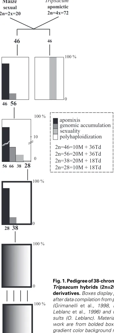
apomixis
genomic accumulation sexuality
2n=46=10M + 36Td 2n=56=20M + 36Td 2n=38=20M + 18Td 2n=28=10M + 18Td polyhaploidization
Maize
sexual 2n=2x=20
Tripsacum
apomictic 2n=4x=72
46
46
46
56
56 66
38
28
10 100 %
0
100 %
0
28
38
100 %
0
100 %
0
100 %
0
mutation have been reported as well (Hair, 1956, Noyes, 2005, Richards, 1996).
Since maize and Tripsacum were first hybridized in the early 30’s (Mangelsdorf and Reeves, 1931), pathways for introgressing Tripsacum genetic material into the crop have been extensively scrutinized (Harlan et al., 1970, Harlan and DeWet, 1977). Nev-ertheless, in spite of several decades of efforts (Kindiger and Sokolov, 1997, Leblanc, 1995, Leblanc et al., 1996, Petrov et al., 1984), no maize germplasm expressing some level of apomixis has been recovered yet. Conventional backcrossing at CIMMYT using a T. dactyloides apomictic donor and maize yielded facul-tative apomictic hybrids possessing two maize (M) genomes and one genome from T. dactyloides (Td) (i.e., 2n=38=20M+18Td) (Leblanc et al., 1996). Here, we report on both female reproduc-tive behavior and genomic characterization in these materials and derivative generations (Figure 1, Table 1). Our observations shed light onto critical obstacles, some of them likely of epigenetic nature, such as developmental abnormalities in apomictic seeds, methylation pattern differences following apomictic reproduction, and unexpected behavior of the chromosomal region that governs apomixis. Collectively, they provide an explanation for the modest success of transfer attempts to date.
Results
Seed developmental abnormalities in maize-Tripsacum and
Tripsacum apomicts
Seed development in maize-Tripsacum individuals with 38 chromosomes (2n=20M + 18Td) hand pollinated using diploid maize individuals initiated for 36 ± 9% of the flowers (mean ± SD; >2000 flowers from 3 different cycles, 10 plants per cycle, 2 inflorescences per plant). Mature kernels were highly variable with regard to endosperm formation (Figure 2A), however with a strong trend toward defective development (65 to 80%). By contrast, 84 ± 9% kernels (mean ± SD, N=744) contained a
morphologically normal embryo (Figure 2B). This suggests that endosperm collapsing occurred after cellularization, a critical step whereby apomictic proembryos resume development (Grimanelli et al., 2003). Unfavorable allelic interactions might have promoted endosperm failure, but a more plausible explanation is that development primarily suffered from maternal genomic excess. In endosperms from 2n=38 individuals pollinated with diploid maize, mother plants typically contributed six genomes (4M and 2Td) for a single paternal genome (1M). This corresponds to a strong distortion of the typical 2 maternal to 1 paternal genomic ratio (2m: 1p) required for normal seed development in many Angiosperms, including maize (Lin, 1984, Nishiyama and Inomata, 1966) and Tripsacum, as indicated by flow cytometrical analyses in normal and imbalanced endosperms. As shown in Figure 2E, endoreduplication in endosperms derived from 4x apomicts crossed with 4x male progenitors (2n=10x, 8m: 2p) occurred precociously compared to 6x normal endosperms (4m:2p; 4x sexuals X 4x progenitors), a difference in cell cycle progression already reported in maize as a mark for maternal excess (Leblanc et al., 2002). Dosage effects incidence in the endosperm of 2n=38 apomicts was corroborated by observations made from sectioned developing kernels. Cell growth was affected similarly to that of maize endosperms suffering from maternal genomic excess (Cooper, 1951): i. e., enlarged central cells, undifferentiated basal cell layer, and poor subepidermal differentiation (compare Figure 2C and 2D). Nevertheless, deleterious effects appeared less Fig. 2. Seed development in 2n=38 maize-Tripsacum (38C clone) and Tripsacum dactyloides apomicts. (A) Typical inflorescence of 38C clone pollinated using diploid maize. (B) Section in mature kernel of 38C showing a normal-looking embryo (emb) but surrounded by atypical endosperm cells (end). (C) Peripheral section of 38C endosperm with altered cell differentiation. (D) Peripheral section of normal endosperm from a sexual Tripsacum dactyloides strain: (al) aleurone layer cells, and (se) starchy endosperm cells. (E) Cell cycle analysis in endosperms from apomictic and sexual tetraploid Tripsacum strains. Bars, 300 µm.
T. dactyloides ecotypes Improved maize populations
11-36 BS13 (S) C8
61-664 BSSS (R) C13
65-1234 BS10 (FR) C11
112-1327 BS26 (S) C13
112-1328 BSCB1 (R) C13
Maize lines (CMLs) BS11 (FR) C11
CML62 (2) POB.21C6 x BS10 (FR) C11
CML78 POB.43C10 x BS10 (FR) C11
CML135 (1,2) POB.24C9 x BS10 (FR) C11
CML139 (1) POB.27C10 x BS26 (S) C3
CML204 POB.25C4 x BSCB1(R) C13
CML216 POB.27C10 x BSCB1(R) C13
CML258 POB.21C6 CML341 POB.43C10 CML346 POB.24C9 CML408, POB.25C4 CML413 POB.32C6 CML416 POB.27C10
TABLE 1
LIST OF THE TRIPSACUM AND MAIZE STOCKS USED AND
OBTAINED FROM CIMMYT GERMPLASM BANK
(1) and (2): lines used to derive H1and H3 maize hybrids, respectively. emb
end
se al
3C 6C 12C 24C apomict
3C 6C 12C sexual
B
C
D
E
critical in our materials than those reported for maize in which <1% of the 4x X 2x kernels produce an endosperm (Cooper, 1951, Leblanc et al., 2002). Developmental adaptations that ultimately restore the endosperm balance requirement are frequent in apomicts (Grimanelli et al., 1997, Grossniklaus et al., 2001, Nogler, 1984). Among these, the union of polar nuclei with two sperm nuclei could have contributed to dosage effect alleviation in 38C X 2x endosperms. To further investigate the importance of such fertilization events in seed development of apomicts, we directly estimated DNA contents in growing endosperms from normally developed kernels obtained 15 days after pollination with diploid maize (H3). Flow cytometry analyses indicated that 1x paternal contribution (fertilization by a single sperm cell) was predominant (80%, n=86). Therefore, fertilization of the central cell by two sperm cells unlikely served as a critical factor to mitigate dosage effects in endosperms of 2n=38 apomicts.
Defects were also common during embryo germination. De-spite the use of an in vitro procedure, germination of kernels with
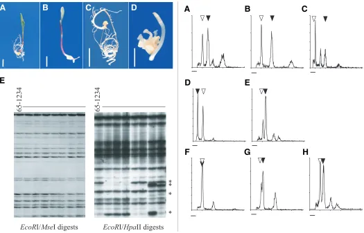
Fig. 3 (Left). Maternal embryo defects in maize-Tripsacum and Tripsacum apomicts.(A-D) Germination of maternal seedlings from seeds from maize-Tripsacum apomict 38C showing normal (A) and aberrant (B-D) developments. Bars, 1 cm. (E) Genomic cloning (EcoRI/ MseI digests) and detection of methylation-associated polymorphisms ( EcoRI/ HpaII digests) through AFLP fingerprinting in maternal progenies of 65-1234, a tetraploid, apomictic T. dactyloides ecotype. Asterisks indicate differentially methylated restriction sites.
Fig. 4 (Right). Progeny sorting of apomictic clone 38C after DNA content analyses in young seedlings. (A) Maternal clone with 2n=38. (B,C) Off type progeny with higher DNA contents than that of maternal clones: 2n=48 (B) and 2n=76 (C). (D-H) Off type progeny with lower DNA contents than that of maternal clones: 2n=10 (D), 2n=28 (E), 2n=20 (F), 2n=22 (G), 2n=27 (H). Open and dark triangles indicate 2C peaks produced by nuclei isolated from CML62, the diploid maize reference, and from the sample, respectively. Chromosome numbers were determined by root tip counts. Bar, 50-channel intensity.
65-1234
EcoR
I/
Mse
I digests
EcoR
I/
Hpa
II digests
65-1234
*
*
*
*
B
C
D
E
A
G
B
C
D
E
F
H
A
developed endosperm usually expanded over several weeks and produced less than 60% viable seedlings (Table 2). Low seed fertility resulted from lack of germination but also from develop-mental alterations, including early developdevelop-mental arrest, undiffer-entiated growth, and differentiation failure of shoot or roots (Fig-ures 3A to 3D). Polyembryonic kernels were frequent (Table 2), a trend that is usually associated to apomictic developments (Hanna and Bashaw, 1987) and for which we have provided recently a detailed characterization in Tripsacum and maize-Tripsacum hy-brids (Grimanelli et al., 2003). Finally, although development initiated to a lesser extent than in embryos accompanied by an endosperm, embryos dissected from defective kernels and cul-tured on a hormone-free medium exhibited similar defective developmental behaviors (data not shown).
phenomenon perturbs embryo development in apomicts, we studied progeny from five apomictic Tripsacum dactyloides indi-viduals. First, similarly to that observed in maize-Tripsacum hybrids, germination rates of normal seeds were low (33 to 70%) and a significant proportion (4 to 55%) of the germinating seeds were affected in development (Table 3). Altogether, viable adult plants were recovered for only 25% of the seeds. Late-occurring defects were also observed in adult plants, including sterility and delayed flowering time (respectively, 8% and 4%, n=70). We further performed a genowide characterization of DNA me-thylation for each progeny. As control experiments, we used two populations representing non-apomictically derived, but genetic replica, of the mother plant; the first one (n=10) consisted of cuttings of a single apomictic genotype, 65-1234 while the second one (n=12) was generated by selfing CML216, a maize homozy-gous line. We first verified that the three procedures we used reproduced faithfully mother plant genotypes by generating AFLP fingerprints from DNAs cleaved using EcoRI and–MseI, two methyl-insensitive restriction enzymes. All loci scored (n > 250) were strictly monomorphic in all cases studied (mother plant vs. progeny/replica, amongst progeny; Figure 3E). Then, we as-sessed methylation status of CCGG sites using a modified AFLP procedure using EcoRI/MspI or EcoRI/HpaII digests as preamplification DNA templates (Cervera et al., 2002). For both EcoRI/HpaII and EcoRI/MspI digests, we observed identical fingerprints among replica used for control experiments, indicat-ing conservation of genome methylation patterns (data not shown). By contrast, patterns of digestion among clonal individuals de-rived apomictically showed methylation-sensitive polymorphisms: firstly, pairwise comparisons of methylation patterns revealed that a significant fraction of the fragments detected using EcoR1/
HpaII digests (5.8%; n > 250) were polymorphic (Figure 3E) whilst they appeared monomorphic using EcoR1/Msp1 digests. Fur-thermore, we also detected polymorphisms for 2.4% of the fragments amplified from EcoR1/Msp1 digests. We thus conclude that apomictic clones differed for methylation patterns within CCGG regions despite genomic identity. These results suggest that inaccurate erasing, maintenance or establishment of DNA methylation might occur during apomictic reproduction and that, as observed in animals (Dean et al., 2001), this could explain the defects noted in maternal embryos.
Reproduction in 38-chromosome apomictic clones can oc-cur through a variety of mechanisms
Variation in the reproductive behavior within a single individual is common in both natural and artificial apomicts (Koltunow et al., 2000, Leblanc, 1995, Nogler, 1984, Noyes, 2005, Richards, 1996, Van Dijk et al., 1999). In order to assess the nature and the extent of variation in reproductive behavior in 38C, an apomictic clone, we collected flow cytometrical, cytogenetical and molecular data within progenies obtained after pollination using 2x maize pro-genitors. Although the majority of viable seedlings consisted of maternal individuals (similar DNA content and fingerprinting pat-terns, Figures 4A and 5A respectively), progeny also contained a mixture of non-maternal genotypes (9%, Table 2; Figures 4 and 5). The prevailing triggering mechanism consisted of unreduced egg cell fertilization as shown by DNA content and fingerprinting analyses, both indicative of the addition of one maize haploid genome to that of 38C (Figures 4B and 5A). The remaining non-maternal progeny were infrequent (<0.2% overall, Table 2) and split into two categories regarding DNA content: (1) values twice that of the mother clone (Figure 4C) resulting from parthenogen-esis in egg cells derived from unreduced megaspores that under-went spontaneous chromosome doubling (n=218; 2n=76, data not shown), and (2) a wide range of values significantly lower than that of the mother clone (n=249; Figures 4D to 4H).
Accession number
Germinating seeds (%)
Early arrest (%) a
Undifferentiated growth (%) a
Viable adult plant (%)
65-1234 (n=145) 35 33 22 16
11-36 (n=34) 68 0 4 65
61-664 (n=22) 73 19 31 36
122-1327 (n=42) 33 7 14 26
112-1338 (n=38) 32 17 33 16
Total (n=281) 41 20 20 25
TABLE 3
DEVELOPMENTAL DEFECTS IN APOMICTIC
TRIPSACUM DACTYLOIDES PROGENIES
a values shown were determined for germinated seeds
Fig. 5. AFLP genotyping of progeny from maize-Tripsacum apomic-tic clone 38C. (A) Derivatives through unreduced female gametes. Dark and open circles respectively indicate maternal progeny or off type progeny after fertilization (genomic accumulation, open circles). Arrows show maize fragments specific to the male progenitor. (B) Sexual derivatives. (C) Sporadic chromosome loss in one individual derived from unreduced, parthenogenetical female gametes (star). The arrow indi-cates one missing fragment.
Male Nt Ge (%) Me (%) Npr Mp (%) Inc (%) Dec (%)
H1(1) 7,393 54 17.3 4,691 93.7 6.1 0.2
H3(2) 10,206 49.5 13.0 5,740 88.2 11.5 0.3
All - - - >100,000 91.0 8.8 0.2
TABLE 2
PROGENY CLASSES FROM 38C APOMICTS POLLINATED USING DIPLOID MAIZE AS DETERMINED BY DNA CONTENT ANALYSES
Nt: total number of kernels; Ge: kernels that produced viable seedlings; Me: multiple embryos; Npr: number of viable progeny recovered; Mp: maternal progeny (same DNA content as mother plants); Inc: off type progeny showing increase in DNA content; Dec: off type progeny showing decrease in DNA content. (1) 2001 winter cycle; (2) 2002 summer cycle
Maize 38C Maize 38C
38C
B
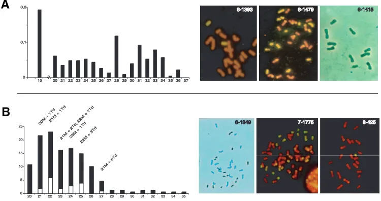
Fig. 6. Cytogenetical characterization of off type progeny obtained from apomictic 38C clones (A) and from highly facultative 2n=43 hybrids (B). (A) Chromosome number frequencies (n=228) and chromosome painting in three archetypal derivatives with regard to their origin: 6-1393, sexually derived with 2n=20M+2Td; 6-1479, asexually derived, but chromosomally imbalanced, with 2n=31=17M+14Td, and; 6-1415, a maize haploid. (B) Chromosome number distribution (n=127) and chromosome painting in 6-1349 (2n=33=18M+15Td); in 7-1775, one derivative of 6-1349 through genomic accumulation (2n=43=28M+15Td), and; in 8-425, a sexual offspring of 7-1775 (2n=23=22M+1Td). Sexual individuals that carried the DNA segment associated with diplospory distributed according to white bars and chromosome complements are indicated on top of corresponding bars. Chromosome painting was obtained using digoxygenin labeled Tripsacum DNA as a probe detected with either FITC or horseradish peoxidase as anti-digoxygenin conjugate ( Tripsacum chromosomes are yellow and dark blue respectively).
9-547
H3 9-547 X H3
Fig. 7. Sexual behavior in 9-547, a maize-Tripsacum hybrid with 2n=22=19M+3Td and carrying the chromosome associated with apomixis. Progeny were derived after crossing with a maize hybrid (H3) and amplicons were obatined using a SSR primer combination (bnlg339 locus).
Figure 6A displays chromosome numbers in 228 out of the 249 individuals of the latter class. Further cytogenetical and molecular characterization provided more comprehensive views for their nature and origin. A first set (n=83) consisted of individuals derived sexually as molecular analyses revealed both segregat-ing maternal markers and paternally inherited markers (Figure 5B). In addition, all plants determined for chromosomal comple-ments (n=62) combined a diploid maize genome with a few extra Tripsacum chromosomes (i.e., 6-1393, Figure 6A). The remain-ing individuals were categorized as follows: hybrids carryremain-ing aneuploid chromosome sets of maize, Tripsacum or both (n=80; 2n=29 to 2n=36, i.e., 6-1479; Figure 6A) with fingerprint patterns indicating no paternal DNA (absence of karyogamy) and sporadic loss of maternal markers (Figure 5C); maize haploids (n=40; 2n=10, i.e., 6-1415; Figure 6A) of maternal and paternal origin (data not shown), and; allodihaploid hybrids with 2n=10M+18Td (n=25). All allodihaploids genotyped (n=9) lacked paternal frag-ments indicating that they arose parthenogenetically from allodihaploid egg cells. However, we could not resolve unambigu-ously whether these eggs derived meiotically or ameiotically because of the lack of heterozygous maize markers detectable in 38C and of the small number of plants analyzed.
Finally, to test whether the viable seedlings we analyzed consisted in a biased sample for inferring reproductive behavior of 38C clones, we also estimated DNA contents in embryos dissected from defective kernels and in embryos that exhibited growth defects (≥ 250 for each type; data not shown). In both cases, they arranged in a similar manner to that of viable seed-lings.
reduced male gametes. However, recent progress toward the molecular deciphering of plant reproduction has revealed pivotal roles for female gametophytic factors during early seed develop-ment. Therefore, conceivably, maternal and paternal transmission might impact differently critical regulatory factors governing the apomictic phenotype. With regards to this, the collection of non-maternal off-types we produced from clone 38C allowed us to examine maternal transmission of apomixis for a variety of modali-ties. In particular, we determined reproductive behaviors in non maternal derivatives with lower DNA content values than that of 38C based on progeny tests and Southern hybridizations using two diagnosis probes for diplospory, umc28 and cdo202 (Leblanc et al., 1995a). Data were in agreement for most hybrids analyzed (n=106), except for three sexually derived individuals carrying the chromo-some harboring the diplospory locus (Table 4). Considering the small number of incongruent individuals, we further wanted to Here, as in many other apomic-tic species, 38C clones reproduced through facultative apomixis. Al-though genomic accumulation was predominant, genetic variation within progeny arose through a number of mechanisms. While in-complete nucleus restitution at meiosis I is a known side effect of diplosporous pathways (Nogler, 1984, Noyes, 2005, Richards, 1996) likely promoted by meiotic asyn-chrony between the maize and Tripsacum chromosomes in our ma-terials (Grimanelli et al., 2003, Harlan et al., 1970), parthenogen-esis in reduced gametes and spon-taneous chromosome doubling did not occur at greater incidences than that observed in many sexual plant species (de Wet, 1979, Kimber and Riley, 1963) or in interspecific hy-brids (Nasrallah et al., 2000, Soltis and Soltis, 1999).
6-1393 6-14796-1479 6-1415 6-1415
validate these observations using a larger population. To achieve this, the unusual reproductive behavior of derivatives of 6-1349, which reproduced through highly facultative apomixis (see next section for a detailed analysis), allowed the recovery of 166 individuals through sexuality. Southern hybridizations using umc28 identified 29 individuals that carried the DNA segment linked to diplospory, out of which 18 set seeds after crosses with diploid maize (see Figure 6B and next section for cytogenetical character-ization). Lack of progeny in the remaining inviduals resulted from early developmental arrest, flowering failure, or seed defects. With regards to this, we observed a similar fertile-to-sterile ratio in the 137 inviduals diagnosed as sexuals (18:29 and 58:79, respec-tively), thus discarding strong deleterious effects induced by the apomixis-associated chromosome. Flow cytometrical and molecu-lar data collected from a total of 1190 progenies indicated that all 18 fertile individuals selected as putative apomicts reproduced sexually (Figure 7). Although we cannot discard potential for functional apomixis in the sterile individuals, these results show that the segment defined by umc28 and cdo202 is not sufficient to confer apomixis by it-self. This suggests that apomixis might depend on critical Tripsacum genetic factors, unidentified in previ-ous studies, and segregated away during meiosis. Alternatively, loss of apomixis in these materials might denote trans-acting chromosomal dosage effects modulating gene expression within the umc28/cdo202 segment (i.e., aneuploid syndrome).
Assessment of the impact of genomic and chromosomal dosage variation on apomixis
In natural populations, apomixis and polyploidy are usually associated. However, artificial ploidy variation in apomicts and
related sexuals has produced ambiguous data, i.e., isolation of diploid or dihaploid apomicts (Bicknell, 1997, Dujardin and Hanna, 1986, Leblanc et al., 1996, Sharbel and Mitchell-Olds, 2001) vs. apomixis induction after ploidy rise in sexual Paspalum notatum (Quarin et al., 2001). Thus, whether polyploidy has a role in reshaping the sexual pathway towards apomixis remains to be elucidated. In higher organisms, many regulatory complexes for gene expression are dosage-dependent, mainly because of stoechiometric requirements among components (Birchler et al., 2001). As a result, changes in genome structure (aneuploidy, ploidy variation) often impact gene expression levels and, ulti-mately, phenotypes. Genomic accumulation and chromosome losses allowed the recovery of maize-Tripsacum lineages conve-nient for addressing the influence of dosage variation in tuning the balance between sexual and apomictic reproduction. First, we addressed the effects of aneuploidy through a chromosome deletion mapping approach by taking advantage of the chromo-somally imbalanced apomicts previously determined for the mode of reproduction (Table 4). They were genotyped using a set of seventy-seven AFLP and twenty-seven SSR markers specifically amplified from the Tripsacum complement of 38C genome (Fig-ure 8A). As shown in Fig(Fig-ure 8B, most markers (92%) failed to amplify the expected fragment in at least one individual. Despite small scale discrepancies in grass genome synteny (Devos, 2005, Goff et al., 2002), we assume that the marker set we used likely covered the entire Tripsacum genome. Therefore, chromo-somal variation, except for that harboring the umc28/cdo202 region, had no significant effect on diplospory; although their developmental fate could vary across genotypes, most female gametes were unreduced. Secondly, we examined the impact of ploidy variation throughout genomic accumulation series derived from clones of 38C and chromosomally imbalanced derivatives that showed contrasted Tripsacum genome landscapes (Table 5). In order to limit the impact of allelic variation introduced through the male, plants were pollinated with H3, the male progenitor used in 38C pedigree. All progenies, but that obtained from hybrid 6-1349 (2n=33=18M+15Td, Figure 6B), showed a propensity for unreduced female gamete formation similar to that of their respective mother plant (Table 5). Therefore, although variations for parthenogenesis vs. fertilization in unreduced egg cells could be observed in some instances (a phenomenon noticed in most hybrid generations we generated), apomictic reproduction was prevalent in these materials. In derivatives of 6-1349 with 2n=43=28M+15Td (Figure 6B), DNA content estimates
Off type nature N RFLP Pr. test
Allodihaploids 9 + A
Maize haploids 25 - S
Asexually-derived aneuploids 39 + A
Asexually-derived aneuploids 2 - S
Sexually-derived addition lines 28 - S
Sexually-derived addition lines 3 + S
TABLE 4
REPRODUCTIVE MODE IN FERTILE, NON-MATERNAL DERIVATIVES OF 38C AS DIAGNOSED BY RFLP ANALYSES AND PROGENY TESTING USING AT LEAST 50 INDIVIDUALS
A: Apomictic. S: Sexual.
A – Apomictic clones B – 2n+n derivatives
Clone 2n N Npr UR N Npr UR
38C 38 nd - 0.99 27 726 0.93*
6-1313 36 4 310 1.00 5 41 0.96*
6-1349 33 >50 5150 0.99 66 630 0.18
6-1394 34 12 1660 1.00 19 137 0.97*
6-1439 31 3 51 0.95 1 7 1.00nd
6-1544 35 9 1096 1.00 45 493 0.95*
6-1580 36 10 1112 1.00 25 278 0.97*
6-1583 36 15 1100 1.00 18 199 0.95*
TABLE 5
DNA-CONTENT-BASED PROGENY TESTS IN APOMICTIC CLONES WITH DIFFERENT CHROMOSOMAL COMPLEMENTS
revealed that only a small fraction originated from unreduced megagametophytes through either apomixis or genomic accumu-lation (18%, Table 5B). The remainders arose sexually; they displayed a wide range of chromosome numbers and cytogeneti-cal analyses indicated aneuploid maize chromosome comple-ments (Figure 6B). Interestingly, 26 out of the 66 individuals with 43 chromosomes produced 100% sexual progeny. However, progeny size was generally low and it remained unclear whether reversion to sexuality was complete or partial. We further inves-tigated possible male effects by pollinating clones of the seven hybrids originally selected (Table 5) using a set of 8 diverse maize CIMMYT lines (CML78, CML204, CML258, CML341, CML346, CML408, CML413 and CML416). Derivatives through genomic accumulation, including those derived from 6-1349, showed simi-lar behavior to that of their original counterparts produced using H3 (data not shown), thus discarding that the phenomenon
resulted from specific allelic interactions.
These findings indicate that neither chromosome depletion (except for that causing the removal of the segment homologous to maize 6L) nor maize genome addition can promote reversion towards sexuality, when endured by maize-Tripsacum chromo-some complements proper for functional apomixis. Collectively, they suggest that apomixis responds little to genome plasticity, a behavior also documented recently in alloploid apomicts of the Boechera holboellii complex (Kantama et al., 2007). However, although the general mechanisms governing apomictic reproduc-tion in maize-Tripsacum apomicts unlikely operate in a dosage dependent manner, the recurring reproductive shift between 6-1349 and its derivatives after genomic accumulation suggests that dosage dependency occurs to some extent and may account, at least partially, for variations in facultativeness commonly ob-served in apomictic species.
Discussion
Although the proposition for an epigenetic role have been put forward for the emergence of apomixis from the sexual pathway (regulation through epialleles, relaxation of genomic imprinting), its relevance remains largely unexplored (Grossniklaus et al., 2001, Koltunow and Grossniklaus, 2003, Ranganath, 2004). As discussed below, our observations lend support to epigenetics as a critical component in the biology of apomicts along two different lines: seed development and transgenerational propagation of apomixis. However, apomixis was expressed in our materials with no dependency for polyploidy nor for chromosomal variation, two mechanisms usually epigenetically regulated (Chen, 2007). This suggests that epigenetic effects associated with polyploidy un-likely participate in re-directing the sexual development towards apomixis, supporting the proposition that the usual association of polyploidy and apomixis can be regarded as an evolutionary adaptation, i.e. for balancing natural selection against apomictic genomes that accumulate deleterious mutations generation-wise (Mogie, 1992) and/or for preventing deleterious effects induced by apomictic DNA segments when transmitted through haploid gametes (Nogler, 1984).
Two epigenetic mechanisms controlling sexual plant repro-duction act as barriers for apomictic seed development
Development of both the embryo and the endosperm was affected in 38-chromosome apomictic clones. Deleterious dos-age effects in endosperm development likely resulted from differ-ential parental imprinting, a well described, but molecularly unde-termined as yet, epigenetic phenomenon in seed plants. On the other hand, the epigenetic nature of embryo defects is less clear. The developmental defects we observed may denote genome wide effects resulting from the genetic makeup of this material as reported in newly formed allopolyploids (reviewed in Chen, 2007, Rapp and Wendel, 2005). In such hybrids, certain gene expres-sion patterns vary suddenly and often reflect nonequivalent parental contributions to the transcriptome. Heritable changes, some of them of epigenetic nature (DNA methylation, chromatin structure), have been documented to explain the novel transcriptome observed in hybrids as well as its potential to persist over time (Adams et al., 2003, Kellogg, 2003). Since the genome of the maize-Tripsacum generations we described here likely has Fig. 8. Tripsacum genome landscapes in chromosomally
imbalanced, asexual derivatives of maize-Tripsacum apomictic hybrid 38C. (A) Segregating pattern of one AFLP fragment specific to 65-1234, the Tripsacum dactyloides donnor. Mz: maize male progeni-tor. Td: 65-1234. 38C: 2n=38 maize-Tripsacum mother plant. (B) Tripsacum genomic contributions as determined by maize SSR mark-ers detecting a fragment specific of the Tripsacum genome in 38C clones. Each column shows one individual, which Tripsacum chromo-some number is indicated on top. m: maternal clone. chr. imb. pr.: chromosomally imbalanced progeny. mdh: maternal dihaploid (2n=28). ph: paternal haploid (2n=10). Gray areas: not determined.
been subjected to allopolyploidization-induced effects, this mate-rial seems by nature unsuitable to investigate the impact of apomictic reproduction on DNA methylation patterns. On the other hand, although the polyploid origin of apomictic genomes in Tripsacum still needs clarification (Grimanelli et al., 1998) and in the light of recent data in resynthesized Brassica napus lines (Gaeta et al., 2007), we assume that possible epigenetic effects of polyploidization have been long stabilized. We show that Tripsacum apomictic progeny suffer from developmental defects and that methylation patterns within a single genomic clone (same or different generations) vary significantly. Aberrant genome methylation patterns have been shown to induce large pleiotropic effects on development in plants, including after cloning proce-dures (Cao and Jacobsen, 2002, Finnegan et al., 1996). All together, this suggests that apomictic progeny in Tripsacum carry an epigenetic load that arises from improper genome reprogram-ming of methylation states during apomixis. Therefore, in the light of the impact of the phenomenon in Tripsacum apomicts (Table 3), we believe that it explained most embryo developmental defects we recorded in maize-Tripsacum progeny.
Furthermore, the difference in viability between sexually and asexually derived offspring in Tripsacum likely results from the maintenance of critical epigenetic mechanisms acting during sexual plant reproduction. Whether the phenomenon is exclusive to our materials or expands to other apomictic systems remains to be investigated. Notably, short-circuiting of sexual programs in some apomicts allows to override epigenetic constraints for endosperm development (Grimanelli et al., 1997, Grossniklaus et al., 2001, Nogler, 1984), but data in aposporous Hieracium also support a role for epigenetics in apomictic embryo developmental defects (Koltunow et al., 2000). Clearly, further research will be required to determine the nature of the mechanisms responsible for both endosperm and embryo alterations in apomictic seeds.
Restoration of sexuality upon transmission through fe-male sexual gametes
Data obtained from asexual, chromosomally imbalanced, derivatives of 2n=38 apomictic hybrids strongly suggest that expression of apomixis in maize-Tripsacum hybrids relies upon one single Tripsacum chromosomal region: its removal caused reversion to sexuality and expression of the trait was indepen-dent of its chromosomal complement recipient as no response after genome or chromosomal dosage variation was observed. In view of these results, sexual reproduction in hybrids that derived sexually from apomictic mother plants and that carried the chromosomal segment associated with diplospory unlikely resulted from a missing chromosomal region critical for apo-mixis to function, but segregated away during meiosis. Like-wise, chromosomal rearrangements were also discarded as we found only one rearranged chromosome among all the comple-ments we determined cytogenetically (n=37; data not shown). Apart from its chromosomal environment (that appeared neu-tral with regards to completion of the apomictic phenotype), another distinctive feature for the chromosome harboring the diplospory locus between sexually derived hybrids and chro-mosomally imbalanced hybrids stands in its transgenerational propagation, i.e. reduced, fertilized vs. unreduced, parthenogenetical female gametes, respectively. With regard to unreduced gametes, fertilization had little impact on the
reproductive mode in the resulting derivatives as shown from data collected in genomic accumulation series. Finally, since our materials were fully male sterile, we could not perform reciprocal (sexual X apomict) crosses to examine transmission through male gametes. However, it is worth mentioning that transmission through male Tripsacum gametes did not result in segregation distortion against apomixis (Leblanc et al., 1995a). Therefore, our data strongly suggest non-equivalency of the DNA region controlling apomixis when transmitted through reduced female gametes on one side and through unreduced female gametes and reduced male gametes on the other side, i. e. non functional vs. functional, respectively. These findings support the proposition for an epigenetic mechanism by which sexuality is restored when apomixis regulatory components are inherited through female reduced gametes. Such female germ line specific epigenetic repression has been reported in Droso-phila as responsible for hybrid dysgenesis (Blumenstiel and Hartl, 2005) and telomeric Trans-Silencing Effect or TSE (Josse et al., 2007). Both phenomena result from maternal repression of transposable elements (TE) through a RNA silencing path-way while the paternal germ line transmits active TEs. In addition, TSE repressing mechanism is induced for TEs, or trans-genes, inserted into subtelomeric heterochromatin and shows variegation when silencing is not complete (Josse et al., 2007). Whether maternal repression of apomixis mechanisti-cally relies on similar bases is unknown, but note that, in most cases investigated including Tripsacum, “apomixis DNA seg-ments” consist of non-recombining chromosomal blocks. Fi-nally, reports in Pennisetum indicate similar strong segregation distortion for the apospory-specific genomic region (AGSR) when inherited through sexual female gametes (Roche et al., 2001). Interestingly, the two DNA segments associated with apomixis, and cytogenetically best characterized to date, con-tain rearrangements of high repetitive sequences derived from transposable elements (Akiyama et al., 2004, Calderini et al., 2006). It is thus conceivable that the phenomenon has evolved in species reproducing through different apomictic pathways. A role for epigenetic repression of apomixis may be the mainte-nance of sexual reproduction within agamic complexes, a critical evolutionary force for population dynamics and adapta-tion.
biology of sexual plants, therefore strongly limiting the effi-ciency of the conversion of sexual crop into apomicts.
Materials and Methods
Plant material
All Tripsacum and maize materials were obtained from CIMMYT Plant Genetic Resources Center (Table 1). The five apomictic Tripsacum dactyloides accessions used in this report (65-1234, 11-36, 61-664, 112-1327, 112-1328) have been previously characterized for the mode of reproduction and classified as diplosporous apomicts of the Antennaria type (Leblanc, 1995, Leblanc et al., 1995b).
The hybridization scheme we followed for the production of sexual and apomictic 38-chromosome maize-Tripsacum hybrids (2n=20M+18Td, M and Td indicate a maize and a Tripsacum origin, respectively) is detailed elsewhere comprehensively (Leblanc et al., 1996, Savidan, 2000) and summarized in Figure 1. The T. dactyloides donor used in the initial cross was 65-1234, a tetraploid apomict with 2n=72. Note also that all maize-Tripsacum hybrids used in this work derived from individuals of a single apomictic allodihaploid clone (2n=28=10M+18Td) and that 28- and 38-chromosomes individuals are fully male sterile and were used as pistillate progenitors exclusively.
Occasional fertilization of unreduced female gametes in all dihaploid clones by a maize sperm cell (15%; Leblanc et al., 1996) yielded 38-chromosome hybrids. Male progenitors were as follows: H1 and H3, two maize F1 hybrids respectively derived from CML135 and CML139, and from CML135 and CML62, and individuals from 18 CIMMYT improved maize populations listed in Table 1. Subsequent backcrossing genera-tions were derived using H1, H3 and CIMMYT Maize lines (CMLs, Table 1).
In most cases, Tripsacum and maize-Tripsacum progeny were grown from endosperm-filled kernels, but embryos dissected out of defective kernels were also rescued. Kernels were germinated in a dark growth chamber at 27°C on agar-solidified N6 medium (Chu et al., 1975) while dissected embryos were grown under similar conditions on N6 medium supplemented with 4% sucrose. Once selected, seedlings were acclima-tized into Jiffy pots prior to transplanting into 20 inches pots.
Determination of reproductive behavior in maize-Tripsacum hybrids Female-fertile hybrids were systematically progeny-tested. Evalua-tion of genetic hetero / homogeneity within progeny was primarily as-sessed by comparing DNA content estimates (collected through flow cytometry, see section below) in mother plants to that of offspring. In the backcrossing scheme we followed, similar DNA contents to that of the mother plant denoted apomixis whereas a significant difference indicated its failure. Typically, individuals resulting from the fertilization of an unreduced gamete showed an increase in DNA content while sexuality or haploidization caused a reduction (Figure 1). Further characterization was carried out for selected individuals by gathering cytogenetic and molecular data (see corresponding sections for experimental proce-dures). Molecular analyses consisted in detecting the chromosomal segment responsible for diplospory through RFLP analyses using umc28 and cdo202 clones (Leblanc et al., 1995a), and in genotyping progeny using maize Simple Sequence Repeat (SSR) and Amplified Fragment Length Polymorphism (Vos et al., 1995).
Flow cytometric analyses
DNA content values were estimated based on flow cytometric analy-ses (FCM) performed using a Partec CA-II device (PARTEC, Munster, Germany) and following a procedure adapted from Galbraith et al. (1983). Nuclei preparations consisted of filtrates (90µm mesh) of small pieces of tissues (e.g. leaf from young seedlings, dissected embryos, dissected endosperms) chopped using a razor blade into Galbraith’s buffer contain-ing 1mg/l of 33342 Hoechst DNA dye (Sigma Aldricht, France).
Typically, fluorescence signals were collected from at least 2000
intact excited nuclei and they were plotted into intensity (x axis, 512 channels) X nuclei counts (y axis) histograms. The flow cytometer was calibrated regularly by adjusting on channel 50 the fluorescence intensity of 2C peaks produced by leaf nuclei extracted from a diploid maize inbreed (CML62, CIMMYT line). Finally, the ratio of the mean intensity of the 2C peak (CV<5%) produced by the mother plant to that of each individual within progeny were used to assay the clonal nature of offspring, i.e. maternal clones produce a ratio of 1 while for off type individuals it deviates from 1.
Chromosomal analyses
Chromosome numbers were obtained from root tip preparations collected from young seedlings following Jewel and Islam-Faridi (1994). Chromosomal complements were determined using genomic in-situ hybridization as described by Islam-Faridi and Mujeeb-Kazi (1995).
Molecular procedures
Total genomic DNA extraction, Southern hybridizations and SSR detection were performed following standard procedures for maize.
Twenty-seven maize SSRs out of 235 were selected based on DNA amplification of T. dactyloides 65-1234, of maize (H1, H3), and of several individuals of a 2n=38 clone (data not shown) using the following criteria: detection of loci spread out in the maize genome and production of amplicons specific to the T. dactyloides genome in 2n=38 maize-Tripsacum hybrids. Loci (name and bin) in the final selection were as follows: phi97 (1.01); bnlg1866 (1.03); bnlg400 (1.09); bnlg1018 (2.04); bnlg1225 (2.06); bnlg1940 (2.08); bnlg1523 (3.03); dup5 (3.04); bnlg2118 (3.09); bnlg1162 (4.03); bnlg1337 (4.11); bnlg1006 (5.00); bnlg2323 (5.04); phi85 (5.07); bnlg1867 (6.01); bngl657 (7.02); bnlg1161 (7.04); phi116 (7.06); bnlg1834 (8.03); bnlg1863 (8.04); bnlg1782 (8.05); phi022 (9.03); bnlg1884 (9.05); bnlgl128 (9.08); phi117 (10.00); bnlg236 (10.06); bnlg2190 (10.06) (for details see http:// www.maizegdb.org/probes).
Fingerprinting was performed using an AFLP procedure modified from Vos et al. (1995) that allowed chemiluminescent visualization of the amplified fragments. Ligation reactions were performed using DNA fragments produced by the EcoRI and MseI restriction enzymes. For pre-selective amplifications, non-selective primers complementary to EcoR1 and Mse1 adapters were used (5’-GACTGCGTACCAATTC-3’. 5’-GATGAGTCCTGAGTAA-3’, respectively). Selective amplifications were performed using identical primers with three additional nucle-otides at the 3’ ends (ACA for EcoR1 and CAA, CAT, CAC, CAG, CTC, CGG, and CTT for Mse1).
For assessing genome-wide DNA methylation, we followed the modified AFLP procedure of Cervera et al. (2002) to compare DNA fingerprints obtained from EcoRI/MseI digests (methylation-insensitive polymorphisms) and from EcoRI/HpaII and EcoR1/Msp1 digests (me-thylation-sensitive polymorphisms). In order to avoid polymorphisms arising from differences in developmental stages (Finnegan et al., 1998), all DNA samples were obtained from the terminal leaf beneath the inflorescence.
Histology
Freshly collected kernels were fixed in FAA for 24 hours. Dehydratation, paraffin infiltration, sectioning (10 µm), and staining (safranin / fast green) were made following standard procedures (Freeling and Walbot, 1994). Preparations were examined under a Zeiss Axiophot or Leitz Aristoplan light microscope.
Acknowledgments
Station) for field assistance. We thank Daphné Autran for helpful discussions.
References
ADAMS, K.L., CRONN, R., PERCIFIELD, R. and WENDEL, J.F. (2003). Genes duplicated by polyploidy show unequal contributions to the transcriptome and organ-specific reciprocal silencing. Proc. Natl. Acad. Sci. USA 100: 4649-4654. AKIYAMA, Y., CONNER, J.A., GOEL, S., MORISHIGE, D.T., MULLET, J.E., HANNA, W.W. and OZIAS-AKINS, P. (2004). High-resolution physical mapping in Pennisetum squamulatum reveals extensive chromosomal heteromorphism of the genomic region associated with apomixis. Plant Physiol. 134: 1733-1741. ALBERTINI, E., MARCONI, G., BARCACCIA, G., RAGGI, L. and FALCINELLI, M. (2004). Isolation of candidate genes for apomixis in Poa pratensis L. Plant Molecular Biology 56: 879-894.
ASKER, S. and JERLING, L. (1992). Apomixis in plants. CRC Press, Boca Raton, USA.
BABCOCK, E.L. and STEBBINS, G.L. (1938). The American species of Crepis: Their interrelationships and distribution as affected by polyploidy and apomixis. Carnegie Institution of Washington Publication 504: 1-119.
BANTIN, J., MATZK, F. and DRESSELHAUS, T. (2001). Tripsacum dactyloides (Poaceae): A natural model system to study parthenogenesis. Sexual Plant Reprod. 14: 219-226.
BERGQUIST, R.R. (1981). Transfer from Tripsacum dactyloides to corn of a major gene locus conditioning resistance to Puccinia sorghi. Phytopathology 71: 518-520.
BERTHAUD, J., SAVIDAN, Y., BARRÉ, M. and LEBLANC, O. (1997). Maize, Tripsacum, and teosinte. B. Tripsacum. In Biodiversity in Trust, (ed. FUCILLO, D.SEARS, L. and STAPELTON, P.). University Press, Cambridge, pp. 227-235. BICKNELL, R.A. (1997). Isolation of a diploid, apomictic plant of Hieracium
aurantiacum. Sexual Plant Reprod. 10: 168-172.
BICKNELL, R.A. and KOLTUNOW, A.M. (2004). Understanding apomixis: Recent advances and remaining conundrums. Plant Cell 16 Suppl: S228-S245. BIRCHLER, J.A., BHADRA, U., BHADRA, M.P. and AUGER, D.L. (2001).
Dosage-dependent gene regulation in multicellular eukaryotes: Implications for dosage compensation, aneuploid syndromes, and quantitative traits. Dev. Biol. 234: 275-288.
BLUMENSTIEL, J.P. and HARTL, D.L. (2005). Evidence for maternally transmitted small interfering RNA in the repression of transposition in Drosophila virilis. Proc. Natl. Acad. Sci. USA 102: 15965-15970.
BROWN, W.V. and EMERY, H.P. (1958). Apomixis in the Gramineae: Panicoideae. Am. J. Botany 45: 253-263.
CALDERINI, O., CHANG, S.B., DE JONG, H., BUSTI, A., PAOLOCCI, F., ARCIONI, S., DE VRIES, S.C., ABMA-HENKENS, M.H., LANKHORST, R.M., DONNISON, I.S. et al. (2006). Molecular cytogenetics and DNA sequence analysis of an apomixis-linked BAC in Paspalum simplex reveal a non pericentromere location and partial microcolinearity with rice. Theor. Appl. Genetics 112: 1179-1191. CAO, X. and JACOBSEN, S.E. (2002). Locus-specific control of asymmetric and
CpNpG methylation by the DRM and CMT3 methyltransferase genes. Proc. Natl. Acad. Sci. USA 99 Suppl 4: 16491-16498.
CARMAN, J.G. (1997). Asynchronous expression of duplicate genes in angiosperms may cause apomixis, bispory, tetraspory, and polyembryony. Biol. J. Linnean Soc. 61: 51-94.
CERVERA, M.T., RUIZ-GARCIA, L. and MARTINEZ-ZAPATER, J.M. (2002). Analysis of DNA methylation in Arabidopsis thaliana based on methylation-sensitive AFLP markers. Molec. Genet. Genomics 268: 543-552.
CERVIGNI, G.D., PANIEGO, N., PESSINO, S., SELVA, J.P., DIAZ, M., SPANGENBERG, G. and ECHENIQUE, V. (2008). Gene expression in diplosporous and sexual Eragrostis curvula genotypes with differing ploidy levels. Plant Mol Biol 67: 11-23.
CHEN, Z.J. (2007). Genetic and epigenetic mechanisms for gene expression and phenotypic variation in plant polyploids. Annu. Rev.Plant Biol. 58: 377-406. CHU, C.C., WANG, C.C., SUN, C.S., HSU, C., YIN, K.C., CHU, C.Y. and BI, F.Y.
(1975). Establishment of an efficient medium for anther culture of rice, through comparative experiments on the nitrogen sources. Scientia Sinica 18: 659-666.
COOPER, D.C. (1951). Caryopsis development following matings between diploid and tetraploid strains of Zea mays. Am. J. Botany 38: 702-708.
DE WET, J.M. (1979). Origins of polyploids. Basic Life Sciences 13: 3-15. DEAN, W., SANTOS, F., STOJKOVIC, M., ZAKHARTCHENKO, V., WALTER, J.,
WOLF, E. and REIK, W. (2001). Conservation of methylation reprogramming in mammalian development: Aberrant reprogramming in cloned embryos. Proc. Natl. Acad. Sci. USA 98: 13734-13738.
DEVOS, K.M. (2005). Updating the ‘crop circle’. Curr. Opin. Plant Biol. 8: 155-62. DUJARDIN, M. and HANNA, W.W. (1986). An apomictic polyhaploid obtained from a pearl millet x Pennisetum squamulatum apomictic interspecific hybrid. Theor. Appl. Genet. 72: 33-36.
FARQUHARSON, L.I. (1955). Apomixis and polyembryony in Tripsacum dactyloides. Am. J. Botany 42: 737-743.
FINNEGAN, E.J., GENGER, R.K., PEACOCK, W.J. and DENNIS, E.S. (1998). DNA methylation in plants. Ann. Rev. Plant Physiol. Plant Molec. Biol. 49: 223-247. FINNEGAN, E.J., PEACOCK, W.J. and DENNIS, E.S. (1996). Reduced DNA methylation in Arabidopsis thaliana results in abnormal plant development. Proc. Natl. Acad. Sci. USA 93: 8449-8454.
FREELING, M. and WALBOT, V. (1994). The Maize Handbook. Springer Verlag, New York.
GAETA, R.T., PIRES, J.C., INIGUEZ-LUY, F., LEON, E. and OSBORN, T.C. (2007). Genomic changes in resynthesized Brassica napus and their effect on gene expression and phenotype. Plant Cell 19: 3403-3417.
GALBRAITH, D.W., HARKINS, K.R., MADDOX, J.M., AYRES, N.M., SHARMA, D.P. and FIROOZABADY, E. (1983). Rapid flow cytometric analysis of the cell cycle in intact plant tissues. Science 220: 1049-1051.
GOFF, S.A., RICKE, D., LAN, T.H., PRESTING, G., WANG, R., DUNN, M., GLAZEBROOK, J., SESSIONS, A., OELLER, P., VARMA, H. et al. (2002). A draft sequence of the rice genome (Oryza sativa l. ssp. japonica). Science 296: 92-100.
GRIMANELLI, D., GARCIA, M., KASZAS, E., PEROTTI, E. and LEBLANC, O. (2003). Heterochronic expression of sexual reproductive programs during apomictic development in Tripsacum. Genetics 165: 1521-1531.
GRIMANELLI, D., HERNANDEZ, M., PEROTTI, E. and SAVIDAN, Y. (1997). Dosage effects in the endosperm of diplosporous apomictic Tripsacum (Poaceae). Sexual Plant Reprod. 10: 279-282.
GRIMANELLI, D., LEBLANC, O., ESPINOSA, E., PEROTTI, E., GONZALEZ DE LEON, D. and SAVIDAN, Y. (1998). Non-mendelian transmission of apomixis in maize-Tripsacum hybrids caused by a transmission ratio distortion. Heredity 80 (Pt 2): 40-47.
GRIMANELLI, D., LEBLANC, O., PEROTTI, E. and GROSSNIKLAUS, U. (2001). Developmental genetics of gametophytic apomixis. Trends Genet. 17: 597-604. GROSSNIKLAUS, U., SPILLANE, C., PAGE, D.R. and KOHLER, C. (2001). Genomic imprinting and seed development: Endosperm formation with and without sex. Curr. Opin. Plant Biol. 4: 21-27.
GUSTAFSSON, A. (1946). Apomixis in higher plants. Part I. The mechanisms of apomixis. Lunds Univ. Årsskr. N. F. Avd 42: 1-66.
HAIR, J.B. (1956). Subsexual reproduction in Agropyron. Heredity 10: 129-160. HANNA, W.W. and BASHAW, E.C. (1987). Apomixis: Its identification and use in
plant breeding. Crop Science 27: 1136-1139.
HARLAN, J.R., DE WET, J.M.J., NAIK, S.M. and LAMBERT, R.J. (1970). Chromo-some pairing within genomes in maize-Tripsacum hybrids. Science 167: 1247-1248.
HARLAN, J.R. and DEWET, J.M.J. (1977). Pathways of genetic transfer from Tripsacum to Zea mays. Proc. Natl. Acad. Sci. USA 74: 3494-3497. ISLAM-FARIDI, M.N. and MUJEEB-KAZI, A. (1995). Visualization of Secale cereale
DNA in wheat germplasm by in situ hybridization. Theor. Appl. Genetics 90: 595-600.
JEWELL, D.C. and ISLAM-FARIDI, M.N. (1994). A technique for somatic chromo-some preparation and c-banding of maize. In The Maize Handbook, (ed. FREELING, M. and WALBOT, V.). Spinger-Verlag, New York, pp. 484-492. JOSSE, T., TEYSSET, L., TODESCHINI, A.L., SIDOR, C.M., ANXOLABEHERE, D.
3: 1633-1643.
KAEPPLER, S.M., KAEPPLER, H.F. and RHEE, Y. (2000). Epigenetic aspects of somaclonal variation in plants. Plant Molec. Biol. 43: 179-188.
KANTAMA, L., SHARBEL, T.F., SCHRANZ, M.E., MITCHELL-OLDS, T., DE VRIES, S. and DE JONG, H. (2007). Diploid apomicts of the Boechera holboellii complex display large-scale chromosome substitutions and aberrant chromo-somes. Proc. Natl. Acad. Sci. USA 104: 14026-14031.
KELLOGG, E.A. (2003). What happens to genes in duplicated genomes. Proc. Natl. Acad. Sci. USA 100: 4369-4371.
KIMBER, G. and RILEY, R. (1963). Haploid angiosperms. Botanical Rev. 29: 480-531.
KINDIGER, B. and SOKOLOV, V. (1997). Progress in the development of apomictic maize. Trends Agronomy 1: 75-94.
KOLTUNOW, A.M. (1993). Apomixis: Embryo sacs and embryos formed without meiosis or fertilization in ovules. Plant Cell 5: 1425-1437.
KOLTUNOW, A.M. and GROSSNIKLAUS, U. (2003). Apomixis: A developmental perspective. Ann. Rev. Plant Biol. 54: 547-574.
KOLTUNOW, A.M., JOHNSON, S.D. and BICKNELL, R.A. (2000). Apomixis is not developmentally conserved in related, genetically characterized Hieracium plants of varying ploidy. Sexual Plant Reprod. 12: 253-266.
LEBLANC, O. (1995). Modes de reproduction dans le complexe agamique des Tripsacum, et analyse d’hybrides maïs-Tripsacum pour le transfert de l’apomixie chez le maïs. PhD thesis, Institut National Agronomique Paris-Grignon and Université Pierre et Marie Curie, Paris.
LEBLANC, O., GRIMANELLI, D., GONZALEZ DE LEON, D. and SAVIDAN, Y. (1995a). Detection of the apomictic mode of reproduction in maize-Tripsacum hybrids using maize RFLP markers. Theor. Appl. Genetics 90: 1198-1203. LEBLANC, O., GRIMANELLI, D., ISLAM-FARIDI, N., BERTHAUD, J. and SAVIDAN,
Y. (1996). Reproductive behavior in maize-Tripsacum polyhaploid plants: Implications for the transfer of apomixis into maize. J. Heredity 87: 108-111. LEBLANC, O., PEEL, M.D., CARMAN, J.G. and SAVIDAN, Y. (1995b).
Megasporo-genesis and megagametoMegasporo-genesis in several Tripsacum species (Poaceae). Am. J. Botany 82: 57-63.
LEBLANC, O., POINTE, C. and HERNANDEZ, M. (2002). Cell cycle progression during endosperm development in Zea mays depends on parental dosage effects. Plant J. 32: 1057-1066.
LIN, B.Y. (1984). Ploidy barrier to endosperm development in maize. Genetics 107: 103-115.
MANGELSDORF, P.C. and REEVES, R.G. (1931). Hybridization of maize, Tripsacum and Euchlaena. J. Heredity 22: 339-343.
MOGIE, M. (1992). The evolution of asexual reproduction in plants. Chapman & Hall, London.
NASRALLAH, M.E., YOGEESWARAN, K., SNYDER, S. and NASRALLAH, J.B. (2000). Arabidopsis species hybrids in the study of species differences and evolution of amphiploidy in plants. Plant Physiol. 124: 1605-1614.
NISHIYAMA, I. and INOMATA, N. (1966). Embryological studies on cross-in-compatibility between 2x and 4x in Brassica. Japan J. Genetics 41: 27-42. NOGLER, G.A. (1984). Gametophytic apomixis. In Embryology of angiosperms,
(ed. JOHRI, B. M.). Springer-Verlag, Berlin, pp. 475-518.
NOYES, R.D. (2005). Inheritance of apomeiosis (diplospory) in fleabanes ( Eri-geron, Asteraceae). Heredity 94: 193-198.
OZIAS-AKINS, P. (2006). Apomixis: Developmental characteristics and genetics. Crit. Rev. Plant Sci. 25: 199-214.
OZIAS-AKINS, P. and VAN DIJK, P.J. (2007). Mendelian genetics of apomixis in plants. Ann. Rev. Genetics 41: 509-537.
PETROV, D.F., BELOUSOVA, N.I., FOKINA, E.S., LAIKOVA, L.I., YATSENKO, R.M. and SOROKINA, T.P. (1984). Apomixis and its role in evolution and breeding. Oxonian Press, Ltd., New Dehli, India.
QUARIN, C.L., ESPINOZA, F., MARTINEZ, E.J., PESSINO, S. and BOVO, O.A. (2001). A rise in ploidy level induces the expression of apomixis in Paspalum notatum. Sexual Plant Reprod. 13: 243-249.
RANDOLPH, L.F. (1970). Variation among Tripsacum populations of Mexico and Guatemala. Brittonia 22: 305-337.
RANGANATH, R.M. (2004). Harnessing the developmental potential of nucellar cells: Barriers and opportunities. Trends Biotech. 22: 504-510.
RAPP, R.A. and WENDEL, J.F. (2005). Epigenetics and plant evolution. New Phytologist 168: 81-91.
REIK, W. and DEAN, W. (2001). DNA methylation and mammalian epigenetics. Electrophoresis 22: 2838-2843.
RICHARDS, A.J. (1996). Genetic variability in obligate apomicts of the genus Taraxacum. Folia Geobotanica et Phytotaxonomica 31: 405-414.
RIDEOUT, W.M., EGGAN, K. and JAENISCH, R. (2001). Nuclear cloning and epigenetic reprogramming of the genome. Science 293: 1093-1098. ROCHE, D., CHEN, Z., HANNA, W. and OZIAS-AKINS, P. (2001). Non-mendelian
transmission of an apospory-specific region in reciprocal crosses between sexual pearl millet (Pennisetum glaucum) and an apomictic F1 (P. glaucum x P. squamulatum). Sexual Plant Reprod. 13: 217-223.
SAVIDAN, Y. (2000). Apomixis: Genetics and breeding. In Plant breeding reviews, vol. 18 (ed. JANICK, J.). John Wiley & Sons, Inc., pp. 13-86.
SHARBEL, T.F. and MITCHELL-OLDS, T. (2001). Recurrent polyploid origins and chloroplast phylogeography in the Arabis holboellii complex (Brassicaceae). Heredity 87: 59-68.
SOLTIS, D.E. and SOLTIS, P.S. (1999). Polyploidy: Recurrent formation and genome evolution. Trends Ecol. Evolution 14: 348-352.
SPILLANE, C., CURTIS, M.D. and GROSSNIKLAUS, U. (2004). Apomixis technol-ogy development-virgin births in farmers’ fields? Nature Biotech. 22: 687-691. TUCKER, M.R., ARAUJO, A.C., PAECH, N.A., HECHT, V., SCHMIDT, E.D., ROSSELL, J.B., DE VRIES, S.C. and KOLTUNOW, A.M. (2003). Sexual and apomictic reproduction in Hieracium subgenus pilosella are closely interrelated developmental pathways. Plant Cell 15: 1524-1537.
VAN DIJK, P.J., TAS, I.C., FALQUE, M. and BAKX-SCHOTMAN, T. (1999). Crosses between sexual and apomictic dandelions (Taraxacum). II. The break-down of apomixis. Heredity 83: 715-721.
Further Related Reading, published previously in the
Int. J. Dev. Biol.
See our Special Issue Plant Development edited by José Luis Micol and Miguel Angel Blázquez at: http://www.ijdb.ehu.es/web/contents.php?vol=49&issue=5-6
See our recent Special Issue Fertilization, in honor of David L. Garbers and edited by Paul M. Wassarman and Victor D. Vacquier at: http://www.ijdb.ehu.es/web/contents.php?vol=52&issue=5-6
Self-incompatibility systems: barriers to self-fertilization in flowering plants
Anne C. Rea and June B. Nasrallah Int. J. Dev. Biol. (2008) 52: 627-636
From phenotypic to molecular polymorphisms involved in naturally occurring variation of plant development
Carlos Alonso-Blanco, Belén Mendez-Vigo and Maarten Koornneef Int. J. Dev. Biol. (2005) 49: 717-732
Historical perspectives on plant developmental biology
Mieke Van Lijsebettens and Marc Van Montagu Int. J. Dev. Biol. (2005) 49: 453-465
Gametophyte interaction and sexual reproduction: how plants make a zygote
Leonor C. Boavida, Ana Maria Vieira, Jörg D. Becker and José A. Feijó Int. J. Dev. Biol. (2005) 49: 615-632
The making of gametes in higher plants
Leonor C. Boavida, Jörg D. Becker and José A. Feijó Int. J. Dev. Biol. (2005) 49: 595-614
Control of reproduction by Polycomb Group complexes in animals and plants
Anne-Elisabeth Guitton and Frederic Berger Int. J. Dev. Biol. (2005) 49: 707-716
Seed maturation: developing an intrusive phase to accomplish a quiescent state
Jesús Vicente-Carbajosa and Pilar Carbonero Int. J. Dev. Biol. (2005) 49: 645-651
Flower and fruit development in Arabidopsis thaliana
Pedro Robles and Soraya Pelaz Int. J. Dev. Biol. (2005) 49: 633-643
Young microspore-derived maize embryos show two domains with defined features also present in zygotic embryogenesis.
2006 ISI **Impact Factor = 3.577**
Pilar S Testillano, Carmen Ramírez, Jezabel Domenech, Maria-José Coronado, Phillipe Vergne, Elisabeth Matthys-Rochon and María C Risueño
Int. J. Dev. Biol. (2002) 46: 1035-1047
Epigenetic reprogramming of the genome—from the germ line to the embryo and back again.
K L Arney, S Erhardt, R A Drewell and M A Surani Int. J. Dev. Biol. (2001) 45: 533-540
Characterization of seed storage proteins and their synthesis during seed development in Olea europaea
W Wang, J De-Dios-Alche, AJ Castro, MI Rodriguez-Garcia Int. J. Dev. Biol. (2001) 45: S63-S64
Mechanisms of the proliferation and differentiation of plant cells in cell culture systems.
H Fukuda, M Ito, M Sugiyama and A Komamine Int. J. Dev. Biol. (1994) 38: 287-299
Molecular genetic approaches to plant development.