Epithelial-Mesenchymal Transitions
in development and disease: old views and new perspectives
M. ANGELA NIETO*
Instituto de Neurociencias, CSIC-UMH, San Juan de Alicante, Spain
ABSTRACT The epithelial to mesenchymal transition (EMT) is a fascinating phenotypic change
that is undertaken by embryonic and adult cells in physiological and pathological conditions,
respectively. This change in cell behavior involves the loss of epithelial characteristics and the
acquisition of migratory properties. While it has long been established as a fundamental process
in the generation of many different embryonic tissues, its significance during tumor progression
as an initial determining step in the metastatic cascade has remained a matter of debate. Recent
molecular analyses coupled with state-of-the-art imaging technology have helped to define the
EMT as an important landmark, not only during tumor progression, but also during the
develop-ment of other pathologies such as organ fibrosis. Spanish groups have contributed to the analysis
of EMT both from the developmental and the pathological point of view, in particular assessing
the implication of the Snail genes in this process. Interestingly, the contribution of Spanish
scientists to the existence of EMT in tumors possibly goes back more than 100 years, when Cajal
referred to some “pear-like cells, not attached to each other” in his description of human breast
carcinomas.
KEY WORDS:
snail transcription factors, EMT, cell migration
The EMT in embryonic development
The epithelial to mesenchymal transition (EMT) involves
pro-found changes in the morphology and behavior of epithelial cells.
Not only do epithelial cells loose contact with their neighbors but
they also become motile and can break through the basement
membrane that separates different tissues within the embryo.
Decades ago, embryologists rapidly became aware of this
trans-formation (Hay, 1968; Thiery, 1984; Bellairs, 1987; Hay, 1989) in
part because EMT occurs repeatedly during embryonic
develop-ment for the generation of tissues and organs whose precursors
originate far from their final destination. EMT is necessary for the
embryo to allow epithelial cells to migrate over what may be very
long distances. It is important to note that EMT refers to epithelial
cells that adopt a mesenchymal cell phenotype and thus, neither
the process nor the molecules that trigger it (see below) are
generally used to promote the movement of other cell types such
as migration of neurons within the developing brain. Another
interesting aspect of EMT that must be considered occurs once
the cells have reached their destination, where their
differentia-tion into different cell types very often involves the reverse
process, a mesenchymal to epithelial transformation (MET). The
BIOLOGY
www.intjdevbiol.com*Address correspondence to: M. Angela Nieto. Instituto de Neurociencias, CSIC-UMH, San Juan de Alicante, E-03550, Spain. Tel: +34-96-591-92-43. e-mail: anieto@umh.es
Final author-corrected PDF published online: 10 September 2008
ISSN: Online 1696-3547, Print 0214-6282
© 2009 UBC PressPrinted in Spain
Abbreviations used in this paper:
EMT, epithelial to mesenchymal transition;
MET, mesenchymal to epithelial transition.
transient nature of the transformation facilitates the formation of
many different embryonic derivatives and explains why the term
transition is preferred to that of transformation.
the full process both
in vitro and in vivo. Accordingly, the
expres-sion of many molecules that influence both cell morphology and
behavior are regulated by the Snail
proteins (Fig. 2).
The first indication that
Snail genes were involved in triggering
EMT came form studies in the early chick embryo. Antisense
oligonucleotides against Slug (now called Snail2) prevented
neural crest and mesoderm delamination (Nieto
et al., 1994) and
subsequently, it was confirmed that
Snail genes are crucial for the
induction of EMT in different species and tissues (reviewed in
Hemavathy
et al., 2000; Nieto, 2002; Ip and Gridley, 2002; De
Craene
et al., 2005; Barrallo-Gimeno and Nieto, 2005). As such,
in addition to the neural crest and the mesoderm,
Snail genes
participate in the EMT necessary for the formation of the heart
cushions (Romano and Runyan, 2000; Carmona
et al., 2000;
Timmerman
et al., 2004), the parietal endoderm (Velmaat et al.,
2000) and the closure of the palate (Martinez-Alvarez
et al., 2004;
Murray
et al., 2007), as well as events in other tissues and organs.
Moreover, the
Snail genes have been the subject of numerous
evolutionary studies due to peculiarities such as the evolutionary
interchange of the expression patterns of the different
Snail genes
in different species, and their association with the origin of the
neural crest (Sefton
et al., 1998; Locascio et al., 2002; Manzanares
and Nieto, 2003). Likewise the EMT has been studied across
evolution given its ancestral role in triggering cell movements in
Metazoa (Fritzenwanker
et al., 2004). Other developmental genes
that are important in EMT include the transcription factors
Twist
(Yang
et al., 2004), E47 (Pérez-Moreno et al., 2001) and Sip-1
(Comijn
et al., 2001). In this review, we will focus our attention on
the
Snail genes since several groups working in Spain have
contributed significantly to our understanding of how these genes
participate in the EMT process, both during embryonic
develop-ment and in the adult.
The EMT in tumor progression
As discussed in the first publication that described a
relation-ship between
Snail genes and EMT, their pathological activation
could contribute to the onset of an invasive or metastatic
pheno-type during the progress of epithelial cancers (Nieto
et al., 1994).
At the cellular level, the delamination of malignant cells from the
primary tumor is reminiscent of that undertaken by neural crest
cells and the mesoderm. Indeed, Snail is activated in the invasive
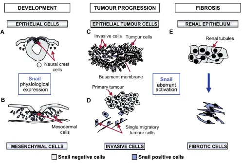
Fig. 1. Snail and the Epithelial-Mesenchymal Transition (EMT) in health and disease. (A,B)
The EMT is fundamental for the development of many
tissues and organs, including the neural crest and the mesoderm of amniotes.
(C)
Snail is activated
in vivo
at the invasive front of chemically-induced
mouse skin tumors and it is present in human carcinomas of different etiologies, where it is inversely correlated with the degree of differentiation
and is associated with lymph-node metastasis (see Barrallo-Gimeno and Nieto, 2005, for a review).
(D)
Multiphoton intravital microscopy has facilitated
the visualization of individual primary tumor cells migrating away from the tumor mass (Wang
et al.,
2002).
(E)
Snail is maintained silent in the adult
and its pathological activation in the kidney leads to renal fibrosis (Boutet
et al.,
2006).
DEVELOPMENT
EPITHELIAL CELLS
MESENCHYMAL CELLS
Neural crest
cells
Mesodermal
cells
Snail
physiological
expression
Snail negative cells
Snail positive cells
RENAL EPITHELIUM
FIBROSIS
FIBROTIC CELLS
Snail
aberrant
activation
Snail
aberrant
activation
Renal tubules
TUMOUR PROGRESSION
EPITHELIAL TUMOUR CELLS
INVASIVE CELLS
Invasive cells
Basement membrane
Tumour cells
Single migratory
tumour cells
Primary tumour
B
C
D
areas of tumors generated in the skin of mice (Cano
et al., 2000;
Fig. 1C), in undifferentiated breast tumors (Blanco
et al., 2002)
and in other carcinomas from different etiologies (see for instance
Rosivatz
et al., 2002; Saito et al., 2004; Miyoshi et al., 2005;
Kuphal
et al., 2005; Franci et al., 2006; Boutet et al., 2007). Thus,
Snail is now regarded as a marker of tumor malignancy and a
target of anti-invasive drugs. Furthermore, Snail has also been
implicated in the promotion of tumor recurrence (Moody
et al.,
2005) and regulates the expression of other molecules unrelated
to EMT, such as the vitamin D receptor, with implications in cancer
therapy (Palmer
et al., 2004).
The molecular analysis of Snail-induced EMT showed that
Snail is a strong repressor of
E-cadherin transcription (Batlle et
al., 2000; Cano et al., 2000), which very much influences cell
behavior both in embryonic development and tumor progression.
Indeed, the loss of E-cadherin expression is clinically regarded as
poor prognostic sign, since it is associated with the transition to an
invasive phenotype (Perl
et al., 1998). In addition to Snail, other
E-cadherin repressors that contribute to EMT and tumor
progres-sion have subsequently been described. These include members
of the ZEB and HLH families that are differentially distributed in
tumors of different origins (see Peinado
et al., 2007 for a review).
It is important to note that E-cadherin repression is not
suffi-cient to induce EMT or invasive properties, as its re-expression in
mesenchymal cells does not induce the reversion to the epithelial
phenotype (Navarro
et al., 1993). Indeed, E-cadherin repressors,
and in particular Snail, directly or indirectly regulate the
expres-sion of many additional target genes (Fig. 2) in order to repress the
epithelial character and provoke the mesenchymal transition.
Moreover, the different Snail family members may be functionally
equivalent (Del Barrio and Nieto, 2002) and as well as their many
common targets, each may also have specific targets
(Moreno-Bueno
et al., 2006).
Snail can also promote tumor progression by activating
angio-genesis (Peinado
et al., 2004b) and there are indications to
suggest that silencing Snail can revert invasion (Olmeda
et al.,
2007). Recent studies are contributing to our knowledge of the
mechanisms that control Snail activity as a transcription factor
(Peinado
et al., 2004a; Peinado et al., 2005).
Animals models generated to study the role of Snail in
tumori-genesis are now available and should prove very useful to further
study these processes (Perez-Mancera
et al., 2005a; 2005b).
However, it is important to bear in mind that these models must
take into account the fundamental roles played by Snail during
embryonic development, which can jeopardize such studies. In
summary, EMT is an important step in the acquisition of the
invasive phenotype of tumors, providing an example of an
impor-tant developmental process that adopts a sinister role in the adult,
as discussed by Jean Paul Thiery (Thiery, 2002).
The EMT in organ fibrosis
The sinister role of EMT in the adult is not restricted to tumor
progression and indeed, adult non-transformed epithelial cells
exposed to Snail undergo EMT, disrupting tissue homeostasis.
Their aberrant activation in the adult kidney is sufficient to induce
Snail1/Snail2
tubular EMT and the development of fibrosis in transgenic mice
(Fig. 1E) and pathological Snail expression is observed in the
fibrotic areas of human kidneys (Boutet
et al., 2006). Until
re-cently, renal fibrosis was thought to be provoked by the activation
of interstitial fibroblasts that deposit an excess of collagen fibers.
However, recent studies have shown that renal tubular epithelial
cells also undergo EMT (Iwano
et al., 2002). Furthermore, Snail
is upregulated during the EMT suffered by hepatocytes (Valdés
et
al., 2002) and mesothelial cells in patients treated with peritoneal
dialysis (Yañez-Mo
et al., 2003). Thus, it appears that Snail genes
must be maintained silent in the adult. Snail activity is not only
regulated at the level of transcription but its subcellular
localiza-tion is also subject to strict control (Dominguez
et al., 2003; Zhou
et al., 2004). Epigenetic mechanisms are likely to be fundamental
in silencing Snail since an increase in its expression can be
attributed to promoter demethylation, and such increases have
been correlated with the invasive properties of carcinoma cell
lines (Fraga
et al., 2004). Interestingly, this reactivation of Snail
can be considered—as a return to the embryonic cellular state
since it fulfills the same function in the adult as in the embryo, the
induction of EMT.
The EMT in tumor progression: A hundred years later?
Although the magnificent contribution of Santiago Ramón y
Cajal to modern Neurobiology has been well acknowledged, not
that many scientists are aware that he also made significant
contributions in other fields. As well as his scientific publications
he also wrote some extremely successful books, the best known
of which is the
“Textura del sistema nervioso del hombre y los
vertebrados” first published in Spanish in 1899. However, he also
wrote an excellent and comprehensive Manual of Pathological
Anatomy for which he generated all the histological slides and
drawings. It was in this book that he described the histopathology
of many diseases in detail including an extremely interesting
chapter on carcinomas. His description of the cells in mammary
tumors is not only extremely detailed but also, it is illuminating and
far ahead of its time in terms of his ideas about tumor malignancy.
When describing the characteristics of an invasive breast tumor
he mentions: “
The epithelial islands are not surrounded by a
basement membrane.. We must mention the fusiform, pear-like
and star-like forms.. These cells are not attached to each other..
This explains their invasive ability” (Ramon y Cajal, 1900). Even
today, it would be difficult to better describe the epithelial cells that
acquire invasive properties. These cells were superbly illustrated
by Cajal and Fig. 3 includes some drawings adapted from Figures
62 and 68 of the third edition of his Manual, published in 1900. It
is fascinating to see how accurately he illustrated both a
differen-tiated and an undifferendifferen-tiated and invasive human breast
carci-noma (Fig. 3A and C, respectively). Interestingly enough, in
infiltrating ductal breast carcinomas (IDC) Snail expression is
inversely correlated with the degree of differentiation (Blanco
et
al., 2002). What is more, the Snail-expressing cells in these
tumors are very reminiscent in location and shape to those
described by Cajal more than 100 years ago (Fig. 3G-H). Thus, it
seems that Cajal “understood” the process of EMT many years
before its significance in tumor malignancy was established.
Old views and new perspectives
Contrary to the long established concept of the role of EMT in
the formation of embryonic tissues, the significance of EMT in
metastasis remains a matter of debate (Tarin, 2005). As a matter
of fact, there is still little convincing evidence that this process
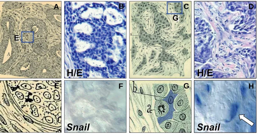
Fig. 3. From Cajal's “pear-like” cells to the Snail-expressing cells in tumors.
Cajal’s drawing of human breast carcinoma biopsies.
(A,E)
Differentiated and undifferentiated ductal breast carcinomas, respectively.
(C,G)
Details of these drawings to better visualize the morphology of cells.
The letters in
(G)
indicate a mitotic cell (labeled “a” by Cajal) and a fusiform or pear-like cell, described as invasive and capable of migrating due to
the lack of the cellular cement (labeled “b” and shaded in blue). Adapted from Figs. 68 and 62 in (Ramón y Cajal, 1900). Histological staining
(B,D)
and
in situ
hybridization
(F,H)
showing that
Snail
is expressed in dedifferentiated breast carcinomas. Note that the labeled cells closely resemble those
described by Cajal. Pictures adapted from Blanco
et al.,
2002.
H/E
H/E
H/E
H/E
Snail
Snail
Snail
Snail
G
G
E
E
G
B
C
D
E
F
H
occurs in human tumors. In the first place, EMT is likely to be a
focal event at the initial stages of tumorigenesis and in addition,
it is a dynamic process making the visualization of malignant cells
with a migratory mesenchymal phenotype extremely difficult. It is
very clear that for a metastasis to form, cells must colonize a
distant site. During embryonic development, migratory
mesen-chymal cells cease their migration and differentiate when they
have reached their destination (Sefton
et al., 1998). They
con-comitantly downregulate
Snail expression and undergo MET,
losing their mesenchymal phenotype. This makes it also very
difficult to see them in the mesenchymal state, but it is in
agreement with the re-expression of E-cadherin observed in
axillary lymph node metastases (Bukholm
et al., 2000) and in
some experimentally generated metastases (Mareel
et al., 1991).
The recent development of intravital multiphoton microscopy
together with novel approaches in the use of fluorescent cell
markers has enabled the initial steps of tumour dissemination to
be analysed
in vivo in animal models. Accordingly, single
carci-noma cells that have lost their epithelial polarity can be seen to
migrate out of primary tumors (Wang
et al., 2002; Condeelis and
Segal, 2003; Fig. 1D). These data provide the first direct evidence
of EMT at the initial stages of the metastatic cascade
in vivo.
Another interesting concept that should be addressed is the
proliferative state of the malignant cells that delaminate from the
primary tumor. At first sight, one always thinks of cancer being
associated with high rates of proliferation. However, there is little
proliferation at the invasive front of carcinomas (Jung
et al., 2001)
and the dramatic cytoskeletal changes that take place during EMT
are probably incompatible with cell division (Barrallo-Gimeno and
Nieto, 2005). Indeed, cells transiently stop dividing before
under-going migration during embryonic development. Thus, although
unregulated proliferation is fundamental for tumors to form and
grow, this is not the case during their malignant phase.
Interest-ingly, Snail blocks cell cycle progression by repressing the
ex-pression of the
Cyclin D gene and increasing the expression of the
cell cycle inhibitor p21 (Vega
et al., 2004). The capacity to
visualize fluorescent cells disseminating from tumors has also
enabled them to be isolated and purified, permitting their
molecu-lar signature to be defined. Significantly, such analyses have
demonstrated that invasive cells are not proliferative (Condeelis
et al., 2005) indicating that the proliferation of tumor cells can be
dissociated from malignancy.
Finally, another property associated to Snail-induced EMT is
the resistance of cells to apoptosis or cell death. Indeed, these
cells become resistant to the loss of survival factors, the action of
direct apoptotic stimuli and to genotoxic stress (Inoue
et al., 2002;
Perez-Losada
et al., 2003; Vega et al., 2004, Kajita et al., 2004).
Thus, Snail confers a selective advantage to embryonic cells
migrating towards their final destination and to invasive malignant
cells in their attempts to disseminate and form metastasis. Again,
the
in vivo purification of disseminating cells has confirmed that
these cells are resistant to conventional chemotherapy (Goswami
et al., 2004).
In summary, we have significantly advanced our knowledge in
the last decade about the cellular processes that have been
hijacked from normal developmental networks and aberrantly
employed in adult pathologies. The combination of sophisticated
animal models and the development of fluorescent probes and
nanodevices that can be incorporated into state-of-the-art
intravi-tal microscopes seem to offer promise in our fight against one of
the most devastating aspects of cancer, the metastatic process,
as well as against degenerative organ diseases.
Acknowledgements
I would like to thank all members of the lab for very stimulating and
fruitful discussions and the Cajal Institute (CSIC) where I learned to
admire Cajal´s contributions and where his original drawings and slides
are kept. Work in the lab is supported by Spanish Ministry of Education
and Science Grants BFU2005-05772, NAN2004-09230-C04-04 and
CONSOLIDER-INGENIO 2010 CSD2007-00017 and CSD2007-00023.
References
BARRALLO-GIMENO, A., and NIETO, M.A. (2005) The Snail Genes as Inducers of Cell Movement and Survival: Implications in Development and Cancer. Devel-opment. 132:3151-3161.
BATLLE, E., SANCHO, E., FRANCI, C., DOMINGUEZ, D., MONFAR, M., BAULIDA, J., and GARCIA DE HERREROS, A. (2000) The Transcription Factor Snail Is a Repressor of E-Cadherin Gene Expression in Epithelial Tumour Cells. Nat Cell. Biol 2:84-89.
BELLAIRS, R. (1987). The primitive streak and the neural crest: comparable regions of cell migration? In: Developmental and Evolutionary aspects of the neural crest. Wiley and Sons, Inc. p. 123-145
BLANCO, M. J., MORENO-BUENO, G., SARRIO, D., LOCASCIO, A., CANO, A., PALACIOS, J., and NIETO, M.A. (2002) Correlation of Snail Expression with Histological Grade and Lymph Node Status in Breast Carcinomas. Oncogene.
21:3241-3246.
BOUTET, A., DE FRUTOS, C. A., MAXWELL, P. H., MAYOL, M. J., ROMERO, J., and NIETO, M. A., (2006) Snail Activation Disrupts Tissue Homeostasis and Induces Fibrosis in the Adult Kidney. EMBO J. 25:5603-5613.
BOUTET, A., ESTEBAN, M. A., MAXWELL, P. H., and NIETO, M. A. (2007) Reactivation of Snail Genes in Renal Fibrosis and Carcinomas: A Process of Reversed Embryogenesis? Cell Cycle. 6:638-642.
BUKHOLM, I. K., NESLAND, J. M., and BORRESEN-DALE, A. L. (2000) Re-Expression of E-Cadherin, Alpha-Catenin and Beta-Catenin, but Not of Gamma-Catenin, in Metastatic Tissue from Breast Cancer Patients. J Pathol. 190:15-19.
CANO, A., PEREZ-MORENO, M. A., RODRIGO, I., LOCASCIO, A., BLANCO, M. J., DEL BARRIO, M. G., PORTILLO, F., and NIETO, M.A. (2000) The Transcrip-tion Factor Snail Controls Epithelial-Mesenchymal TransiTranscrip-tions by Repressing E-Cadherin Expression. Nat Cell Biol. 2:76-83.
CARMONA, R., GONZALEZ-IRIARTE, M., MACIAS, D., PEREZ-POMARES, J. M., GARCIA-GARRIDO, L., and MUNOZ-CHAPULI, R. (2000) Immunolocalization of the Transcription Factor Slug in the Developing Avian Heart. Anat Embryol (Berl) 201:103-109.
COMIJN, J., BERX, G., VERMASSEN, P., VERSCHUEREN, K., VAN GRUNSVEN, L., BRUYNEEL, E., MAREEL, M., HUYLEBROECK, D., and VAN ROY, F. (2001) The Two-Handed E Box Binding Zinc Finger Protein Sip1 Downregulates E-Cadherin and Induces Invasion. Mol Cell. 7:1267-1278.
CONDEELIS, J. AND SEGALL. (2003). Intravital imaging of cell movemet in tumors. Nat. Rev. Cancer 3: 921-930.
CONDEELIS, J., SINGER, R. H., and SEGALL, J.E., (2005) The Great Escape: When Cancer Cells Hijack the Genes for Chemotaxis and Motility. Annu Rev Cell Dev Biol. 21:695-718.
DE CRAENE, B., VAN ROY, F., and BERX, G. (2005) Unraveling Signalling Cascades for the Snail Family of Transcription Factors. Cell Signal. 17:535-547.
DEL BARRIO, M.G. AND NIETO M.A. (2002). Overexpression of Snail family members highlights their ability to promote chick neural crest formation. Devel-opment 129: 1583-1593.
DOMINGUEZ, D., MONTSERRAT-SENTIS, B., VIRGOS-SOLER, A., GUAITA, S., GRUESO, J., PORTA, M., PUIG, I., BAULIDA, J., FRANCI, C., and GARCIA DE HERREROS, A. (2003) Phosphorylation Regulates the Subcellular Location and Activity of the Snail Transcriptional Repressor. Mol Cell Biol. 23:5078-5089.
BALMAIN, A., CANO, A., and ESTELLER, M. (2004) A Mouse Skin Multistage Carcinogenesis Model Reflects the Aberrant DNA Methylation Patterns of Human Tumors. Cancer Res. 64:5527-5534.
FRANCI, C., TAKKUNEN, M., DAVE, N., ALAMEDA, F., GOMEZ, S., RODRIGUEZ, R., ESCRIVA, M., MONTSERRAT-SENTIS, B., BARO, T., GARRIDO, M., BONILLA, F., VIRTANEN, I., and GARCIA DE HERREROS, A. (2006) Expres-sion of Snail Protein in Tumor-Stroma Interface. Oncogene. 25:5134-5144.
FRITZENWANKER, J. H., SAINA, M., and TECHNAU, U. (2004) Analysis of Forkhead and Snail Expression Reveals Epithelial-Mesenchymal Transitions During Embryonic and Larval Development of Nematostella Vectensis. Dev Biol. 275:389-402.
GOSWAMI, S., WANG, W., WYCKOFF, J. B., and CONDEELIS, J. S. (2004) Breast Cancer Cells Isolated by Chemotaxis from Primary Tumors Show Increased Survival and Resistance to Chemotherapy. Cancer Res. 64:7664-7667.
HAY, E. D. (1968). Organization and fine structure of epithelium and mesenchyme in the developing chick embryo. In: Fleischmajer, R. and Billingham, R. E. Eds. Epithelial-mesenchymal interactions. Williams and Wilkins. p31-55
HAY, E. D. (1989) Theory for Epithelial-Mesenchymal Transformation Based on The «Fixed Cortex» Cell Motility Model. Cell Motil Cytoskeleton 14:455-457.
HAY, E. D. (1995) An Overview of Epithelio-Mesenchymal Transformation. Acta Anat (Basel) 154:8-20.
HEMAVATHY, K., ASHRAF, S. I., and IP, Y. P. (2000) Snail/Slug Family of Repressors: Slowly Going into the Fast Lane of Development and Cancer.
Gene. 257:1-12.
INOUE, A., SEIDEL, M. G., WU, W., KAMIZONO, S., FERRANDO, A. A., BRONSON, R. T., IWASAKI, H., AKASHI, K., MORIMOTO, A., HITZLER, J. K., PESTINA T. I., JACKSON, C. W., TANAKA, R., CHONG, M. J., MCKINNON, P. J., INUKAI, T., GROSVELD, G. C., and A. LOOK, T. (2002) Slug, a Highly Conserved Zinc Finger Transcriptional Repressor, Protects Hematopoietic Progenitor Cells from Radiation-Induced Apoptosis in Vivo. Cancer Cell. 2:279-288.
IP, Y. T., and GRIDLEY, T. (2002) Cell Movements During Gastrulation: Snail Dependent and Independent Pathways. Curr Opin Genet Dev. 12:423-429.
IWANO, M., PLIETH, D., DANOFF, T. M., XUE, C., OKADA, H., and NEILSON, E. G. (2002) Evidence That Fibroblasts Derive from Epithelium During Tissue Fibrosis. J Clin Invest. 110:341-350.
JUNG, A., SCHRAUDER, M., OSWALD, U., KNOLL, C., SELLBERG, P., PALMQVIST, R., NIEDOBITEK, G., BRABLETZ, T., and KIRCHNER, T. (2001) The Invasion Front of Human Colorectal Adenocarcinomas Shows Co-Localiza-tion of Nuclear Beta-Catenin, Cyclin D1, and P16ink4a and Is a Region of Low Proliferation. Am J Pathol. 159:1613-1617.
KAJITA, M., MCCLINIC, K. N., and WADE, P. A. (2004) Aberrant Expression of the Transcription Factors Snail and Slug Alters the Response to Genotoxic Stress.
Mol Cell Biol. 24:7559-7566.
LOCASCIO, A., MANZANARES, M., BLANCO, M.J. AND NIETO, M.A. (2002). Modularity and reshuffling of Snail and Slug expression during vertebrate evolution. Proc. Natl. Acad. Sci. USA. 99: 16841-16846.
KUPHAL, S., PALM, H. G., POSER, I., and BOSSERHOFF, A. K. (2005) Snail-Regulated Genes in Malignant Melanoma. Melanoma Res. 15:305-313. MANZANARES, M., and NIETO, M.A. (2003) A Celebration of the New Head and
an Evaluation of the New Mouth. Neuron. 37:895-898.
MAREEL, M. M., BEHRENS, J., BIRCHMEIER, W., DE BRUYNE, G. K., VLEMINCKX, K., HOOGEWIJS, A., FIERS, W. C., and VAN ROY, F. M. (1991) Down-Regulation of E-Cadherin Expression in Madin Darby Canine Kidney (Mdck) Cells inside Tumors of Nude Mice. Int J Cancer 47:922-928.
MARTÍNEZ-ÁLVAREZ, C., BLANCO, M.J., PÉREZ, R., APARICIO, M., RESEL, E., RABADÁN, M.A., MARTÍNEZ, T., AND NIETO, M.A. (2004). Snail family members and cell survival in physiological and pathological cleft palates. Dev. Biol. 265: 207-218.
MIYOSHI, A., KITAJIMA, Y., KIDO, S., SHIMONISHI, T., MATSUYAMA,S., KITAHARA, K., and MIYAZAKI, K. (2005) Snail Accelerates Cancer Invasion by Upregulating Mmp Expression and Is Associated with Poor Prognosis of Hepatocellular Carcinoma. Br J Cancer. 92:252-258.
MOODY, S. E., PEREZ, D., PAN, T. C., SARKISIAN, C. J., PORTOCARRERO, C. P., STERNER, C. J., NOTORFRANCESCO, K. L., CARDIFF, R. D., and CHODOSH, L.A. (2005) The Transcriptional Repressor Snail Promotes Mam-mary Tumor Recurrence. Cancer Cell. 8:197-209.
MORENO-BUENO, G., CUBILLO, E., SARRIO, D., PEINADO, H., RODRIGUEZ-PINILLA, S. M., VILLA, S., BOLOS, V., JORDA, M., FABRA, A., PORTILLO, F., PALACIOS, J., and CANO, A. (2006) Genetic Profiling of Epithelial Cells Expressing E-Cadherin Repressors Reveals a Distinct Role for Snail, Slug, and E47 Factors in Epithelial-Mesenchymal Transition. Cancer Res. 66:9543-9556.
MURRAY, S. A., ORAM, K. F., and GRIDLEY, T. (2007) Multiple Functions of Snail Family Genes During Palate Development in Mice. Development.
134:1789-1797.
NAVARRO, P., LOZANO, E., and CANO, A. (1993) Expression of E- or P-Cadherin Is Not Sufficient to Modify the Morphology and the Tumorigenic Behavior of Murine Spindle Carcinoma Cells. Possible Involvement of Plakoglobin. J Cell Sci. 105 (Pt 4):923-934.
NIETO, M. A. (2002) The Snail Superfamily of Zinc-Finger Transcription Factors.
Nat Rev Mol Cell Biol 3:155-166.
NIETO, M. A., SARGENT, M. G., WILKINSON, D. G., and COOKE, J. (1994) Control of Cell Behavior During Vertebrate Development by Slug, a Zinc Finger Gene. Science. 264:835-839.
OLMEDA, D., JORDA, M., PEINADO, H., FABRA, A., and CANO, A. (2007) Snail Silencing Effectively Suppresses Tumour Growth and Invasiveness. Oncogene.
26:1862-1874.
PALMER, H. G., LARRIBA, M. J., GARCIA, J. M., ORDONEZ-MORAN, P., PENA, C., PEIRO, S., PUIG, I., RODRIGUEZ, R., DE LA FUENTE, R., BERNAD, A., POLLAN, M., BONILLA, F., GAMALLO, C., DE HERREROS, A. G., and MUNOZ, A. (2004) The Transcription Factor Snail Represses Vitamin D Receptor Expression and Responsiveness in Human Colon Cancer. Nat Med.
10:917-919.
PEINADO, H., BALLESTAR, E., ESTELLER, M. and CANO, A. (2004a) Snail Mediates E-Cadherin Repression by the Recruitment of the Sin3a/Histone Deacetylase 1 (Hdac1)/Hdac2 Complex. Mol Cell Biol. 24:306-319.
PEINADO, H., DEL CARMEN IGLESIAS-DE LA CRUZ, M., OLMEDA, D., CSISZAR, K., FONG, K. S., VEGA, S., NIETO, M. A., CANO, A., and PORTILLO, F. (2005) A Molecular Role for Lysyl Oxidase-Like 2 Enzyme in Snail Regulation and Tumor Progression. EMBO J. 24:3446-3458.
PEINADO, H., MARIN, F., CUBILLO, E., STARK, H. J., FUSENIG, N., NIETO, M.A., and CANO, A. (2004b) Snail and E47 Repressors of E-Cadherin Induce Distinct Invasive and Angiogenic Properties in Vivo. J Cell Sci. 117:2827-2839.
PEINADO, H., OLMEDA, D., and CANO, A. (2007) Snail, Zeb and Bhlh Factors in Tumour Progression: An Alliance against the Epithelial Phenotype? Nat Rev Cancer. 7:415-428.
LOSADA, J., SANCHEZ-MARTIN, M., CARO, M., PEREZ-MANCERA, P. A., and SANCHEZ-GARCIA,I. (2003) The Radioresistance Biological Function of the Scf/Kit Signaling Pathway Is Mediated by the Zinc-Finger Transcription Factor Slug. Oncogene. 22:4205-4211.
PEREZ-MANCERA, P. A., GONZALEZ-HERRERO, I., PEREZ-CARO, M., GUTIERREZ-CIANCA, N., FLORES, T., GUTIERREZ-ADAN, A., PINTADO, B., SANCHEZ-MARTIN, M., and SANCHEZ-GARCIA, I. (2005a) Slug in Cancer Development. Oncogene. 24:3073-3082.
PEREZ-MANCERA, P. A., PEREZ-CARO, M., GONZALEZ-HERRERO, I., FLORES, T., ORFAO, A., DE HERREROS, A. G., GUTIERREZ-ADAN, A., PINTADO, B., SAGRERA, A., SANCHEZ-MARTIN, M., and SANCHEZ-GARCIA, I. (2005b) Cancer Development Induced by Graded Expression of Snail in Mice. Hum Mol Genet. 14:3449-3461.
PEREZ-MORENO, M. A., LOCASCIO, A., RODRIGO, I., DHONDT, G., PORTILLO, F., NIETO, M. A., and CANO, A. (2001) A New Role for E12/E47 in the Repression of E-Cadherin Expression and Epithelial-Mesenchymal Transi-tions. J Biol Chem. 276:27424-27431.
PERL, A. K., WILGENBUS, P., DAHL, U., SEMB, H., and CHRISTOFORI, G. (1998) A Causal Role for E-Cadherin in the Transition from Adenoma to Carcinoma.
Nature. 392:190-193.
RAMON Y CAJAL, S. (1900). Manual de Anatomía Patológica.
RAMON Y CAJAL, S. (1899). Textura del sistema nervioso del hombre y los vertebrados. Imprenta y Librería de Nicolás Moya. Madrid.
ROMANO, L. A., and RUNYAN, R. B. (2000) Slug Is an Essential Target of Tgfbeta2 Signaling in the Developing Chicken Heart. Dev Biol. 223:91-102.
Epithe-lial-Mesenchymal Transition Regulators Snail, Sip1, and Twist in Gastric Cancer. Am J Pathol. 161:1881-1891.
SAITO, T., ODA, Y., KAWAGUCHI, K., SUGIMACHI, K., YAMAMOTO, H., TATEISHI, N., TANAKA, K., MATSUDA, S., IWAMOTO, Y., LADANYI, M., and TSUNEYOSHI, M. (2004) E-Cadherin Mutation and Snail Overexpression as Alternative Mechanisms of E-Cadherin Inactivation in Synovial Sarcoma.
Oncogene. 23:8629-8638.
SEFTON, M., SANCHEZ, S., and NIETO, M. A. (1998) Conserved and Divergent Roles for Members of the Snail Family of Transcription Factors in the Chick and Mouse Embryo. Development. 125:3111-3121.
TARIN, D., THOMPSON, E. W., and NEWGREEN, D. F. (2005) The Fallacy of Epithelial Mesenchymal Transition in Neoplasia. Cancer Res. 65:5996-6000;
discussion 00-1.
THIERY, J.P. (1984) Mechanisms of Cell Migration in the Vertebrate Embryo. Cell Differ. 15:1-15.
THIERY, J. P. (2002) Epithelial-Mesenchymal Transitions in Tumour Progression.
Nat Rev Cancer. 2:442-454.
THOMPSON, E. W., NEWGREEN, D. F., and TARIN, D. (2005) Carcinoma Invasion and Metastasis: A Role for Epithelial-Mesenchymal Transition? Can-cer Res. 65:5991-5; discussion 95.
TIMMERMAN, L. A., GREGO-BESSA, J., RAYA, A., BERTRAN, E., PEREZ-POMARES, J. M., DIEZ, J., ARANDA, S., PALOMO, S., MCCORMICK, F., IZPISUA-BELMONTE, J. C., and DE LA POMPA, J. L. (2004) Notch Promotes Epithelial-Mesenchymal Transition During Cardiac Development and Onco-genic Transformation. Genes Dev. 18:99-115.
VALDES, F., ALVAREZ, A. M., LOCASCIO, A., VEGA, S., HERRERA, B., FERNANDEZ, M., BENITO, M., NIETO, M. A., and FABREGAT, I. (2002) The
Epithelial Mesenchymal Transition Confers Resistance to the Apoptotic Effects of Transforming Growth Factor Beta in Fetal Rat Hepatocytes. Mol Cancer Res.
1:68-78.
VEGA, S., MORALES, A. V., OCANA, O. H., VALDES, F., FABREGAT, I., and NIETO, M. A. (2004) Snail Blocks the Cell Cycle and Confers Resistance to Cell Death. Genes Dev. 18:1131-1143.
VELTMAAT, J.M., ORELIO, C.C., WARD-VAN OOSTWAARD, D., VAN ROOIJEN, M.A., MUMMERY, C,L, AND DEFIZE, L.H. (2000). Snail is an immediate early target gene of parathyroid hormone related peptide signaling in parietal endo-derm formation. Int. J. Dev. Biol. 44:297-307.
WANG, W., WYCKOFF, J. B., FROHLICH, V. C., OLEYNIKOV, Y., HUTTELMAIER, S., ZAVADIL, J., CERMAK, L., BOTTINGER, E. P., SINGER, R. H., WHITE, J. G., SEGALL, J. E., and CONDEELIS, J. S. (2002) Single Cell Behavior in Metastatic Primary Mammary Tumors Correlated with Gene Expression Pat-terns Revealed by Molecular Profiling. Cancer Res. 62:6278-6288.
YANEZ-MO, M., LARA-PEZZI, E., SELGAS, R., RAMIREZ-HUESCA, M., DOMINGUEZ-JIMENEZ, C., JIMENEZ-HEFFERNAN, J. A., AGUILERA, A., SANCHEZ-TOMERO, J. A., BAJO, M. A., ALVAREZ, V., CASTRO, M. A., DEL PESO, G., CIRUJEDA, A., GAMALLO, C., SANCHEZ-MADRID, F., and LOPEZ-CABRERA, M. (2003) Peritoneal Dialysis and Epithelial-to-Mesenchymal Tran-sition of Mesothelial Cells. N Engl J Med. 348:403-413.
YANG, J., MANI, S. A., DONAHER, J. L., RAMASWAMY, S., ITZYKSON, R. A., COME, C., SAVAGNER, P., GITELMAN, I., RICHARDSON, A., and WEINBERG, R. A. (2004) Twist, a Master Regulator of Morphogenesis, Plays an Essential Role in Tumor Metastasis. Cell. 117:927-939.
Further Related Reading, published previously in the Int. J. Dev. Biol.
See our recent Special Issue Fertilization, in honor of David L. Garbers and edited by Paul M. Wassarman and Victor D. Vacquier at:
http://www.ijdb.ehu.es/web/contents.php?vol=52&issue=5-6
See our recent Special Issue Ear Development edited by Fernando Giraldez and Bernd Fritzsch at:
http://www.ijdb.ehu.es/web/contents.php?vol=51&issue=6-7
Tbx1 is expressed at multiple sites of epithelial-mesenchymal interaction during early development of the facial complex
Maria Zoupa, Maisa Seppala, Thimios Mitsiadis and Martyn T. Cobourne
Int. J. Dev. Biol. (2006) 50: 504-510
Migration of neural crest-derived enteric nervous system precursor cells to and within the gastrointestinal tract
Alan J. Burns
Int. J. Dev. Biol. (2005) 49: 143-150
Regulation of cell adhesion and migration in lens development
Peggy S. Zelenka
Int. J. Dev. Biol. (2004) 48: 857-865
Primordial germ cell migration
Kathleen Molyneaux and Christopher Wylie
Int. J. Dev. Biol. (2004) 48: 537-543
The migration and differentiation of a chemist entangled in developmental and
cancer biology. An interview with Jean-Paul Thiery.
Fred T. Bosman
Int. J. Dev. Biol. (2004) 48: 529-535
From here to there; a life based on migration. An interview with Isaiah J. Fidler
Ian R. Hart
Int. J. Dev. Biol. (2004) 48: 457-462
Collective cell migration in morphogenesis and cancer
Peter Friedl, Yael Hegerfeldt and Miriam Tusch
Int. J. Dev. Biol. (2004) 48: 441-449
Cytoskeletal mechanisms responsible for invasive migration of neoplastic cells
Jury M. Vasiliev
Int. J. Dev. Biol. (2004) 48: 425-439
Regulation of cell migration during tracheal development in Drosophila
melanogaster.
Valérie Petit, Carlos Ribeiro, Andreas Ebner and Markus Affolter
Int. J. Dev. Biol. (2002) 46: 125-132
Developmental expression of Smad1-7 suggests critical function of TGF-beta/BMP
signaling in regulating epithelial-mesenchymal interaction during tooth
morphogenesis.
Xun Xu, Lesley Jeong, Jun Han, Yoshihiro Ito, Pablo Bringas and Yang Chai
Int. J. Dev. Biol. (2003) 47: 31-39
Patterning parameters associated with the branching of the ureteric bud regulated
by epithelial-mesenchymal interactions.
Yanfeng Lin, Shaobing Zhang, Juha Tuukkanen, Hellevi Peltoketo, Taina Pihlajaniemi
and Seppo Vainio
Int. J. Dev. Biol. (2003) 47: 3-13
Snail is an immediate early target gene of parathyroid hormone related peptide
signaling in parietal endoderm formation.
J M Veltmaat, C C Orelio, D Ward-Van Oostwaard, M A Van Rooijen, C L Mummery and
L H Defize
Int. J. Dev. Biol. (2000) 44: 297-307