Dissertation der Fakultät für Biologie der Ludwig-Maximilians-Universität
München
Transient tetraploidy as a route to chromosomal
instability
vorgelegt von
Anastasia Yurievna Kuznetsova
aus Moskau, Russland
Erklärung
Die vorliegende Arbeit wurde zwischen October 2008 und Mai 2013 unter Anleitung von Frau. Dr. Zuzana Storchova - -
Wesentliche Teile dieser Arbeit sind in folgenden Publikationen veröffentlicht:
Abnormal mitosis triggers p53-dependent cell cycle arrest in human tetraploid cells
Eidestattliche Erklärung
Diese Dissertation wurde selbstständig, ohne unerlaubte Hilfe erarbeitet.
Martinsried, am 23.05.13
Anastasia Kuznetsova
Dissertation eingereicht am: 23.05.13
1. Gutachter: Herr Prof. Dr. Stefan Jentsch
2. Gutachter: Herr Prof. Dr. Peter Becker
Table of Contents
Summary ... 7
Introduction ... 8
1. Tetraploidy: causes and proliferation control. ... 8
2. Tetraploid state as an intermediate to aneuploidy, chromosomal instability and tumorigenesis. ... 11
3. Molecular mechanisms triggering CIN. ... 15
3.1. Aneuploid state per se as a trigger of CIN. ... 15
3.2. Loss of sister chromatid cohesion as a cause of CIN. ... 16
3.3. Alterations in the spindle assembly checkpoint (SAC). ... 17
3.4. Multiple centrosomes and multipolar division. ... 20
3.5. Alteration in mitotic spindle function. ... 22
3.5.1. Defects in kinetochore organization and function. ... 22
3.5.2. Alterations in the mitotic spindle machinery. ... 23
3.5.2.1. MAPs and their role in MT dynamics ... 26
3.5.2.2. Kinesins and their role in MT dynamics ... 27
3.5.3. Defects in mitotic error correction. ... 32
3.6. Deregulation of the cell cycle arrest pathways. ... 33
Aim of This Study ... 37
Results ... 38
1. Isolation and characterization of posttetraploid cells. ... 38
1.1. In vitro evolution of cells after tetraploidization. ... 38
1.2. Cell cycle and growth characteristics of the posttetraploid cells. ... 39
2. Aneuploidy and chromosomal instability of the posttetraploid cells. ... 41
2.1. Chromosome numbers in the posttetraploid cells. ... 41
2.2. Chromosomal instability in the posttetraploid cells. ... 42
2.3. Chromosome segregation errors in the posttetraploids. ... 49
3. Causes of chromosomal instability in the posttetraploids. ... 52
3.1. Contribution of supernumerary centrosomes to chromosomal instability. ... 52
3.3.2. Altered mitotic spindle geometry of posttetraploid cells. ... 60
3.3.3. Other changes potentially causing chromosomal instability. ... 62
3.4. Spindle assembly checkpoint alterations in the posttetraploids. ... 63
3.5. Tolerance to chromosome missegregation in the posttetraploids. ... 66
Discussion ... 71
Tetraploidization drives chromosomal instability independently of the p53 status. .... 71
Erroneous mitosis is a source of CIN. ... 74
Supernumerary centrosomes are not the sole source of CIN in posttetraploid cells. . 76
Sister chromatid cohesion is not altered in posttetraploids. ... 78
Altered levels of mitotic kinesins change the spindle geometry and enhance the frequency of segregation errors. ... 78
Increased tolerance to mitotic errors contributes to CIN in posttetraploid cells. ... 83
Supplementary Information ... 88
Materials and Methods ... 101
1. Materials ... 101
1.1. Cell lines. ... 101
1.2. Primary antibodies. ... 101
1.3. Sodium dodecyl sulfate-polyacrylamide (SDS-PAGE) gel electrophoresis and immunoblotting materials. ... 102
1.4. Other materials. ... 103
2. Methods ... 103
2.1. Cryopreservation and cultivation of cells. ... 103
2.2. Generation of posttetraploid cell lines. ... 104
2.3. Determination of non-viable cells in culture. ... 104
2.4. Protein biochemistry methods. ... 105
2.4.1. Cell lysis and protein concentration measurement. ... 105
2.4.2. SDS-PAGE and immunoblotting. ... 105
2.5. Microscopy. ... 106
2.5.1. Live cell imaging. ... 106
2.5.1.1. Live imaging of untreated cells and cells treated with mitotic poisons. ... 106
2.5.2. Determination of the chromosome copy number and chromosomal
structural aberrations in cells. ... 107
2.5.2.1. Chromosome spreads (standard karyotyping). ... 107
2.5.2.2. Fluorescence in situ hybridization (FISH) on centromeric region. ... 108
2.5.2.3. Whole chromosome multicolor FISH (mFISH) ... 108
2.5.3. Mitotic error analyses in fixed cells. ... 109
2.5.3.1. Mitotic abnormalities scoring in anaphase and early telophase. ... 109
2.5.3.2. Micronucleation test. ... 110
2.5.4. Immunofluorescent staining. ... 110
2.5.4.1. Mitotic spindle staining. ... 110
2.5.4.2. Staining for interkinetochore distance, kinetochore distribution measurements and high-resolution mitotic error visualization. ... 111
2.5.4.3. Centrosome staining. ... 111
2.6. High-throughput methods. ... 112
2.6.1. Array comparative genomic hybridization (aCGH). ... 112
2.6.2. mRNA microarray-based gene expression analysis. ... 112
2.7. Statistical analysis. ... 113
2.8. Image processing. ... 114
Figure list ... 115
References ... 117
Abbreviations ... 138
Acknowledgements ... 140
Summary
Summary
Aneuploidy, defined as alterations in both chromosome number and structure, along with chromosomal instability (CIN) are common hallmarks of cancer. Growing evidence suggests that aneuploidy and CIN facilitate carcinogenesis in both mice and humans. One of the routes to CIN can be via an unstable tetraploid intermediate. However, the mechanisms contributing to the development of CIN in the post-tetraploid progeny remain elusive.
Introduction
Introduction
1. Tetraploidy: causes and proliferation control.
A whole genome multiplication or polyploidy (for example, three fold – triploidy, four fold – tetraploidy, etc.) is currently regarded as one of the driving forces of biological diversity. Polyploidization, or paleopolyploidy, was proposed to occur in plant evolution (Masterson, 1994) and early in the vertebrate evolution (Van de Peer et al., 2009). It can provide organisms and their cells with additional genetic material for adaptation to changes in the environment (Aleza et al., 2011; Otto and Whitton, 2000), as well as robustness against lethal mutations and loss of chromosomes. To date, polyploidy has been described to frequently occur in plants and fungi (Albertin and Marullo, 2012). In animals, polyploidy occurs predominantly in lower forms, such as flatworms. However, polyploidy was also reported in some higher forms of Animalia, such as African clawed frog (Xenopus laevis), salamanders, salmon; at the same time, so far in only one mammalian species red vizcacha rat (Tympanoctomys barrerae)and related species (Gallardo et al., 1999).
Polyploidy can also occur in the tissues of otherwise diploid organisms. For example, polyploidization frequently takes place as a part of a developmental and differentiation program in human organisms. Prominent examples are human heart muscle cells and megakaryocytes, where a single polyploid cell can give rise to many thrombocytes. A programmed cytokinesis failure results in polyploidization in liver hepatocytes (Guidotti et al., 2003). A large body of evidence suggests that polyploidy occurs through endoreplication (i.e. duplication of the genome without subsequent cell division) as a stress response mechanism (Lee et al., 2009a).
Introduction
Three major routes to aberrant polyploidization, well documented for tetraploidy, are described up to date, namely, cytokinesis failure, cell-cell fusion and mitotic slippage (Figure 1).
Figure 1. Three main routes to aberrant tetraploidy(from Storchova and Kuffer, 2008)
Introduction
can also lead to tetraploidization (Rieder and Maiato, 2004; Storchova and Kuffer, 2008). In contrast to the first two mechanisms, mitotic slippage results in a mononucleated tetraploid cell. Thus, tetraploidization can occur due to various mechanisms, such as different types of abortive cell division as well as virus-induced cell-cell fusion.
Since aberrant tetraploidization is poorly tolerated by human organism, it suggests the existence of mechanisms restricting further proliferation of spontaneously formed tetraploids (Ganem and Pellman, 2007). Initially, tetraploidy was proposed to trigger a so- “ p y p ”, b b q y cytokinesis failure in a p53-dependent manner in mammalian cells (Andreassen et al., 2001; Margolis et al., 2003). However, follow-up studies using lower, less toxic concentrations of dihydrocytochalasin B (DCB) to induce tetraploidization, proved that DNA replication and mitotic entry takes place in tetraploid RPE1-hTERT (retinal pigment epithelium cell line) and HDF (human diploid fibroblasts) (Uetake and Sluder, 2004; Wong and Stearns, 2005).
The p53 pathway plays an essential role in the proliferation inhibition of tetraploid cells after mitotic slippage (Rieder and Maiato, 2004). In addition, p53-proficient DCB-treated and sorted newly formed tetraploid murine cells did not proliferate in culture, whereas p53-deficient cells did (Fujiwara et al., 2005). Similarly, absence of p53 in the tetraploids was shown to promote subtetraploid aneuploidy (Vitale et al., 2010). The fact that tetraploid cells, formed through different mechanisms, arrest in a p53-dependent manner raised a question of the nature of upstream triggers of this arrest.
Introduction
p38/MAP stress kinase pathway; the role of p38/MAP pathway in proliferation restriction of tetraploids was shown in at least two independent studies (Mikule et al., 2007; Vitale et al., 2008). Defective spindle assembly checkpoint (SAC) can lead to chromosome missegregation and further cause p53-dependent cell cycle arrest and/or death, as reported for defects in some of key SAC proteins Bub1 (Budding uninhibited by benzimidazoles 1), BubR1 (Budding uninhibited by benzimidazoles-related 1) and TTK/hMps1 kinase (Huang et al., 2009b; Jeganathan et al., 2007; Shin et al., 2003). It is also possible that chromosome missegregation after multipolar mitosis that leads to imbalanced gene copy number and protein abundance, in turn might cause proteotoxic stress. The proteotoxic stress was shown in budding yeast with few additional copies of chromosomes; however, these changes were not associated with cell cycle arrest (Oromendia et al., 2012). Since the protein imbalance after chromosome missegregation in multipolar mitosis is substantially higher than described in aneuploid yeast, this imbalance can potentially impair cell proliferation.
Complex mitotic spindle organization of tetraploids, i.e. the increased amount of chromosomes and centrosomes, impedes the identification of the arrest triggers. Moreover, multiple factors functioning either together or independently might cause cell cycle arrest. Therefore, the pathways restricting the proliferation of tetraploids, as well as the consequences of the escape from the restriction control remain enigmatic.
2. Tetraploid state as an intermediate to aneuploidy, chromosomal
instability and tumorigenesis.
Introduction
intermediate formation followed by chromosome missegregation (Nigg, 2002; Shackney et al., 1989; Storchova and Pellman, 2004) (Figure 2).
Figure 2. Tetraploid state as an intermediate to complex numerical aneuploidy.
The hypothesis is further supported by an important characteristic of aneuploid tumors – a frequent presence of multiple centrosomes that can originate after cytokinesis failures during tetraploidization. Multiple centrosomes were proposed to be a major source of chromosome missegregation and tumorigenesis by Theodor Boveri at the beginning of the last century (Boveri, 2008). Subsequent studies showed a strong correlation between amplification of centrosome numbers and tumorigenesis in majority of cancers (Nigg, 2002). Moreover, tetraploid cells were identified in early stages of cervical cancer (Kirkland et al., 1967; Olaharski et al., 2006) , p ’ p w w v rise to aneuploids upon p53 inactivation (Galipeau et al., 1996). A mutation in APC gene frequently found in colorectal tumors leads to cytokinesis failure followed by aneuploidy in vivo (Caldwell et al., 2007). Other described cases providing the evidence for tetraploidization in tumors are bladder- (Shackney et al., 1995), breast- (Dutrillaux et al., 1991; Shackney and Silverman, 2003) and prostate cancers (Deitch et al., 1993; Montgomery et al., 1990), and hyperplastic lesions in the pancreatic cancers (Tanaka et al., 1984). Thus, tetraploidy can frequently occur in different tumors, in particular during early stages of tumorigenesis. According to the tetraploid intermediate model, tetraploidization leading to aneuploidy might eventually facilitate tumorigenesis (Storchova and Pellman, 2004).
Introduction
with cell transformation; de novo aneuploidy is more likely to trigger tumorigenesis. Trisomic and tetrasomic aneuploids, generated by chromosome transfer, display rather an antiproliferative response, manifesting in a slower growth rate and a slow growth-related changes in gene expression (Lengauer et al., 1997; Pfau and Amon, 2012; Sheltzer et al., 2012; Stingele et al., 2012; Williams et al., 2008). Furthermore, chromosome gains, occuring during chronic inflammatory processes, such as D p y ’ , (Gisselsson, 2011). Down syndrome (trisomy of chromosome 21) patients, on one hand display increased risk of leukaemia, but on the other hand a remarkably lower frequency of solid tumors in comparison to the general population (Satge et al., 1998).
Overall, the trisomic and tetrasomic aneuploids frequently (but not necessarily) maintain stable karyotypes, in a stark contrast to karyotypically divergent near-tetraploid cells observed in most aneuploid cancers. These complex aneuploidies are likely linked to tumor formation, hence supporting a role of tetraploidization prior to aneuploidy in cell transformation.
Complex aneuploidy is usually a steady-state manifestation of CIN – dynamic changes in the number of the chromosomes during propagation. CIN is the hallmark of the majority of solid tumors (Haruki et al., 2001; Lengauer et al., 1997; Yoon et al., 2002). Clinical studies show that CIN in cancer is associated with resistance to drug treatment and poor prognosis (Carter et al., 2006; Duesberg et al., 2000; Walther et al., 2008). This can be explained by inherent ability of CIN cells to easily adapt to changes in their growth environment (Bakhoum and Compton, 2012). Interestingly, the frequency of CIN in non-diploid (near-triploid and near-tetraploid) tumors is substantially higher than in near-diploid tumors (Storchova and Kuffer, 2008). A possible explanation is that increased ploidy can provide reserves in chromosome copy number and decrease a probability of lethal nullisomies (loss of both chromosome copies) after chromosome missegregation in CIN cells. Taken together, tetraploidization is associated with CIN and tumorigenesis. However, it remains unclear what are the mechanisms causing CIN and development of cancer in tetraploid cells.
The role of polyploidization in triggering CIN was shown in budding yeast
Introduction
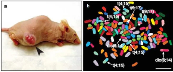
direct evidence of the role of unstable tetraploid intermediates in CIN and tumorigenesis was demonstrated upon injection of tetraploid p53-deficient murine mammary epithelial cells (MMEC) into nude mice (Fujiwara et al., 2005). Remarkably, only tetraploid cell injections led to tumors, in contrast to control diploid cell injections. The resulting tumors displayed near-tetraploid aneuploidy with chromosome gains and losses as well as structural chromosome rearrangements, suggesting ongoing CIN (Figure 3).
Figure 3. Tetraploidy facilitates tumorigenesis and CIN (adapted from Fujiwara et al., 2005).
(a) Tumor indicated by an arrowhead in a nude mouse at the site of injection of tetraploid
p53-deficient MMECs; (b) Representative spectral karyotyping data from one tumor, showing
near-tetraploid chromosome number in a cell, non-reciprocal translocations (t) and dicentric chromosomes (dic) indicated by arrowheads; b 10 μ
Later study further confirmed the role of tetraploidy in tumorigenesis in mice. Upon prolonged passaging in vitro, diploid mouse ovarian surface epithelial cells (MOSEC) undergo cytokinesis failure at a high frequency, form tetraploid and subsequently, aneuploid cells (Lv et al., 2012). The intraperitoneal injection of aneuploids (late passages) into C57BL/6 mice induced tumor formation on the intestinal surface, whereas injection of diploids (early passages) did not. Of note, the p53 status in the cells from resulting tumors was not investigated. Thus, possible p53 pathway deregulation could allow the proliferation in aneuploid state and tumor growth.
Introduction
Similarly to previously discussed study from Lv and colleagues, the authors did not investigate the p53 status of the meningiomas, however, they suggest defective tetraploidy checkpoint as a cause of observed CIN and proliferation after tetraploidization.
Expression of transcription factor cut homeobox 1 (CUX1) was shown to activate a program causing aneuploidy after tetraploidization (Sansregret et al., 2011). Upon induction of tetraploidy with transient cytokinesis failure by blebbistatin treatment cells, mock-expressing cells underwent predominantly multipolar mitosis and died, whereas cells overexpressing CUX1 were shown to undergo predominantly bipolar mitosis. In this scenario, the proposed mechanism of action is prolongation of mitosis, which allows more time for pair-wise clustering of centrosomes. Observed progeny displayed subtetraploid aneuploidy associated with tumorigenesis upon injection into mice. Similarly to the studies of Lv and colleagues and Baia and colleagues, p53 status of the resulting aneuploid cells was not addressed. Moreover, the observed aneuploidy could be attributed to CUX1 overexpression but not tetraploidization itself.
In summary, evidence suggests the oncogenic potential of transient tetraploidy and association with complex aneuploidy and CIN. However, little is known about the molecular mechanisms underlying the transitions from tetraploidy to CIN.
3. Molecular mechanisms triggering CIN.
The mechanisms driving chromosomal instability likely affect both the chromosome segregation fidelity as well as the ability to arrest after chromosome missegregation. Up to date, different proteins were shown to be associated with whole chromosome numerical instability. They function in spindle assembly checkpoint (SAC), formation of kinetochore-microtubule interactions, mitotic spindle organization, cytokinesis, centrosome number control, sister chromatid cohesion and cell cycle regulation.
3.1. Aneuploid state
per se
as a trigger of CIN.
Introduction
insufficient functions of the protein machineries involved in chromosome segregation and maintenance, thus destabilizing the genome. According to this hypothesis, aneuploidy itself can be a catalyst of persistent changes in the karyotype without a gene mutation prerequisite (Duesberg and Li, 2003). Moreover, even a slight increase in the instability of a newly formed aneuploid might be sufficient to promote stronger CIN, if the initial chromosome changes favor chromosome missegregation (Anderson et al., 2001; Matzke et al., 2003).
The possibility that aneuploidy can drive genetic instability was directly tested in single chromosome-disomic yeast Saccharomyces cerevisiae strains: approximately 70% (9 out of 13) disomic strains displayed increased levels of chromosome missegregation in comparison to euploid controls (Sheltzer et al., 2011). Another study showed that the extent of CIN correlates with a deviation from euploid DNA content: haploid (1N) yeast strains were more stable than strains with 1.5 to 2N ploidy (Zhu et al., 2012).
In summary, results suggest that aneuploidy itself at least in budding yeast can further promote CIN. Whether similar scenario is taking place in higher eukaryotes remains poorly investigated.
3.2. Loss of sister chromatid cohesion as a cause of CIN.
Introduction
Deficiency in key components of cohesion complex Ssc1/Mcd1 causes chromosome misalignment in metaphase and subsequent chromosome missegregation (Morrison et al., 2003). Similarly, Smc1 downregulation leads to higher frequency of micronucleation and aneuploidy (Musio et al., 2003). Mutational inactivation of another member of cohesin complex STAG2/Scc3 also promotes CIN (Solomon et al., 2011). RNAi-mediated depletion, as well as haploinsufficiency of Sgo1 (one of the key regulators of cohesion establishment), causes CIN in colorectal cancers (Iwaizumi et al., 2009; Yamada et al., 2012). Moreover, mutations, downregulation and overexpression of separase, which cleaves cohesins at the onset of anaphase, was reported to trigger CIN (Shepard et al., 2007; Wirth et al., 2006; Xu et al., 2011; Zhang et al., 2008a). Interestingly, high levels of separase were identified in human breast cancer. Cells derived from these tumors displayed premature chromosome disjunction and lagging chromosomes (Zhang, Ge et al. 2008). Similarly, depletion of securin, a separase inhibitor, also instigates CIN (Jallepalli et al., 2001). Finally, a moderate but recurrent cohesion defect associated with CIN was observed in tetraploid yeast (Storchova et al., 2006).
Taken together, the levels of cohesin complex proteins and their cofactors should be tightly regulated to ensure chromosome segregation fidelity. However, since cohesin complex has been also implicated in several other cellular functions (Dorsett, 2011), the mechanistic link between cohesion defects and CIN development remains to be investigated.
3.3. Alterations in the spindle assembly checkpoint (SAC).
Another mechanism that can contribute to CIN is the defective spindle assembly checkpoint (SAC), complex protein machinery that controls proper execution of mitotic events and ensures faithful chromosome segregation. SAC arrests or delays cell division until all chromosome kinetochores are stably and properly attached to the microtubules emanating from the opposite poles of the mitotic spindle. In recent years the key components of the SAC have been characterized in great detail (for review see Musacchio and Salmon, 2007). One major SAC gene group comprises
Introduction
complex/cyclosome). The latter mediates the polyubiquitination of its substrates, such as cyclin B and securin, subsequently destructed by the 26S proteasome. Upon protease-mediated degradation of cyclin B, major mitotic kinase CDK1 is inactivated, leading to a rapid onset of anaphase.
Introduction
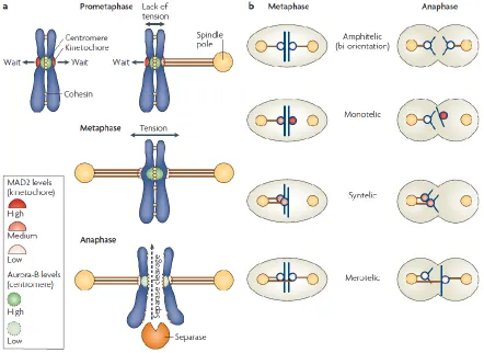
Figure 4. Chromosome attachment in mitosis and SAC activation (from Musacchio and Salmon, 2007).
(a) Mad2 levels are high at the unattached kinetochores, moderately high at a kinetochore attached in
a monotelic chromatid pair, and very low upon establishment of a proper amphitelic attachment.
Aurora B is activated by a lack of tension on sister kinetochores, upon tension establishment Aurora B
activity at the sister kinetochores is low. When all KT-MT attachments are amphitelic, the SAC signal
ceases, separase cleaves cohesins, thus promoting sister chromatid separation in anaphase. (b)
Examples of proper (amphitelic) and improper (synthelic, merotelic) KT-MT attachments. Monotelic is
a normal condition during prometaphase before biorientation establishment.
Introduction
This aneuploidy was shown to be associated with CIN and tumorigenesis (Matsuura et al., 2000). Taken together, the deregulation or partial inactivation of SAC components attenuates SAC and triggers CIN. Notably, complete inactivation of the SAC is lethal in different cell lines and homozygous deletion of key checkpoint components is embryonic lethal in mice (Thompson et al., 2010).
Despite the accumulating experimental evidence, the role of the attenuated SAC response in the common occurrence of CIN and cancer is largely debated in the field. CIN cancer cell lines show rather a robust SAC response to spindle poisons (Tighe et al., 2001); moreover, CIN cell lines halt anaphase onset in the presence of misaligned chromosomes (Gascoigne and Taylor, 2008). Furthermore, CIN cells frequently die during prolonged mitotic block (Brito and Rieder, 2009) and cannot survive without functional SAC (Kops et al., 2004). Lastly, analysis of 132 colorectal cancer cell lines showed no mutations in SAC genes (Barber et al., 2008). These findings support the view that SAC functions normally in the majority of cancer CIN cell lines.
3.4. Multiple centrosomes and multipolar division.
Aberrant chromosome and centrosome numbers frequently coincide in cancer cells (Hardy and Zacharias, 2005). For a long time multiple centrosomes were considered to be the major source of CIN in a wide range of human cancers from solid tumors to hematological malignancies (Boveri, 2008; Chan, 2011; Lingle et al., 2002; Nigg, 2002; Salisbury et al., 1999). Several different mechanisms were described to cause centrosome amplification (Doxsey, 2001; Ko et al., 2005; Mussman et al., 2000). Four main models of centrosome amplification were proposed: centrosome overduplication, abortive cell division, cell fusion and de novo centriole formation (Nigg, 2002).
Introduction
during multipolar division frequently displayed sister chromatid nondisjunction and nullisomies. A follow-up study on the Wilms tumor explained the reason for high frequency of trisomies in cancer cells (Gisselsson et al., 2010). Cells can progress through anaphase in a tripolar manner (the most frequent type of multipolar anaphase), when each of the daughter nuclei gets nearly a third of parental DNA material. This step is often followed by an asymmetric cytokinesis, resulting in two daughter cells, thus generating a trisomy and monosomy in diploid cells. However, in tetraploid cells the chromosome distribution in tripolar mitosis increases the probability of random segregation. Importantly, multipolar divisions are poorly tolerated and the viability of the progeny after multipolar mitoses is low (Ganem et al., 2009). This suggests that multipolar divisions might not be the sole source of CIN in cells with multiple centrosomes.
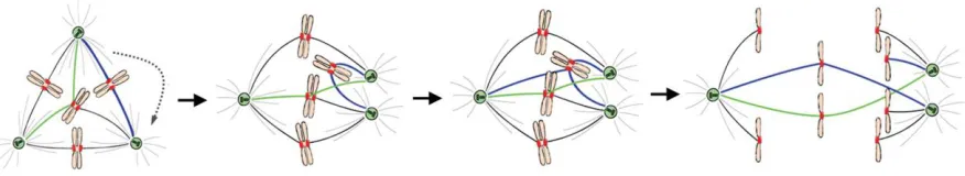
Interestingly, many cancer cells develop an adaptation to suppress multipolar cell divisions – clustering of centrosomes. Clustering leads to the reduction of spindle pole numbers, in particular, it leads to bipolar spindle formation (Brinkley, 2001; Kwon et al., 2008; Murphy, 2003; Quintyne et al., 2005). A stable propagation and maintenance of the karyotypes in the presence of multiple clustered centrosomes was reported for Drosophila melanogaster S2 cells (Basto et al., 2008); however, other reports suggest that centrosome clustering is often associated with chromosome missegregation. Cells that form a multipolar intermediate in metaphase often display lagging chromosomes in a bipolar anaphase (Ganem et al., 2009; Silkworth et al., 2009). The proposed mechanism for formation of lagging chromosomes in anaphase is an accumulation of unresolved merotelic kinetochore-microtubule attachments due to the extra centrosome coalescence at the anaphase onset (Figure 5). This mechanism is consistent with the presence of multiple centrosomes and proliferation in CIN state, and has been proposed as a possible cause of CIN in cells with multiple centrosomes.
Introduction
Merotelic attachments are formed in a multipolar (here, tripolar) intermediate. They can remain
unresolved before extra centrosome clustering. Clustering of extra centrosomes promotes additional
merotelic attachment. If remaining incorrected, merotelically attached chromosomes lag in anaphase
and cause chromosome missegregation.
Bipolar division strongly reduces the chances of both lethal nullisomies and newly arising complex aneuploidies, thus supporting the proliferation in contrast to multipolar division. Clearly, a bipolar spindle is more robust and less missegregation-prone in the cells with normal centrosome numbers than in the cells with multiple clustered centrosomes. This view is supported by a recent work from our laboratory, where we show that similar frequency of cell cycle arrest was observed after chromosome missegregation in tetraploids in bipolar anaphase, as well as in multipolar metaphase followed by bipolar anaphase and by multipolar anaphase alone. Our data suggest that the formation of a multipolar intermediate followed by bipolar division can trigger cell cycle arrest to similar levels as multipolar division (Kuffer et al., 2013).
Therefore, it is reasonable to speculate that cells that reduced their centrosome numbers early after centrosome amplification get a selective growth advantage. This view is further supported by the fact that rounds of repeated cytokinesis failure did not lead to centrosome amplification in various cell lines (Krzywicka-Racka and Sluder, 2011). Likely, centrosome amplification is just a transient condition leading to aneuploidy, but cannot be a sole source of CIN due to deleterious consequences of multipolar mitosis on the chromosome copy number and associated protein imbalance.
In summary, centrosome amplification causing an increase in merotelic attachment formation represents one of the common mechanisms of CIN. However, the fact that increased centrosome numbers are not maintained for a long period after formation argues against the role of extra centrosomes as the exclusive triggers of CIN.
3.5. Alteration in mitotic spindle function.
3.5.1.
Defects in kinetochore organization and function.
Introduction
undergo persistent association and dissociation with the kinetochores, therefore constantly attempting to establish proper attachments and allowing possible mitotic error correction (Nicklas and Ward, 1994).
Kinetochore consists from inner kinetochore, associated with centromeric DNA, and more dynamic outer kinetochore, which is formed around the time of the nuclear envelope breakdown (reviewed in Maiato et al., 2004). The innermost part of the kinetochore is composed of histone variant CENPA and other CENP proteins (reviewed in Musacchio and Salmon, 2007). The outermost part of kinetochore is responsible for interaction with the MTs of the spindle; this layer consists of outer kinetochore or KNL1 complex/Mis12 complex/Ndc80 complex (KMN) and fibrous corona. The corona proteins are generally more labile than other kinetochore proteins and the protein content is dependent whether the MTs are anchored to the KT or not. Upon establishment of the KT-MT interactions, the amount of SAC proteins diminishes and the amount of such as proteins of Ran pathway, APC and other proteins involved in proper MT anchoring increases (Joseph et al., 2004; Kaplan et al., 2001; Salina et al., 2003; Shah et al., 2004; Tirnauer et al., 2002).
Alterations in the kinetochore structure cause defects in chromosome attachments, and, subsequently, mitotic abnormalities. For example, the dysfunction in Ndc80 complex manifests itself in the absence of KT-MT attachments, suggesting its essential role in establishment of stable attachments (DeLuca et al., 2002; Martin-Lluesma et al., 2002). Similarly, depletion of Mis12 complex subunits prolongs mitosis and leads to defects in chromosome alignment and biorientation (Kline et al., 2006). Inhibition of an Hsp90 using RNAi triggered delocalization of several key kinetochore proteins such as CENP-H, CENP-I, and BUB1 and caused chromosome misalignment and aneuploidy (Niikura et al., 2006). Interestingly, RNAi silencing of kinetochore-bound MT-associated proteins APC and EB1 was shown to cause misalignment of sister kinetochores, anaphase lagging and aneuploidy, without affecting the KT-MT binding per se (Draviam et al., 2006).
In summary, impairment of the kinetochore structure might cause defects in mitotic progression and subsequently lead to aneuploidy.
Introduction
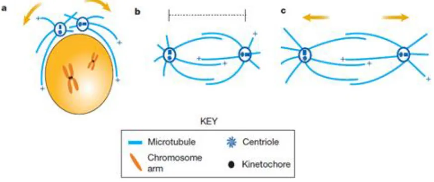
dynamics in faithful chromosome segregation maintenance. This emerging interest can be readily explained by the fact, that mitotic spindle orchestrates virtually all chromosome movements in mitosis (Compton, 2000). Thus, high liability and dynamicity are prerequisites for the mitotic spindle organization. It is ensured by two important mechanisms. First, mitotic MTs display a higher turnover rate than interphase MTs and undergo continuous GTP hydrolysis-dependent lengthening and shortening, necessary for spindle formation and force generation within the spindle (Mitchison and Kirschner, 1984; Saxton et al., 1984). The dynamic instability is intrinsic to tubulin itself as was shown by in vitro experiments (Hyman and Karsenti, 1996). Second, mitotic MTs cross-bridge and slide relatively to adjacent MTs (Figure 6). Spatial and temporal control of MT dynamics is modulated by many microtubule-associated proteins (MAPs) and microtubule motor proteins, kinesins and dyneins, directly interacting with tubulin subunits. Growing evidence suggests that microtubule dynamics in mitosis is not driven by one single protein: instead, a balance of synergic and antagonistic forces generated by multiple motors is necessary to drive spindle movements.
Figure 6. Spindle pole and chromosome movements during mitosis (adapted from Sharp et al., 2000).
MTs and MT motors orchestrate spindle assembly, maintenance and elongation. (a) Bipolar spindle b y (b) p p b ( ) Sp p “+”
plus-ends of the MTs.
Introduction
Introduction
other with the help of plus-end-directed kinesins (kinesin-5 proteins), thus pushing the spindle poles further apart. This movement is strengthened by dynein-mediated pulling forces on astral microtubules bound to cell cortex (poleward flux). Chromosome separation is followed by telophase and reconstitution of nuclear envelope. Cytokinesis, executed by combined action of actin and myosin, finalizes cell division. Astral MTs in the spindle midzone play an essential role in guiding a cleavage furrow to the former spindle equator by stimulating the cell cortex. However, the exact mechanism of cortex stimulation by MTs in cytokinesis remains unclear.
The orchestration of mitosis and cytokinesis requires finely balanced activity of motor and non-motor MT-associated proteins, however, it remains enigmatic. An in-depth understanding of this machinery can be beneficial for investigation of the chromosome segregation and CIN.
3.5.2.1.
MAPs and their role in MT dynamics
Microtubule associated proteins (MAPs) are non-motor proteins that can directly bind to the MTs and regulate their assembly and stability. Destabilization of the MTs can occur not only by depolymerization from a MT end, but also through severing mechanism that generates an internal break in the MT. This reaction requires an activity of the MT severing MAP enzymes, namely fidgetin, katanin, spastin and Op18/stathmin (Cassimeris, 2002; Roll-Mecak and McNally, 2010; Roll-Mecak and Vale, 2008). In Drosophila melanogaster, severing proteins were described to act in a Pac-Man flux. Fidgetin and spastin contribute to MT minus-end depolymerization and flux at centrosomes, whereas katanin localizes at the centromeric region and stimulates chromosome motility by Pac-Man mechanism (Zhang et al., 2007). Inhibition of these factors causes elongation of the MTs. For example, inhibitory phosphorylation of katanin, that occurs in allotetraploid Xenopus laevis leads to longer spindles in comparison to the related diploid species Xenopus tropicalis
Introduction
enzymes can result in increased stability of MTs and thus impair proper KT-MT dynamics.
Many types of up-to-date characterized stabilizing MAPs specifically function at the microtubule plus-ends and regulate their stability. In addition, many MAPs can have other functions in spindle organization. For example, MAP TPX2 plays an important role in MT nucleation and stabilization. Additionally, TPX2 recruits Aurora A, and this recruitment is important for proper chromosome alignment to the spindle (Kufer et al., 2002). TPX2 conditional knockout and haploinsufficiency have detrimental effects on MT nucleation that leads to aberrant mitotic spindle formation and chromosome segregation errors. Moreover, TPX2-haploinsufficient mice are prone to developing aneuploid adenocarcinomas and lymphomas (Aguirre-Portoles et al., 2012). Remarkably, overexpression of TPX2 was described for many tumors and strongly correlates with CIN in several human cancers (Asteriti et al., 2010; Carter et al., 2006). This suggests that tight regulation of TPX2 is essential for maintenance of the genome stability. Overexpression of another MAP XMAP215/ch-Tog is observed in liver and colon tumors (chTOG, Colonic Hepatic Tumor Overexpressed Gene) (Charrasse et al., 1995). It plays an important role in spindle pole organization and spindle MT stabilization by crosslinking K-fibers and dampening MT disassembly (Barr and Gergely, 2008; Booth et al., 2011; Gergely et al., 2003). Overall, abundant presence of MT-stabilizing proteins in tumors might cause MT over-stabilization effect. This defect in MT dynamics might potentially lead to slower disassembly of the incorrectly attached MTs at the kinetochore, thus increased rates of chromosome missegregation.
3.5.2.2.
Kinesins and their role in MT dynamics
MT-Introduction
interacting outer surface (Su et al., 2012), and a short neck domain, binding to KLCs. Processive movement of kinesins along the MT utilizes one ATP molecule per step. Most kinesins have uni-directional motility and are plus-end-directed, with the exception of minus-end-directed members of kinesin-14 family (Ambrose et al., 2005).
Kinesins have been implicated in various activities in the cell. For example, in interphase, kinesins transport cargo along the MTs, such as organelles and other structures. Mitotic kinesins (12 out of around 40 kinesins in total) function in polar ejection force generation on chromosome arms, chromosome congression, spindle pole separation and cleavage furrow positioning.
An active MT depolymerization is essential to carry out some of these functions. The best characterized to date is the kinesin-13 family of nonmotile depolymerases, consisting of three members in mammalian cells: Kif2A, Kif2B and Kif2C/MCAK (mitotic centromere-associated kinesin) (Moore and Wordeman, 2004; Wordeman, 2005). In contrast to other kinesins, kinesin-13 proteins show a different microtubule-binding pattern: only one molecule out of two binds to adjacent filaments (Mulder et al., 2009). This binding, and subsequently the depolymerization function, can be inhibited upon the detyrosination of -tubulin, leading to MT stabilization (Peris et al., 2009). Members of kinesin-13 family have distinct and non-overlapping functions in mitosis. Kif2A localizes to the minus-ends of the centrosome MTs and is involved in the formation of a bipolar spindle (Ganem and Compton, 2004). Kif2B also localizes to centrosomes and the spindle midzone, where it plays role in establishment of bipolarity, chromosome movement, and cytokinesis (Manning et al., 2007). Furthermore, Kif2B localizes to the kinetochore, where it promotes the correction of KT-MT attachment errors specifically in prometaphase acting downstream of Aurora B-mediated phosphorylation of the KMN network (Bakhoum et al., 2009b).
Introduction
correction and promoting chromosome missegregation (Bakhoum et al., 2009b). Remarkably, overexpression of Kif2B or Kif2C/MCAK suppresses the incidence of lagging chromosomes in CIN cell lines, providing compelling evidence that stimulation of MT disassembly, at least partially, rescues CIN phenotype (Bakhoum et al., 2009b). Further support of this evidence was provided by the finding that KT-MT attachments in CIN cancer cells are inherently more stable than in normal cells (Bakhoum et al., 2009a). Additionally, Kif2C/MCAK helps focusing microtubules at aster centers and facilitates asters to bipolar spindles transition in an Aurora A kinase-dependent manner (Zhang et al., 2008b). Taken together, kinesin-13 proteins represent a family of potent MT depolymerases whose activity is essential for efficient KT-MT attachment error correction.
Another well-characterized family of plus-end directed kinesins essential for mitotic progression, is kinesin-8 family: Kif18A, Kif18B and Kif19 in mammals, as well as S. cerevisiae homolog Kip3 and S. pombe Klp5 and Klp6 (Lawrence et al., 2004). In contrast to kinesins-13, kinesin-8 proteins, such as Kif18A and yeast Kip3, are highly processive motors involved in the regulation of the MT length. Importantly, they act both as stabilizers and destabilizers of the MTs, depending on the context. For example, it was shown that Kif18A could directly depolymerize MTs (Mayr et al., 2007). Other works report that Kif18A dampens the growth of MT plus-ends by slowing down both polymerization and depolymerization of the MT plus-ends (Du et al., 2010; Stumpff et al., 2011). Similarly, yeast Kip3 is described to act as a plus-end-depolymerase for growing astral MTs during spindle positioning, and a stabilizer for shrinking MTs (Gupta et al., 2006; Varga et al., 2009). The integrated effect from opposing functions of kinesins-8 is explained by a concentration-threshold model, where high velocity and processivity of kinesins-8 leads to accumulation at the plus-ends and rapid depolymerization of MTs; furthermore, lower concentration of kinesins-8 reaches shrinking MTs, thus favoring MT stabilization (Stumpff et al., 2008; Su et al., 2011). Hence, kinesin-8 members serve as essential regulators of MT length in mitosis and the effects of kinesins-8 seem to be concentration-dependent.
Introduction
1998; West et al., 2001). In parallel to increased spindle length, Kif18A depletion impairs chromosome congression to metaphase plate, causes loss of tension on sister kinetochores and activates Mad2-dependent SAC response (Mayr et al., 2007). Further studies revealed an unexpected role for Kif18A in chromosome oscillations in mitosis. Kif18A reduces the amplitude of kinetochore oscillations by pausing MT growth, thus slowing down poleward movement in anaphase (Stumpff et al., 2008). Hence, Kif18A, together with polar ejection forces, promotes chromosome alignment in metaphase (Stumpff et al., 2011; Stumpff et al., 2012). Observed defects in chromosome alignment upon Kif18A depletion were proposed to be mediated by kinesin Kif10/CENP-E (kinesin-7 family). Kif10/CENP-E levels are reduced upon Kif18A depletion and re-expression of Kif10/CENP-E rescues the chromosome misalignment (Huang et al., 2009a).
In contrast to mitotic spindle-associated and kinetochore-associated kinesins, a subgroup of mitotic kinesins, called chromokinesins, can bind to chromosome arms. Some chromokinesins localize to the mitotic spindle as well as to the midzone. A well-characterized kinesin-4 family consists of Kif4A and Kif4B in humans; members of this family were also identified in other organisms (Lawrence et al., 2004; Mazumdar and Misteli, 2005). A representative kinesin Kif4A was shown to be essential for the chromosome condensation and faithful chromosome segregation: RNAi silencing of Kif4A causes early hypercondensation of chromosomes, misalignment of chromosomes, abnormal spindle geometry such as multipolarity and unfocused spindles during prometaphase and metaphase. Furthermore, Kif4A together with Kif22/Kid (kinesin-10) that agonize and antagonize polar ejection force, respectively, and abovementioned Kif18A cooperate in promoting congression of bioriented chromosomes. In anaphase, Kif4A depletion leads to MT elongation and lagging chromosomes, causing aneuploidy (Mazumdar et al., 2004; Shrestha et al., 2012; Zhu et al., 2005). A complete loss of Kif4A was also reported to induce aneuploidy and tumorigenesis in mice (Mazumdar et al., 2006). At the final stages of cell division, Kif4 deficiency manifests in mislocalization of key cytokinesis kinesins and CPC proteins and, ultimately, in cytokinesis failure (Kurasawa et al., 2004).
Introduction
inhibition of Kif11/Eg5 by monastrol leads to monopolar spindles and thus tensionless chromosome attachments (Cochran et al., 2005; Kapoor et al., 2000). Moreover, further study on Kif11/Eg5 revealed a role of spindle elongation forces in merotelic attachment correction. Partial inhibition of Kif11/Eg5 that reduces spindle length decreased anaphase lagging in primary and unstable cancer cell lines, thus suggesting that longer spindles might be more merotelic attachment-prone (Choi and McCollum, 2012). An important, although non-essential role in establishing spindle bipolarity is carried out by Kif15/Hklp2 kinesin that is complementary to Kif11/Eg5. An important, although non-essential role in establishing spindle bipolarity is carried out by Kif15/Hklp2 kinesin that is complementary to Kif11/Eg5. Presence of Kif15/Hklp2 is sufficient to prevent metaphase spindle collapse when Kif11/Eg5 is inhibited and to promote spindle elongation (Tanenbaum et al., 2009; Vanneste et al., 2009). A minus-end-directed KifC1/HSET opposes Kif11/Eg5 activity and cross-links MTs (Cai et al., 2009); moreover, KifC1/HSET is essential for centrosome clustering function in cells with multiple centrosomes. Silencing the KifC1/HSET leads to centrosome de-clustering and multipolar anaphase (Kwon et al., 2008). It also manifests in an increase in lagging chromosomes and abnormal karyotypes, suggesting its important role in the genome stability maintenance (Kim and Song, 2013). Kif22/Kid is involved in the polar ejection force generation on chromosome arms; upon inhibition of Kif22/Kid the chromosomes fail to oscillate in metaphase (Antonio et al., 2000; Levesque and Compton, 2001). Kif10/CENP-E is important for monooriented chromosome gliding along the K-fibers of already bioriented chromosomes upon alignment in metaphase (Kapoor et al., 2006). Kif23/CHO1/MKLP1 and KLP3A kinesins function in a midzone formation together with GTPases and members of CPC (Straight and Field, 2000).
Introduction
mitotic slippage or death in mitosis. In contrast to that, at lower concentrations of nocodazole mitosis is prolonged, but cells eventually divide with mitotic errors and either die in a subsequent cell cycle or further proliferate and accumulate aneuploidy (Jordan et al., 1992). Similarly, whereas higher concentration of taxol is efficiently killing tumor cells, lower concentrations cause delay in mitosis that subsequently leads to formation of aneuploid cells (Ikui et al., 2005). Therefore, a better understanding of changes in MT dynamics in tumorigenesis might help to develop new strategies for cancer treatment. In this context, kinesins that cooperate with each other, dyneins and MAPs in the spindle positioning and chromosome movement represent a good target for cancer treatment. This suggestion is further supported by the fact that deregulation of many kinesins was identified in different cancers. The kinesin expression analysis may play an important role in tumor detection, cancer prognosis, and may help to establish novel strategies for cancer treatment (Huszar et al., 2009; Yu and Feng, 2010).
3.5.3.
Defects in mitotic error correction.
Correction of faulty KT-MT attachments is rate-dependent on the release of the kinetochore from MTs. Once released, the unattached kinetochore triggers the SAC response that in turn provides time for the error correction. The key orchestrator of mitotic error correction is a serine-threonine kinase Aurora B. Together with Survivin, Borealin and INCENP, Aurora B localizes at the centromeric region and forms a Chromosome Passenger Complex (CPC) (Ruchaud et al., 2007). Common concept of Aurora B activity is that it phosphorylates targets localized in the outer kinetochore KMN network. The phosphorylation destabilizes faulty attachments and facilitates the incorrect attachment release (Cheeseman et al., 2002; Welburn et al., 2010). The released kinetochore can be eventually re-captured by the MTs emanating from the correct pole. Aurora B phosphorylation of the KMN network creates a gradient of MT binding activity. Once a correct KT-MT attachment is established and sister chromatids are properly bioriented, kinetochores are pulled out outside of the Aurora B activity zone. Hence, the correct KT-MT attachments escape from the zone with high Aurora B activity and get stabilized (Liu et al., 2009; Vader et al., 2006).
Introduction
latter in turn promoted tumorigenesis upon injection into nude mice (Nguyen et al., 2009). Another study showed that overexpression of Aurora B increases the frequency of lagging chromosomes, thus causing near-diploid aneuploidy and CIN (Ota et al., 2002). Interestingly, RNAi depletion of Aurora B and INCENP in
Drosophila melanogaster S2 cells inhibited chromosome alignment at the metaphase plate and caused high frequency of lagging in anaphase (Adams et al., 2001). Similar chromosome alignment and segregation errors, followed by subsequent cytokinesis failures upon Aurora B depletion were also observed in chicken DT40 cells (Hegarat et al., 2011). Interestingly, a recent study showed that a SAC protein Mad2 has a SAC-independent function. CIN cells frequently overexpress Mad2 and display lagging in anaphase. Higher levels of Mad2 are responsible for Aurora B mislocalization at the centromere in a Cdc20-dependent manner, thus possibly leading to defects in mitotic error correction (Kabeche and Compton, 2012). Finally, inhibition of Aurora B in human tetraploid cells leads to massive cell death (Marxer et al., 2012). Since tetraploids have substantially higher frequency of incorrect KT-MT attachments because of increased merotely, it is likely that they have a stronger requirement of functional error correction machinery and hence for CPC.
Depletion of borealin in human cells leads to formation of bipolar spindles associated with ectopic microtubule asters and followed by chromosome missegregation (Gassmann et al., 2004). However, the role and targets of CPC in chromosome segregation as well as the exact mechanisms of CPC-mediated error correction still remain to be studied in more detail.
In summary, alterations in KT-MT error correction machinery and kinetochore defects, in particular in Aurora B kinase deregulation is frequently linked to CIN (Giet et al., 2005; Katayama et al., 1999). However, the role of Aurora B and CPC proteins in carcinogenesis remains elusive, as mutations in this machinery are rather rare in cancer.
3.6. Deregulation of the cell cycle arrest pathways.
Introduction
2011). p53 haploinsufficiency results in Li-Fraumeni syndrome associated with a very high predisposition to tumorigenesis (Varley, 2003). Moreover, p53 mutants frequently not only lose tumor suppression function but even obtain an oncogenic potential (Brosh and Rotter, 2009). However, targeted inactivation of p53 alone is not sufficient to promote CIN (Bunz et al., 2002). Instead, CIN was shown to develop upon inactivation of both the Mad2- and p53-dependent checkpoints (Burds et al., 2005). Therefore, p53 loss is not sufficient to promote CIN and requires additional changes (for example, on mitotic level) in human cells.
In turn, p53 proficiency is important for abrogating the proliferation of cells with abnormal karyotypes (Andreassen et al., 2001; Donehower et al., 1995; Ganem and Pellman, 2007; Livingstone et al., 1992), when missegregation even of a few chromosomes triggers p53 accumulation in the nucleus (Thompson and Compton, 2010). This p53 activation prevents highly missegregating tetraploid cells from further proliferation already after the first tetraploid mitosis (Kuffer et al., 2013). Thus, it becomes clear why tetraploid progeny has been analyzed mostly in p53-negative cells so far (Fujiwara et al., 2005; Lv et al., 2012; Vitale et al., 2010). Interestingly, p53-proficient tetraploid cells that escape the arrest fate maintained chromosomal stability, suggesting absence of other defects that can contribute to CIN (Ho et al., 2010).
Introduction
A downstream target of p53, p21, serves as a direct inhibitor of Cdk1 and an executor of p53-mediated arrest: activation of p21 even in absence of p53 is sufficient to suppress aneuploidy (Barboza et al., 2006). Decrease in p21 levels strongly correlates with CIN in high and low grade premalignant liver lesions as well as hepatocarcinomas (Lee et al., 2009b), suggesting potential role of p21 deregulation in the development of liver cancers.
Other tumor suppressor Rb protein, which is mutated in retinal cancer (retinoblastoma) and some other cancers, act as a regulator of E2F family of transcription factors. Deregulation of Rb pathway and abnormal activation of E2F transcription factors lead to E2F-dependent Mad2 overexpression, causing CIN in p53-deficient cells (Hernando et al., 2004; Schvartzman et al., 2011). In addition, proper function of Rb is important to limit the proliferation of tetraploid cells (Andreassen et al., 2001; Borel et al., 2002).
Deregulation of cyclins was also reported to promote CIN: for example, steady expression of cyclin E, a regulator of Cdk2 (cyclin-dependent kinase 2) leads to abnormalities in S-phase, CIN and tumorigenesis (Spruck et al., 1999; Willmarth et al., 2004). Notably, in this case the S-phase defect does not manifest in abnormally high centrosome numbers that could explain CIN. However, another study reports centrosome overamplification upon cyclin E overexpression (Nakayama et al., 2000). Continuous expression of another regulator of G1 to S transition cyclin D1 was linked to enrichment of the genes of the CIN signature (Casimiro et al., 2012; Casimiro and Pestell, 2012).
Introduction
Aim of This Study
Aim of This Study
Whole chromosome instability (CIN) is a common hallmark of many cancers that is associated with a poor clinical prognosis. In recent years, significant advance has been made in deciphering the mechanisms leading to persistent chromosome missegregation. A route to CIN through a tetraploid intermediate formation has been proposed previously. Mounting evidence supports the view that tetraploidy is a transient state that results in aneuploidy, CIN, and, eventually, in tumorigenesis, at least in p53-deficient cells (Fujiwara et al., 2005; Lv et al., 2012). However, the route from tetraploidy to aneuploidy and CIN remains largely elusive. Initially, multiple centrosomes were suspected to be the major cause of CIN in tetraploid cells. Yet, the fact that multiple rounds of cytokinesis failures do not establish centrosome amplification (Krzywicka-Racka and Sluder, 2011) argues against the role of multiple centrosomes as the sole source of CIN in tetraploid progeny.
The aim of my study was to determine which adaptations allow the cell proliferation after tetraploidization and what mechanisms contribute to chromosomal instability in posttetraploid progeny. Furthermore, I aimed to investigate whether single tetraploidization alone is sufficient to trigger CIN and whether it depends on p53 presence and function in posttetraploid cells. In more detail, my objective was to: 1. Generate posttetraploid progenies (PTs) after induced cytokinesis failure in stable
diploid cell lines.
2. Investigate the chromosome segregation fidelity in the PTs in comparison to progenitor diploid and tetraploid (immediately after cytokinesis failure) cell lines. 3. Assess the contribution of extra centrosomes to CIN in PTs.
4. Explore further alterations that can be involved in CIN development such as: 4.1. Changes in the microtubule dynamics and spindle geometry.
4.2. Alterations in the spindle assembly checkpoint.
Results
Results
1. Isolation and characterization of posttetraploid cells.
1.1.
In vitro
evolution of cells after tetraploidization.
In order to investigate the consequences of tetraploidization, two p53-proficient near-diploid and chromosomally stable cell lines were used: HCT116 and hTERT RPE1. HCT116 is a human colorectal carcinoma cell line characterized by microsatellite instability (MIN). hTERT RPE1 (hereafter, RPE1) is a retinal pigment epithelium cell line immortalized by the expression of human telomerase. To allow chromatin visualization, both cell lines stably express histone 2B conjugated to GFP (H2B-GFP).
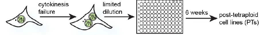
Cytokinesis failure induced by actin inhibitor dihydrocytochalasin (DCD) treatment, results in the formation of tetraploid binucleated cells with the frequency of binucleation reaching 70-80%. Estimated 600 cells for each cell line were plated by limiting dilution on 96-well plates (approximately 0.5 cell/well) and allowed to propagate for six weeks (Figure 7).
Figure 7. Generation of the PTs: a schematic depiction of the experimental strategy.
Results
Figure 8. DNA content profile of HPTs and RPTs
DNA content profile of HPTs (top row) or RPTs (bottom row) assessed by flow cytometry after
propidium-iodide staining of DNA.
This approach allowed me to obtain proliferating near-tetraploid cell populations from cells that underwent transient tetraploidization.
1.2. Cell cycle and growth characteristics of the posttetraploid
cells.
Results
Figure 9. The length of interphase and time from NEBD to OA in HCT116 and its derivatives.
(A) Time in interphase in HCT116 and HPTs measured from the anaphase onset to the next NEBD,
Tukey range and median are plotted, four experiments. (B) Time in mitosis in diploid and tetraploid
HCT116 and HPTs measured from NEBD to the anaphase onset, Tukey range and median are
plotted, four experiments. Numbers above the box and whiskers in both (A) and (B) plots indicate the
number of cells analyzed. .
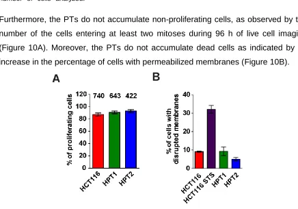
Furthermore, the PTs do not accumulate non-proliferating cells, as observed by the number of the cells entering at least two mitoses during 96 h of live cell imaging (Figure 10A). Moreover, the PTs do not accumulate dead cells as indicated by no increase in the percentage of cells with permeabilized membranes (Figure 10B).
Figure 10. Numbers of non-proliferating and dead cells in culture.
Results
Taken together, posttetraploid cells do not display prominent changes in the interphase and mitosis duration (nuclear envelope to onset of anaphase); nor they arrest and die more frequently than progenitor diploid cells.
2. Aneuploidy and chromosomal instability of the posttetraploid
cells.
2.1. Chromosome numbers in the posttetraploid cells.
Results
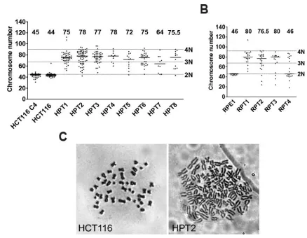
Figure 11. Chromosome numbers in HCT116, RPE1 and their posttetraploid derivatives.
Distribution of chromosome numbers and median values counted from metaphase spreads of (A)
HCT116- and (B) RPE1-derived cell lines. Dashed lines mark diploid (2N), triploid (3N) and tetraploid
(4N) values. The numbers above the scatter bars indicate the medians. (C) Representative
metaphase chromosome spreads obtained from HCT116 and HPT2.
Thus, posttetraploid cells display a numerical aneuploidy with variable karyotype compositions that are compatible with survival.
2.2. Chromosomal instability in the posttetraploid cells.
Results
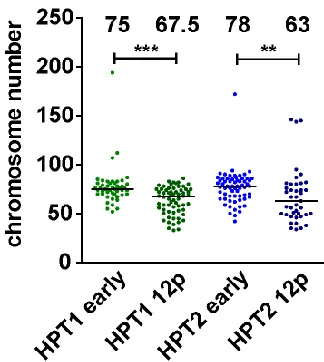
Figure 12. Chromosome number upon propagation for 12 additional passages in posttetraploid cells.
Distribution of chromosome numbers in early passages and 12 passages later (12p), median values
of two independent experiments, differences are statistically significant (Mann-Whitney test, p<0.05).
The numbers above the scatter bars indicate the medians.
Results
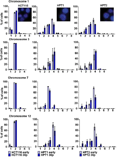
Figure 13. Chromosome numbers upon propagation in posttetraploid cells.
Comparison of chromosome number distribution of chromosomes 1, 3, 7, and 12 in early passages
and 36 passages later. Mean and SEM of two independent FISH experiments. Numbers on x-axis
indicate chromosome copy numbers. Insets: representative staining of chromosomes 1 (red) and 7
Results
Previously reported findings on unstable malignant tumors show that abnormal nuclear shapes may serve as markers of ongoing CIN (Gisselsson et al., 2001). This feature was also shared in posttetraploids: the cells displayed remarkable changes in the nuclear shape such as prominent nuclear blebbing and micronucleation (Figure 14, Figure 20).
Figure 14. Nuclear blebbing in PTs.
DNA is stained with Sytox Green. Nuclei with blebbing are marked with white arrowheads.
Results
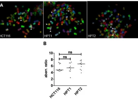
Figure 15. Total frequency of chromosomal translocations in HCT116 and its posttetraploid derivatives.
(A) Representative mFISH spreads images. Arrowheads indicate chromosome translocations. (B)
Quantification of the chromosomal structural alteration frequency in HCT116 and its posttetraploid
derivatives. Median is plotted; 10 cells were analyzed for each cell line; statistical significance was
Results
cell line translocatio n
cell ID type
of transl ocatio n 1 2 3 4 5 6 7 8 9 10
HCT116
t(17;18) 1 1 1 1 1 1 1 1 c
t(6;17) 1 s
t(10;16) 1 1 1 1 1 1 1 1 c
t(8;16) 1 1 1 1 1 1 1 1 c
HPT1
t(1;13) 1 s
t(17;18) 2 2 2 2 1 1 2 2 2 4 c
t(6;16) 1 s
t(19;22) 1 s
t(5;15) 1 s
t(2;13) 1 s
t(10;18) 1 s
t(13;18) 1 s
t(10;16) 1 2 2 1 c
t(8;16) 1 1 1 1 1 1 2 2 c
t(14;21) 1 s
HPT2
t(8;16) 2 1 1 1 1 2 4 1 3 c
t(10;16) 2 1 1 1 1 2 4 2 2 c
t(17;18) 2 1 1 1 1 2 2 3 1 c
t(14;17) 1 s
t(3;16) 1 s
t(5;8) 1 s
t(10;19) 1 s
Table 1. Frequency of translocations for individual chromosomes in HCT116 and its posttetraploid derivatives.
Types of translocations: c – constitutive, s – sporadic. Constitutive translocation was defined when
more than 50% of the cells contained at least one translocation of this type.
Results
Figure 16. Chromosome content of HCT116 2N, HPT1 and HPT2
(A) Left: HCT116 2N, right: representative chromosomes 4, 6, 19 without translocations and
chromosome translocation t(17;18), (B) HPT1 and (C) HPT2. Total of 10 cells was analyzed for each
cell line. Median and interquartile range are plotted.
Results
2.3. Chromosome segregation errors in the posttetraploids.
To identify the source of chromosomal instability in posttetraploid clones, I analyzed occurrence of chromosome segregation errors in posttetraploid cells. First, I quantified the frequency of abnormal mitosis in anaphase and telophase in fixed cells. 4.7% of all mitoses in HCT116 cells are aberrant, whereas 15.8 % and 15.0 % of bipolar anaphases in HPT1 and HPT2, respectively, displayed aberrancies such as lagging chromosomes and anaphase bridges (Figure 17).
Figure 17. Lagging chromosomes and anaphase bridges in HCT116 and its posttetraploid derivatives.
(A) Percentage of abnormal mitoses evaluated in fixed images, mean and SD of three experiments.
The numbers above the bars indicate the number of analyzed cells. (B) Examples of scored mitotic , b 10 μ
Results
Figure 18. Abnormal mitosis in HCT116 and its posttetraploid derivatives.
(A) Percentage of erroneous cell divisions evaluated from time-lapse imaging in HCT116. Mean and
SD of four experiments. The numbers above the bars indicate the number of analyzed cells. (B)
Lagging chromosomes followed by micronucleus formation, bar 10 μ . Note that mitosis was
classified as erroneous, if any defect was visible at least in one mitotic timelapse capture.
Thus, although the frequency of mitotic errors in PTs decreased in comparison to newly formed 4Ns, the frequency remains higher than in diploid HCT116, and is predominantly due to the lagging chromosomes and anaphase bridges. Frequent mitotic errors were also observed in RPT1 and RPT2, with a two-fold and four-fold increase, respectively, in comparison to RPE1 (Figure 19).
Results
Figure 19. Abnormal mitosis in RPE1 and its posttetraploid derivatives.
Percentage of erroneous cell divisions evaluated by time-lapse imaging; mean and SEM are plotted;
three experiments in RPE1 and RPT1 and two in RPT2, thus only RPT1 could be statistically
evaluated. The numbers above the bars indicatethenumberofanalyzedcells.
Figure 20. Micronucleation frequency in HCT116 and HPTs.
Percentage of interphase cells with micronuclei, median and Tukey range of seven independent
experiments. The numbers above the bars indicate the number of analyzed cells.