D
EVELOPMENT AND
V
ALIDATION OF A
NOVEL APPROACH FOR THE ANALYSIS OF
M
ARINE
B
IOTOXINS
Bing Cheng Chai
College of Engineering and Science
Victoria University
Submitted in fulfilment of the requirements of the degree of Doctor of Philosophy
ii man & M or ga n 2 01 4
Harmful algal blooms (HABs) which can produce a variety of marine biotoxins are a prevalent and growing risk to public safety. The aim of this research was to investigate, evaluate, develop and validate an analytical method for the detection and quantitation of five important groups of marine biotoxins in shellfish tissue. These groups included paralytic shellfish toxins (PST), amnesic shellfish toxins (AST), diarrheic shellfish toxins (DST), azaspiracids (AZA) and neurotoxic shellfish toxins (NST).
A novel tandem liquid chromatographic (LC) approach using hydrophilic interaction chromatography (HILIC), aqueous normal phase (ANP), reversed phase (RP) chromatography, tandem mass spectrometry (MSMS) and fluorescence spectroscopic detection (FLD) was designed and tested. During method development of the tandem LC setup, it was found that HILIC and ANP columns were unsuitable for the PSTs because of the lack of chromatographic separation power, precluding them from being used with MSMS detection. In addition, sensitivity for the PSTs at regulatory limits could not be achieved with MSMS detection, which led to a RP-FLD combination. The technique of RP-MSMS was found to be suitable for the remaining four groups of biotoxins. The final method was a combination of two RP columns coupled with FLD and MSMS detectors, with a valve switching program and injection program.
A novel sample preparation method was also developed for the extraction and clean-up of biotoxins from mussels. It was determined that Strata-X was a suitable sorbent for use in the clean-up of mussel extracts. A validation study was carried out on the developed method via analysis of certified reference materials for AZAs, DSTs and ASP, and naturally contaminated mussel material for PSTs. A major limitation to this research was the scarcity and the restrictions in obtaining and receiving biotoxin reference materials. In addition, no reference materials were available for brevetoxins. Therefore, spiking trials were conducted for brevetoxins and it was found that no recoveries could be observed, possibly due to irreversible binding to matrix components.
iii
ed
m
an
&
M
or
ga
n
2
01
4
D
ECLARATION OF
O
RIGINALITY
I, Bing Cheng Chai, declare that the Ph.D. thesis entitled “Development and Validation of a novel approach for the analysis of Marine Biotoxins” is no more than 100,000 words in length including quotes and exclusive of tables, figures, appendices, bibliography, references and footnotes. This thesis contains no material that has been submitted previously, in whole or in part, for the award of any other academic degree or diploma. Except where otherwise indicated, this thesis is my own work.
Signed:______________________________________________________________ Bing Cheng Chai
iv man
&
M
or
ga
n
2
01
4
Dedicated to my parents and to Eleanor
Whatever your hand finds to do, do it with your might, for there is no work or thought or knowledge or wisdom in Sheol, to which you are going.
— Ecclesiastes 9:10 ESV
And whatever you do, in word or deed, do everything in the name of the Lord Jesus, giving thanks to God the Father through him.
v
ed
m
an
&
M
or
ga
n
2
01
4
A
CKNOWLEDGEMENTS
Firstly, I would like to express my sincere gratitude to my supervisors at VU, Dr. Rohani Paimin for initiating this project. Also, to Dr. Nicholas Milne and Professor Stephen Bigger for their help and support throughout the writing process. I would not have been able to complete this work without your valuable contributions.
I would also like to thank those at the National Measurement Institute, Dr. Saman Buddhadasa, who provided the environment and resources without which this project would not be possible. Also to the NMI General Manager, James Roberts, and the Port Melbourne Branch Manager, Shyam Kumaran, as well as Section Manager, Timothy Stobaus, and Katherine Stockham, the Technical Development Coordinator. Their guidance and support throughout this project was crucial. Many thank also to Peter Anstis and Paul Armishaw for their help in helping me understand measurement uncertainty and their patience in walking me through the calculations.
To James Pyke and Jim Tsiotinas from Agilent Technologies, for providing the hardware and technical expertise when I had questions about chromatography and tandem LC, which brought the tandem LC setup into fruition.
To Allison Turnbull and Tom Madigan from the South Australia Research and Development Institution, for supplying the toxic shellfish material critical for the research.
vi man
&
M
or
ga
n
2
01
4
P
RESENTATIONS ARISING FROM
R
ESEARCH
Chai B.C. (2012) “Developing a more sensitive and rapid method for the detection of Paralytic Shellfish Toxins”, Australian Institute of Food Science and Technology (AIFST) Young Members’ Presentation Evening, Deakin University, Burwood, VIC, Australia.
Chai B.C., Paimin R., Stockham K. (2012) “Developing a Rapid Liquid Chromatography Tandem Mass Spectrometry (LC-MS/MS) method for the detection of Paralytic Shellfish Toxins (PST)”, 20th Royal Australian Chemical Institute Research &
Development Topics Conference, Deakin University, Geelong, VIC, Australia.
Chai B.C (2013) “Development of a HILIC-MS/MS method for the determination of Paralytic Shellfish Toxins in mussel”, 24th Conference for Residue Chemists (CRC), Victoria University, Melbourne, VIC, Australia.
Chai B.C., Paimin R., Buddhadasa S. (2013) “Marine Biotoxins” — Collaborative Research Partnerships Symposium (CRPS), Victoria University, Werribee, VIC, Australia.
Chai B.C., Paimin R., Stockham K. (2013) “Characterisation of matrix components which suppress ionisation of Paralytic Shellfish Toxins for analysis with Hydrophilic Interaction Chromatography Tandem Mass Spectrometry”, 40th International Symposium on High Performance Liquid Phase Separations and Related Techniques Hobart 2013 (HPLC 2013), Hobart, TAS, Australia.
Chai B.C. (2014) “The Trouble With Toxins”, 3 Minute Thesis (3MT) Competition, Victoria University, Footscray, VIC, Australia.
vii ed m an & M or ga n 2 01 4
T
ABLE OF
C
ONTENTS
1 GENERAL INTRODUCTION ... 11.1HARMFUL ALGAL BLOOMS ... 1
1.1.1 Factors Affecting Biotoxin Production ... 4
1.1.2 Effects on Humans ... 6
1.1.3 Impact on Animals ... 7
1.1.4 Economic Impacts ... 8
1.2CONTROL AND MONITORING OF HARMFUL ALGAL BLOOMS ... 10
1.2.1 Establishing regulatory limits ... 11
1.2.2 Emerging Toxins ... 12
1.2.2.1 Cyclic Imines ... 13
1.2.2.2 Yessotoxins and Pectenotoxins ... 13
1.2.2.3 Palytoxin ... 14
1.2.2.4 Tetrodotoxin ... 14
1.2.2.5 Ciguatera Fish Poisoning ... 14
1.3SIGNIFICANCE AND AIM OF RESEARCH ... 15
1.4THESIS ORGANISATION ... 16
2 LITERATURE REVIEW ... 17
2.1INTRODUCTION ... 17
2.2HYDROPHILIC BIOTOXINS ... 17
2.2.1 Amnesic Shellfish Toxins (AST) ... 17
2.2.1.1 Chemistry and Sources ... 18
2.2.1.2 Toxicity and Mechanism of Action ... 19
2.2.2 Paralytic Shellfish Toxins (PST) ... 20
2.2.2.1 Chemistry and Sources ... 20
2.2.2.2 Toxicity and Mechanism of Action ... 21
2.3LIPOPHILIC BIOTOXINS ... 22
2.3.1 Diarrheic Shellfish Toxins (DST) ... 22
2.3.1.1 Chemistry and Sources ... 23
2.3.1.2 Toxicity and Mechanism of Action ... 23
2.3.2 Azaspiracids (AZA) ... 24
2.3.2.1 Chemistry and Sources ... 24
viii man & M or ga n 2 01 4
2.3.3.1 Chemistry and Sources ... 26
2.3.3.1 Toxicity and Mechanism of Action ... 27
2.4METHODS OF ANALYSIS ... 27
2.4.1 Reference Materials ... 29
2.4.2 Biological Methods ... 30
2.4.2.1 Bioassay-based Methods ... 30
2.4.2.2 Immuno-based Methods ... 31
2.4.3 Analytical Methods of Detection ... 33
2.4.3.1 Hydrophilic Toxins ... 33
2.4.4 Mass Spectrometry ... 34
2.5TANDEM LC ... 37
2.5.1 Reversed Phase Chromatography ... 40
2.5.2 Hydrophilic Interaction Chromatography ... 41
2.5.3 Aqueous Normal Phase (ANP) Chromatography ... 43
2.6TOXICITY EQUIVALENCE FACTORS ... 45
2.7CONCLUSION ... 47
3 GENERAL REAGENTS AND MATERIALS ... 48
3.1INTRODUCTION ... 48
3.1.1 Chemicals and Reagents ... 48
3.1.2 Equipment ... 50
3.1.3 Shellfish Samples ... 51
4 DESIGNING THE TANDEM LC-FLD-MSMS ... 53
4.1INTRODUCTION ... 53
4.2DETERMINATION OF BIOTOXIN MULTIPLE REACTION MONITORING TRANSITIONS 54 4.2.1 Materials and Methods ... 55
4.2.2 Results and Discussion ... 56
4.3COLUMN SELECTION FOR LIPOPHILIC AND HYDROPHILIC BIOTOXINS ... 58
4.3.1 Materials and Methods ... 59
4.3.1.1 Hydrophilic Toxins ... 59
4.3.1.2 Lipophilic Toxin Chromatography ... 59
4.3.2 Results and Discussion ... 60
4.3.2.1 Hydrophilic Biotoxins ... 60
ix ed m an & M or ga n 2 01 4 4.3.2.3 Discussion ... 63
4.4FLUORESCENCE CHROMATOGRAPHY FOR PARALYTIC SHELLFISH TOXINS ... 65
4.4.1 Materials and Methods ... 65
4.4.2 Results and Discussion ... 66
4.5SETTING UP TANDEM LC SYSTEM WITH SELECTED COLUMNS AND DETECTORS .... 67
4.6COMBINED LIPOPHILIC AND HYDROPHILIC EXTRACTS ON TANDEM LC-FLD-MSMS ... 70
4.6.1 Material and Methods ... 70
4.6.2 Results and Discussion ... 71
4.7FINAL TANDEM LC-FLD-MSMSDESIGN ... 74
4.8CONCLUSION ... 77
5 COMBINED SAMPLE PREPARATION FOR HYDROPHILIC AND LIPOPHILIC MARINE BIOTOXINS ... 78
5.1INTRODUCTION ... 78
5.1.1 Biotoxin extraction methods ... 80
5.1.2 Solid Phase Extraction ... 82
5.1.3 Combined Toxin Extraction and Cleanup Method ... 85
5.2COMPARISON OF CARBON AND POLYMERIC SPE ... 87
5.2.1 Materials and Methods ... 88
5.2.2 Results and Discussion ... 88
5.2.3 Conclusion ... 90
5.3OPTIMISATION OF SPE ELUTION VOLUMES ... 90
5.3.1 Materials and Methods ... 91
5.3.2 Results and Discussion ... 91
5.3.3 Conclusion ... 92
5.4OPTIMISATION OF EXTRACTION SAMPLE MASS ... 92
5.4.1 Materials and Methods ... 93
5.4.2 Results and Discussion ... 93
5.4.3 Conclusions ... 95
5.5VERIFICATION OF STRATA-X-CWFRACTIONATION OF PSTS ... 95
5.5.1 Materials and Methods ... 95
5.5.2 Results and Discussion ... 95
5.5.3 Conclusions ... 96
x man & M or ga n 2 01 4
5.6.2 Results and Discussion ... 97
5.7FINAL SAMPLE PREPARATION METHOD ... 100
5.8PRELIMINARY VALIDATION OF SAMPLE PREPARATION METHOD ... 101
5.8.1 Preliminary Recovery Data for PSTs ... 101
5.8.2 Preliminary Recovery Data for DSTs ... 102
5.8.3 Preliminary Recovery Data for ASTs ... 102
5.8.4 Matrix Effects ... 103
5.8.5 Preliminary Recovery Data for PbTx-1 and PbTx-2 ... 104
5.8.6 Preliminary Recovery Data for AZA-CRM-Mus ... 106
5.9CONCLUSION ... 109
6 VALIDATION OF TANDEM LC-FLD-MSMS METHOD ... 110
6.1INTRODUCTION ... 110
6.1.1 Linearity ... 111
6.1.2 Selectivity ... 111
6.1.3 Limit of Detection/Quantitation ... 111
6.1.4 Repeatability ... 111
6.1.5 Measurement Uncertainty ... 112
6.2VALIDATION RESULTS ... 113
6.2.1 Linearity ... 113
6.2.2 Selectivity ... 114
6.2.3 Limit of Detection and Quantitation ... 117
6.2.4 Repeatability/Reproducibility ... 118
6.2.5 Accuracy ... 119
6.2.6 Measurement Uncertainty ... 120
6.3CONCLUSION ... 124
7 CONCLUSIONS AND FUTURE WORK ... 126
7.1CONCLUSIONS AND RECOMMENDATIONS ... 126
7.2FUTURE WORK ... 128
8 REFERENCES ... 131
APPENDICES ... 168
xi
ed
m
an
&
M
or
ga
n
2
01
4
B. SUMMARISED EXTRACTION METHOD FROM AOAC (2006) AND CLEANUP METHOD FROM HARWOOD ET AL. (2013) ... 170 C. SUMMARISED EXTRACTION AND CLEANUP METHOD FOR
LIPOPHILIC BIOTOXINS (GERSSEN ET AL., 2009A) ... 171 D. SUMMARISED EXTRACTION AND CLEANUP METHOD FOR
xii man
&
M
or
ga
n
2
01
4
L
IST OF
T
ABLES
Table 1-1 Marine biotoxin intoxication syndromes and associated organisms ... 2
Table 1-2 Biotoxin profiles of different dinoflagellates ... 6
Table 1-3 Symptoms of shellfish poisoning in humans (Munday and Reeve, 2013) ... 7
Table 1-4 Biotoxin limits set for bivalve molluscs in Australia (Australian Government, 2015) and Europe (EFSA, 2009a) ... 11
Table 2-1 Currently adopted TEF values (EFSA, 2009a) ... 46
Table 3-1 Chemical properties, logP values and supplied concentration for chemicals used in this research ... 49
Table 4-1 MRM transitions determined by LC-MSMS ... 57
Table 4-2 Flow rates, solvent ratios and valve positions for the tandem LC-FLD-MSMS system ... 68
Table 5-1 Summarised marine biotoxin extraction methods ... 82
Table 5-2 Comparison of toxin peak areas between 5 grams and 1 gram extraction mass (± 1 Std Dev), n= 3 ... 94
Table 5-3 Recoveries of several PSTs from spiking experiment (* denotes the recommendation for Recovery Correction) ... 101
Table 5-4 Recoveries for CRM-DSP-Mus ... 102
Table 5-5 Recoveries for CRM-AZA-Mus ... 106
Table 6-1 Linearity of standards ... 113
Table 6-2 Limit of Detection and Quantitation determined for marine biotoxins ... 118
Table 6-3 Repeatability and reproducibility data for AZA and PSTs (n=3) ... 119
Table 6-4 Toxin recoveries from CRMs and spiked mussel tissues... 119
xiii ed m an & M or ga n 2 01 4
L
IST OF
F
IGURES
Figure 2-1 Chemical structures of Amnesic Shellfish Toxins (FAO, 2004b) ... 18Figure 2-2 General structure of PSTs (Suarez-Isla, 2015) ... 21
Figure 2-3 Chemical structures of Okadaic Acid and Dinophysistoxins (Holmes and Teo, 2002) ... 23
Figure 2-4 Chemical structure of Azaspiracids 1-5 ... 25
Figure 2-5 Chemical structures of Brevetoxins (Turner, Higgins, et al., 2015) ... 27
Figure 2-6 Diagram of ion formation in ESI source (Cech and Enke, 2002) ... 35
Figure 2-7 Scheme of tandem mass spectrometer (Agilent Technologies, 2015) ... 37
Figure 2-8 Schematic of a tandem LC setup (Pyke et al., 2015) ... 39
Figure 2-9 Partition and adsorption mechanism in HILIC retention (Heaton and Smith, 2012) ... 42
Figure 2-10 Chemical surface composition of underivatised or bare silica (McCalley, 2007). ... 42
Figure 2-11 Functional groups of ZIC HILIC (a) and TSKgel Amide80 (b) stationary phases (Guo and Gaiki, 2011) ... 43
Figure 2-12 Chemical surface composition of silica hydride (Pesek and Matyska, 2009) ... 44
Figure 2-13 Mechanism of ANP retention (Kulsing et al., 2014) ... 45
Figure 4-1 Structure and logP values of Eprinomectin compared to some lipophilic toxins ... 55
Figure 4-2 Chromatograms of PST reference standards at 800 µg/mL on trialed columns. Elution regions are shown for C toxins (A), GTX toxins (B) and STX, dcSTX, NEO and dcNEO (C) ... 61
xiv man & M or ga n 2 01 4
(b)(Quilliam et al., 1993) ... 65
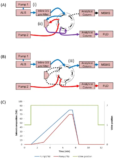
Figure 4-6 Fluorescence chromatograms of PST contaminated mussels with Poroshell column (a) and Polaris C18 column (b). Dashed lines indicate corresponding toxin peaks between the two columns. ... 66 Figure 4-7 Schematic of tandem LC –FLD-MS setup with fluorescence detector and
C18 trap ... 69 Figure 4-8 Loss of Domoic Acid when exposed to water (a), periodate oxidant (b) and
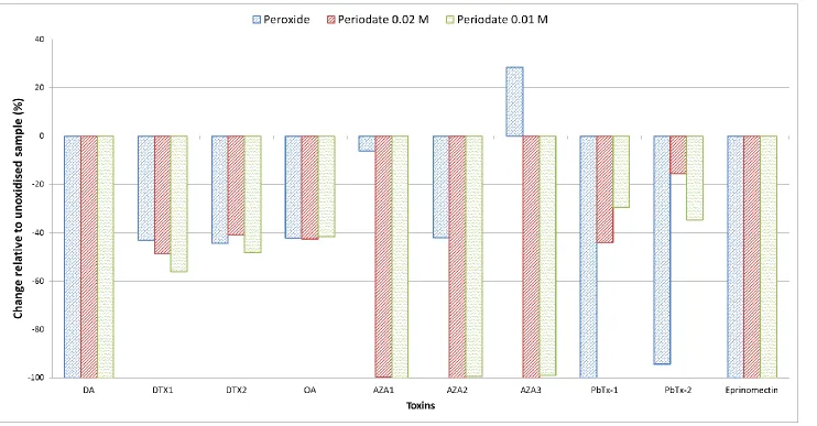
peroxide oxidant (c) ... 72 Figure 4-9 Decrease in lipophilic toxins peak areas when exposed to periodate and
peroxide oxidation agents ... 73 Figure 4-10 Scheme of final tandem LC-FLD-MS setup with inline filter replacing C18
trap ... 76 Figure 5-2 Timeline of method development process ... 79 Figure 5-1 General scheme of SPE cleanup, showing the sequence of conditioning, load,
wash and elution steps with position of analytes and matrix components at each stage (Majors, 2013) ... 84 Figure 5-3 Flowchart of shellfish tissue cleanup methods from the AOAC 2005.06
method for hydrophilic PSTs (in blue) and from Gerssen et al. (2009a) (in red) for lipophilic toxins. The proposed combined method is shown in purple. ... 87 Figure 5-4 FLD chromatograms of peroxide oxidised PST LRMs after EnviCarb SPE
(a) and Strata-X SPE (b) cleanup ... 89 Figure 5-5 Chromatogram of eprinomectin recovery from EnviCarb SPE (a) and
Strata-X SPE (b) ... 90 Figure 5-6 FLD chromatograms of six consecutive 1 mL elutions collected from
Strata-X SPE cartridge ... 92 Figure 5-7 FLD chromatograms of 5 g and 1 g PST LRM after periodate and peroxide
oxidation ... 94 Figure 5-8 Peroxide oxidation and controls of PST LRM after fractionation using
xv
ed m an & M or ga n 2 01 4 Figure 5-9 Overlaid chromatograms of 6 replicates of PST LRM extracts using regenerated Strata-X SPE cartridges ... 98
Figure 5-10 Peak areas of PSTs from 6 load, elute and regeneration cycles of Strata-X SPE cartridges ... 99
Figure 5-11 Final extraction and clean-up scheme ... 100
Figure 5-12 Change in peak area f domoic acid in different matrix concentrations 103 Figure 5-13 Peak area of brevetoxin 1 (PbTx-1) at different concentrations of matrix extract in solution ... 105
Figure 5-14 Peak area of brevetoxin 2 (PbTx-2) at different concentrations of matrix extract in solution ... 106
Figure 5-15 Peak areas of CRM-AZA-Mus at various dilution levels (average of two replicates) ... 108
Figure 6-1 MSMS and FLD chromatograms of CRM-Zero-Mus ... 115
Figure 6-2 MSMS and FLD chromatograms of CRM-DSP-Mus ... 115
Figure 6-3 MSMS and FLD chromatograms of CRM-AZA-Mus ... 116
Figure 6-4 MSMS and FLD chromatograms of PST LRM ... 117
Figure 6-5 Measurement Uncertainty charts for AST ... 121
Figure 6-6 Measurement Uncertainty charts for PSTs ... 122
Figure 6-7 Measurement Uncertainty charts for AZAs ... 122
xvi man
&
M
or
ga
n
2
01
4
L
IST OF
A
BBREVIATIONS
AOAC Association of Analytical Communities AST Amnesic Shellfish Toxins
AZA Azaspiracid
CCFFP Codex Committee on Fish and Fishery Products
CITAC Cooperation on International Traceability in Analytical Chemistry CRM Certified Reference Material
DA Domoic Acid
DTX Dinophysitoxin
EFSA European Food Safety Authority ESI Electrospray ionisation
FLD Fluorescence Detection GCB Graphitised carbon black
GUM The ISO Guide to the Expression of Uncertainty in Measurement HAB Harmful algal bloom
HPLC High Performance Liquid Chromatography ISO International Standards Organisation
LC-FLD Liquid Chromatography — Fluorescence detection LC-MSMS Liquid Chromatography — Tandem Mass Spectrometry LLE Liquid-liquid extraction
LOD Limit of detection LOQ Limit of quantitation LOR Limit of reporting
LRM Laboratory Reference Material m/z Mass to charge ratio
xvii
ed
m
an
&
M
or
ga
n
2
01
4
MRM Multiple Reaction Monitoring MSMS Tandem Mass Spectrometry NMI National Measurement Institute NRCC National Research Council Canada NST Neurotoxic Shellfish Toxins
OA Okadaic Acid
PbTx Brevetoxin
PP Polypropylene
PST Paralytic Shellfish Toxins PVDF Polyvinylidene Difluoride
QC Quality Control
RM Reference Material
RSD Relative standard deviation
SARDI South Australian Research and Development Institute SD Standard deviation
SPE Solid Phase Extraction SSR Sample to Solvent Ratio
STX Saxitoxin
xviii man
&
M
or
ga
n
2
01
4
L
IST OF
A
PPENDICES
A. Sample injection program for Tandem LC-FLD-MSMS ... 169 B. Summarised extraction method from AOAC (2006) and cleanup method from
Harwood et al. (2013) ... 170
C. Summarised extraction and cleanup method for Lipophilic biotoxins (Gerssen et al., 2009a) ... 171
1
G
ENERAL
I
NTRODUCTION
1.1
Harmful Algal Blooms
Cyanobacteria, dinoflagellates, algae and diatoms are small aquatic organisms which are found globally, from freshwater lakes to temperate oceans. Collectively, these organisms can be grouped as phytoplankton. Of the phytoplanktons, 300 marine species are able to multiply rapidly to form dense biomasses known as Harmful Algal Blooms (HABs)(Daneshian et al., 2013; Gerssen et al., 2010a). HABs can cause mass fish deaths either through oxygen depletion or by physically clogging gills, but the greatest risk to fauna is presented by toxic secondary metabolites produced by phytoplankton. These biotoxins may affect the liver (hepatotoxins), nervous system (neurotoxins), and skin (dermatoxins)(Zanchett and Oliveira-Filho, 2013). In the temperate latitudes of Europe, South Africa, Asia, Australia, North America and South America, toxin-producing species of HABs can cause amnesic, azaspiracid, diarrhetic, neurotoxic and paralytic shellfish poisonings (Berdalet et al., 2015). Thus, HABs pose a significant risk to public health, recreation, tourism, aquaculture and marine ecosystems due to their adverse effects on the environment, which threatens water quality, health of living resources and economies of nearby populations (McLean and Sinclair, 2012).
Table 1-1 Marine biotoxin intoxication syndromes and associated organisms
Toxic syndrome Toxins Producer species References
Paralytic Shellfish
Poisoning Saxitoxins and analogs
Alexandrium, Gymnodinium, Pyrodinium bahamense var. compressum, Cyanobacteriaspecies(Lyngbya, Anabaena, Cylindrospermopsis, Aphanizomenon, Phlanktothrix)
(Wiese, 2010) (Amade, 2014)
Amnesic Shellfish
Poisoning Domoic Acid and analogs
Pseudo-nitzschia spp.,Red algae such as Chondria armata,
Digenea simplex, and Alsidium corallinum (Lefebvre, 2010)
Diarrhetic Shellfish Poisoning
Okadaic Acid,
Dinophysistoxins Dinophysis spp., Prorocentrum (Valdiglesias, 2013)
Azaspiracid Poisoning Azaspiracids and analogs Azadinium spp., Protoperidinium spp. (Kilcoyne, 2014)
(Kalaitzis ,2010)
Neurotoxic Shellfish
example, Paralytic Shellfish Toxins (PSTs) are neurotoxins which can cause paralysis, while brevetoxins are termed because they are produced by the diatom Karenia brevis. Some species of the diatom Pseudo-nitzschia are capable of producing the biotoxin responsible for Amnesic Shellfish Poisoning (ASP). In freshwater, several species of cyanobacteria are also known to produce PSTs (Wörmer et al., 2011). Crustaceans, shellfish and fish that feed on these phytoplankton accumulate biotoxins and transmitting them to humans and animals further up the food chain (Farre et al., 2013). They can also be transmitted through the ingestion of contaminated water (Batoréu et al., 2005).
In recent decades, an increase in toxic bloom events has been observed. Analysis of HAB trends in the North American region has revealed that PST-producing dinoflagellates and AST-producing diatoms have been observed in varying intensities and frequencies along the west coast of Canada, USA, and Mexico (Lewitus et al., 2012). In Turkey, more frequent observances of AST contamination have been reported (Dursun et al., 2015), while in Ireland, there have been increasing closures of shellfish harvesting sites due to AZAs and DSPs found over the permitted levels (James et al., 2002; Kilcoyne, 2015). The occurrence of HAB-forming organisms usually suited to milder waters have also been observed in waters off Norway (Edwards et al., 2006). As a result, phytoplankton and their associated biotoxins are also increasingly being found in places with no record of HABs: In Iceland, the north coast of Eyjafjordur and west coast of Breidafjordur experienced a bloom of Alexandrium spp. in June 2009, contaminating blue mussels with paralytic shellfish toxins (PSTs), which led to extensive closures of harvesting sites, the first biotoxin related closure reported in Iceland (Burrell et al., 2013). It was recently reported that oyster harvests were closed for the first time in the Gulf of Mexico due to confirmed presence of okadaic acid (DSTs) (Deeds et al., 2010). Similarly, first reports have been published of AST detected in abalone species off the southern coastline of Australia (Malhi et al., 2014).
Smayda, 2007). Climate change has also been cited as one of the major drivers of the increase in HABs (Silva et al., 2015). Increasing sea surface temperatures have also been linked to the increase in dinoflagellate populations in the north-east Atlantic, where they outcompete native diatom populations (Edwards and Richardson, 2004). Warmer sea surface temperatures and water stratification increases the growth rate of dinoflagellates, and nutrient depletion at the surface favours the survival of dinoflagellates (Bopp et al., 2005). Climate models predict more frequent blooms of biotoxin producing dinoflagellates (van der Fels-Klerx et al., 2012), leading to an increase in the window of opportunity for blooms, with earlier and more persistent blooms (Moore et al., 2011).
1.1.1
Factors Affecting Biotoxin Production
Biotoxin production by phytoplankton is affected by environmental and genetic factors (Pistocchi, 2014). Nutrient availability factors such as iron content in water, environmental parameters such as irradiance, temperature, salinity or inorganic nutrients have been shown to affect biotoxin content and composition for several different
Alexandrium strains (Etheridge and Roesler, 2005; He et al., 2010). Relationships between photosynthesis or growth rate and total toxicity were not found, suggesting that environmental factors directly influence toxicity.
Biotoxin production can also be a survival response to presence of predator signalling compounds: In response to zooplankton lipids, Selander et al. (2015) have found that some Alexandrium species become 20 times more toxic. Holland and Kinnear (2013) have proposed that biotoxins acts as a defense mechanism against predators and also as a physiological aid, participating in nutrient absorption pathways.
Apart from extreme nutrient deprivation conditions that may cause a shift in the biotoxin profile, isolates in exponential growth phase tend to maintain their molecular fingerprint of biotoxins in culture. This makes it possible to compare biotoxin profiles from Alexandrium species and strains in long-term cultures, even if the isolates were not collected at the same time. These biotoxin profiles have been proposed to be used to trace the source organisms in biotoxin outbreaks (Wong et al., 2011). However, significant changes in biotoxin composition have been reported in cells exposed to different stresses (Etheridge and Roesler, 2005; Poulton et al., 2005). Nevertheless, diverse biotoxin profiles have been observed in different phytoplankton populations globally.
In Australia, HAB species that present a risk to shellfish include Pseudo-nistzchia
Table 1-2 Biotoxin profiles of different dinoflagellates
Organism Dominant Toxins References
G. catenatum GTX6, C3, C4 Costa et al., 2014
Alexandrium spp. GTX1, GTX4 Lefebvre et al., 2008
Alexandrium spp. STX, NEO, GTX2, GTX3 Etheridge et al., 2005
A. minutum GTX2, GTX3, GTX1, GTX4, STX Abouabdellah et al., 2008
A catenella C2, GTX2, GTX3, dcGTX2 Alvarez et al., 2009, Krock et al., 2007
A tamarense, A
ostenfeldii GTX2, GTX3, STX Burrell et al., 2013 Alexandrium
tamarense/catenella
C12, GTX1, GTX4, GTX2, GTX3,
NEO Montoya et al., 2010
Alexandrium tamarense C2, GTX4 Kim and Shin, 2015
There is a lag time between peak of the bloom and toxicity maximum. For example, the maximum toxicity of mussels was measured 13 days after peak cell counts of a bloom of Gymnodinium catenatum (Costa et al., 2014). Quantification becomes particularly important during this period since the highest risk of human poisoning occurs at this stage. After the peak of the bloom, a gradual decrease of C3 & C4 and GTX6 concentration, suggesting a depuration process in mussels.
1.1.2
Effects on Humans
when the wave action near beaches break up cells of brevetoxin-producing diatoms (Backer et al., 2003; Fleming et al., 2009; Pierce et al., 2003). Exposure to aerosols containing brevetoxins have been linked with aggravated asthma symptoms (Bean et al., 2011; Kirkpatrick et al., 2011).
Table 1-3 Symptoms of shellfish poisoning in humans (Munday and Reeve, 2013)
Toxin Reported effects in humans
Paralytic Shellfish Toxins Nausea, paresthesia, tachycardia, muscular paralysis, respiratory failure, death
Amnesic Shellfish Toxins Vomiting, diarrhoea, abdominal pain, confusion, memory loss, seizure, coma, death
Diarrhetic Shellfish Toxins Nausea, vomiting, diarrhoea, abdominal pain
Azaspiracids Nausea, vomiting, diarrhoea, abdominal pain
Brevetoxins
Nausea, vomiting, diarrhoea, chill, sweating,
dysaesthesia, hypotension, paresthesia of lips, face and extremities, cramps, paralysis, seizures and coma after ingestion. Rhinorrhoea, cough, bronchoconstriction after inhalation
1.1.3
Impact on Animals
Substantial mortalities of aquacultured Atlantic salmon at two sites in the Bay of Fundy (New Brunswick, Canada) in September 2003 were associated with a bloom of
Alexandrium fundyense, a PST producing dinoflagellate. The zooplankton sampled contained PSTs matching the profile of blooming A. fundyense cells (Sephton et al., 2007).
In the Portuguese coastal region, sardines are the most abundant planktivorous fish and a major component of the marine food web. Sardines consume phytoplankton, which include G. catenatum. The PST profile characterised in sardine samples in a study conducted by Costa et al. (2010) showed same sulfocarbamoyl and decarbamoyl toxins found in the consumed algae with minor differences in relative abundance of each biotoxin.
Intracellular biotoxins can be released into the environment via excretion or lysis of phytoplankton cells, posing another route of exposure via direct absorption from the water by aquatic animals. Studies conducted by Lefebvre (2008) have confirmed that the ingestion of PSTs via algal or zooplankton vectors is a route of PST exposure causing acute toxicity in adult and larval fish during toxic blooms. Extracellular STX exposure has been shown to impair the physiology and behaviour of developing fish larvae, causing a complete loss of sensorimotor function, caused delayed hatching and malformations and mortalities in zebrafish larvae (Lefebvre, 2008). However, the stability of extracellular PSTs in water may also be affected by upwelling events or river plumes and may not persist long at seawater pH unless stabilisation is achieved by complexation with other substances (Rue and Bruland, 2001).
1.1.4
Economic Impacts
aquaculture practices caused a voluntary export ban to be enforced until internationally recognised shellfish sanitation programs were established (Chalermwat et al., 2003).
The growing HAB threat will affect countries with large aquaculture industries such as Iceland, where marine products accounted for 42% of total export value in 2009 (Burrell
et al., 2013). New Zealand also has a large aquaculture industry valued at NZ$1.5 billion in 2010 (Rhodes et al., 2013). Losses to local businesses such as shellfish farms, beach businesses, and other seafood related industries from one local PSP outbreak in Maine, USA were estimated at US$6 million per year (Boesch et al., 1997). In Malaysia, fish die-offs due to HAB was estimated to cause RM$20 million loss in one incident (Lim et al., 2012). In Australia, a PST contamination of Tasmanian shellfish during an unexpected HAB event was estimated to have a total financial impact of AU$23 million across the commercial fisheries and marine farming sector (Campbell et al., 2013).
In addition, the cost of operating a monitoring program is also significant: In America, most states operate their own monitoring program, which cost up to US$200,000 annually. In Australia, shellfish safety monitoring is operated by each state, guided by a federal framework. States which have a large shellfish and aquaculture industry such as New South Wales, South Australia, Tasmania, and Victoria have larger operating costs (Ajani et al., 2012; FRDC, 2011). The total costs of managing cyanobacterial risks were estimated to cost AUD$180-240 million annually, although this figure includes costs associated with joint management, urban and rural extractive users and non extractive users of Australian water resources (Steffensen, 2008).
1.2
Control and Monitoring of Harmful Algal Blooms
Many of the biotoxins which have been identified in the past half century are now monitored with various chemical, biochemical or molecular-based methods (Moreira et al., 2014). Due to monitoring programs being implemented by governments around the world, a decrease in the number of cases of poisoning has been observed (Bean et al., 2005). A report by Hallegreaeff (2014) places the fatality rate of poisoning cases at 15% (300 out of 2000 cases reported annually worldwide).
New tools have been developed to aid in monitoring the conditions of water and likelihood of toxic HABs. One of them is Solid Phase Adsorbent and Toxin Tracking (SPATTs) which are passive bags of adsorbent material which are left in open water for a time to adsorb toxins and can be analysed by chemical methods (Fux, 2008; McCarthy
et al., 2014; Zendong et al., 2014).
Additionally, measures can be implemented to minimise human exposure to these chemicals. One technique involves removal of HAB or toxins from water bodies when they occur. This can be done by application of modified clay to blooms to flocculate algae in water (Lu et al., 2015), which was found to reduce one group of PST (gonyautoxins) by 82% and simultaneously reduce phosphate and nitrate concentration, which are macronutrients needed for phytoplankton growth. This method has been reported to have some success in China (Pan et al., 2011) and Malaysia (Lim et al., 2012). More recently, removal of PSTs via probiotic lactic acid bacteria (Lactobacillus rhamnosus GG and LC-705) have been reported by Vasama et al. (2014). The mechanism of this removal is thought to be a binding of biotoxins to components of the bacteria, as no differences were found between viable and non-viable forms of the bacteria. This finding is a positive initial result for management of biotoxin levels in water bodies.
The driving force for progress in the area of marine biotoxins monitoring and control is the fact that there are no current known antidotes to marine biotoxin intoxication (Silva
all monitoring programs are measurement techniques to determine risk and toxicity. Monitoring algal cell counts in water is an indirect measurement of toxicity risk as a high algal cell count may not necessarily mean that shellfish are toxic. Direct measurement of shellfish tissue remains a critical tool in ensuring edible shellfish safety.
1.2.1
Establishing regulatory limits
In Australia, the Imported Foods Inspection Scheme (IFIS) is a food inspection program overseen by the Department of Agriculture. Its purpose is to monitor imported food for compliance with Australian food reference standards. Bivalve molluscs such as mussels and oysters are categorised as risk foods. This means that it has been assessed as having medium to high risk to consumer health (Imported Food, 2015). Limits of biotoxin content have been established to protect human health. Australia follows international guidance in setting regulatory limits for marine biotoxins (Table 1-4). However, there are some differences in the levels established by Australia and Europe.
The maximum allowed limits of phycotoxins are established based on data derived from past poisoning incidents (FAO, 2004a). Toxicology data from animal trials are also used to determine toxicity and are used to revise as more information is gathered. Risk assessments take into account epidemiological data such as consumption frequencies, portion size variations between populations, and toxicological information. Using this data, limits are also set designed to provide covers up to the 97.5th percentile of global population (Toyofuku, 2006).
Table 1-4 Biotoxin limits set for bivalve molluscs in Australia (Australian Government, 2015) and Europe (EFSA, 2009a)
Toxin Legislated toxins Australian Limits European Limits Domoic
Acid DA and analogs 20 mg DA/kg 20 mg DA/kg
STX STX and analogs 0.8 mg STX equivalents/kg
Toxin Legislated toxins Australian Limits European Limits
DST OA, DTX1/2/3 0.2 mg OA
equivalents/kg
0.16 mg OA equivalents/kg
NST Brevetoxin 1/2 and derivatives thereof
0.8 mg BTX2
equivalents/kg Not Regulated
AZAs Azaspiracid 1/2/3 Not Regulated 0.16 mg AZA1 equivalents/kg
The current regulatory limit for PSTs was established in the 1930s based on experiments on mice. The initial results of PST testing were expressed in terms of Mouse Units (MU), one unit being the amount of total biotoxin that killed a 20 g mouse within 15 min (EFSA, 2009b). As chemical methods became viable and reference standards of PSTs became available, the relationship between MU and toxin amount was determined: 1 MU was found to be equivalent to 0.2 µg of STX (Wekell et al., 2004). This relationship was used to convert the mouse units from mouse bioassay into microgram equivalents. The mouse bioassay has a detection limit of 200 MU, which is 40 µg STX eq/100 g shellfish. The 80 µg STX eq/100 g shellfish is thought to have originated from Californian authorities who instituted quarantine measures when 2 mg of shellfish extract contained 2 MU (Wekell et al., 2004).
Amnesic shellfish poisoning (ASP) caused by domoic acid and its isomers is a relatively newer syndrome, first being observed in 1987 in Prince Edward Island, Canada. After this episode, two workshops held in Ottawa, Canada and California, USA established the current regulatory limit of 20 mg/kg based on analysis of uneaten mussels recovered from the outbreaks and symptoms exhibited by victims (Wekell et al., 2004).
1.2.2
Emerging Toxins
(FAO, 2004a; Paredes et al., 2011; Picot et al., 2011; van Egmond, 2004). Risk assessments have yet to be done on many groups of toxins, which include hazard monitoring of HAB species, risk assessment and management of biotoxins in foods and non-foods, and registration of analytically-verified intoxications in humans (Daneshian
et al., 2013). In addition to the major groups of monitored marine biotoxins discussed above, there are also compounds that have recently been identified but have not yet reached the stage of having quantitative limits placed on them, due to a lack of toxicological data or no established link between illness and exposure to these emerging toxins e.g. cyclic amines, the yessotoxins and pectenotoxins, palytoxins and tetrodotoxin.
1.2.2.1 Cyclic Imines
Cyclic imines (CI) are a newly-described family of structurally related marine toxins (Silva et al., 2015). Within the cyclic imine group are spirolides, gymnodimines, pinnatoxins, and pteriatoxins. The main common feature of members in this group is the presence of an imine moiety as part of a bicyclic ring system. Some organisms that produce cyclic imines also produce other marine biotoxins, such as Karenia (NSTs),
Alexandrium and Gymnodinium (PSTs). Cyclic imine intoxication leads to neurological symptoms in mice (Paredes et al., 2011). To date, human intoxications have not been reported (McNabb et al., 2012).
1.2.2.2 Yessotoxins and Pectenotoxins
1.2.2.3 Palytoxin
Palytoxins (PlTx) are a class of potent non-protein marine toxins, whose main biological target is the biological mechanism which maintains cellular ionic concentrations critical to normal cell functions (Ciminiello et al., 2010). Originally isolated from the marine zoanthid Palythoa, it was subsequently found in dinoflagellates of the genus Ostreopsis (Lenoir et al., 2004). Analogs of palytoxin have also been identified: ovatoxins, ostreocins, and mascarenotoxin. Mouse studies have provided evidence of lower oral toxicity compared to intravenous or intraperitoneal injection (Ciminiello et al., 2010). Electrospray ionization-tandem mass spectrometry (ESI-MS/MS) shows great potential for rapid and sensitive identification of marine biotoxins in contaminated material (Ciminiello et al., 2010).
1.2.2.4 Tetrodotoxin
Tetrodotoxin (TTX) is a well-known toxin occurring in pufferfish (Jang and Yotsu-Yamashita, 2006). They have a similar mode of action to PSTs, binding to the sodium channel in nerves and blocking normal mode of signal transfer. The source of TTX in pufferfish has been found to be endosymbiotic bacteria which naturally inhabit the gut of pufferfish (Bane et al., 2014). Increasingly, TTX has been found outside of Asia in gastropods and fish (McNabb et al., 2014). Although saxitoxin has been found in pufferfish flesh (Landsberg et al., 2006), TTX has not been reported in shellfish. Consequently, TTX is only a concern in fugu or pufferfish and there are no limits established for shellfish.
1.2.2.5 Ciguatera Fish Poisoning
finfish are currently determined by mouse bioassay. Although it is a validated method, the tests have to be performed on a per-fish basis. Unlike shellfish, fish are mobile and ciguatera precursors undergo a complex biotransformation before final ciguatoxin formation and accumulation in predator finfish.
1.3
Significance and Aim of Research
There is a limited expertise in marine biotoxin analysis due to difficulties in obtaining reference materials and the costs associated with operating test methods. Currently there is only one major supplier of marine biotoxin reference materials and reference standards for global testing bodies. These limitations prevent progress in developing efficient and effective techniques to support food security and comply with regulations for public safety. Global trade will also benefit by having more efficient closing and opening of fisheries and harvesting sites, reducing the economic impact of HABs globally. Although there are methods that are validated for lipophilic and hydrophilic toxins, none are able to analyse both classes of toxins simultaneously. Therefore, there is a need for an improved and rapid analytical methods to respond to increasing threats of HABs.
The novel tandem LC setup used by Pyke et al. (2015) for metabolomics studies has been demonstrated to be able to separate both hydrophilic and lipophilic compounds for analysis by MSMS in a short amount of time. This tandem LC setup shows great potential for application to the analysis of marine biotoxins as they may also suitable for MS detection.
Based on the gaps highlighted above, the aim of this research is to develop a tandem LC method for the simultaneous detection and quantification of PSTs, ASTs, DSTs, NSTs and AZAs, with a focus on the ability to detect and quantify these five major groups at levels relevant to current regulatory limits.
To achieve this aim, a three phase approach was taken:
instrument configuration and integration with traditional Reversed Phase (RP) chromatography.
2. Development and optimisation of a new extraction method for the extraction of both lipophilic and hydrophilic toxins from shellfish tissue.
3. Validation of the novel tandem LC method and assessment of suitability for routine monitoring of marine biotoxins.
1.4
Thesis Organisation
2
L
ITERATURE
R
EVIEW
2.1
Introduction
The aim of this chapter is to give more detailed information on the chemistries of the five groups of marine biotoxins studied in this research project. Following that, methods for the analysis of these biotoxins will be summarised and the complexities in analysis of these compounds will be discussed, along with progress made in recent years.
2.2
Hydrophilic Biotoxins
2.2.1
Amnesic Shellfish Toxins (AST)
2.2.1.1 Chemistry and Sources
Domoic acid is a water-soluble cyclic amino acid (Figure 2-1). It belongs to the kainoid class of compounds, and has been isolated from several red algae species and a number of diatom species (FAO, 2004b). More than 10 species of diatoms are known to produce this toxin, mostly from the genus Pseudonitzschia (Lefebvre and Robertson, 2010). To date, several isomers of DA have also been identified (Tasker, 2014). Isodomoic acid A, B, and C were identified as minor constituents in the red alga Chondria armata. Other known isomers of domoic acid are isodomoic acid A–H and 5’-epi-Domoic acid (Figure 2-1). Romero et al. (2011) have shown that AST composition is not dependent on geographical latitude, but is a characteristic of diatom strain and sub-strain. For example, strains of the diatom Nitzschia navis-varingica collected in the Philippines were found to contain mostly isodomoic acid B while those collected from Indonesia and Japan contained mostly domoic acid. It was previously thought that isodomic acid B would be in higher proportion at lower latitudes.
2.2.1.2 Toxicity and Mechanism of Action
Amnesic Shellfish Poisoning manifests itself via gastrointestinal (vomiting, diarrhoea, and abdominal cramps) and neurological effects (headaches and short-term memory loss). Domoic acid is a glutamate receptor activator, specifically targeting ionotropic glutamate receptors (Pulido, 2014). These receptors form cation-specific ion channels and regulate fast excitatory transmission in the central nervous system. In the hippocampus (the region associated with memory), domoic acid exposure can lead to amnesia and memory loss issues (Lefebvre and Robertson, 2010).
The toxicity of isodomoic acid A (IA), Isodomoic acid B (IB), and Isodomoic acid C (IC) is reported to be significantly lower than DA alone. In mouse studies conducted by Munday et al., (2008), intraperitoneal administration of the toxins found LD50 of DA at
6.0 mg/kg while no deaths occurred for IA, IB and IC at the same dosage. IC was also dosed at 20 mg/kg with no deaths observed. These isomers are not included in sample toxicity tests due to their low presence compared to domoic acid. Nevertheless, because of structural similarities of these isomers, there is a possibility that marine animals such as shellfish, fish and mammals may act as vectors of the toxin isomers, which could potentially be converted from IB to IA and finally to DA in the animal tissue via
enzymes or other chemical mechanisms, although there is no report showing these bioconversions in the animal tissue.
2.2.2
Paralytic Shellfish Toxins (PST)
PSTs cause paralytic shellfish poisoning (PSP). They are a class of biotoxins that bind to sodium channels within the cell, disrupting normal cell signaling pathways. The disruption of cell signaling pathways may lead to nausea, tingling sensations around the lips and fingers, paralysis, and potentially death. These toxins are produced by both prokaryotes and eukaryotes (Orr et al., 2013).
PSTs were originally detected on the Pacific Coast of the United States in 1937 and described by Schantz and Magnussen (1961). Since then, PSTs have been found in many other locations. This is partially due to the progressive implementation of monitoring programs, which have grown in parallel with the development of shellfish aquaculture, but also perhaps due to a true increase in the frequency of toxic outbreaks (Álvarez et al., 2009).
2.2.2.1 Chemistry and Sources
Dinoflagellates from the genus Alexandrium, Gymnodinium, and Pyrodinium have been identified as PST producers (Shumway et al., 2003). Several genera of cyanobacteria are also known to produce this toxin (Lyngbya, Anabaena, Cylindrospermopsis,
Aphanizomenon, and Phlanktothrix)(Amade et al., 2014).
Fifty-seven analogs of PST have been identified to date (Wiese et al., 2010). These biotoxins have a common 3,4,6-trialkyltetrahydropurine structure and the following subgroups are formed by modifications of the four R-group side chains (Figure 2-2): These analogs fall under the following subgroups: N-sulfocarbamoyl (C1-4, GTX5 and GTX6), carbamate (GTX1-4, STX, NEO), decarbamoyl (dcGTX1-4, dcSTX, dcNEO), Deoxydecarbamoyl (doGTX2,3, doSTX), benzoate (GC1-6; GC1a-6a; GC1b-6b), C11-hydroxy (M1-5), Angola (A-D), and Lyngbya (LWT1-6)(Humpage et al., 2010). The recently identified benzoate analogs have been found to have a slight lipophilic property (Baker et al., 2003).
+2)(Dell’Aversano et al., 2005). This zwitterionic property of the PSTs has been used for separation and isolation of PSTs toxins from a sample based on cationic exchange mechanisms (Turner et al., 2009).
Figure 2-2 General structure of PSTs (Suarez-Isla, 2015)
2.2.2.2 Toxicity and Mechanism of Action
PSTs are neurotoxins, binding irreversibly with Site IV of the sodium channel in cells, which prevent action potentials from propagating along nerves and leads to paralysis (Mattei and Legros, 2014). They are also known to interact with calcium and potassium channel (Cusick and Sayler, 2013). Saxitoxin is one of the most potent naturally-occurring toxins known, having a fatal dose of 1 mg for a standard human model weight of 70 kg (Wiese et al., 2010). However, other analogs of saxitoxin have different toxicities due to structural differences which directly influence its binding ability to sodium channel receptors. PST exposure also triggers the oxidative stress response in animals, exacerbating its toxic effect (Ramos et al., 2014). PST exposure studies in cats have suggested that these toxins move freely between the extracellular and intercellular space (Andrinolo et al., 2002) and also the blood-brain barrier (Cianca et al. 2007), which may explain why PST intoxication is so rapid.
2011). However, other studies have shown no effects of pH on toxin content (Holland and Kinnear, 2013): STX is a known sodium channel blocker, which slows down Na+ uptake in highly saline environments. Ion channels are not only found in nerve cells, but are also used for motility and nutrient uptake in simple organisms.
Conversion of N-sulfocarbamoyl toxins such as C1/C2 to the corresponding carbamoyl analogues (GTX2/GTX3) can occur in shellfish tissues due the activity of glutathione-S-transferase enzymes (Nogueira et al., 2004). These mechanisms are yet to be fully elucidated. These conversions have been found to occur spontaneously in response to physicochemical factors such as pH (Krock et al., 2007). PSTs are highly water soluble and degrades rapidly in alkaline solutions (Stewart and McLeod, 2014).
The current regulatory limit for PSTs in shellfish is 800 µg STX.diHCl equivalents/kg. (EFSA, 2009a). “DiHCl” refers to the dichloride salt form of saxitoxin, which includes two chlorine ions.
2.3
Lipophilic Biotoxins
For this research, three lipophilic marine biotoxin groups were studied: Diarrheic Shellfish Toxins (DSTs), Azaspiracids (AZAs), and Neurotoxic Shellfish Toxins (NSTs).
2.3.1
Diarrheic Shellfish Toxins (DST)
The DST group consists of okadaic acid and dinophysistoxins (DTXs). OA was originally identified in black sponges, but later traced to Prorocentrum and Dinophysis
2.3.1.1 Chemistry and Sources
They are a group of lipophilic heat soluble polyethers (Sosa and Tubaro, 2015). DTX1 is the methylated derivative of OA while DTX2 is an isomer of OA (Louzao et al., 2015). DTX3 describes a group of toxins with OA modified at the R1 group with long chain fatty acids (Figure 2-3).
Figure 2-3 Chemical structures of Okadaic Acid and Dinophysistoxins (Holmes and Teo, 2002)
2.3.1.2 Toxicity and Mechanism of Action
Okadaic acid and its analogs are specific inhibitors of serine/threonine protein phosphatase 1 and 2A, binding to a hydrophobic groove near the active sites of these enzymes (Sosa and Tubaro, 2015). Severe effects on the gastrointestinal system are characteristic of DST poisoning: Diarrhoea, nausea, vomiting, abdominal pain, and chills can appear between 30 mins to 4 hours after ingestion of DSTs (Valdiglesias et al., 2013). However, full recovery usually occurs within three days (FAO, 2004c). This may lead to underreporting of poisoning incidents as the symptoms may be confused with indigestion or food spoilage.
neuroblastoma cells (Louzao et al., 2015). Neuropeptide Y inhibits gastrointestinal motility and water and electrolyte secretion throughout the intestine, and interactions with OA may trigger metabolic pathways which cause diarrhoea invivo.
New toxicological data published by Louzao et al. (2015) have shown that DTX1 is more likely to disrupt and pass through the intestinal monolayer, which increases its toxicity compared to OA. In light of this information, there may be a need to revise the Toxicity Equivalence Factor (TEF) for DTX1. TEFs will be further discussed in Section Error! Reference source not found.. The current regulatory limit for DST in shellfish is 160 µg OA equivalents/kg (EFSA, 2009a).
2.3.2
Azaspiracids (AZA)
Azaspiracids (AZAs) are polyether marine toxins that have been associated with severe gastrointestinal intoxications. It was first associated with human intoxications in 1995 after several people in the Netherlands consumed Irish mussels from Killary Harbour (James, Moroney, et al., 2003). Azaspiracids were originally grouped together with the DSTs due to similar symptoms, but it was subsequently recognised as unique from DSTs (Twiner, 2008).
2.3.2.1 Chemistry and Sources
AZAs are known to be produced by marine dinoflagellates from the genera Azadinium
and Amphidoma (Hess et al., 2015) and are so called due to their chemical structure: a cyclic amine (Aza group), a unique tri-spiro-assembly and a carboxylic acid group (Twiner, 2008). Shortly after determination of the first azaspiracid toxin (AZA1), four additional analogs were discovered, AZA2 and AZA3 differing only in the methyl group positions (Figure 2-4), while AZA4 and AZA5 are hydroxyl analogs of AZA3 (Hess et al., 2015). The carboxylic acid group and cyclic imine structure within AZAs cause the toxin to be ionised over the whole pH range, which gives it a slight water-soluble property.
Twiner, et al., 2015; Krock et al., 2012; Rehmann et al., 2008). However, the toxicities of these now analogs are unknown and therefore not considered when testing for toxicity of samples. Analytical reference standards are currently available only for AZA 1, 2 and 3.
AZAs have been shown to depurate only slowly in shellfish, which could be due to toxin-binding proteins (Twiner et al., 2012). Shellfish are also known to transform AZAs via hydroxylation and carboxylation reactions, possibly facilitated by enzymes (Rehmann et al., 2008).
Figure 2-4 Chemical structure of Azaspiracids 1-5
2.3.2.2 Toxicity and Mechanism of Action
greater possibilities for interaction of AZA with its biological target, and slow down elimination from the body. AZAs have also been found to bind to proteins, which gives it protection against extreme pH conditions. However, when exposed to simulated human conditions, the amount of AZAs observed increased. This implies that after ingestion of contaminated shellfish, AZAs can become more toxic in the gut after ingestion of shellfish (Alfonso et al., 2008)
The current regulatory limit for AZAs in shellfish is 160 µg AZA1 equivalents/kg (EFSA, 2009a).
2.3.3
Neurotoxic Shellfish Toxins (NST)
Exposure to brevetoxins causes Neurotoxic Shellfish Poisoning (NSP). These toxins have been detected in cockles, mussels and oysters (Ishida, Nozawa, Nukaya, et al., 2004), and persist in sediments and the community of small organisms around seagrass (Hitchcock et al., 2012).
2.3.3.1 Chemistry and Sources
Brevetoxins are a group of lipophilic polyether toxins produced by the dinoflagellate species Karenia brevis. Two structural backbones (Type A and B) are the basis of all known analogs of brevetoxin. Brevetoxins 1, 7 and 10 have the Type A backbone while Brevetoxins 2, 3, 5, 6, 8, and 9 have the Type B backbone. Figure 2-5 shows one example of Type A and Type B brevetoxins. Upon ingestion by shellfish, brevetoxins can undergo transformation, such as transformation of Brevetoxin 5 to Brevetoxin B5 (Ishida, Nozawa, Hamano, et al., 2004). Brevisulcatic toxins and Karenia brevisculata
Figure 2-5 Chemical structures of Brevetoxins (Turner, Higgins, et al., 2015)
2.3.3.1 Toxicity and Mechanism of Action
Brevetoxins target Site V of the α-subunit of voltage-gated sodium channels, but unlike other neurotoxins like saxitoxin and tetrodotoxin, brevetoxins are channel activators. Although it is called Neurotoxic Shellfish Poisoning, intoxication also produces gastrointestinal symptoms along with neurological (abdominal pain, vomitting, nausea, ataxia, paresthesia, reversal of temperature sensation)(Plakas and Dickey, 2010). In toxicity studies on mice, intraperitoneal injection of brevetoxins, abdominal breathing, along with elevated respiration rates were observed.
The current regulatory limit set for NSTs in shellfish is 800 mg PbTX-2 equivalents/kg (Brovedani et al., 2015). However, to date, this level has only been adopted in the USA, New Zealand and Australia (Turner, Higgins, et al., 2015).
2.4
Methods of Analysis
locations. Early test methods relied on introduction of suspected toxic samples to various animal models (Campbell et al., 2011). Useful bioassays were then refined and modified to serve as quantitative tools, and have since become ubiquitous for detecting harmful concentrations of marine algal toxins (Rourke and Murphy, 2014; Stewart and McLeod, 2014).
Methods that are currently employed for the detection of marine toxins are divided into analytical and biological groups (Vilariño et al., 2010). Analytical methods enable identification of toxins measured based on their physiochemical properties, giving qualitative as well as quantitative information on the shellfish toxin profiles. Biological methods do not reveal toxin profiles within a sample but give an overall estimate of the toxin content. Under the biological methods group are immuno-based techniques, receptor-based techniques, and cell or tissue-based techniques.
2.4.1
Reference Materials
Reference materials play a critical role in validating any newly developed method, both biological and analytical. For marine biotoxin analysis, certified mussel tissue and purified toxin reference standards enable the development and further refinement of methods, increasing confidence in the results obtained from these methods. Commercial availability of certified reference materials are critical to the maintenance and continuous validation of analytical methods (Otero and Alfonso, 2014). Reference materials are used to routinely calibrate measurement systems to support the enforcement and inspection of biotoxin risk sites.
Hydrophilic and lipophilic toxins can be chemically synthesized, produced by laboratory biosynthesis, or isolated and purified from harvest of toxic phytoplankton from the environment. Synthesis pathways for several PSTs (Bhonde and Looper, 2011; Fleming and Bois, 2006; Iwamoto and Nagasawa, 2010; Tsuchiya et al., 2015) and AZAs have been published (Nicolaou, Frederick, et al., 2006; Nicolaou, Koftis, et al., 2006), but the processes are costly and carries a high risk due to the product being a highly potent toxin. Raw material for purification and standard production can be sourced from natural toxic blooms, but is more unpredictable and variable in toxin content and profile (Vilariño et al., 2010). Therefore, standard production is still primarily through culture of toxic dinoflagellates and large scale chromatography and fractionation (Kilcoyne et al., 2014).
2.4.2
Biological Methods
2.4.2.1 Bioassay-based Methods
The Mouse Bioassay (MBA) was first created by Sommer and Myer in the 1930s (Humpage et al., 2010) and is a prime example of a biological method for marine toxin detection. In this assay, shellfish extracts are injected into the intraperitoneal cavity of mice, and time to death is recorded in the case of toxic samples. This result is compared against a dose-death time curve. However, it is now known that there is no correlation between the specific activity of saxitoxin analogs and lethal doses in mouse bioassays (Munday and Reeve, 2013), implying that dose-death time curves cannot be applied to all toxins and have to be determined individually for each toxin. Previously, it was assumed that the relative toxicities of the saxitoxin analogs could be predicted by their behavior in the mouse bioassay.
In addition, the MBA does not comply with modern standards of animal welfare. Despite the progress away from the MBA, it still remains an important method for developing countries without the resources to use advanced chemical methods, as well as in situations where there are unidentified toxins (Vilariño et al., 2010). The MBA is able to detect biotoxins at regulatory levels and has a long history of consumer protection. It is also an established and validated method. In some countries where cost, regulatory inertia and the lack of analytical chemistry skills and infrastructure continue to be issues, the use of MBA for shellfish safety testing appears likely to continue. Considering that the MBA is likely to be used for shellfish testing for some years to come, regulated hypothermia and other nonlethal endpoints has been suggested in place of time of death measurements (Stewart and McLeod, 2014).
In places where a move away from the MBA has been encouraged, other methods have been developed for the testing of marine biotoxins. Bioassays based on other animal models such as locusts and cockroaches have been trialled (Cook et al., 2006; Ruebhart
In recent years, the MBA has come under criticism from the scientific community, animal welfare groups as well as risk assessment authorities, mostly in Europe and America (Campbell et al., 2011). Multiple weaknesses of the MBA have also been highlighted: non-specificity, false positives, interference by certain metals, effects of sex, strain and weight of the animals, the effects of pH of the injected solution and poor inter-laboratory agreement (Guy and Griffin, 2009). Up until 2011, the MBA was the reference method for lipophilic toxins, but work on validation of alternative assays have led to the adoption of LC-MSMS by the European Union as the reference method for such toxins (Otero and Alfonso, 2014). This decision took effect on 1 July 2011, and the mouse bioassay was phased out after 31 December 2014. The number of mice needed for bioassays is expected to decrease substantially over the coming years as alternative testing methods are adopted (Turrell et al., 2007). New Zealand’s response following its first shellfish biotoxin event in 1993 used an estimated 80,000 mice annually; however, this declined considerably over ensuing years with the introduction of phytoplankton monitoring and chemical methods for toxin detection.
2.4.2.2 Immuno-based Methods
Immuno-based methods depend on biotoxins binding to specific antibodies which are then measured to give a “Yes or No” tests for the presence of biotoxins. In some cases, an ELISA test can give semi-quantitative responses by measuring the intensity of the band formed on the strip. These tests have been applied for use as a shipboard screening method on board harvesting ships (Jellett et al., 2002; Turner, Tarnovius, et al., 2015). Examples of immuno-based methods are ELISA kits (Campbell et al., 2009; Sato et al., 2014; Tsumuraya et al., 2014), magnetic microspheres (Devlin et al., 2011), receptor binding assay (AOAC, 2011a), membrane biosensors (Campàs et al., 2007), tissue culture assays (Kogure et al., 1987), fluorescence planar waveguide biosensors (Meneely et al., 2013), and most recently, the optoelectronic mouse (Campbell et al., 2014).