Available Online atwww.ijcsmc.com
International Journal of Computer Science and Mobile Computing
A Monthly Journal of Computer Science and Information Technology
ISSN 2320–088X
IMPACT FACTOR: 6.017
IJCSMC, Vol. 7, Issue. 6, June 2018, pg.23 – 30
Medical Image De-noising and
Compression via a 2-D Wavelet Transform
Ibrahim Abdulai Sawaneh
1¹Department of Computer Science, Institute of Advanced Management and Technology, Sierra Leone
1
Abstract— Medical Images generally exhibits high level artefacts called noise. De-noising a medical image is essential in medical field. Transmitting medical image over the internet needs to be compressed and also remove noise to produce excellent result that can be easily viewed and interpreted by medical professionals. Wavelet transform enhances the superiority of an image and reduces noise level that is been transmitted. The image to be process is loaded and decomposed into level 3 using wavelet type known as biorthogonal via 2D wavelet transform. Furthermore, soft threshold is chosen to reduce the noise in the image. Unlike, hard threshold is an opposite of soft thresholding. Soft thresholding minimizes the coefficients above the threshold value. Medical image compression & de-noising using wavelet-based decomposition without discarding image originality describes horizontal, vertical and diagonal details of the image.
Keywords— Discrete Wavelet Transform, Medical Images, De-Noising, 2D Wavelet, Image Compression.
I. INTRODUCTION
Wavelet –based algorithm is an efficient and cost-effective method that is applied to several sub fields of discrete wavelet transform such as Image Compression, Image De-noising, Image Enhancement, Image Recognition, Feature Detection, Face Detection & Extraction, and Texture Classification [1].Image Compression is a procedure that minimizes an image to a reduced bit of pixel points within the image to be stored for further analysis and for fast transmission over the internet. Essentially, medicare image compression methods are lossy and lossless compression [2], [3] respectively.
Lossy image compression method is applied in several health applications. It encompasses numerous schemes including Transform coding, DCT and DWT. Lossy compression is not efficient as compressed image cannot be recovered source image but instead gives an approximation image. Here, sometime the programmer makes choose whether to tradeoff image data size over image quality.
De-noising normally removes noise in an image to maintain the clarity within the original image data, regardless of the frequency contents of the signal. Moreover, the low frequency contents are retained and the high frequency contents discarded. Several hard and soft thresholdings are applied to enhance the quality of the processed image.
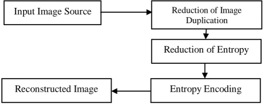
Figure 1 Image Compression Structural Model
II. MEDICAL IMAGE COMPRESSION OVERVIEW
The widely used applications of images in medicare fields ranging from MRI, CTs, medical tele-conferencing, video-conferencing and e-therapist consultancy raises certain questions which need answers. Huge healthcare records such as images, graphics, and files are transmitted over a communication links [4], [5] in raw form result to poor data transmission interval, increase in storage space on disk and increase in cost of operation. Medicare imageries constituents of many artifacts called redundancy or duplication which consume more of storage space on disks, increases transmission bandwidth and require more time for both uploading and downloading processes.
III.WAVELET MODEL
Most medical image signals portray attributes the time space domain with no information on the frequency. This poses difficulty for both time and frequency information is needed to attain optimal compression result. Wavelet analysis split image signal into approximation and detail information. Approximation portion indicates the image pixel values at the top left hand corner with the other three details(horizontal, vertical and diagonal) showing the changes the medicare image undergo from one stage to another. Normally smaller detail coefficients are set to zero and exhibits negligible to the image. Thresholding assign values below detail which is said to be small to zero. Many zeros or sparsity in the medicare image provides superior compression. Power compaction is number of records achieved for both compression and reconstruction process equivalent to sum of the square pixel values. Wavelet, a computational method that reduces and partition the image data into many sub-band frequencies
In wavelet transform, detail frequency of image signal is achievable at any particular interval and in Fourier transform, only the amplitude signal is noted while time frequency is discarded [6]. Features like matching pursuit method, basis pursuit, ℓ1 optimization method, mean square error, and normalized mean square error
play fundamental function in data reconstruction scheme. Each representation present different feature and
therefore, programmer need to select appropriate scheme for particular task. Wavelet function contains two main features [7];
(2)
For oscillatory function or wavy situation.
(3) Input Image Source Reduction of Image
Duplication
Reduction of Entropy
The speedily technological development in compression of big dataintegration [6] creates an enhanced optimization scheme to attain superior data reconstruction routine. WT splits medical records into block wavelets and discards unnecessary information, maintaining relevant details for recovering. Many wavelets exist but our focus is DWT though other such as Haar, Fourier transform and Fast Fourier transform were considered to choose the scheme. Compressing image is essential because, most images contain artifacts demanding huge storage capacity and high financial cost. Compression helps eliminate redundancy, reduce transmission rate, and subsequently lesser storage space to accommodate many image signals.
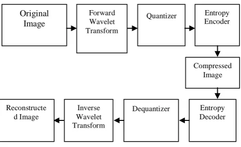
Figure 2 Structure of the Wavelet Transform Compression Scheme
IV.IMAGE DE-NOISING IN WAVELET
Medical Images often contains lot of artifacts either due to muscle's movement or other electrical interference. De-noising is instrumental in removing artifacts whilst preserving image superiority for fast data transmission. Many works is done lately on wavelet thresholding, and image signal de-noising [8]. This is conceivable as wavelet effectively de-noise signal with minimal energy compaction with lesser coefficients occurring owing to the artifacts and bigger coefficients because crucial signal features [9].
Nevertheless, Mutiresolution scrutinizes full signals in same sphere of different resolutions. Higher frequency constituents signal utilize small window in attaining optimum time resolution, and small frequency constituents use larger window in achieving superior frequency details. Vitally, the window has identical capacity regardless of height and width with different wavelet analytics. Heisenberg’s Uncertainty Principle controls the window’s area especially when the frequency resolution increases and correspondingly decreases the time resolution. Mutiresolution is a fascinating pillar for wavelet analytics.
In multiresolution, a low pass filter and high pass filter bands are selected to bisect the frequency range between them. The low pass filter is applied to each data row in order to attain low frequency components of the row. Using Shannon's Sampling Theorem to sub sampled the data into two sub-bands and produces an output data that only contains half the original number of data samples. Furthermore, the high pass filter is applied to the same row of data with high pass partition separated and placed by the side of the low pass components that is applied to all rows [9]. Filtering is then applied to each column producing four bands of data samples as indicated in figure 1 below. The LL band is again decomposed using the same procedure to produce more sub-bands. Original Image Forward Wavelet Transform
1
stLevel 2
ndLevel 3
rdLevel
Figure 3 Wavelet Decomposition
V. COMPRESSION IN 2D WAVELET
This technique uses Matlab GUI wavemenu to remove noise in 2-dimensional digital image. The succeeding phases are performed in de-noising a medical image.
Save image to de-noise in a folder and our image was labeled as "skull_002" JPEG file. Lunch Matlab wavemenu and load the image.
Select db (2) as wavelet type with level 4 decomposition.
Select analyze and click on de-noise
Set threshold to default "fixed form threshold". Select hard threshold. Also set the noise structure to "scaled white noise". Finally set thresholds by level for horizontal, diagonal and vertical coefficients.
Clicks on de-noise and finally save your file
VI.EXPERIMENTAL RESULTS
This can be computed using the Matlab GUI interface. The following steps detail the process;
Generate Matlab code for image de-noising
Type wavemenu into the Matlab command window
LL
HL
HL
LH
HH
LH
HH
LL HL HL HL
LH HH
LH
HH
LH
HH
LL
HL
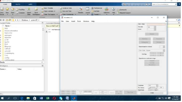
1) On the Wavelet Tool Main Menu, click on wavelet 2-D
Figure 5: Wavelet 2-D
2) Load the Noisy facets 5.5 example indexed image. Using the default biorthogonal wavelet and level 4 decomposition, click De-noise.
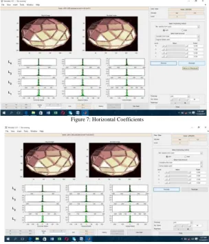
Figure 6: De-noising noisy facets indexed image 5.5
Figure 7: Horizontal Coefficients
Figure 8 Vertical Coefficients
ii. Generate the MATLAB code with File > Generate Matlab Code (De-noising Process).
1. Generating MATLAB Code for Compression
i. Type wavemenu into the Matlab command window
ii. Select Wavelet 2-D.
iii. Select File > Load > Image and load the woman.mat indexed image from the Files\MATLAB\R2016a\toolbox\wavelet\wavedemo folder. When the Loading an Image dialog appears, select No to load the grayscale image.
iv. Select the bior 5.5 wavelet to Level 3.
vi. Click Compress.
vii. Using the default Global thresholding, set Select thresholding method to Balance sparsity-norm
viii.Click Compress
Figure 10: The original and compressed images respectively.
i. File > Generate Code (Compression Process) generates the code.
ii. Save the MATLAB program
iii. Save the compressed image from the Wavelet 2-D Compression tool.
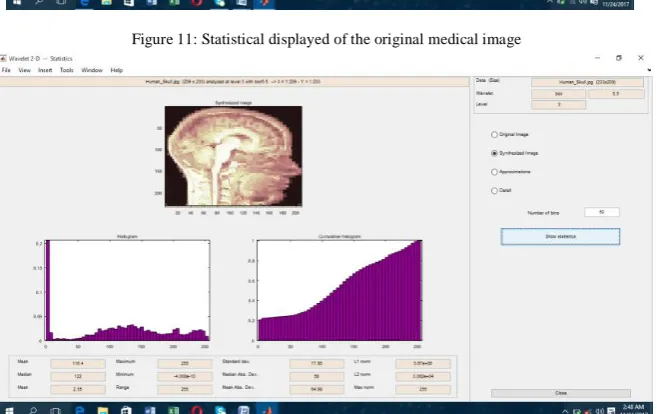
Figure 11: Statistical displayed of the original medical image
VII. CONCLUSION
Wavelet menu toolbox was used to de-noise and compress a 2D medical image in Matlab platform systematically. The result shows that s a result the compressed image with number of zeros = 98.65% noise free in the vertical, horizontal and diagonal details and got energy retention = 96.98%. The experiment shows that almost no traceable distortion of the image quality was lost. The statistical part of the processed image exhibits the following for original image as; mean = 116.4, median = 122, mean = 2.55, maximum = 255, minimum = 0, range = 255, standard dev. =77.95, Median Abs. Dev.=58, mean Abs. Dev.=64.98, Li norm=5.67e+06, L2 norm =3.092e+04, Max norm=255 and the synthesized image as mean = 116.4, median = 122, mean = 2.55, maximum = 255, minimum = -4.008e-10, range = 255, standard dev.=77.95, Median Abs.Dev.=58, mean Abs. Dev. =64.98, Li norm=5.67e+06, L2 norm =3.092e+04, Max norm=255.
VIII. FUTURE WORK
The research will be directed in future work to exploits more wavelet types to e-noise medical image to make the image less noise free with excellent compression scheme that enhances less storage space and speedy up transmission rate thereby saving more lives.
ACKNOWLEDGEMENT
The author would like to thank Prof Paul Kamara, and Professor Prince Sorie Conteh of the Institute of Advanced Management and Technology (IAMTECH), Freetown Sierra Leone for their support and encouragement.
REFERENCES
[1] James S. Walker, ―Wavelets Based Image Processing,‖ Department of Mathematics University of Wisconsin, Eau Claire
[2] Said, A., & Pearlman, W. A. (to appear). An image multiresolution representation for Lossless and lossy compression. IEEE Transactions on Image Processing.
R. C. Gonzalez, R. E. Woods, S. L. Eddins, ―Digital Image Processing using MATLAB‖.
[4] J. Walker and T. Nguyen. Wavelet-based image compression [J]. 2001
[5] S. Grgic, M. Grgic, B. Zovko-Cihlar. Performance analysis of image compression using wavelets [J].2001, 48(3), 682–695
[6] Z. Zhang and B. D. Rao, “Extension of SBL algorithms for the recovery of block sparse signals with intra-block correlation,” IEEE Trans. on Signal Processing, vol. 61, no. 8, pp. 2009–2015, 2013.
[7] S. Bhavani, K. Thanushkodi, “A Survey on Coding Algorithms in Medical Image Compression”, International Journal on Computer Science and Engineering, Vol. 02, No. 05, pp. 1429-1434, 2010. [8] Kanwaljot Singh Sidhu, Baljeet Singh Khaira, Ishpreet Singh Virk, Medical Image Denoising In The
Wavelet Domain Using Haar And DB3 Filtering, International Refereed Journal of Engineering and Science (IRJES).