International Journal of Nanomedicine 2017:12 5443–5460
International Journal of Nanomedicine
Dove
press
submit your manuscript | www.dovepress.com 5443
R e v I e w
open access to scientific and medical research
Open Access Full Text Article
Polyethylenimine-based micro/nanoparticles as
vaccine adjuvants
Chen Shen1
Jun Li1
Yi Zhang1
Yuce Li2
Guanxin Shen3
Jintao Zhu2
Juan Tao1
1Department of Dermatology, Union
Hospital, Tongji Medical College, Huazhong University of Science and Technology, wuhan, China; 2School of
Chemistry and Chemical engineering, Huazhong University of Science and Technology, wuhan, China;
3Department of Immunology, Tongji
Medical College, Huazhong University of Science and Technology, wuhan, China
Abstract: Vaccines have shown great success in treating and preventing tumors and infections, while adjuvants are always demanded to ensure potent immune responses. Polyethylenimine (PEI), as one of the well-studied cationic polymers, has been used as a transfection reagent for decades. However, increasing evidence has shown that PEI-based particles are also capable of acting as adjuvants. In this paper, we briefly review the physicochemical properties and the broad applications of PEI in different fields, and elaborate on the intracellular processes of PEI-based vaccines. In addition, we sum up the proof of their in vivo and clinical applications. We also highlight some mechanisms proposed for the intrinsic immunoactivation function of PEI, followed by the challenges and future perspectives of the applications of PEI in the vaccines, as well as some strategies to elicit the desirable immune responses.
Keywords: cationic polymers, APCs, immunoactivation, danger signals, anti-infection, anticancer
Introduction
Due to their weak immunogenicity, conventional vaccines, especially the subunit vaccines, are always combined with adjuvants to ensure potent immune responses. Polyethylenimine (PEI), as a kind of cationic polymer, has been extensively applied as a nucleotide delivery reagent for decades. In recent years, the robust adjuvanticity of PEI has been continuously documented. Increasing evidence has shown that PEI-based particles are capable of improving the efficiency of conventional vaccines against infections and tumors. These efficiencies are characterized by direct indicators, such as enhanced maturation rates of antigen-presenting cells (APCs) and increased prolifera-tion of effector cells, as well as amplified producprolifera-tion of antigen-specific antibodies and various cytokines and chemokines.
In this review, we first introduce the physicochemical properties of PEI and its general applications in distinct areas. Then, we focus on the effects of neat PEI itself and PEI-based nanoparticles/microparticles (NPs/MPs) in antigen uptake and presentation, which is the foundation for understanding the interplay between PEI and APCs. We then extend the focus on the effects of PEI-relevant adjuvant potency from the cell level to preclinical studies or clinical trials. Subsequently, we are fascinated to figure out some possible underlying mechanisms of its intrinsic immunoactivation functions. Finally, we discuss the challenges and future perspectives of the applications of PEI in vaccines, and explore the promising approaches to optimize it’s immune responses while manipulating the toxicity properly.
Synthesis and broad applications of PEI
PEI is a kind of synthesized cationic polymer with topologies of linear or branched
forms, and its molecular weight ranges from 1 kDa to 1,000 kDa.1 The most common
Correspondence: Juan Tao Department of Dermatology, Union Hospital, Tongji Medical College, Huazhong University of Science and Technology, No 1277 Jiefang Avenue, wuhan 430022, China
Tel +86 027 8572 6195 email tjhappy@126.com
Journal name: International Journal of Nanomedicine Article Designation: Review
Year: 2017 Volume: 12
Running head verso: Shen et al
Running head recto: Polyethylenimine-based micro/nanoparticles as adjuvants DOI: http://dx.doi.org/10.2147/IJN.S137980
International Journal of Nanomedicine downloaded from https://www.dovepress.com/ by 118.70.13.36 on 23-Aug-2020
For personal use only.
Number of times this article has been viewed
This article was published in the following Dove Press journal: International Journal of Nanomedicine
Dovepress Shen et al
and essential characteristic of PEI is its hydrophilic cationic polymeric structure. The strong positive-charged PEI con-denses negative particles (such as the DNA, negative antigens) or plasma membranes in vivo. Moreover, the PEI backbone
contains one nitrogen atom in every three atoms,2 forming
amorphous net structures to work powerfully in lysosomes, as
a “proton sponge”.3,4 These active amino groups, especially the
primary and secondary amines, provide numerous possibilities for structural modifications, which enable them to target the
agents and attenuate the potential toxicity.5
Synthesis and physiochemical
properties of PEI
In general, branched PEI, which contains primary, secondary and tertiary amine groups, can be synthesized through the cat-ionic ring-opening polymerization of aziridine (Figure 1A–C). Linear PEI (LPEI), however, includes secondary amines only, commonly derived from acidic hydrolysis of polyoxazoline (Figure 1D and E). Generally, the synthetic processes of gain-ing LPEI with narrow distribution of the molecular weight
could be rather challenging.5 In PEI, amino groups with
different sorts possess different properties. It is evidenced that primary and secondary amines have strong abilities to bind to nucleic acids, as well as targeting agents, drugs and other functional moieties. Yet, although with less binding capacity, tertiary amines efficiently buffer the pH decline in
acidic conditions.6,7 In general, branched PEI is in the liquid
state and water-soluble, whereas LPEI is solid at room tem-perature and less soluble in cold water, phenol, ethyl, ether, acetone and other solvants, and it turns more soluble in hot water, acidic aqueous solution and organic solutions (such as the methanol, ethanol or chloroform). Though PEIs of these two topologies are quite different, they both possess active amino groups, and the over-positive-charged nitrogens are
always linked to the toxicity of PEI.5
Broad applications of PEI
On the basis of the physicochemical properties, PEI is widely used in broad fields, including effluent treatments, carbon dioxide absorption, separation and purification of proteins,
antibacterial operations and other procedures.8–12 In 1995,
Boussif et al prepared a groundbreaking type of PEI/DNA
+
1 1 +1
+1
+1
1 1+
1
1+
1+ 1 + 1
1+
+1
%UDQFKHG3(,
1
+ /HZLVDFLG
+1 1
+
1 1+
1+
+1
Q R
P
1
+ +
,QLWLDWLRQ 5
+ 1
1 + 3URSDJDWLRQ 5 + 5 3(,
1XF
1XF 1 + 5
Q 7HUPLQDWLRQ
D
±+
1
P 5 %UDQFKLQJ D5 + E5 3(,
1 1 + 5
Q 5
P
'HSURWRQDWLRQ
E5 +
±+
1 1 + 5
Q
P
+1
+
1 1+
Q /LQHDU3(,
1 2
5′
3RO\PHUL]DWLRQ
(
1XF±
5′ +0H(W
( 1 1XF
Q
5′
2
+\GURO\VLV FRQF+&O &
( 1 +
1XF Q
1 1 + 5
Q 1
+
1 + 5
Q
±
$
%
&
'
(
Figure 1 (A) Molecular structure of branched PeI. (B) Polymerization of aziridine to branched PeI. (C) Mechanism of the cationic ring-opening polymerization of aziridine. (D) Molecular structure of linear PeI. (E) Synthesis of linear PeI from substituted 2-oxazolines. Reproduced from Jäger M, Schubert S, Ochrimenko S, Fischer D, Schubert US.
Branched and linear poly(ethylene imine)-based conjugates: syn thetic modification, characterization, and application. Chem Soc Rev. 2012;41(13):4755–4767. with permission of The Royal Society of Chemistry.5
Abbreviations: PeI, polyethylenimine; conc, concentrate.
International Journal of Nanomedicine downloaded from https://www.dovepress.com/ by 118.70.13.36 on 23-Aug-2020
Dovepress Polyethylenimine-based micro/nanoparticles as adjuvants
Table 1 Broad applications of PeI
No Formulations Application Description References
1 Branched/linear PEI; PEI-modified biomass; thiourea-modified PEI
Effluent treatment Cationic flocculants in industrial effluents; anionic
Cr(vI) sorption and reduction in the biomass; the precious metal recovery material
8, 26, 27
2 PEI-modified mesoporous MCM-48/
MCM-41 membranes
Carbon dioxide adsorption
N2/CO2 selectively diminished and high capacity for
CO2 capture
9, 28
3 PEI-PAA bilayer-modified porous PPE
membranes; silica-supported PeI
Proteins separation PeI-PAA bilayer improving IgG-binding ability under
optimized conditions; efficient separation of peptides
with different isoelectric points by adjusting pH value 29, 30
4 Quaternary ammonium PeI NPs; poly(hyaluronic acid)-PeI particles
Antibacterial effects Antibacterial activities of dental composites; antimicrobial property against G−/G+ bacteria strains
10, 12, 31
5 Dishes precoated with PeI Adhesive agents Demonstrating strong cell adhesion which survived washing procedures
11
6 Two-photon fluorophore-conjugated
PeI; commercially magnetic NPs (Gara or G100)/PeI complexes; PeI/ NaYF4 NPs doped with lanthanide ions
Conjugating imaging agents
Confirming the uptake and cytoplasmic localization
of complexes in Hela cells; showing a pronounced hypointense region in tumor tissues after intra-carotid administration; possessing “upconversion
fluorescence” and showing potential in biological
labeling
16, 17, 22
7 PeI/DNA; poly-siRNA/PeI complexes; PeI-siRNA NPs; PeI/pDTA-H19 DNA complexes
Conjugating nucleotides
Delivering oligonucleotides into embryonic neurons;
RFP gene silencing efficiency of ~80% in B16F10 cells; HeR-2 downregulation in breast tumors; complete tumor response of 64.1%, with 90% CI
(49.7%–76.8%) in patients with superficial bladder
cancer
2, 13, 14, NCT00595088
8 CNGRC/PeG/PeI/DNA vector; PeI– glutathione conjugates; PeI-coated albumin NPs for protein delivery
Conjugating peptides/proteins
when conjugating the targeting peptide CNGR protein transduction tool for GST-fused proteins; delivering BMP and promoting osteogenesis in vivo;
specifically deliver β-gal genes to CD13+ cells and
tissues
15, 20, 32
9 HPv16L1-BMP/mPeI/pHPv16L1 NPs Immune adjuvants Significantly enhancing humoral immune response
and the number of IFN-γ-producing CD8+ T cells
18
Abbreviations: Cr(VI), Chrominum(VI); PEI, polyethylenimine; PAA, polyacrylic acid; PPE, polyethylene; NPs, nanoparticles; FITC, fluorescein isothiocyanate; DTA,
diphtheria toxin A; CNGRC, CD13/aminopeptidase N-binding NGR peptide; GST, glutathione S-transferase; BMP, bone morphogenetic protein; mPeI, maltosylated PeI; IFN-γ, interferon-γ.
NPs and successfully transferred DNA into nerve stem cells.2
Subsequently, PEI has drawn more attention in biomedical
field, especially as drug carriers,13–15 biological labels16,17 and
vaccine adjuvants (Table 1).18
Among all the delivered drugs, the most common ones are nucleic acids. PEI is the second acceptable nonviral
nucleic acid transfer agent, besides poly-L-lysine (PLL).2
Nowadays, PEI transfection reagents are commercially
avail-able, which include ExGen500®, jetPEI® and PEIpro™.19
Besides nucleic acids, proteins or peptides are also included
in the category.20,21 PEI also works as a biological label when
conjugated with imaging agents (such as the Fe3O4 and
fluorescent NPs), and reflects the specific cell or sub-cell information, such as migrations and other behaviors, under
certain conditions.16,22,23 The adjuvant effect of PEI is an
emerging area. In a series of recent studies, vaccines com-posed of PEI as the immunostimulants are quite competent
in treating infections or tumors.24,25
Intracellular process of PEI/
modified PEI as vaccine adjuvants
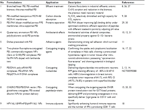
To better manipulate its adjuvanticity, strategies to construct the PEI-based vaccines are highly desirable. PEI could be incorporated into vaccine structures through different ways: directly binding with antigens, coating on antigen-loaded NPs/MPs, coating existing particles with antigens absorbed on the surface or co-encapsulated NPs/MPs with antigens and other constructed forms (Figure 2). During these processes, plenty of cytokines and ligands could also be added in the vaccines based on the desirable applications.Cell uptake of PEI-based vaccines
The first step for a vaccine to take its effect is the endocy-tosis process by APCs. Similar to traditional vaccines, the factors influencing the cell uptake of PEI-based vaccinesinclude size,33 shape,34 charge35 and surface chemistry of the
particles.36,37
International Journal of Nanomedicine downloaded from https://www.dovepress.com/ by 118.70.13.36 on 23-Aug-2020
Dovepress Shen et al
General factors influencing
PEI-based polyplex uptake
With broad range of molecular weights, different topolo-gies and plentiful modification possibilities, PEI-based
polyplexes are rather versatile in size, shape and properties.5
Therefore, PEI-based vaccines demonstrate broad possibili-ties in applications.
Also, the strong positive charge in PEI is beneficial for cellular internalization. PEI can bind to the negative proteoglycan on cell membranes and mediate the uptake
process through the electrostatic interaction.33,35
Gener-ally, phagocytosis by immune cells of foreign particles
is an actin-dependent process.34 The loosely organized
PEI in neat state will disturb the actin remodeling when initiating the internalization. Yet, PEI compresses nega-tive substances to condense small particles, or PEI-coating
strategy will result in a smooth surface.25,38 These spherical
particles are symmetrical and easily uptaken from any point
of attachment.34
Size is one of the useful factors in the toolbox affecting the uptake of PEI-based particles, as well as other poly-meric particulate carriers. Champion et al demonstrated that particle size played a role in phagocytosis only if the
volume was larger than the cell size.34,39 It is relatively
easy to achieve small-sized PEI-based polymers. For the PEI-coated large MPs, the uptake process is restricted; however, the electrostatic forces attach the huge particles to the cell membranes, and perform as a continuous depot
of the antigens.40,41
Effect of surface properties of
PEI-based polyplex on cellular uptake
In addition to the general parameters, significant attention has been paid to the chemical modifications of the surfacechemistry.34 Some modifications would largely influence the
cellular uptake of the PEI-based polyplexes.
In APCs, phagocytosis is frequently mediated by
receptors (via mannose receptor-, complement receptor-, Fcγ
receptor- and scavenger receptor-mediated pathways or other
pathways).34 Targeted modifications can obviously improve
the cellular uptake efficiency and the subsequent biological
effects of the polyplexes.36,37,42 For MPs or less positively
charged particles, the tethering of targeting moieties on the
particles’ surface is even more important for endocytosis.43,44
Hu et al prepared mannose-modified PEI-cell-penetrating peptide (CPP)/DNA particles and found an improved
DC2.4-consuming efficiency.45 It is worth noting that Toll-like
receptors (TLRs) are not included in the phagocytosis-related receptors, but they initiate the phagosome-mediated APCs
maturation and inflammatory secretion,46 which is discussed
in this review. Cho et al reported that the maltosylated PEI-mediated vaccines for cervical cancers were more effective than neat human papillomavirus antigens. Presumably, recep-tor-mediated endocytosis of PEI-based particles by maltose
subsequently enhanced the transfection efficiency.18 For
PEI-coated NPs/MPs, similar patterns of receptor-mediated gene delivery have also been demonstrated. Mesoporous silica NPs coupled with PEI also demonstrated high transfection
efficiency due to the more effective targeting into APCs.47
Besides the specific receptor-mediated targeting strategy, another nonspecific targeting method is the hydrophobic modification of PEI. Given the lipophilic nature of cell membranes, the lipid-like structure facilitates the interactions between the hydrophobic species and plasma membranes
(eg, lysosomal membranes and nuclear membranes).7 It is
demonstrated that 76% and 96% of acetylated branched PEI (BPEI) enhanced the cellular uptake ability by four-fold,
cor-responding to the hydrophobic interaction theory.48,49 When
the lipid components were incorporated, though possessing a relatively decreased positive charge, the electrostatic
interac-tion could still be maintained and aid in binding.48,49 Recently,
Parhiz et al reported that hexanoated-PEI vector was also more effective than the corresponding non-hydrophobic PEI
in enhancing transfection rates.19 Besides the transfection
benefits, Wang et al proved that hydrophobic modification of alkyl chains to PEI vaccines optimized the cross-presentation of antigens and upregulated IL-2 secretion. Yet, correlation
$
%
&
'
3(, $QWLJHQSHSWLGHV $QWLJHQ'1$ 03V
Figure 2 Schematic illustration of PeI-based NPs/MPs. (A) electrostatic nanosized complexes of cationic PeI and anionic peptides or DNA. (B) PeI-coated MPs encapsulated with peptides or DNA. (C) PeI-coated MPs peptides or DNA adsorbed on the surface by static electricity. (D) PeI MPs encapsulated with DNA or peptides.
Abbreviations: PeI, polyethylenimine; NPs, nanoparticles; MPs, microparticles.
International Journal of Nanomedicine downloaded from https://www.dovepress.com/ by 118.70.13.36 on 23-Aug-2020
Dovepress Polyethylenimine-based micro/nanoparticles as adjuvants
between the properties of the synthesized hydrophobic-modified PEI and the vaccine potency still needs further
investigation.50
CPPs are another valuable species that can optimize the interactions between cell membranes and the PEI-based polyplex. CPPs, characterized by the polycationic amino
acid residues (ie, arginine and lysine), have been known
for their specialized ability to penetrate the cellular mem-branes. Until now, most studies demonstrated that CPPs take effect by electrostatic binding to negatively charged cell membranes and induction of vesicle rupture by osmotic
change and/or membrane lysis.19 Truncated Tat peptide and
penetratin are the most popular CPPs, and both are
arginine-rich peptides.19 Recently, Morris and Sharma constructed
arginine-modified oligo-(alkylaminosiloxanes)-grafted PEI and found that the modified PEI/pDNA exhibited 98% cell viability and 150% more gene transfection efficiency than neat BPEI in human nasopharyngeal epidermoid
carcino-ma.51 Inhibitor studies, by use of various cellular uptake
inhibitors such as wortmannin and genistein, indicated the role of arginine moiety in promoting internalization of the polyplex, and the process possibly contained a combination
of multiple pathways.51
More modification strategies will be developed to mag-nify the advantages of PEI-based vaccines in the initiation process. Yet, after certain modifications, the particle size, charge and other parameters of the polymers may vary cor-respondingly. Though each of these areas can be studied separately, the interplay among these parameters must be considered as a whole to identify the best performance in
boosting the uptake.34,52
Antigen presentation of PEI-based
vaccines
After being uptaken, antigens are processed and transported through different cell compartments and are finally presented within the peptide-binding groove of a major histocompat-ibility complex (MHC) molecule (MHC class I molecules or class II molecules). T cell receptors can only recognize antigens presented in this pattern. Mostly, MHC class I molecules present endogenous peptides (such as transformed or infected cell components) and elicit the cell-mediated immunity (CMI), while class II molecules acquire exogenous peptides (extracellular pathogens or vaccine peptides) and
induce abundance of antibodies.53 PEI-based vaccines with
antigen peptides mainly follow the exogenous/class II
path-way and stimulate the humoral immunity.54 PEI-based DNA
vaccines provide the opportunities to synthesize multiple antigens in APCs and stimulate effective cellular immunity and long-lasting memory immunity. However, most DNA is actually delivered to bystander cells (such as myocytes and fibroblasts). In this case, newly synthesized antigen peptides are presented on or secreted out of bystander cells. They are
shuttled to APCs, acting as the exogenous antigens.55,56
The “proton sponge effect” of PEI
makes cross-presentation possible
As mentioned above, MHC class I molecules present endogenous peptides synthesized in the cells, while class II molecules present the exogenous internalized antigens. Occa-sionally, MHC class I molecules also participate in presenting exogenous antigens, known as the cross-presentation, which is very meaningful in biological evolution. In human beings, since the virus invasion does not commonly occur inprofes-sional APCs and the CMI is not naturally generated,57 virus
antigens released from the bystander cells turn into exogenous antigens for APCs. The cross-presentation mechanism in APCs largely increases the efficiency of the immune system to generate CMI and eliminate all the infected cells. Yet, in application of vaccines for treating cancers and infections, which needs the CMI instead of mere humoral immunity,
the cross-presentation is also of great importance.57 In the
processes, phagocytosed or endocytosed antigens escape from the vacuole and gain entry to the cytosol (known as the “lysosomal escape”). Then, they become qualified clients for ubiquitination and subsequent degradation by the proteasome, followed by the transporter associated with antigen process-ing (TAP)-mediated transfer into the endoplasmic reticulum,
and presentation by MHC class I molecules.57–59
There are two representative mechanisms of the “lyso-somal escape”, that are, the variation of osmotic pressure and
membrane lytic activity.19 Some pH-sensitive biomaterials
possess the membrane-destabilizing property; thus, they
are good candidates.60,61 PEI has the unique “proton sponge
effect”, which buffers under acidic conditions.2,62 When
involved in acidic conditions, the ATP-mediated pH- dependent proton pumps open, followed by passive influx of chloride ions and water molecules resulting in
hyperos-molar state instantaneously, causing the vesicles to burst.2,62
The “lysosomal escape” process of PEI-based polyplex
was well recorded by Merdan et al (Figure 3).6,7 PEI/
ribozyme and PLL/ribozyme migrations in the living cells were identified under confocal laser scanning microscope, and they were found to first gather in acidic vesicles, most
International Journal of Nanomedicine downloaded from https://www.dovepress.com/ by 118.70.13.36 on 23-Aug-2020
Dovepress Shen et al
probably lysosomes. Unlike the PLL-based complex, the vesicles containing the PEI-based complex met with a sudden burst while letting out the contents throughout the
cytoplasm.6,7 To verify the function of the pH change in the
“lysosomal escape” process of PEI, they prepared the PEI/ ribozyme group with bafilomycin A, a selective inhibitor of endosomal/lysosomal acidification. It showed no lysosomal rupture, suggesting the major role of acidification in the
procedure.6,7 The escaped antigens (peptides or DNA) in
the PEI-based vaccines in the cytoplasm are then prepared to undergo a cross-presentation process. The process has recently been verified again by Song et al. They found that PEI-coated poly(lactide-co-glycolide) (PLGA) (OVA) NPs induced efficient cross-presentation of antigens on MHC class I molecules through the endosome escape and lysosomal
processing.63
Effect of surface properties of
PEI-based polyplex on antigen
presentation
There is a group of membrane-destabilizing peptides, which have shown the possibility of not only promoting cell uptake but also improving “lysosomal escape” and even nuclear
translocation.64,65 Although PEI has the proton sponge effect
to carry out the “lysosomal escape” itself, sometimes it is incompetent, and addition of membrane-destabilizing pep-tides (such as CPPs) in the vaccines can improve the cross-presentation effect. Ogris et al have explained the conditions
in their study.52 The transfection efficiency was 10-fold
(in B16F10 cells) to more than 100-fold (in Neuro2A cells,
K562 cells) lower in small PEI/DNA particles compared with the large ones. Introduction of the lysosomotropic drug chloroquine or the pH-specific, membrane-active peptide (INF5) really promoted a substantial increase in antigen genes expression, which confirmed the hypothesis and importance
of adding CPP moieties.52 Tan et al bounded two truncated
peptides with penetrating properties to BPEI, resulting in higher transfection efficiency without causing cytotoxicity in
CHO-K1, B16F10 and 293FT cell lines.66 These results can
be ascribed to the enhanced endosomal disrupting activity of CPP-bound PEI carriers than to their parent carriers.
In PEI/DNA vaccines, PEI helps with the nucleic acids protection and endosomal escape. Yet, the transfection
effi-ciency may still be limited in these vaccines.62 Particles of
over 30 nm diameter or over 40 kDa molecular weight will require the active aid of the coupled nuclear localization
signal (NLS) in nuclear translocation.60 NLS peptides are
usu-ally short conservative sequences, which bind to cytoplasmic importins and dock to the nuclear pore complex, thus aiding the nucleic acids transcription. The most commonly used NLS
contains the PKKKRKV sequence.67 In addition, the
well-known arginine-rich peptides, such as Tat and penetratin,
also possess such nuclear transport activities.67 Besides, many
other viral-origin peptides and even histone H1, protamine, ribonucleoprotein A1, high-motility-group proteins and oth-ers containing the above polycationic amino acids also act
as effective NLS.60 Parhiz et al investigated the efficiency
of PEI/DNA vaccines by attaching different arginine-rich sequences. It was found that the arginine-rich derivatives of PEI induced higher DNA and siRNA transfection efficiency
$
%
%HIRUH $IWHUFigure 3 (A) Living cell microscopic visualization of the “proton sponge effect” of polyethylenimine (PEI). PEI is green, while ribozyme is red in this observation. Before burst, the vesicle has a yellow core and green PEI corona, while afterwards, the faint green-yellow fluorescence can be seen evenly distributed throughout the entire cell. The remnant of the vesicles is significantly smaller and deeper red. Images were recorded 28, 31, 37, and 40 min after incubation. Yellow arrows indicate a single vesicle undergoing the process of swelling
and bursting. The scale bar in the upper-left image is 10 μm and can be applied in the other images. (B) The same confocal layer of 1 nm thickness before and after lysosomal burst (37
and 40 min after incubation). Clearly, fluorescence intensity increases throughout the whole cell after burst, whereas there seems to be an area where it is slightly weaker which might
be the nucleus. The right image is an optical microscopy image showing the positions and numbers of the cells. Reproduced from Pharm Res, Intracellular pro cessing of poly(ethylene imine)/ribozyme complexes can be observed in living cells by using confocal laser scanning microscopy and inhibitor experiments. , 2002;19(2):140–146, Merdan T, Kunath K, Fischer D, Kopecek J, Kissel T, (copyright 2002 Springer). with permission of Springer.6
International Journal of Nanomedicine downloaded from https://www.dovepress.com/ by 118.70.13.36 on 23-Aug-2020
Dovepress Polyethylenimine-based micro/nanoparticles as adjuvants
than the groups without modifications. Moreover, PEI/DNA vaccines conjugated with arginine-rich peptides and hydro-phobic derivatives demonstrated the highest DNA transfec-tion efficiency, indicating that ratransfec-tional designs of two or more modifications are promising, to get a better transfection
efficiency (antigen DNA expression) and lower cytotoxicity.68
All the efforts to elevate DNA transfection promise antigenic DNA translation and presentation. The advantages and modi-fication strategies of PEI-based vaccines related to antigen uptake and presentation are illustrated in Figure 4.
In vivo and clinical applications
of PEI and modifiers as vaccine
adjuvants
To assess the performance of the PEI-based vaccines, we must consider the following three levels: in vitro, in vivo and clinical trials. Clearly, the clinical trials of large samples are the most convincing data for evaluating the efficiency of newly developed vaccines. Although we have got much more data from animal trials supporting the superior effects of PEI-based vaccines, only a few trials of PEI-based vaccines have been processed into clinical level. We summarize the application of PEI-based vaccines in animal experiments against infections and cancers, in Tables 2 and 3, respectively.
In vivo applications of vaccines
The in vivo applications of vaccines are confronted with bottlenecks sometimes, for two main reasons. One is the
safety issue, as PEI is widely known for its toxicity. The
other is the complicated microenvironment in vivo.61,69 In
the microenvironment, every immune-active material has its corresponding targeting immune cells. These cells secrete various chemokines, attracting monocytes, granulocytes and other immune-promoting cells, while the attracted cells secrete more cytokines to form a positive feedback. As frequently depicted for many traditional adjuvants, the monocytes ultimately uptake the complexes, differentiate into dendritic cells (DCs) or phagocytes and then migrate
to secondary lymphoid tissues.70,71 Intriguingly in PEI, the
local inflammation seems beneficial to the recruitment and
activation of APCs.72
Though the general process is somewhat clear, the detailed signal communications of one specific vaccine in in vivo applications are still puzzling. For example, it was found that intramuscularly injected MF59 and aluminum adjuvants tar-geted different cell types, while all the recruited cells had simi-lar potential to engulf carrier–antigen complexes. All events occurred in draining lymph nodes (DLNs) at certain time
inter-vals, probably due to a bystander effect.70,71 Further research
indicated that, different from lipopolysaccharide (LPS), MF59 and aluminum took a TLR-independent mechanism to activate immunization and bias the monocytes differentiation to DCs
rather than macrophages.70,71 Yet, as for the PEI-based vaccines,
several issues, such as the local activation of cells and the roles of PEI-based vaccines in it, the secretion of cytokines and the uptake and differentiation processes, along with the lymph drainage situations, need further investigation.
$QWLJHQSUHVHQWDWLRQRI 3(,RUPRGLILHG3(,EDVHGYDFFLQHV
³/\VRVRPDOHVFDSH´ZLWK&33V 0+&,,PROHFXOHV
0+&,PROHFXOHV ³/\VRVRPDOHVFDSH´ DQGFURVVSUHVHQWDWLRQ
1XFOHDUWUDQVSRUWZLWK1/6
&HOOXODUXSWDNHRI
3(,RUPRGLILHG3(,EDVHGYDFFLQHV
355OLJDQGV
+\GURSKRELFPRLHWLHV L'&
P'& P'& 3RVLWLYHFKDUJHV
9DULRXVVL]HV L'&
6SKHUH
&33V
Figure 4 Illustration showing the uptake and presentation processes of based vaccines and the modified forms. Positive charge, round shape and controllable size of PEI-based MPs and NPs are all benefits contributing to the uptake process. Besides, they could be modified with PRR ligands, hydrophobic moieties or CPPs, which facilitate the
polyplex internalization by receptor-mediated routes, membrane fusions, penetration process and other unknown routes. After polyplex degradation in the endosomes and
lysosomes, the released antigens are presented onto MHC molecules directly by transmission or indirectly by gene expression process. PEI itself has the “lysosomal escape” feature, while vaccines with CPPs-modified PEI possess more potent “lysosomal escape” property. They assist with the cross-presentation of delivered antigens. NLS is the
specialized sequence added with nucleic acids to assist nuclear translocation of the polymers.
Abbreviations: PeI, polyethylenimine; MPs, microparticles; NPs, nanoparticles; PRR, pattern recognition receptor; CPPs, cell-penetrating peptides; MHC, major histo-compatibility complex; NLS, nuclear localization signal; iDC, immature dendritic cells; mDC, mature dendritic cells.
International Journal of Nanomedicine downloaded from https://www.dovepress.com/ by 118.70.13.36 on 23-Aug-2020
Dovepress Shen et al
Table 2
The infection applications of P
eI-based vaccines in vivo
No
Particle formulations Animal models
Routes Antigens Main points Infections References 1 25-kDa P eI/pci-S DNA NPs Balb/c i.n. SARS-Co v spikes plasmid(pci-S) H ig he r S-sp ec ifi c Ig G 1 in s er um a nd m uc os al Ig A in lu ng w as h
than pci-S alone; higher number B220
+ cells in spleen; higher
CD80/CD86/MHC II expression on CD11
+ D
C s in c er vi ca l
LNs; higher
IFN-γ
/TNF-α
/IL-2-producing T cells in lung
SARS
54
2
25-kDa BP
eI/
influenza HA polymers
C57BL/6J
i.n.
Influenza HA peptide
Pe
I-immunized mice with enhanced protection in weight
loss than CTB; an enhanced IgG ratio of native to denatured antigens than CTB; similar titers of HA-specific serum IgG1, IgG2
α
and IgA as CTB; HA-specific IgG levels in the serum
were significantly higher for PEI than for CTB after the prime vaccination but were equivalent after the booster
Influenza 25 3 Pe I/pRSC-gD-IL-21 DNA NPs Balb/c
Ocular mucosal administration
HS
v
glycoprotein D
and IL-21 plasmid (pRSC-gD-IL-21)
Inducing higher specific sIgA in tears,
IFN-γ
and IL-4 in
serum; enhancing the cytotoxicity of NK cells, as well as splenocyte proliferative responses to glycoprotein D; less HSK degree than controlled group of murine ocular mucosa
HS v 81 4 25-kDa P eI/PLGA MPs/DNA Balb/c i.m.
DNA encoding three Ags
of
L.
monocytogenes
Better survival rate of mice immunized with a sublethal dose of L. monocytogenes
than naked DNA
L. monocytogenes
77
5
22-kDa, 25-kDa or 87-kDa LP
eI or
JetP
eI/DNA NPs
Balb/c and C57BL/6J
i.m.
Recombinant plasmid L. monocytogenes expressing O
v A Increased in vivo DNA expression 20- to 400-fold; enhanced
DNA-induced epitope-specific CD8
+ T cell responses 10- to
25-fold in vivo; increased numbers of cells secreting type-I cytokines; improved antigen-specific Th1 cell and humoral responses; eliciting memory cellular responses
L. monocytogenes 56 6 JetP eI/gp120 DNA polymers Balb/c
Pulmonary administration
gp120 plasmid
Inducing 10 times greater CD8
+ T cell responses; producing
type-1 cytokines and higher CD4
+ T cell responses;
producing more IL-2 in lungs and draining LNs compared to i.m.; inducing CD8 + T cell responses in gut and vaginal
mucosa, protecting mice better in a lethal recombinant virus challenge
HI
v
74
7
Mannosylated- PeI/pSHI
v NPs (Derma v ir) Naïve rhesus macaques
Topically on skin
SHI
v
plasmid(pSHI
v
)
Inducing antigen-specific CD8
+ and CD4 + T immune
responses and a similar number of transduced DC in LNs in nonhuman primates compared to ex vivo DC vaccination; in PB, a similar number of antigen-specific CD8
+CD3 + and
CD8-CD3
+ cells for Derma
v
ir and ex vivo DCs; in PB,
no Abs are detected; with DTH on skin
HI v 73 8 PLGA MPs containing Pe I/DNA complexes Balb/c i.m. HI v plasmid(Gag, Pol and env)
Inducing significant enhanced Abs and CTL responses to HIv
vaccine DNA prime/M
v
A boost regime
HI v 75 9 22-kDa LP eI/HI v
-gp120 DNA NPs
Balb/c
i.v.
HI
v
-gp120 plasmid
Inducing a rapid elevation of serum level of IL-12,
IFN-γ
;
a single administration eliciting a number of gp120-specific CD8 + T cells 20 times higher than DNA alone; protective
responses against both systemic and mucosal challenges
HI
v
76
International Journal of Nanomedicine downloaded from https://www.dovepress.com/ by 118.70.13.36 on 23-Aug-2020
Dovepress Polyethylenimine-based micro/nanoparticles as adjuvants
10
LP
eI or BP
eI/HI
v
-gp140 polyplexes
Balb/c and C57BL/6J
s.c. and i.p.
HI
v
-1 gp140
Enhancing antigen-specific serum IgG production; more antibodies than aluminum in mice and rabbits; recruiting neutrophils followed by monocytes to the administration site; enhancing antigen uptake by APCs; the bias was modulated by NLRP3 inflammasome toward Th2 response but global adjuvanticity unchanged; adding CpG-ODN, adjuvant potency increasing with Th1 biased responses
HI v 24 11 Mannosylated P eI/ pv AX1-HI v gag DNA NPs Balb/c i.m. pv AX1-HI v gag plasmid
In vitro higher transfection in DC of complexed DNA vaccines; IgG2
α
T cell response percentage, cytokine
production significantly higher even with lower DNA dose
HI v 88 12 25-kDa BP eI/ w Iv polymers
Balb/c and C57BL/6J
i.n.
H9N2 influenza WIV
Higher antigen-specific IgA in local nasal cavity, trachea, lung than pure
w
Iv
; increasing amount of IgG (IgG1, IgG2
α ) in serum H9N2 influenza 78 13 25-kDa P eI-coated PLA MPs/HBsAg Balb/c i.p. HBsAg
Generating a rapid and efficient humoral immune response and cytokine release than aluminum-absorbed or free antigens; strong stimulation to the Th1 response
HB v 82 14 Pe I/ γ -PGA
NPs-coated PyTAM DNA
C57BL
i.p.
PyTAM DNA
Significantly improved survival rate from lethal
Plasmodium
yeolii
challenge than naked PyTAM plasmid; enhanced
antigen-specific IgG1 and IgG2
β
antibody levels, higher
proportion of the
IFN-γ
-producing CD4
+ and CD8 + T cells
in spleen, higher level of IL-4,
IFN-γ
, IL-12 and
TNF-α
levels
in the sera and in the supernatants from ex vivo splenocytes
P. yeolii/ P. malariae
89 15 SPIONs/25-kDa BP eI NPs/MSP1 DNA Balb/c
i.p. and i.m.
MSP1 plasmid
The complexes induced Abs against
P. yeolii
, with higher
responses induced i.p. than i.m., with IgG2
α
subclasses as
the predominance, together with cellular immunity; eliciting high levels of
IFN-γ
, moderate levels of IL-4 and IL-17;
inducing Th1, Th2 and Th17 cell-mediated immunity
P. yeolii/ P. malariae
90 Abbreviations: P eI, polyethylenimine; pci-S, SARS DNA vaccine; i.n., intranasal; SARS, severe acute respiratory syndrome; L. monocytogenes, Listeria monocytogenes ; Co v , coronavir us; S, spike protein; MHC II, major histocompatibility complex class II; DCs, dendritic cells; LNs, lymph nodes; IFN-γ , interferon-γ ; TNF-α , tumor necrosis factor-α ; IL, interleukin; BP eI, branched Pe I; HA, hemagglutinin; CTB, cholera toxin B subunit; pRSC, mock plasmid; HS v , herpes simplex virus; sIgA, serum IgA; NK, natural killer; HSK, herpes stromal keratitis; PLGA, poly(lactide-co-glycolide); MPs, microparticles; i.m., intramusal; Ags, antigens; LP eI, linear Pe I; NPs, nanoparticles; Th, T helper cells; gp, glycoprotein; pSHI v , plasmid of simian human immunodeficiency virus; PB, peripheral blood; Abs, antibodies; DTH, delayed-type hypersensitivity; CTL, cytotoxic T lymphocyte; s.c., subcutaneous; i.p., intraperitoneal; APCs, antigen-presenting cells; NLRP3, NLR family pyrin domain-containing 3; w Iv , whole inactivated virus; HBsAg, hepatitis B surface antigen; HB v , hepatitis B virus; γ -PGA, poly( γ -glutamic acid); P.yelolii ; plasmodium yeolii ; P. malariae , Plasmodium malariae PyTAM,
PyGP18P-transamidase-related protein; SPIONs, superparamagnetic iron oxide NPs; MSP1, merozoite surface protein 1.
International Journal of Nanomedicine downloaded from https://www.dovepress.com/ by 118.70.13.36 on 23-Aug-2020
Dovepress Shen et al
Table 3
The anticancer applications of P
eI-based vaccines in vivo
No Particle formulations Animal models Routes Antigens Main points Tumors References 1 Pe I/p v AX1/C-G250 DNA complexes + C-G250 protein Balb/c i.m. v
AX1/C-G250 plasmid and C-G250 protein
Pe
I/DNA
+
C-G250 group exhibiting the strongest
antibody titer, higher CD8
+ and CD4 + T lymphocyte
proliferation and cytokine production
Renal cell carcinoma
83 2 Chitosan-1,200-Da P eI/gp100 DNA complexes C57BL/6J i.d. gp100 plasmid Chitosan-P
eI helping DNA enter into the cytoplasm and
into the nucleus of DCs; finally improving resistance to the B16BL6 melanoma challenge
B16BL6 melanoma
91
3
2-kDa BP
eI-stearic acid
micelles-coated Trp2
C57BL/6J
s.c.
Trp2 peptide
Trp2-loaded P
eI-stearic acid micelles promoting CD86,
MHC II and CCR-7 expression in the DLNs; enhancing Trp2-specific CTL activity; inhibiting tumor growth in a B16-F10 mouse melanoma model compared to free Trp2
Melanoma 92 4 HP v 16L1-MBP/maltosylated 25-kDa BP eI(mP eI)/pHP v
16L1 DNA NPs
Balb/c
i.m.
HP
v
16L1 plasmid and
HP
v
16L1 peptide
Protein/DNA co-delivery vaccine producing higher systemic IgG and mucosal IgA; inducing higher
IFN-γ
production by splenic CD8
+ T cells than protein alone
and protein/DNA mixture
Cervical and other HP
v -related cancers 18 5 600-Da BP eI-Tat/p e7-NT-gp96 DNA complexes + IP-10 C57BL/6J s.c. HP v 16 e7 plasmid
Co-immunization strategy producing the highest IgG2
α
,
the highest
IFN-γ
response in comparison with other
groups; significantly suppressing TC-1 tumor growth with elevated levels of
IFN-γ
and IL-2 production in the LNs
Cervical and other HP
v -related cancers 93 6 25-kDa BP eI/pAc-neo-O v A DNA complexes C57BL/6J s.c. pAc-neo-O v A DNA Pe
I/DNA complexes inducing class I-restricted antigen
presentation and enhancing CTL activity in vivo; exhibiting
more
pronounced
prophylactic
and
therapeutic
antitumor effects than DNA vaccine; P
eI inducing
NF-κ
B
translocation
H-2b restricted murine thymoma cell line (
eG7-O v A cells) 94 7 70-kDa BP
eI or 25-kDa LP
eI/PLGA
MPs-coated MCP3-sFv20 DNA
Balb/c
i.m. and i.d.
MCP3-sFv20 DNA encoding the B cell idiotype antigen
Higher CD80/MHC II expression in RA
w
264.7 in vitro;
improved survival rates of BP
eI/PLGA MPs compared to
LP
eI/PLGA MPs, both longer survival rates than
saline-injected controls or blank MPs
B cell lymphoma
38 Abbreviations: P eI, polyethylenimine; pv AX1, eukaryotic expression vector; C-G250, renal carcinoma-associated antigens; i.m., intramusal; gp, glycoprotein; i.d., intradermal; DCs, dendritic cells; BP eI, branched Pe I; Trp2, tyrosinase-related protein 2; s.c., subcutaneous; MHC II, major histocompatibility complex class II; DLNs, draining lymph nodes; CTL, cytotoxic T lymphocyte; mP eI, maltosylated Pe I; HP v , human papillomavirus; NPs, nanoparticles; IFN-γ , interferon-γ
; IL, interleukin; LNs, lymph nodes; LP
eI, linear P
eI; PLGA, poly(lactide-co-glycolide); MPs, microparticles; MCP3, monocyte chemotactic protein 3; sFv20, heavy an
d light Ig chains.
International Journal of Nanomedicine downloaded from https://www.dovepress.com/ by 118.70.13.36 on 23-Aug-2020
Dovepress Polyethylenimine-based micro/nanoparticles as adjuvants
Progress of in vivo applications
of PEI-based vaccines
Different types of PEI, antigens, constructed strategies and immune routes are chosen in constructing the PEI-based vaccines in vivo. Frequently researched diseases include
AIDS,24,73–76Listeria monocytogenes infection,56,77
respira-tory diseases,54,78–80 B cell lymphoma,38 herpes simplex
virus attacks,81 viral B hepatitis,82 renal cell carcinoma,83
melanoma84 and others.85 The PEI-based vaccines work
through almost all the known immune routes, and usually mimic the pathogenesis of the diseases themselves. For exam-ple, in respiratory infection, intranasal administration was prone to elicit mucosal secretory antibodies, as well as more
serum antibodies.54 A similar trend was found for the HIV
vaccines which elicited more antibodies in the vagina.74
The detected markers prove the efficacy of these vaccines usually through two main aspects: (1) intermediate indica-tors, such as antigen DNA expression, APCs maturation rates, humoral and cellular immunity responses in serum, lymphoid organs or tissues, the T helper type 1 (Th1)/T helper type 2 (Th2) response bias, the memory T and B cells
production and secretion levels of the related cytokines;56,76,86
(2) comprehensive effects, such as higher animal survival
rates, less weight loss and less tumor metastasis rates.38,54,77
The vaccines have demonstrated positive results in a series of in vivo experiments, while only a few clinical trials have
been performed.40 The only satisfactory example was the
DermaVir (mannosylated-PEI/plasmid Simian HIV DNA). Lisziewicz et al performed the ex vivo DC-based vaccination of DermaVir and found that DermaVir-transduced autolo-gous monocyte-derived DCs induced HIV-specific T cells
in rhesus macaques.87 Moreover, the same group improved
the function of the ex vivo DC vaccination strategy with topical administration. Compared with the previous ex vivo DC-based vaccinations, the vaccination strategy proposed by Lisziewicz et al resulted in a similar number of transduced DCs in the lymph node and induced a similar quantity and quality of HIV-specific Th1-type T cell responses. The DermaVir Patch has been tested in Phase I/II clinical tri-als at present, and is the new promising agent in clinical
applications.73
Intrinsic immune-activating
function of PEI
Though many studies showed the performance of PEI-based vaccines in vitro and in vivo, the immune-activating proper-ties of PEI still need to be revisited. It is not clear from these studies whether the improved immune efficiency was solely
due to antigen protection (vaccine delivery vehicle) or due to the intrinsic adjuvant property (immunostimulator) of PEI.25,56,78,95 Moreover, in the case of PEI-coated polyplex, PEI together with the NPs/MPs should be regarded as a whole to take effects. When encapsulated inside, it was even more difficult to prove the adjuvanticity of PEI. Thus, the observed immunoactivation effects of PEI need further confirmation, and the revelation of the possible molecular pathways of the PEI-based vaccines would help us in understanding and developing new PEI-based vaccines.
Notably, some biomaterials have been regarded as well-known immunostimulants. Such immunologically active materials have been continuously discovered, and the
under-lying mechanisms were partially explained.35,96 Among them,
the clinically approved adjuvants (aluminum and MF59) are
good examples.97 Besides, 2,000-kDa poly(γ-glutamic acid)
(γ-PGA) NPs were proved to activate DCs through TLR4
pathway, similar to γ-PGA-Phenol NPs.98–100 The activation
by polyanhydride NPs and fullerene NPs was mediated by
multiple TLRs on DCs.101–104 Inspiring in vitro and in vivo
results have also suggested the immunoactivating functions of PEI, especially in DNA vaccines. With the understanding of the above-stated characteristics of PEI and PEI-based poly-plex, it is necessary to provide an overview of the adjuvants’ properties and their underlying mechanisms.
Effect of immune activation of PEI
A well-established mode of an adjuvant taking effects is the direct activation of APCs, resulting in costimulatorymole-cules expression and cytokines secretion in vitro.63 Compared
to bare Au nanorods (AuNRs), PEI-coated AuNRs induced a higher level of phenotypic maturation of DCs (Figure 5) and cytokines release. The immune-promoting effects almost matched LPS to some extent, while the subsequent
activa-tion of specific immunizaactiva-tion and the interferon (IFN)-γ
production were strictly Env-dependent.105 Similarly, when
conjugating hepatitis B surface antigen (HBsAg), cationic chitosan and PEI-coated poly(lactic acid) (PLA) MPs also had a stronger capability than PLA MPs or bare antigens to promote macrophages internalization and maturation, followed by stronger humoral immune and Th1 responses
in vivo.82 The anti-B cell lymphoma vaccines made of
70-kDa BPEI and 25-kDa LPEI-functionalized PLGA MPs also resulted in higher CD80 and MHC II expression in
RAW264.7 cells in vitro than bare MPs.38 Moreover, the
immunoactivating effects of PEI should also be assessed to understand if better in vivo effect can be achieved without adding PEI in polyplex compared to control groups.
International Journal of Nanomedicine downloaded from https://www.dovepress.com/ by 118.70.13.36 on 23-Aug-2020
Dovepress Shen et al
Immune-activating mechanisms
of PEI
To reveal the immunoactivating property of PEI, gene expression profiles of in vivo immunization of mice with
PEI were investigated.86 Compared with the control group,
immuno-related genes of two injected groups were acti-vated at considerably high levels regardless of the antigens (Figure 6). Based on the results and relations analyzed by PubGene, Regnström et al speculated that PEI has important immune-activating effects, presumably through the granulo-cyte colony-stimulating factor, and the next step will be the
exploration of their interplay.86
TLR pathways
With many immune-related genes being upregulated in the treatment of PEI, it is interesting to reveal the precise molecular pathways behind this upregulation. The TLRs and their downstream myeloid differentiation primary response gene 88 (MyD88) or TIR-domain-containing adapter-inducing
interferon-β (TRIF) pathways are the most classical
immuno-activation routes, which stimulate the nuclear transcription
factors, nuclear factor-κB (NF-κB), to activate the expression
of a series of cytokines and maturation markers (such as B7
molecules).57 Shokouhi et al studied a series of biomaterials
and found that many of them modulated the maturation and cytokine secretion of DCs to different extents in vitro. The TLRs cascade effects may play an important role in sensing the existence of these materials, and the hydrophobic domains
may be the “danger signals” which start the APCs activation,
which are similar to “pattern antigens”.106 Huang et al and Chen
et al found that cationic polymers (such as cationic dextrans and PEI) promoted the maturation of macrophages via TLR4, and secreted type-I cytokines (Figure 7). Surprisingly, further
3HUFHQWRIPDWXUH'&V
&'F0+&,,&'&'
8QWUHDWHG (QY
&7$%$X15
&7$%$X15(QY3''$&$X15
3(,$X15 3(,$X15(QY
/36
3''$&$X15(QY
Figure 5 Significant increase in the percentage of mature DCs (CD11c+
MHCII+CD80+CD86+DCs) when DCs were treated with PeI-coated AuNRs
polyplex. *P,0.05. Reprinted with permission from Xu L, Liu Y, Chen Z, et al. Surface-engineered gold nanorods: promis ing DNA vaccine adjuvant for HIv-1 treatment. Nano Lett. 2012;12(4):2003–2012. Copyright 2012 American Chemical Society.105 Abbreviations: DCs, dendritic cells; PeI, polyethylenimine; AuNRs, Au nanorods; CTAB, cetyl trimenthyl ammonium bromide; PDDAC, poly(diallydimethyl ammonium chloride); LPS, lipopolysaccharide.
3(,S'1$ 3XUH3(,
-DN
,/
,/5D
&7/$
6/$3
,/
,/5E
,/5
,/5
,QWHJULQD
,QWHJULQE
0,3D
0,3E
*&6)5
,&$0
&'&'
&;&5
&'/5
P51$
OHYHODGMXVWHGLQWHQVLW\
&
P51$
OHYHODGMXVWHGLQWHQVLW\
&'
&' 6\N /)$D
6$7% &'5 % 9 DY
F-81
328
3D[
%
P51$
OHYHODGMXVWHGLQWHQVLW\
,)1J5 <% -DN 6/$3 MXQ' 6WDW
$
Figure 6 Immune-related gene expressions in spleen cells of bare polyethylenimine (PeI)-treated mice as compared to reporter plasmid-conjugated PeI-immunized mice. The mRNA levels are shown as adjusted intensities for each gene. (A) Upregulated markers for the Th1 and Th2 responses. (B) Genes of the adaptive and innate immune responses. (C) Upregulated genes involved in immunogenicity and immunostimulation. Adapted by permission from Macmillan Publishers Ltd: Gene Ther. Regnström K, Ragnarsson eG, Köping-Höggård M, Torstensson e, Nyblom H, Artursson P. PeI – a potent, but not harmless, mucosal immuno-stimulator of mixed T-helper cell response and FasL-mediated cell death in mice. Gene Ther. 2003;10(18):1575–1583. copyright 2003.86
International Journal of Nanomedicine downloaded from https://www.dovepress.com/ by 118.70.13.36 on 23-Aug-2020
Dovepress Polyethylenimine-based micro/nanoparticles as adjuvants
especially the in vivoprocess.25 The NLRP3 inflammasome is
among the nod-like receptors (NLRs), a kind of pattern recog-nition receptors found on APCs. The NLRP3 inflammasome was known to be activated by various stresses, including
K+ efflux, reactive oxygen species (ROS) generation and
lysosome rupture.111 Thus, the NLRP3 inflammasome
acti-vation by PEI-antigen NPs in the experiment is potentially through the lysosomal-destabilizing activity or other damage-associated molecular patterns (DAMPs) caused by the tox-icity of PEI. Interestingly, NLRP3 inflammasome function only biased adaptive immunity toward a Th2 response,
instead of affecting the overall PEI adjuvant activity.25
ROS
ROS generation is always related to tissue or cell damage, which is a kind of “danger signal”. The ROS and innate-me-diated pro-inflammatory cytokines (such as the tumor
necro-sis factor-α and interleukin-1β) were signals in arming the
adaptive immunity, sufficiently demonstrating the adjuvant
effects of PEI.112 Mulens-Arias coated superparamagnetic
iron oxide NPs (SPIONs) with PEI and used them to stimulate macrophages. They found that the TLR4 and ROS signaling were involved. Intriguingly, though PEI-coated SPIONs and LPS both induced macrophages activation and M1 pheno-type genes upregulation, the gene expression profiles were
different.113 It was also reported that the OVA-loaded PLGA
MPs with a PEI-DS polyelectrolyte multilayer induced ROS in attachment with APCs. More positive charges on the out-ermost layer and higher molecular weight resulted in higher
ROS production.114 Many nonpathogenic adjuvants,
includ-ing PEI, are toxic to tissues, under which circumstances ROS is generated. Although ROS are of extensive interest in the exploration of PEI-based vaccine mechanisms, the specific role and relationship with other immunity-related molecules still merit further investigations.
Other factors in the “danger
signals”
The “danger signals” elicited from PEI seem to be the main cause of its adjuvant effects. Regnström et al explored the gene expression profiles of PEI/DNA bronchial vaccines and found that bare PEI upregulated the expressions of genes involved in cell cycle regulation, oncogenesis and differentiation. These findings indicated the cytotoxicity and
risks involved with PEI-based vaccines.86 The hypothesis
went along with the previous explanations of aluminum adjuvants and that the immunoactivation potency may be caused by the physiological reaction toward the sensed
danger signals.115 Meanwhile, the toxicity from adjuvants
investigation revealed that PEI reversed the differentiation of tumor-associated macrophages and modulated the activity of
natural killers (NKs) to kill the tumor cells.107,108 Similarly,
cationic dextran and PEI have also been found to repolarize myeloid-derived suppressor cells (MDSCs) into the antitumor phenotype. Knock-out mice experiments further proved the
requirement of TLR4 signaling.109 Other results found that
in in vitro investigation, LPEI produced the TLR5-inducible
cytokines in wild-type mice instead of TLR5−/− littermates.
Therefore, LPEI was considered as a TLR5 agonist, and it is
speculated that LPEI structurally resembled the flagellin.110
Ma et al found that the PEI/DNA vaccines were much more effective than naked DNA vaccines in antitumor trial. The co-cultivation of PEI with BMDCs in vitro revealed the activation and expression of important nuclear translocation
factors (such as NF-κB p50 and p65), which were important
nuclear transcription molecules of TLRs. Therefore, the enhanced type I-mediated cytotoxic T lymphocyte activation,
type II-mediated Th1/Th2 response and interferon-γ-activated
NKs might relate to the NF-κB-dependent inflammation and
apoptosis induced by PEI.94
NLR family pyrin domain-containing
3 (NLRP3) inflammasome pathways
Wegmann et al also supported the adjuvanticity of PEI, which promoted DCs trafficking to DLNs and led to cytokinessecretion.25 They proved the immunoactivation mechanisms
of PEI related to the interferon regulatory factor 3 (IRF3)-dependent signaling and NLRP3 inflammasome activation,
3%6
3%6
,/RIFRQWURO
&DWLRQLFPDWHULDOV
3%6 ,J* 076
3(, &GH[WUDQ
Figure 7 PeI-induced IL-12 secretion of macrophages was inhibited by TLR4 antibody. In contrast, the addition of isotype control antibody did not inhibit such
IL-12 production significantly. Reprinted from Biomaterials, 2010;31(32), Chen H, Li P, Yin Y, et al, The promotion of type 1 T helper cell responses to cationic polymers in vivo via toll-like receptor-4 medi ated IL-12 secretion. 8172–8180, Copyright (2010), with permission from elsevier.108
Abbreviations: PeI, polyethylenimine; IL-12, interleukin-12; TLR4, Toll-like receptor 4.
International Journal of Nanomedicine downloaded from https://www.dovepress.com/ by 118.70.13.36 on 23-Aug-2020
Dovepress Shen et al
would affect the cell states to exhibit various activities, which will impair the outcome of overall effects of PEI. Recently, Palumbo et al constructed an in vitro co-culture system and demonstrated the feasibility to transfect the PEI/DNA vac-cine via fibroblasts. The vaccination surprisingly resulted in the cross-presentation of antigens and DC maturation. By comparing the high- and less-toxic PEI/DNA compositions and corresponding DC maturation states, as well as MHC I-restricted OVA presentation, the results highlighted that polymer-induced cytotoxicity probably benefited the immune
activation.116 Many endogenous antigens from the apoptotic
or necrotic cells (such as the dsDNA, HMGB1, heat shock proteins and uric acids) have long been known to recruit
and activate immune systems.116 In Wegmann et al’s report,
they proved the strong dependence of double-stranded DNA (dsDNA)-mediated IRF3-triggered adjuvant effect of PEI. The free cellular dsDNA was released from the apoptotic or
dead cells.25 It is a good representative of the DAMPs signal
to investigate an immune response.
Summary of the immunoactivation
mechanisms of PEI
Generally, PEI or PEI-based particles can stimulate the immune system, accompanied by the induction of various sorts of cell stress and immune response-related
transcrip-tion factors.117 Yet, it is notoriously difficult to elucidate the
mechanisms clearly. The evidence for adjuvant effect of PEI or PEI-based particles in different research is generalized and drawn out by our own understanding (Figure 8). Most current evidence supports the “danger signals” or “damage-associated molecular patterns” theory. Both TLRs and NLRs are receptors in APCs that recognize the danger signals. PEI-associated cytotoxicity may be one manifested pattern of such
danger signals.24,25 Some nontoxic biomaterials also have
profound effects on immune cells.106 In some experiments,
PEI was also found to be nontoxic in vivo, but still possessed
the adjuvanticity.25 Thus, other mechanisms may exist to
stimulate immunity in addition to the direct toxicity of PEI. Some products reflecting the danger or damage signals have
%DUH3(, %\VWDQGHUFHOOVGHDWK
+0*% 8ULFDFLG
3(,EDVHG13V 5HOHDVHG'1$
3(,EDVHG03V
&\WRVRO
0LWRFKRQGULRQ
,QIODPPDVRPH
1XFOHXV
1)κ% ,5)
$3&
526
7/5V
05V
0SKHQRW\SH
&'
&'
0+&PROHFXOHV
2WKHU LQIODPPDWRU\F\WRNLQHV
,)1β
,/β, ,/ 75,)
0\'
Figure 8 Intrinsic immunoactivation properties of PeI-based vaccines. PeI-based particles bind the PRRs and trigger the NF-κB and IRF3 factors to elicit a series of APCs
maturation manifestation. They are also observed to elicit ROS in APCs, generating the “danger signal” by various routes. Besides, PEI-based particles cause the bystander cells death, and release a series of products, including uric acid, released DNA, HMGB1 and others. These materials activate IRF3, inflammasome and other pathways to generate the “danger signals” and APCs maturation.
Abbreviations: PeI, polyethylenimine; PRRs, pattern recognition receptors; NF-κB, nuclear factor-κB; IRF3, interferon regulatory factor 3; APCs, antigen-presenting cells; ROS, reactive oxygen species; MPs, microparticles; NPs, nanoparticles; TLRs, Toll-like receptors; MRs, mannose receptors; TRIF, TIR-domain-containing adapter-inducing interferon-β; MyD88, myeloid differentiation primary response gene 88; MHC, major histocompatibility complex; IL, interleukin; IFN, interferon.
International Journal of Nanomedicine downloaded from https://www.dovepress.com/ by 118.70.13.36 on 23-Aug-2020