MOORE, DANIEL TODD. The Influence of Early Nutrition on Muscle
Development in the Poult. (Under the direction of Peter R. Ferket and
Paul E. Mozdziak)
ABSTRACT
The focus of the dissertation is on myonuclear accretion of the early post-hatch poult because an increase in myofiber size is limited to the number of myonuclei present and the ability of the myofiber to acquire new nuclei is not consistent from hatch to market.
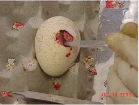
The objective of the first experiment was to develop a technique to manipulate the turkey fetus three days prior to hatch. In order to understand satellite cell mitotic activity, an injection of 5-Bromo-2’-deoxyuridine (BrdU), a thymidine analog, can be given and detected via immunohistochemistry. The post-hatch bird can be given an intra-peritoneal injection of BrdU; however, the hard shell surrounding the avian fetus makes it difficult to administer BrdU to a precise location in the fetus during late development in the turkey. A successful method was accomplished and employed in experiment two.
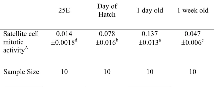
The objectives of the second experiment were to study the injection of nutrients before hatch to the turkey fetus and to determine the influence of myogenic satellite cell mitotic activity and muscle development before hatch and immediately post-hatch. The nutrient injection did not improve muscle development (P ≥ 0.05) following hatch when compared to a saline injected control. However, satellite cell mitotic activity was highest at day of hatch and one day of age (P≤ 0.05) when compared to one week of age
The objective of the third experiment was to determine the influence of β-hydroxy β-methylbutyrate (HMB) and fasting during the first week period on satellite cell mitotic
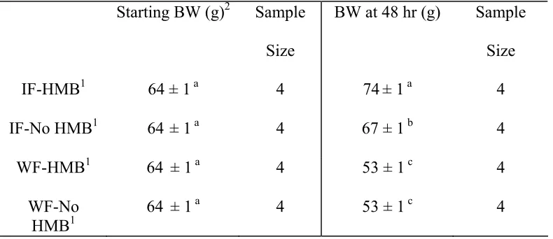
activity, and muscle development. This experiment also employed BrdU injection for satellite cell mitotic activity determination. The detection of the protein Pax7, found in quiescent myogenic satellite cells and recently activated satellite cells, was employed in this experiment.Immediately fed poults given a diet containing HMB had higher body weights (P ≤ 0.01) at 48 hours and one week of age, and had higher satellite cell mitotic activity at 48 hours of age (P ≤ 0.01) compared to the immediately fed poults on a standard industry based starter diet and fasted poults. However, the fasted poults had the lowest amount of satellite cell mitotic activity (P ≤ 0.01) at 48 hours post-hatch than the other two groups. Therefore, HMB may play an anabolic role in early post-hatch muscle development.
THE INFLUENCE OF EARLY NUTRITION ON MUSCLE
DEVELOPMENT IN THE POULT
By
DANIEL TODD MOORE
A dissertation submitted to the Graduate Faculty of North Carolina State University in partial fulfillment of the requirement for the Degree of Doctor of Philosophy
NUTRITION
RALEIGH 2005
APPROVED BY:
_________________
_________________
Dr. Peter Ferket Dr. Jesse Grimes
(Chair of Advisory Committee)
_________________
_________________
Dr. Paul Mozdziak Dr. Jack Odle
BIOGRAPHY
Daniel Todd Moore was born November 17, 1974, in Rockford, Illinois. He graduated from Kirksville Senior High School in Kirksville, Missouri in 1993. He received a Bachelor of Science degree in Animal Science in 1997 from the University of Missouri-Columbia in Columbia, Missouri. In 2000, he completed a Master of Science degree in Animal Science emphasizing poultry nutrition at the University of Missouri-Columbia. From 2000 until the present he has been compiling the requirements for a Ph.D. degree in Nutrition with a minor in Biotechnology at North Carolina State University in Raleigh, North Carolina.
ACKNOWLEDGEMENTS
I thank everyone involved in my doctoral program. I thank Drs. Paul Mozdziak and Peter Ferket for accepting me as their graduate student, and for providing me with excellent guidance and assistance in completing my program. I also thank Drs. Jesse Grimes and Jack Odle for participating as my graduate committee members and for their help and assistance.
I thank Jennifer Bradford, Darell McCoy, and Annette Israel for their advice and technical assistance. Special thanks to Pam Jenkins for statistical assistance. I would also like to thank Debbie Cox and Jennifer Bernhardt for their secretarial assistance. I am thankful to Robbie Upton, Jody Smith, Chris Parks, Scott Crow, and Ondulla Foye and the rest of the graduate students for assisting me when I needed help, and most of all for their friendship.
I would like to thank my parents David and Brenda Moore, my brothers and sisters-in-law, John and Kim Moore, Bryan and Carmen Moore, Adam Moore, and Brett and Jennifer Moore, as well as, my nieces and nephew Abby, Anna, and Levi Moore for sacrificing their son, brother, brother-in-law, and uncle for his pursuit of education and their love throughout the process. I would also like to thank my father-in-law and
mother-in-law, Jim and Melvina Fowler, as well as, my sisters-in-law and brother-in-law, Leigh Anne Fowler, Noah and Myra Spencer, and Lindsey Fowler for being my family away from home and support.
TABLE OF CONTENTS
LIST OF TABLES vi
LIST OF FIGURES vii
1 LITERATURE REVIEW 1
1.1 Breast Muscle Yield in Turkeys 1
1.11 Breast Muscle Production 1
1.12 Influence of Fast Growing Lines on Breast Muscle 2
1.13 Characteristics of Breast Muscle 3
1.2 Impact of Early Nutrition on Poultry 4
1.21 Aspects of Early Nutrition 5
1.22 In Ovo Feeding 5
1.23 β-Hydroxy-β-Methylbutyrate (HMB) in Feed 6 1.24 Impact of Fasting on Avian Muscle Development 7
1.3 Myogenic Satellite Cells 9
1.31 Myogenic Satellite Cells in Muscle Development 10 1.32 Age Related Changes in Myogenic Satellite Cells 11 1.33 Myogenic Satellite Cell Activity in Various Strains of Poultry 12 1.34 Growth Factors Involved in Myogenic Satellite Cell Dynamics 13 1.35 Other Factors Impacting Myogenic Satellite Cell Dynamics 15 1.36 Sub-populations of Myogenic Satellite Cells 17 1.37 Regulatory Factors Involved with Muscle Development and
Myogenic Satellite Cells 18
1.4 Pax7 Involvement in Myogenic Satellite Cells 20
1.41 Structure of Pax7 20
1.42 Pax7 Transcripts 21
1.43 Commitment to the Myogenic Lineage of Cells Expressing Pax7 22 1.44 Pax7 Expression Maintains a Proliferative Reserve of Satellite
Cells 22
1.5 Stem Cells in Skeletal Muscle 24
1.51 Stem Cells Within the Satellite Cell Population 24 1.52 Proteins Expressed in Stem Cells Found in Skeletal Muscle 26
1.6 References 28
2 OVERALL OBJECTIVE 47
3 BROMODEOXYURIDINE ADMINISTRATION TO AVIAN
FETUSES 51
3.1 Abstract 51
3.2 Introduction 52
3.3 Materials and Methods 54
3.4 Results 57
3.5 Discussion 58
3.6 References 60
4 THE EFFECT OF IN OVO NUTRIENT INJECTION ON SATELLITE CELL MITOTIC ACTIVITY 67 4.1 Abstract 67
4.2 Introduction 68
4.3 Materials and Methods 73
4.4 Results 77
4.5 Discussion 78
4.6 References 82
5 THE EFFECT OF EARLY NUTRITION ON SATELLITE CELL DYNAMICS IN THE YOUNG TURKEY 90 5.1 Abstract 90
5.2 Introduction 91
5.3 Materials and Methods 95
5.4 Results 100
5.5 Discussion 103
5.6 References 109
6 THE EFFECT OF EARLY POST-HATCH FASTING ON SATELLITE CELL DYNAMICS IN THE YOUNG TURKEY 122
6.1 Abstract 122
6.2 Introduction 123
6.3 Materials and Methods 126
6.4 Results 131
6.5 Discussion 134
6.6 References 142
LIST OF TABLES
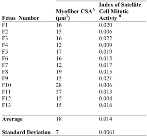
3.1 Myofiber cross-sectional area (CSA), satellite cell mitotic activity, and nuclei per unit area of 25-day fetuses determined using the in ovo
administration of BrdU 62
4.1 Body weight, gain, and Pectoralis thoracicus weight relative to body
weight at one week of age, and Pectoralis supracoracodeus weight
relative to body weight at one week of age 87 4.2 Myofiber diameter, satellite cell mitotic activity, nuclei per unit area
(5-Bromo-2’-deoxyuridine (BrdU) labeled for 2 hours) by treatment 88 4.3 Satellite cell mitotic activity by age 89
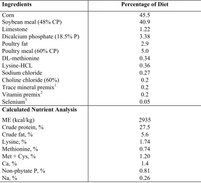
5.1 Basal diet composition 114
5.2 Body weights at 48 hours of age 115
5.3 Body weights at one week of age and one week weight gain 116
5.4 Pectoralis thoracicus weight, Pectoralis supracoracodeus weight,
Pectoralis thoracicus weight/body weight ratio, Pectoralis
supracoracodeus weight/body weight ratio at one week of age 117
5.5 Myofiber cross-sectional area (CSA), satellite cell mitotic activity,
nuclei per unit area by treatment at 24 and 48 hours of age 118 5.6 Myofiber cross-sectional area (CSA), satellite cell mitotic activity,
nuclei per unit area by treatment at one week of age 119 5.7 Pax7 labeling index and Pax7/BrdU labeling index ratio by treatment
at 24 and 48 hours of age 120
5.8 Pax7 labeling index and Pax7/BrdU labeling index ratio by treatment
at one week of age 121
6.1 Basal diet composition 150
6.2 Pectoralis thoracicus weight, Pectoralis supracoracodeus weight,
Pectoralis thoracicus weight/body weight ratio, Pectoralis
LIST OF FIGURES
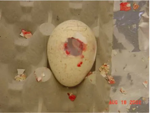
3.1 Injection of BrdU into 25E turkey embryo in ovo 63 3.2 Egg sealed and ready to return to incubator 64
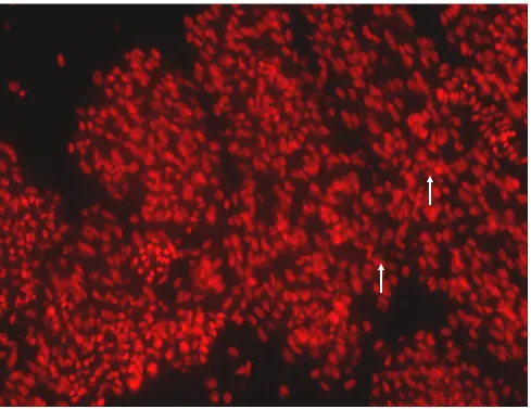
3.3 BrdU labeled nuclei of 25E 65
3.4 Propidium Iodide labeled nuclei 66
6.1 Body weights 152
6.2 Myofiber diameters 153
6.3 Satellite cell mitotic activity 154
6.4 Pax7 labeling index 155
6.5 Bcl-2 labeling index 156
6.6 Nuclei per µm2 157
6.7 Pax7/BrdU labeling index ratio 158
6.8 Pax7+/BrdU+ labeling index 159
6.9 Bcl-2/BrdU labeling index ratio 160
1. LITERATURE REVIEW
1.1 BREAST MUSCLE YIELD IN TURKEYS
Breast meat yield is becoming an important aspect of the turkey industry because the goal of many operations is to maximize the amount of breast meat yield at market age by selecting for fast growing turkey lines and feeding high levels of proteins. The use of varying levels of protein and amino acids for breast meat production has been extensively studied (Noll, 2002), and maximizing production through conventional dietary manipulations will reach a limit. Other avenues of improving breast meat yield will need to be explored to go beyond the conventional nutritional and genetic programs.
1.11 BREAST MUSCLE PRODUCTION
A simplistic way to view breast muscle production is calculating the rate of protein deposition as the difference between protein synthesis and protein degradation. Kang et al. (1985) studied that relationship in turkey poults from one to eight weeks of age using regression equations of protein content and muscle weights on body size. Protein synthesis decreased from 56% to 10%/day and protein degradation decreased from 22% to 7%/day in the Pectoralis thoracicus over the seven week period (Kang et
assume the deposition rate will remain lowered throughout the growth period beyond eight weeks of age.
Another method of evaluating breast muscle production is to observe breast muscle yield as a percentage of carcass weight or live weight. Adams and Stadelman (1991) measured breast meat yield as a percentage of preslaughter and chilled carcass weight in turkeys of three different size categories at market age. It was found that breast meat yield increases as a percentage of both weights in larger turkeys of the same age (Adams and Stadelman, 1991). Essentially, the amount of breast meat yield is directly related to the size of the bird. Interestingly, the percentage of breast meat yield continues to increase as the birds become larger, which is not true of other muscle groups in the turkey (Adams and Stadelman, 1991). This may be due to the increased levels of protein deposition in the breast muscle compared to other muscles at market age (Kang et al., 1985). However, Kang et al. (1985) used mixed sex samples for their determination, in contrast to Adams and Stadelman (1991) who employed only tom turkeys, which may influence breast muscle yield and protein deposition. Since an increase in breast meat yield is related to bird size, turkeys can be marketed at a younger age if a target size is reached without negatively influencing breast meat yield.
1.12 INFLUENCE OF FAST GROWING LINES ON BREAST
MUSCLE
that were heavier at hatch had higher pectoralis muscle weights at market age (Sklan et al., 2003), indicating a similar trend in chickens. To further the understanding of
selecting rapid growth lines of chickens, Remignon et al. (1995) studied the difference in characteristics of the Pectoralis thoracicus between fast growing and slow growing lines
of chicken. The authors found no differences in the distribution of fiber types between the lines (Remignon et al., 1995). However, birds from the faster growing lines exhibit larger myofiber areas (Remignon et al., 1995). In turkeys, differences in muscle
organization between sexes and between lines selected for different growth rates exist at 25 days of incubation (Velleman et al., 2002), suggesting an embryonic frame for programming muscle.
1.13 CHARACTERISTICS OF BREAST MUSCLE
Turkey breast muscle is composed of 90-100% white muscle fibers (Wiskus et al., 1976). Interestingly, fibers in other muscles in the turkey appear red from a high
soft, and exudative (PSE) meat in pork (Greaser, 1986). The PSE condition is also found in turkeys, and may be more prominent in fast growing lines of turkeys (Sosnicki and Wilson, 1991). Elevated levels of postmortem glycolysis in pigs and turkeys results in a rapid decrease in postmortem pH (Pietrzak et al., 1997). In turkeys PSE, may be
counteracted by quickly chilling the carcass (Dransfield and Sosnicki, 1999).
1.2 IMPACT OF EARLY NUTRITION ON POULTRY
In normal commercial production practices, poults often do not have access to feed and water for 48 to 72 hours. The energy required to emerge from the shell leaves the newly hatched poult in a nutrient deficient state (Uni and Ferket, 2004). Glycogen is the primary energy source available to the fetus when hatching; however, upon
1.21 ASPECTS OF EARLY NUTRITION
Providing poults and chicks with feed, aids in the development of the
gastrointestinal tract, and can up-regulate brush border enzymes (Uni, 1998). Organ systems also have high levels of growth during the first week post-hatch in poults relative to body weight, indicating a need for nutrients (Lilburn, 1998). Different sources of protein and energy have varying levels of impact on poults (Lilburn, 1998), showing a need for more digestible nutrients. Noy and Sklan (1999a) found that offering nutrients to poults in solid, semi-solid, or liquid form immediately post-hatch improved body weight and breast meat percentage of body weight at market age. Finally, without access to feed and water, the development of the early poult is dependent on residual nutrients found in the yolk sac that have been depleted during the hatching process (Uni and Ferket, 2004), which can result in a mortality rate of about 5%, poor growth, decreased disease
resistance, and impaired levels of muscle development (Uni and Ferket, 2004).
1.22 IN OVO FEEDING
The term in ovo feeding refers to the administration of nutrients to the embryo before hatch. It is proposed that in ovo feeding will help bridge the nutrient gap between hatch and placement at the farm (Uni and Ferket, 2004). Al-Murrani (1982) injected amino acids in ovo to chicken embryos and found an increase in body weight from hatch
macrophage response in poults and chicks (Gore and Qureshi, 1997). Recombinant human insulin-like growth factor-I (rhIGF-1) was injected into incubating chicken eggs to determine the influence on skeletal muscle development (Kocamis et al., 2002). The rhIGF-I injection altered the expression of skeletal muscle development factors,
myostatin and transforming growth factor-β2 (TGF-β2) in ovo (Kocamis et al., 2002). However, little other research has been performed providing nutrients in ovo or via in ovo feeding.
1.23 β-HYDROXY-β-METHYLBUTYRATE (HMB) IN FEED
One possible nutrient that may be used in early post-hatch diets to improve muscle development is HMB, which is a leucine metabolite that is formed endogenously by the oxidation of α-ketoisocaproate (KIC) and is finally converted to HMG-CoA used in cholesterol synthesis needed for membrane production and repair (Nissen and
Abumrad, 1997). An alternative pathway for HMB utilization that has not been shown or tested is through its action as a butyrate. Butyrates have been shown to inhibit the
activity of the enzyme histone deacetylase (HDAC; Davie, 2003). The decrease in HDAC results in an increase in follistatin and ultimately an increase in muscle growth following a period of stress (Iezzi et al., 2004).
well as, decrease muscle damage during a period of stress to the muscle (Gallagher et al., 2000; Panton et al., 2000; Vukovich et al., 2001), which may be beneficial to animal agriculture.
In vitro, cultures of whole chicken muscles showed a decrease in proteolysis after
the administration of HMB (Ostaszewski, 2000). It has also been shown that HMB decreases proteolysis in young rat muscle following a period of stress by altering the activity of proteolytic enzymes (Jank et al., 2000). However, HMB does not seem to be as effective in more mature animal muscle because broiler chickens showed no increase in body weight and carcass yield at 42 days following HMB supplementation, and weanling pigs did not show an increase in average daily gain following HMB supplementation (Nissen et al., 1994; Gatnau et al., 1995).
1.24 IMPACT OF FASTING ON AVIAN MUSCLE DEVELOPMENT
Glycogen is considered a primary energy source available to the embryo while hatching; therefore, glycogen stores are greatly reduced during the emergence from the shell (John et al., 1987; John et al., 1988), resulting in a newly hatched poult with low glycogen reserves. However, phosphorylase a, a glycogen degradation enzyme involved in the mobilization of glycogen, is at a high level in the one-day-old poult when
possible increase in glycogen reserves needed for a period of stress. If glycogen reserves are depleted at hatch and mild stressors can cause an increase in plasma glucose levels (Donaldson and Christensen, 1991), few energy sources remain for secondary
physiological functions such as muscle development. It also has been shown that both chicks and poults with delayed access to feed and water for 48 hours resulted in a severe shortage of energy that altered body composition (Pinchasov and Noy, 1993).
Body weights are also lower because of the shortage of energy as a result of fasting. Considerable research has shown that chicks with delayed access to feed and water for the first 48 hours post-hatch had lower body weights that were present at market age (Nir and Levanon, 1993; Noy and Sklan, 1999b; Vieira and Moran 1999a). This change in body weight following fasting immediately post-hatch also has resulted in a reduction in meat yield at market age (Vieira and Moran, 1999b). A decrease in all aspects of intestinal development also has been shown to exist following immediate post-hatch fasting (Geyra et al., 2001), which along with decreased energy status results in a lower performance to market age.
Withholding feed during the immediate post-hatch (2-3 days post-hatch) period also negatively impacts muscle development of poultry. In chickens not provided feed for 48 hours following hatch, protein synthesis in the Pectoralis thoracicus was
significantly reduced when compared to fed chicks (Yaman et al., 2000). Also,
Pectoralis thoracicus IGF-I and myostatin mRNA levels increased from hatch to two
Apoptosis of chicks fasted for three days was 88.8% of all nuclei with the amount of apoptosis occurring in fed chicks at the same age was only 1.1% (Mozdziak et al., 2002), giving further indication to the detrimental effect of immediate post-hatch fasting on muscle development.
1.3 MYOGENIC SATELLITE CELLS
Postnatal or post-hatch myofibers contain myonuclei that are incapable of mitotic division (Stockdale and Holtzer, 1961).However, the acquisition of new DNA units, considered the myonucleus and the surrounding cytoplasm, or the increase of existing DNA unit size are the only two mechanisms for post-hatch myofiber hypertrophy. Since there is a limit to DNA unit size and myonuclei are post-mitotic, satellite cell fusion is the mechanism driving muscle growth (Horsley et al., 2001; McCall et al., 1998; Roy et al., 1999). Myonuclear accretion is regulated by the surrounding myofibers (Bischoff, 1990; Mozdziak et al., 1998a). Satellite cells are located between the basal lamina and the sarcolemma of the myofiber and run the entire length of the fiber (Mauro, 1961;
1.31 MYOGENIC SATELLITE CELLS IN MUSCLE
DEVELOPMENT
Embryological muscle development begins with the fusion of mononucleated myoblasts to form myotubes, which mature into myofibers (Schultz and McCormick, 1994). During avian development, a distinct population of satellite cells is present at the midfetal stages of development (Feldman and Stockman, 1992), indicating the
importance of satellite cells from an early developmental time. The developmental origin of satellite cells remains unclear (Buckingham et al., 2003; Chen and Goldhamer, 2003). However, a majority skeletal muscle progenitors arise in the somites (Chen and
1.32 AGE RELATED CHANGES IN MYOGENIC SATELLITE
CELLS
Satellite cells are first associated with myofibers at 19 days of gestation in the mouse with an estimated decrease in satellite cell mitotic activity from 32% to 6% from birth to adulthood (Cardasis and Cooper, 1975), showing a decrease in age-related satellite cell mitotic activity. Mozdziak et al. (1994) show a similar age-related in vivo
decrease in satellite cell mitotic activity from 3 to 9 weeks of age in the turkey. As a result of high levels of postnatal satellite cell mitotic activity, a reduction in muscle size at maturity can result from a short-term reduction in satellite cell mitotic activity early in life (Mozdziak et al., 1997; Mozdziak et al., 2000). In vitro, satellite cells from mature
turkeys retain their proliferative capacity (Doumit et al., 1990), indicating that satellite cells have the ability to proliferate in the older animal but the in vivo environment may
not favor a proliferative state. The classical model for myofiber growth was that satellite cells were orderly added to the fibers to maintain a constant DNA unit size (Moss, 1968). However, from 9 to 26 weeks of age, myofiber growth in turkeys occurred mainly
1.33 MYOGENIC SATELLITE CELL ACTIVITY IN VARIOUS
STRAINS OF POULTRY
If satellite cell mitotic activity plays a major role in governing mature muscle size, then it is possible that genetic lines of poultry selected for different levels of production may have varying levels of satellite cell mitotic activity. Chicks that had the same body weight at hatch, showed no differences in satellite cell mitotic activity in the Pectoralis
thoracicus (Sklan et al., 2003). However, chicks that were heavier in body weight at
hatch had higher satellite cell mitotic activity in the pectoralis muscles that and had heavier pectoralis muscle weights at five days post-hatch and at market age (Sklan et al., 2003). In turkeys, birds selected for fast growth showed an increase in satellite cell proliferation in vitro when compared to a randomly bred line of turkeys (Velleman et al.,
2000), suggesting a difference in satellite cell dynamics between two different lines of turkeys. The dynamics of satellite cells in the Pectoralis thoracicus was also studied in
vitro between Nicholas poults (a fast growing line) and Merriam’s poults (a slow growing
line) (McFarland et al., 1993a). A greater growth rate of the Nicholas poults was
observed when compared to the Merriam’s poults that was associated with an increase in satellite cell proliferation and an increased responsiveness of satellite cells to mitogenic stimuli (McFarland et al., 1993a), suggesting a relationship to satellite cell dynamics at muscle development. Interestingly, the Merriam’s poults had a higher responsiveness to signals associated with differentiation in vitro (McFarland et al., 1993a), which may
1.34 GROWTH FACTORS INVOLVED IN MYOGENIC SATELLITE
CELL DYNAMICS
Growth factors are small polypeptides that are usually secreted by cells to influence themselves or adjacent cells via autocrine or paracrine regulation (Dodson et al., 1996). A family of growth factors is the insulin-like growth factors (IGF), which is found in two forms, IGF-I and IGF-II (McFarland, 1999). In rat satellite cells in vitro,
IGF-I alone stimulated proliferation and differentiation (Allen and Boxhorn, 1989). Other
in vitro research involving rat satellite cells supports these findings showing IGF-I
activates proliferation then stimulates events leading to differentiation (Engert et al., 1996). IGF-I and IGF-II both stimulate proliferation of turkey satellite cells (McFarland et al., 1993b), which was further supported by the fact that the turkey satellite cell cultures showed higher levels of mRNA expression for IGF-II and insulin-like growth factor binding protein-2 (IGFBP-2) during proliferation with decreased levels at the onset of differentiation (Ernst et al., 1996). The influence of growth factors on satellite cell development and muscle growth is evident because satellite cells from Nicholas poults are more responsive to IGFs when compared to Merriam’s poults (McFarland et al., 1995).
Nicholas poults (McFarland et al., 1995). In contrast to the evidence presented by Allen and Boxhorn (1989), Marcelle et al. (1995) found that the avian FGF receptor, FREK, was prominently expressed during differentiation, suggesting a possible role of FGF in differentiation in the bird.
A more recently identified growth factor that influences satellite cell dynamics is hepatocyte growth factor (HGF). HGF stimulates satellite cell proliferation of rat muscle
in vitro (Allen et al., 1995). The HGF receptor c-met is present in quiescent satellite cells
(Allen et al., 1995). Gal-Levi et al. (1998) discovered that HGF increases cell
proliferation and inhibits cell differentiation of cultured chicken myoblasts. HGF in vitro
has also been shown to stimulate turkey satellite cell proliferation and inhibit satellite cell differentiation (Zeng et al., 2002).
Another mitogenic growth factor is platlet-derived growth factor (PDGF). Turkey
Pectoralis thoracicus satellite cells show a greater responsiveness to PDGF, and a greater
binding affinity to PDGF than satellite cells derived from the Biceps femoris (McFarland
et al., 1997), suggesting a greater proliferation rate of Pectoralis thoracicus satellite cells.
Finally, the growth factor transforming growth factor beta (TGF-β) has been shown to decrease proliferation and inhibit differentiation of rat satellite cells in vitro
(Allen and Boxhorn, 1989). Similarly, TGF-β inhibited proliferation and differentiation in turkey satellite cells (Yun et al., 1997).
cells. TGF-β continued to inhibit differentiation in the presence of IGF-I and FGF; however, TGF-β could not suppress the proliferation stimulated by FGF. The combination of IGF-I and FGF resulted in the highest level of proliferation and the highest level of fusion. HGF when combined with IGF-I and FGF did not have any additive of synergistic effects on the proliferation of satellite cells (Zeng et al., 2002). It is important to understand the varying levels of growth factor combination when
determining satellite cell dynamics.
1.35 OTHER FACTORS IMPACTING MYOGENIC SATELLITE
CELL DYNAMICS
Satellite cells in vivo respond to a variety of stimuli that can increase or decrease satellite cell mitotic activity. The irradiation of turkey Pectoralis thoracicus at two
cell mitotic activity on mature muscle size. Feed deprivation of chicks early post-hatch resulted in a decreased level of satellite cell proliferation and decreased muscle weight at market age (Halevy et al., 2000), which is important to the poultry industry that has a practice of delaying the placement of poults and chicks.
Interestingly, new evidence is emerging that suggests a mild stress to the bird may be beneficial to satellite cell mitotic activity and muscle development. Chicks that were exposed to a mild heat stress at three days of age resulted in an increase in satellite cell mitotic activity that resulted in increased body weight and percent breast muscle yield at market age (Halevy et al., 2001). Pophal et al., (2004) fed chicks varying levels of digestible lysine ranging from a slightly deficient level to a level well over the
requirement. The results showed the highest level of satellite cell mitotic activity was in the chicks fed a diet slightly deficient in lysine. The idea of mildly stressing young animals to increase satellite cell mitotic activity is a relatively novel idea that may have a major impact in animal agriculture.
1.36 SUB-POPULATIONS OF MYOGENIC SATELLITE CELLS
Original studies of satellite cells described them as a population of cells residing between the basal lamina and sarcolemma of the myofiber, but more recent studies suggest there are sub-populations of satellite cells. Schultz and Lipton (1982) were the first researchers to suggest satellite cell heterogeneity. Morgan and Partridge (2003) suggest that a population of satellite cells exist that may be derived from a more primitive stem cell that can adopt a more divergent phenotypic fate than a majority of the
population of satellite cells committed to muscle repair. However, even though satellite cell heterogeneity may exist, a majority of cells contributing to muscle do not appear to arise from hematopoietic or other bone marrow precursors (Sherwood et al., 2004). A recent study has confirmed the idea of a sub-population by using a clonal culture system of mouse muscle satellite cells and identifying two types of cells, “round cells” and “thick cells”, with the round cells forming clusters and producing the thick cells that committed to myogenic and osteogenic pathways (Hashimoto, 2004). Schultz (1996) found that 80% of the myogenic satellite cell population provided myonuclei to
myofibers and underwent a more rapid level of proliferation. The remaining 20% of the satellite cells underwent a slower level of proliferation, suggesting that this group of cells is a proliferative reserve maintained to create new satellite cells. Other research also found a satellite cell sub-population consisting of 28% of all satellite cells that was composed primarily of large type cells (Molnar et al., 1996). Through the use of satellite cell expression factors, it has been determined that a majority of satellite cells are
again become quiescent satellite cells (Zammit et al., 2004) displaying a proliferative reserve of satellite cells that maintain the ability to replenish the satellite cell population. Finally, it has been determined that a proliferative reserve of satellite cells exist in turkey muscle that can be identified by their proliferation rate and fusion capabilities (Rouger et al., 2004).
1.37 REGULATORY FACTORS INVOLVED WITH MUSCLE
DEVELOPMENT AND MYOGENIC SATELLITE CELLS
Myogenic regulatory factors (MRFs) are DNA binding proteins that induce a myogenic phenotype. The main MRFs include MyoD, Myf-5, myogenin, and MRF4. It has been shown in mice lacking MyoD and Myf-5 that myoblasts are not present, and myoblast found in mice lacking myogenin and MRF4 cannot differentiate (Rudnicki and Jaenisch, 1995). In tissue culture, most myoblasts express MyoD and Myf-5, but up-regulate myogenin during differentiation (Weintraub, 1993), further indicating
myogenin’s role downstream of MyoD and Myf-5. Cornelison and Wold (1997) showed that once satellite cells were activated MyoD or Myf-5 were expressed, followed by the expression of myogenin. At some time points, all four MRFs were expressed (Cornelison and Wold, 1997), suggesting a cascade of regulatory factors during muscle development.
proliferation. During proliferation, satellite cells express MyoD and a portion of the population expresses Myf-5 (Cooper et al., 1999). In regenerating mouse muscle following a muscle graft, MyoD and myogenin were not detected until after fusion into the myotubes, and they were not express in nonregenerating muscle (Fuchtbauer and Westphal, 1992), suggesting the presence of MyoD and myogenin only in activated satellite cells.
Quiescent satellite cells have been suggested to express c-met, m-cadherin, Pax7, and CD34, (Irintchev et al., 1994; Allen, 1995; Beauchamp et al., 2000; Zammit and Beauchamp, 2001; Asakura et al., 2002). However, c-met and Myf5 have been found in satellite cells further down the cascade of fusion with myofibers (Cornelison and Wold, 1997). The most important function of m-cadherin is to regulate the fusion of the satellite cell with the myofiber (Irintchev et al., 1994). CD34 expression has also been suggested to indicate muscle-derived stem cells (Hwang et al., 2004). Pax7 has been shown to be expressed in quiescent satellite cells and in a small population of satellite cells that quickly return to a quiescent state (Zammit, 2004), making Pax7 an ideal marker for studying the proliferative reserve of satellite cells.
indicates terminally differentiation (differentiation) of the muscle precursor cells into myotubes.
1.4 PAX7 INVOLVEMENT IN MYOGENIC SATELLITE CELLS
Pax7 is a protein expressed in quiescent satellite cells (Zammit and Beauchamp, 2001; Asakura et al., 2002). However, more recently it has been discovered to be present in satellite cells that are active for a brief period before reentering the quiescent state (Zammit, 2004), making Pax7 a reliable marker for studying and understanding a proliferative reserve of satellite cells (Schultz, 1996). Understanding the role of Pax7 is an integral part of understanding satellite cell dynamics in postnatal muscle and is made easier by the development of a Pax7 antibody (Kawakami et al., 1997).
1.41 STRUCTURE OF PAX7
Pax transcription factors have a conserved octapeptide and a transactivation domain that is encoded by eight or nine exons (Balczarek et al., 1997). The
DNA pre-initiation complex (Lamey et al., 2004). In binding, the paired box domain utilizes its two helix-turn-helix motif, and the homeodomain utilizes its helix-turn-helix motif (Lamey et al., 2004). Binding specificity of the homeodomain is influenced by the binding specificity of the paired domain (Mishra et al., 2002).
1.42 PAX7 TRANSCRIPTS
It has been known that Pax7 is involved in muscle development, as well as, development of the nervous system (Jostes et al., 1991). However, recently it appears that there are varying forms of Pax7 that may perform different functions. First, four alternative forms of Pax7 were discovered, and different forms were found in different tissues (Ziman et al., 1997). The four alternative forms were found to differ in their paired-box sequence (Ziman et al., 2001). All forms were expressed in the myogenic lineage, but only two forms were expressed in the neurogenic lineage (Ziman et al., 2001), suggesting the importance for Pax7 in skeletal muscle. Alternative forms of Pax7 were found to be expressed in myogenic cell lines versus tumor cell lines, which
1.43 COMMITMENT TO THE MYOGENIC LINEAGE OF CELLS
EXPRESSING PAX7
Once a cell expresses Pax7, it becomes committed to the myogenic lineage (Seale et al., 2000). Pax7 has been shown to be expressed in quiescent satellite cells (Zammit and Beauchamp, 2001; Asakura et al., 2002). Mice that are Pax7-/- do not contain satellite cells (Seale et al., 2000), which suggests that Pax7 is necessary for the formation or maintenance of satellite cells. Recently, stem cells resident in adult muscle underwent myogenic specification following the expression of Pax7 (Seale et al., 2004). These studies demonstrate the importance of Pax7 to satellite cell dynamics.
1.44 PAX7 EXPRESSION MAINTAINS A PROLIFERATIVE
RESERVE OF SATELLITE CELLS
nuclei to the existing myofiber and Pax7 may be a marker for the maintenance of a renewal population of satellite cells. The overexpression of Pax7 also down-regulates the expression of MyoD and prevents myogenin expression (Olguin and Olwin, 2004), once again demonstrating the role of Pax7 to maintain a proliferative reserve population. Further showing the need for Pax7 in maintaining a self-renewal population, cells that escape differentiation and exit the cell cycle up-regulate the expression of Pax7 (Olguin and Olwin, 2004). During the activation process of quiescent satellite cells, there is a brief period of co-expression of Pax7 and MyoD, but Pax7 is quickly down-regulated during proliferation (Zammitt et al., 2004). However, some cells during proliferation continue to express Pax7, but not MyoD, and then leave the cell cycle prior to
differentiation (Zammitt et al., 2004). Once they leave the cell cycle, they regain their quiescent satellite cell status (Zammitt et al., 2004). It may be possible that this
population of cells produces satellite cells during proliferation to further differentiate, but retain the ability to divide again at a later time. Similar findings that indicate Pax7 is an early marker of proliferation have been observed in chickens (Halevy et al., 2004). In
vitro, Halevy et al. (2004) found that at all time frames tested there was a population of
Pax7+/MyoD- cells indicating the proliferative reserve is maintained throughout the early post-hatch period. A similar relationship of Pax7 to myogenin found in the mouse has also been observed in the chicken (Halevy et al., 2004). All of these recent data indicates the most important role of Pax7 in postnatal muscle is to maintain a proliferative
1.5 STEM CELLS IN SKELETAL MUSCLE
Stem cells are cells that have a high level of plasticity and can differentiate into a variety of cell types. Stem cells in skeletal muscle can either be referred to as stem cells that give rise to skeletal muscle, presumably through a satellite cell lineage, or cells that originate in muscle and differentiate into other tissues. Both situations have been
suggested by previous work (Asakura et al., 2001, Ferrari et al., 1998; Grounds et al., 2002). However, the location of these cells and their status as satellite cells remains unclear because many of the studies have not identified cellular location before isolating cells and determining stem cell capabilities.
1.51 STEM CELLS WITHIN THE SATELLITE CELL POPULATION
that myogenic satellite cells possess mesenchymal stem cell capabilities because cells expressing Pax7 committed to myogenic and non-myogenic pathways in vitro. However,
it also has been observed in isolated muscle fibers that markers present in multipotent adult skeletal muscle-derived stem cells were not coexpressed in cells expressing Pax7 (Zammit and Beauchamp, 2001), suggesting a population of stem cells that is separate from satellite cells lying outside the lamina. Grounds et al. (2002) through an
investigation of skeletal and cardiac muscle, and the literature concluded that satellite cells did not represent a population of multipotential stem cells. At this time, it is difficult to dissect the literature to identify satellite cells with stem cell capabilities.
Stem cells also have been proposed to originate from connective tissue found in skeletal muscle. In skeletal muscle isolated from chicken embryos, it was concluded that a population of mesenchymal stem cells was present in connective tissue (Young et al., 1993). Postnatal mammals also contain pluripotent mesenchymal stem cells derived from skeletal muscle connective tissue (Young et al., 2001a). Finally, mesenchymal stem cells present in skeletal muscle connective tissue derived from fetal, adult, and geriatric donors differentiated into muscle, adipocytes, carilage, bone, fibroblasts, and endothelial cells (Young et al., 2001b), indicating the presence of stem cells within the skeletal muscle tissue.
Finally, bone marrow-derived stem cells have been implicated in muscle
regeneration. Corbel et al. (2003) found that hematopoietic stem cells gave rise to both blood and muscle tissue. A low incidence of bone marrow-derived stem cells
cells into muscle tissue (Bittner et al., 1999; Gussoni et al., 1999). LaBarge and Blau (2002) suggested that bone marrow-derived stem cells not only differentiate into muscle but also contribute to the satellite cell population.
It is difficult to determining if cells with stem cell capabilities found in muscle arise from the heterogeneous satellite cell population or are associated with connective tissue because they have not clearly been identified. A weakness of these intriguing studies was that many employed whole muscle homogenates and did not identify cellular location before identifying multipotential cells in muscle.
1.52 PROTEINS EXPRESSED IN STEM CELLS FOUND IN
SKELETAL MUSCLE
Some proteins expressed in myogenic cells with stem cell potential are CD45, stem cell antigen-1 (SCA-1), CD34, and Bcl-2. Most of the stem cell properties
found outside the basal lamina, and CD45 and Sca-1 are not specifically myogenic (Asakura et al., 2002).
CD34+/CD45- cells isolated from skeletal muscle possessed a small potential to differentiate into adipocytes and endothelial cells (Tamaki et al., 2003), suggesting the stem cell capabilities of CD34+/CD45- cells. In the same experiment, colony forming units that were CD34-/CD45- expressed CD34 prior to the expression of MyoD when forming myogenic cells (Tamaki et al., 2003), indicating its upstream location in the myogenic cascade. Jankowski et al. (2002) found that CD34+ cells, that were considered to be in the stem cell compartment by plating techniques, led to myogenic progenitors and differentiation in regenerating muscle. CD34+/Sca-1+ cells exhibited stem cell properties when injected into mdx mice resulting in the restoration of dystrophin
following an injection of the cells (Torrente et al., 2001). Hwang et al. (2004) showed the stem cell capabilities of skeletal muscle CD34+ or Sca-1+ and CD45- cells by co-culture cells with smooth muscle resulting in the differentiation into smooth muscle.
1.6 REFERENCES
Adams, R.L. and W.J. Stadelman. 1991. Research note: yield of large tom turkeys. Poult. Sci. 70:2379-2381.
Allen, R.E., and L.K. Boxhorn. 1989. Regulation of skeletal muscle satellite cell
proliferation and differentiation by transforming growth factor-beta, insulin-like growth factor 1, and fibroblast growth factor. J. Cell. Physio. 138:311-315.
Allen, R.E., R.A. Merkel, and R.B. Young. 1979. Cellular aspects of muscle growth: myogenic cell proliferation. J. Ani. Sci. 49:115-127.
Allen, R.E., S.M. Sheehan, R.G. Taylor, T.L. Kendall, and G.M. Rice. 1995. Hepatocyte growth factor activates quiescent skeletal muscle satellite cells in vitro. J. Cell Physio. 165:307-312.
Al-Murrani, W.K., 1982. Effect of injecting amino acids into the egg on embryonic and subsequent growth in the domestic fowl. Br. Poult. Sci. 23:171-174.
Asakura, A., M. Komaki, and M.A. Rudnicki. 2001. Muscle satellite cells are multipotential stem cells that exhibit myogenic, osteogenic, and adipogenic differentiation. Diff. 68:245-253.
Asakura, A., P. Seale, A. Girgis-Gabardo, and M.A. Rudnicki. 2002. Myogenic
specification of side population cells in skeletal muscle. J. Cell Bio. 159(1):123-134.
Barr, F.G., J.C. Fitzgerald, J.P. Ginsberg, M.L. Vanella, R.J. Davis, and J.L. Bennicelli. 1999. Predominant expression of alternative pax3 and pax7 forms in myogenic and neural tumor cell lines. Canc. Res. 59:5443-5448.
Beauchamp, J.R., L. Heslop, D.S.W. Yu, S. Tajbakhsh, R.G. Kelly, A. Wernig, M.E. Buckingham, T.A. Partridge, and P.S. Zammit. 2000. Expression of CD34 and Myf5 defines the majority of quiescent adult skeletal muscle satellite cells. J. Cell. Bio. 151(6):1221-1233.
Bischoff, R. 1990. Interaction between satellite cells and skeletal muscle fibers. Development 109:943-952
Bittner, R.E., C. Schofer, K. Weipoltshammer, S. Ivanova, B. Streubel, E. Hauser, M. Freilinger, H. Hoger, A. Elbe-Burger, and F. Wachtler. 1999. Recruitment of bone-marrow-derived cells by skeletal and cardiac muscle in adult dystrophic mdx mice. Anat. Embryol. 199:391-396.
Buckingham, M., L. Bajard, T. Chang, P. Daubas, J. Hadchouel, S. Meilhac, D.
Montarras, D. Rocancourt, and F. Relaix. 2003. The formation of skeletal muscle: somite to limb. J. Anat. 202:59-68.
Campion, D.R. 1984.The muscle satellite cell: a review. Int. Rev. Cytol. 87:225-251.
Cardasis, C.A., and G.W. Cooper. 1975. An analysis of nuclear numbers in individual muscle fibers during differentiation and growth: a satellite cell-muscle fiber growth unit. J. Exp. Zoo. 191:347-351.
Chen, J.C., and D.J. Goldhamer. 2003. Skeletal muscle stem cells. Repro. Bio. Endo. 1:101.
Chi, N. and J.A. Epstein. 2002. Getting your pax straight: Pax proteins in development and disease. Trends. Genet. 18:41-47.
Cooper, R.N., S. Tajbakhsh, V. Mouly, G. Cossu, M. Buckingham, and G.S. Butler-Browne. 1999. In vivo satellite cell activation via myf5 and myoD in regenerating mouse skeletal muscle. J. Cell Sci. 112:2895-2901.
Corbel, S.Y., A. Lee, L. Yi, J. Duenas, T.R. Brazelton, H.M. Blau, and F.M.V. Rossi. 2003. Contribution of hematopoietic stem cells to skeletal muscle. Nature Med. 9(12):1528-1532.
Cornelison, D.D. and B.J. Wold. 1997. Single-cell analysis of regulatory gene expression in quiescent and activated mouse skeletal muscle satellite cell. Dev. Bio. 191:270-283.
Darr, K.C. and E. Schultz. 1989. Hindlimb suspension suppresses muscle growth and satellite cell proliferation. J. Appl. Physio. 67(5):1827-1834.
Dangott. B., E. Schultz, and P.E. Mozdziak. 2000. Dietary creatine monohydrate supplementation increases satellite cell mitotic activity during compensatory hypertrophy. Int. J Sports Med. 21:13-16.
Deasy, B. M., R. Jankowski, and J. Huard. 2001. Muscle-derived stem cells:
characterization and potential for cell-mediated therapy. Blood Cells Mol. Dis. 27(5):924-933.
Dodson, M.V., D.C. McFarland, A.L. Grant, M.E. Doumit, and S.G. Velleman. 1996. Extrinsic regulation of domestic animal-derived satellite cells. Dom. Ani. Endo. 13(2):107-126.
Dominov, J.A., J.J. Dunn, and J.B. Miller. 1998. Bcl-2 expression identifies an early stage of myogenesis and promotes clonal expansion of muscle cells. J. Cell Bio. 142(2):537-544.
Donaldson, W.E., and V.L. Christensen. 1991. Dietary carbohydrate level and glucose metabolism in turkey poults. Comp. Biochem. Physiol. 98A (2):347-350.
Doumit, M.E., D.C. McFarland, and R.D. Minshall. 1990. Satellite cells of growing turkeys: influence of donor age and sex on proliferation and differentiation in vitro. Exp. Cell Res. 189:81-86.
Dransfield, E. and A.A. Sosnicki. 1999. Relationship between muscle growth and poultry meat quality. Poult Sci. 78:743-746.
Engert, J.C., E.B. Berglund, and N. Rosenthal. 1996. Proliferation precedes
differentiation in IGF-I-stimulated myogenesis. J. Cell Bio. 135(2):431-440.
Feldman, J.L., and F.E. Stockdale. 1992. Temporal appearance of satellite cells during myogenesis. Dev. Biol. 153:217-226.
Ferrari, G., G. Cusella-DeAngelis, M. Coletta, E. Paolucci, A. Stornaiuolo, G. Cossu, and F. Mavillo. 1998. Muscle regeneration by bone marrow-derived myogenic
progenitors. Sci. 279:1528-1530.
Fuchtbauer, E.M. and H. Westphal. 1992. MyoD and myogenin are coexpressed in regenerating skeletal muscle of the mouse. Dev. Dyn. 193:34-39.
Gallagher, P.M., J.A. Carrithers, M.P. Godard, K.E. Schulze, and S.W. Trappe. 2000. β -hydroxy-β-methylbutyrate ingestion, Part I: effects on strength and fat free mass. Med Sci. Sports Exerc. 32(12):2109-2115.
Gal-Levi, R., Y. Leshem, S. Aoki, T. Nakamura, and O. Halevy. 1998. Hepatocyte growth factor plays a dual role in regulating skeletal muscle satellite cell proliferation and differentiation. Biochi. Biophy. Acta 1402:39-51.
Gatnau, R., D.R. Zimmerman, S.L. Nissen, M. Wanneuhler, and R.C. Ewan. 1995. Effects of excess dietary leucine an leucine catabolites on growth and immune responses in weanling pigs. J. Anim. Sci. 73:159-165.
Geyra, A., Z. Uni, and D. Sklan. 2001. The effect of fasting at different ages on growth and tissue dynamics in the small intestine of the young chick. Br.J. Nutr. 86:53-61.
Greaser, M.L. 1986. Conversion of muscle to meat. In: P. Bechtel (Ed.) Muscle as Food. 37-102. Academic Press, New York.
Grounds, M.D., J.D. White, N. Rosenthal, and M.A. Bogoyevitch. 2002. The role of stem cells in skeletal and cardiac muscle repair. J. of Histo. Cyto. 50(5):589-610.
Guernec, A., B. Chevalier, and M. J. Duclos. 2004. Nutrient supply enhances both IGF-I and MSTN mRNA levels in chicken muscle. Dom. Ani. Endo. 26:143-154.
Gussoni, E. Y. Soneoka, C.D. Strickland, E.A. Buzney, M.K. Khan A.F. Flint, L.M. Kunkel, and R.C. Mulligan. 1999. Dystrophin expression in the mdx mouse restored by stem cell transplantation. Nature 401:390-394.
Halevy, O., A. Geyra, M. Barak, Z. Uni, and D. Sklan. 2000. Early posthatch starvation decreases satellite cell proliferation and skeletal muscle growth in chicks. J. Nutr. 130:858-864.
Halevy, O., A. Krispin, Y. Leshem, J.P. McMurtry, and S. Yahav. 2001. Early-age heat exposure affects skeletal muscle satellite cell proliferation and differentiation in chicks. Am. J. Physio. Reg. Int. Comp. Physio. 281:R302-R309.
Halevy, O., Y. Nadel, M. Barak, I. Rozenboim, and D. Sklan. 2003. Early posthatch feeding stimulates satellite cell proliferation and skeletal muscle growth in turkey poults. J. Nutr. 133:1376-1382.
Halevy, O., Y. Piestun, M.Z. Allouh, B.W.C. Rosser, Y. Rinkevich, R. Reshef, I.
Hartley, R.S., E. Bandman, and Z. Yablonka-Reuveni. 1992. Skeletal muscle satellite cells appear during late chicken embryogenesis. Dev. Bio. 153:206-216.
Hashimoto, N., T. Murase, S. Kondo, A. Okuda, and M. Inagawa-Ogashiwa. 2004. Muscle reconstitution by muscle satellite cell descendants with stem cell-like properties. Dev. 131:5481-5490.
Horsley, V., B.B. Friday, S. Matterson, K.M. Kegley, J. Gephardt, and G.K. Pavlath. 2001. Regulation of the growth multinucleated muscle cells by an NFATC2-dependent pathway. J. Cell Bio. 153(2):329-338.
Hwang, J.H., S.H. Yuk, J.H. Lee, W.S. Lyoo, S. Ghil, S.S. Lee, I.G. Khang, S.Y. Paik, and J.Y. Lee. 2004. Isolation of muscle derived stem cells from rat and its smooth muscle differentiation. Mol. Cells 17(1):57-61.
Iezzi, S., M.D. Padova, C. Serra, G. Caretti, C. Simone, E. Maklan, G. Minetti, P. Zhao, E.P. Hoffman, P. L. Puri, and V. Sartorelli. 2004. Deacetylase inhibitors increase muscle cell size by promoting myoblast recruitment and fusion through induction of follistatin. Dev. Cell 6:673-684.
Irintchev, A., M. Zeschnigk, A. Starzinski-Powitz, and A. Wernig. 1994. Expression pattern of m-cadherin in normal, denervated, and regenerating mouse muscles. Dev. Dyn. 199:326-337.
Jank, M., P. Ostaszewski, S. Rosochacki, J. Wilczak, and B. Balasinska. 2000. Effect of 3-hydroxy-3-methylbutyrate (HMB) on muscle cathepsin and calpain activities during the post-dexamethasone recovery period in young rats. Polish J. of Vet. Sci. 3(4):213-218.
Jankowski, R.J., B.M Deasy, B. Cao, C. Gates, and J. Huard. 2002. The role of CD34 expression and cellular fusion in the regeneration capacity of myogenic progenitor cells. J. Cell Sci. 115:4361-4374.
John, T.M., J.C. George, and E.T. Moran Jr. 1987. Pre- and post-hatch ultrastructure and metabolic changes in the hatching muscle of turkey embryos from antibiotic and glucose treated eggs. Cytobios 49:197-210.
John, T.M., J.C. George, and E.T. Moran Jr. 1988. Metabolic changes in pectoral muscle and liver of turkey embryos in relation to hatching: influence of glucose and antibiotic-treatment of eggs. Poult. Sci. 67:463-469.
Jostes, B., C. Walther, and P. Gruss. 1990. The murine paired box gene, pax7, is expressed specifically during the development of the nervous and muscular system. Mech. Dev. 33:27-37.
Kang, C.W., M.L. Sundae, and R.W. Swick. 1985. Characteristics of growth and protein turnover in skeletal muscle of turkey poults. Poult. Sci. 64:380-387.
Kocamis, H., S.A. Gahr, J. Richter, D.C. Kirkpatrick-Keller, and J. Killefer. 2002. Myostatin and TGF-β2 gene expression patterns in response to in ovo
administration of rhIGF-I during chicken embryonic development. Growth, Dev., Aging. 66:3-10.
Labarge, M.A., and H.M. Blau. 2002. Biological progression from adult bone marrow to mononucleate muscle stem cell to multinucleate muscle fiber in response to injury. Cell 111:589-601, 2002.
Lamey, T.M., A. Koenders, and M. Ziman. 2004. Pax genes in myogenesis: alternate transcripts add complexity. Histol. Histopath. 19:1289-1300.
Lee, J.Y., Z. Qu-Peterson, B. Cao, S. Kimura, R. Jankowski, J. Cummins, A. Usas, C. Gates, P. Robbins, A. Wernig, and J. Huard. 2000. Clonal isolation of muscle-derived cells capable of enhancing muscle regeneration and bone healing. J. Cell Bio. 150(5):1085-1099.
Lilburn, M.S. 1998. Practical aspects of early nutrition for poultry. J. Appl. Poult. Res. 7:420-424.
Marcelle, C., J. Wolf, and M. Bronner-Fraser. 1995. The in vivo expression of the FGF
receptor FREK mRNA in avian myoblasts suggests a role in muscle growth and differentiation. Dev. Bio. 172:100-114.
McCall, G.E., D.L. Allen, J.K. Linderman, R.E. Grindeland, R.R. Roy, V.R. Mukku, and V.R. Edgerton. 1998. J. Appl. Physiol. 84(4):1407-1412.
McFarland, D.C. 1999. Influence of growth factors on poultry myogenic satellite cells. Poult Sci. 78:747-758.
McFarland, D.C., K.K. Gilkerson, J.E. Pesall, R.H. Wellenreiter, N.H. Ferrin, W.V. Ye, Y.Yun, and L.S. Vander Wal. 1997. Comparison of growth factor receptors and metabolic characteristics of satellite cells derived from the biceps femoris and pectoralis major muscles of the turkey. Gen. Comp. Endo. 105:114-120.
McFarland, D.C., J.E. Pesall, and K.K. Gilkerson. 1993b. The influence of growth factors on turkey embryonic myoblasts and satellite cells in vitro. Gen. Comp. Endo.
89:415-424.
McFarland, D.C., J.E. Pesall, K.K. Gilkerson, and N.H. Ferrin. 1995. The response to growth factors of cultured satellite cells derived from turkeys having different growth rates. Cytobios 82:229-238.
McFarland, D.C., J.E. Pesall, K.K. Gilkerson, and T.A. Swenning. 1993a. Comparison of the proliferation and differentiation of myogenic satellite cells derived from merriam’s and commercial variety of turkeys. Comp. Biochem. Physio. 104A:455-460.
Molnar, G., M.L. Ho, and N.A. Schroedl. 1996. Evidence for multiple satellite cell populations and a non-myogenic cell type that is regulated differently in regenerating and growing skeletal muscle. Tissue Cell 28(5);547-556.
Morgan, J.E., and T.A. Partridge. 2003. Muscle satellite cells. Int. J. Biochem. Cell Biol. 35:1151-1156.
Moss, F.P. 1968. The relationship between the dimensions of the fibres and the number of nuclei during normal growth of skeletal muscle in the domestic fowl. Am. J. Anat. 122:555-564.
Mozdziak, P.E., J.J. Evans, and D.W. McCoy. 2002. Early posthatch starvation induces myonuclear apoptosis in chickens. J. Nutr. 132:901-903.
Mozdziak, P.E., M.L. Greaser, and E. Schultz. 1998b. Myogenin, myoD and myosin expression after pharmacology and surgically induced hypertrophy. J. Appl. Physio. 84(4):1359-1364.
Mozdziak, P.E., E. Schultz, and R.G. Cassens. 1994. Satellite cell mitotic activity in posthatch turkey skeletal muscle growth. Poult. Sci. 73:547-555.
Mozdziak, P.E., E. Schultz, and R.G. Cassens. 1997. Myonuclear accretion is a major determinant of avian skeletal muscle growth. Am. J. Physiol. 272:C565-571.
Mozdziak, P.E., Q. Truong, A. Macius, and E. Shultz. 1998a. Hindlimb suspension reduces muscle regeneration. Eur. J. Appl. Physiol. 78:136-140.
Nir, I., and M. Levanon. 1993. Research note: effect of posthatch holding time on performance and on residual yolk and liver composition. Poult. Sci. 72:1994-1997.
Nissen, S.L. and N.N. Abumrad. 1997. Nutritional role of the leucine metabolite β
-hydroxy β-methylbutyrate (HMB). J. Nutr. Biochem. 8:300-311.
Nissen, S.L., J.C. Fuller Jr., J. Sell, P.R. Ferket, and D.V. Rives. 1994. The effect of β
-hydroxy-β-methylbutyrate on growth, mortality, and carcass qualities of broiler
chickens. Poult. Sci. 73:137-155.
Noll, S.L. 2002. Feeding for live performance and breast meat yield. Proceedings Multi-State Poultry Meeting.
Noy, Y., and D. Sklan. 1999a. Different types of early feeding and performance in chicks and poults. J. Appl. Poult. Res. 8:16-24.
Noy, Y., and D.Sklan. 1999b. Energy utilization in newly hatched chicks. Poult. Sci. 78:1750-1756.
Olguin, H.C. and B.B. Olwin. 2004. Pax-7 up-regulation inhibits myogenesis and cell cycle progression in satellite cells: a potential mechanism for self-renewal. Dev. Bio. 275:375-388.
Ostaszewski, P., S. Kostiuk, B. Balasinska, M. Jank, I. Papet, and F. Glomot. 2000. The leucine metabolite 3-hydroxy-3-methylbutyrate (HMB) modifies protein turnover in muscles of laboratory rats and domestic chickens in vitro. J. Anim. Physiol.
Anim. Nutr. 84:1-8.
Oustanina, S., G. Haus, and T. Braun. 2004. Pax7 directs postnatal renewal and propagation of myogenic satellite cells but not their specification. EMBO J. 23(16):3430-3439.
Panton, L.B., J.A Rathmacher, S. Baier, and S. Nissen. 2000. Nutritional supplementation of the leucine metabolite β-hydroxy-β-methylbutyrate (HMB) during resistance training. Nutrition 16:734-739.
Pietrzak, M., M.L. Greaser, and A.A. Sosnicki. 1997. Effect of rapid rigor mortis
processes on protein functionality in pectoralis major muscle of domestic turkeys. J. Ani. Sci. 75:2106-2116.
Pinchasov, Y. and Y. Noy. 1993. Comparison of post-hatch holding time and subsequent early performance of broiler chicks and turkey poults. Br. Poult. Sci. 34:111-120.
Pophal, S., P.E. Mozdziak, and S.L. Vieira. 2004. Satellite cell mitotic activity of broilers fed differing levels of lysine. Int. J. Poult. Sci. 3(12):758-763.
Qu-Peterson, Z., B. Deasy, R. Jankowski, M. Ikezawa, J. Cummins, R. Pruchnic, J. Mytinger, B. Cao, C. Gates, A. Wernig, and J. Huard. 2002. Identification of a novel population of muscle stem cells in mice: potential for muscle regeneration. J. Cell Bio. 157(5):851-864.
Remignon, H., M.F. Gardahaut, G. Marche, and F.H. Ricard. 1995. Selection for rapid growth increases the number and the size of muscle fibres without changing their typing in chickens. J. Mus Res Cell Mot. 16:95-102.
Rudnicki, M.A. and R. Jaenisch. 1995. The MyoD family of transcription factors and skeletal myogenesis. BioEssays 17(3):203-209.
Rosebrough, R.W., E. Geis, K. Hendersen, and L.T. Frobish. 1978. Glycogen metabolism in the turkey embryo and poult. Poult. Sci. 57:747-751.
Rouger, K., M. Brault, N. Daval, I. Leroux , L. Guigand, J. Lesoeur, B. Fernandez, and Y. Cherel. 2004. Muscle satellite cell heterogeneity: in vitro and in vivo evidences for populations that fuse differently. Cell Tissue Res. 317:319-326.
Roy, R.R., S.R. Monke, D.L. Allen, and V.R. Edgerton. 1999. Modulation of myonuclear number in functionally overloaded and exercised rat plantaris fibers. J. Appl. Physiol. 87(2):634-642.
Schultz, E. and B.H. Lipton. 1982. Skeletal muscle satellite cells: changes in proliferation potential as a function of age. Mech. Age. Dev. 20:377-383.
Schultz, E. and K.M. McCormick. 1994. Skeletal muscle satellite cells. Rev. Phys. Biochem. Pharm. 123:213-57.
Seale, P., J. Ishibashi, A. Scime, and M.A. Rudnicki. 2004. Pax7 is necessary and sufficient for the myogenic specification of CD45+:Sca1+ stem cells from injured muscle. PloS 2(5):664-672.
Seale, P., L.A. Sabourin, A. Girgis-Gabardo, A. Mansouri, P. Gruss, and M.A. Rudnicki. 2000. Pax7 is required for the specification of myogenic satellite cells. Cell 102:777-786
Shefer, G., M. Wleklinski, and Z. Yablonka-Reuveni. 2004. Skeletal muscle satellite cells can spontaneously enter an alternative mesenchymal pathway. J. Cell Sci.
117:5393-5404.
Sherwood, R.I., J.L. Christensen, I.M. Conboy, M.J. Conboy, T.A. Rando, I.L. Weissman, and A.J. Wagers. 2004. Isolation of adult mouse myogenic
progenitors: functional heterogeneity of cells within and engrafting muscles. Cell 119:543-554.
Sosnicki, A.A. and B.W. Wilson. 1991. Pathology of turkey skeletal muscle: implications for the poultry industry. Food Structure 10:317-326.
Stockdale, F.E. and H. Holtzer. 1961. DNA synthesis and myogenesis. Exp. Cell Res. 24:508-520.
Tamaki, T., A. Akatsuka, Y. Okada, Y. Matsuzaki, H. Okana, and M. Kimura. 2003. Growth and differentiation potential of main- and side-population cells derived from murine skeletal muscle. Exp. Cell Res. 291:83-90.
Torrente, Y., J. Tremblay, F. Pisati, M. Belicchi, B. Rossi, M. Sironi, F. Fortunato, M. El Fahime, M.G. D’Angelo, N.J. Caron, G. Constantin, D. Paulin, G. Scarlato, and N. Bresolin. J. Cell. Bio. 152(2):335-348.
Uni, Z. 1998. Impact of early nutrition on poultry: review of presentations. J. Appl. Poult. Res. 7:452-455.
Uni, Z., and P.R. Ferket. 2004. Methods of early nutrition and their potential. World’s Poult. Sci. J. 60:101-111
Van Koevering, M. and S. Nissen. 1992. Oxidation of leucine and α-ketoisocaproate to β -hydroxy-β-methylbutyrate in vitro. Am. J. Physiol. 262(End. Met. 25)E27-E31.
Velleman, S.G., X. Liu, K.E. Nestor, and D.C. McFarland. 2000. Heterogeneity in growth and differentiation characteristics in male and female satellite cells
isolated from turkey lines with different growth rates. Comp. Biochem. Phys. Part A 125:503-509.
Vieira, S.L. and E.T. Moran, Jr. 1999a. Effects of delayed placement and used litter on broiler yields. J. Appl. Poult. Res. 8:75-81.
Vieira, S.L. and E.T. Moran, Jr. 1999b. Effects of egg origin and chick post-hatch nutrition on broiler live performance and meat yields. World’s Poult. Sci. J. 55:125-142.
Vorobyov, E. and J. Horst. 2004. Expression of two protein isoforms of pax7 is
controlled by competing cleavage-polyadenylation and splicing. Gene 342:107-112.
Vukovich, M.D., N.B. Stubbs, and R.M. Bohlken. 2001. Body composition in 70-year-old adults responds to dietary β-hydroxy-β-methylbutyrate similarly to that of young adults. J. Nutr. 131:2049-2052.
Wientraub, H. 1993. The MyoD family and myogenesis: redundancy, networks, and thresholds. Cell 75:1241-1244.
Wiskus, K.J., P.B. Addis, and R.T. Ma. 1976. Distribution of βR, αR and αW fibers in turkey muscles. Poult. Sci. 55:562-572.
Young, H.E., E.M. Ceballos, J.C. Smith, M.L. Mancini, R.P. Wright, B.L. Ragan, I. Bushell, and P.A. Lucas. 1993. Pluripotent mesenchymal stem cells reside within avian connective tissue matrices. In Vitro Cell. Dev. Bio. 29A:723-736.
Young, H.E., C. Duplaa, T.M. Young, J.A. Floyd, M.L. Reeves, K.H. Davis, G.J. Mancini, M.E. Eaton, J.D. Hill, K. Thomas, T. Austin, C. Edwards, J. Cuzzourt, A. Parikh, J. Groom, J. Hudson, and A.C. Black. 2001a. Clonogenic analysis reveals reserve stem cells in postnatal mammals: I. Pluripotent mesenchymal stem cells. Anat. Rec. 263:350-360.
Young, H.E., T.A. Steele, R.A. Bray, J. Hudson, J.A. Floyd, K. Hawkins, K. Thomas, T. Austin, C. Edwards, J. Cuzzourt, M. Duenzl, P.A. Lucas, and A.C. Black. 2001b. Human reserve pluripotent mesenchymal stem cells are present in the connective tissue of skeletal muscle and dermis derived from fetal, adult, and geriatric donors. Anat. Rec. 264:51-62.
Yun, Y., D.C. McFarland, J.E. Pesall, K.K. Gilkerson, L.S. Vander Wal, and N.H. Ferrin. 1997. Variation in response to growth factor stimuli in satellite cell populations. Comp. Biochem. Physio. 117A:463-470.
Zammit, P.S. and J.R. Beauchamp. 2001. The skeletal muscle satellite cell: stem cell or son of stem cell? Differentiation 68:193-204.
Zeng, C. J.E. Pesall, K.K. Gilkerson, and D.C. McFarland. 2002. The effect of hepatocyte growth factor on turkey satellite cell proliferation and differentiation. Poult. Sci. 81:1191-1198.
Zhao, P. and E.P. Hoffman. 2004. Embryonic myogenesis pathways in muscle regeneration. Dev. Dyn. 229:380-392.
Ziman, M.R., S. Fletcher, and P.H. Kay. 1997. Alternate pax7 transcripts are expressed specifically in skeletal muscle, brain and other organs of adult mice. Int. J. Biochem. Cell Bio. 29(7):1029-1036.
2. OVERALL OBJECTIVE
The goal of many turkey operations is to maximize the amount of breast meat yield at market age by management and feeding high levels of protein, which have been extensively studied (Noll, 2002). However, maximizing production through conventional dietary manipulations and management will reach a limit, and other avenues of increasing meat production must be explored.
An area scientifically important for the turkey industry is the muscle development of the turkey. Post-hatch myofibers contain myonuclei that are incapable of mitotic division (Stockdale and Holtzer, 1961), which means an external source of myonuclei must be available for the myofiber to acquire myonuclei. Satellite cells are all cells found between the basal lamina and the sarcolemma of the myofiber (Mauro, 1961), and are mitotically active post-hatch (Feldman and Stockdale, 1992). In the turkey, satellite cell mitotic activity decreased from 3 to 9 weeks of age, and from 9 to 26 weeks of age, myofiber growth mainly occurs through an increase in DNA unit size (Mozdziak et al., 1994). Therefore, the number of myonuclei present in the muscle early in life can influence mature muscle size.
Finally, the influence of nutrition on satellite cell dynamics is important in improving muscle development. Feed deprivation in chicks early post-hatch has a negative impact satellite cell proliferation and a decrease in muscle weight at market age (Halevy, et al., 2000). Numerous studies have shown similar results (Nir and Levanon, 1993; Vieira and Moran 1999a; Vieira and Moran 1999b). Conversely, offering nutrients to poults in solid, semi-solid, or liquid form immediately post-hatch improves body weight and breast meat percentage of body weight at market age Noy and Sklan (1999a).
The specific objectives of this thesis were:
1. To develop a new method for BrdU delivery into the avian fetus that allows for the precise location of the delivery to each fetus.
2. To test the in ovo injection of nutrients to the turkey embryo and the influence
in ovo feeding has on satellite cell mitotic activity immediately post-hatch.
3. To examine the influence of HMB on satellite cell mitotic activity in the newly hatched poult.
4. To examine the influence of fasting on skeletal muscle dynamics of the immediately post-hatch poult.
2.1 REFERENCES
Asakura, A., M. Komaki, and M.A. Rudnicki. 2001. Muscle satellite cells are multipotential stem cells that exhibit myogenic, osteogenic, and adipogenic differentiation. Diff. 68:245-253.
Feldman, J.L., and F.E. Stockdale. 1992. Temporal appearance of satellite cells during myogenesis. Dev. Biol. 153:217-226.
Halevy, O., A. Geyra, M. Barak, Z. Uni, and D. Sklan. 2000. Early posthatch starvation decreases satellite cell proliferation and skeletal muscle growth in chicks. J. Nutr. 130:858-864.
Mauro, A. 1961. Satellite cells of skeletal muscle fibres. J. Biophys. Biochem. Cyto. 9:493-495.
Morgan, J.E., and T.A. Partridge. 2003. Muscle satellite cells. Int. J. Biochem. Cell Biol. 35:1151-1156.
Mozdziak, P.E., E. Schultz, and R.G. Cassens. 1994. Satellite cell mitotic activity in posthatch turkey skeletal muscle growth. Poult. Sci. 73:547-555.
Nir, I., and M. Levanon. 1993. Research note: effect of posthatch holding time on performance and on residual yolk and liver composition. Poult. Sci. 72:1994-1997.