INVESTIGATION
The
Drosophila
CPEB Protein Orb2 Has a Novel
Expression Pattern and Is Important for Asymmetric
Cell Division and Nervous System Function
Nathaniel Hafer,*,1Shuwa Xu,*,1Krishna Moorthi Bhat,†and Paul Schedl*,2 *Department of Molecular Biology, Princeton University, Princeton, New Jersey 08544, and†Department of Neuroscience and Cell Biology, University of Texas Medical Branch School of Medicine, Galveston, Texas 77555
ABSTRACTCytoplasmic polyadenylation element binding (CPEB) proteins bind mRNAs to regulate their localization and translation. While thefirst CPEBs discovered were germline specific, subsequent studies indicate that CPEBs also function in many somatic tissues including the nervous system.Drosophilahas two CPEB family members. One of these,orb, plays a key role in the establishment of polarity axes in the developing egg and early embryo, but has no known somatic functions or expression outside of the germline. Here we characterize the otherDrosophilaCPEB,orb2. Unlikeorb,orb2mRNA and protein are found throughout development in many different somatic tissues. While orb2mRNA and protein of maternal origin are distributed uniformly in early embryos, this pattern changes as development proceeds and by midembryogenesis the highest levels are found in the CNS and PNS. In the embryonic CNS, Orb2 appears to be concentrated in cell bodies and mostly absent from the longitudinal and commissural axon tracts. In contrast, in the adult brain, the protein is seen in axonal and dendritic terminals. Lethal effects are observed for both RNAi knockdowns andorb2
mutant alleles while surviving adults display locomotion and behavioral defects. We also show thatorb2funtions in asymmetric division of stem cells and precursor cells during the development of the embryonic nervous system and mesoderm.
C
YTOPLASMIC polyadenylation element binding (CPEB) family proteins bind to target sequences in the 39-UTR of mRNAs and control their localization and translation. CPEB proteins function in many different biologocial contexts in-cluding oogenesis in Xenopus and Drosophila (Lantz et al.1992, 1994; Christerson and McKearin 1994; Hake and Richter 1994), synaptic plasticity in the rat hippocampus (Wu et al. 1998), and long-term memory in Aplysia (Si
et al.2003, 2010). InXenopus oocytes, CPEB regulates oo-cyte maturation. Prior to progesterone stimulation, it func-tions to repress target mRNAs in conjunction with proteins such as Maskin. After hormone stimulation, CPEB is phos-phorylated, and the phosphorylated isoform recruits factors that stimulate poly(A) addition and translational activation of mRNAs that mediate the maturation process. In
Drosoph-ila, the CPEB protein Orb is required for oocyte determina-tion and the establishment of the anterior–posterior and dorsal–ventral axes of the developing egg chamber. It func-tions by promoting the on-site translation of mRNAs that are localized in the developing oocyte, and like the Xenopus
oocyte CPEB, its activity is regulated by phosphorylation. Most animals have two or more CPEB genes. Completed genome sequences reveal that humans, mice, and Caeno-rhabditis eleganshave four CPEB genes, but only two CPEBs inDrosophila. The homology between the CPEB proteins is limited to the C- terminal half of the protein, which has two RNA-recognition motif (RRM) domains and a zinc finger domain, while the N-terminal half is highly divergent. Phy-logenetic trees indicate that the CPEB genes fall into two different subgroups (Luitjens et al. 2000; Mendez and Richter 2001; Huang et al. 2006). One subgroup includes
Drosophila orb, mouse CPEB1, and the canonical Xenopus
CPEB, while the other subgroup contains the second Dro-sophila CPEB gene,orb2, as well as mammalian CPEB2–4.
In the subgroup that containsorband theXenopusCPEB, most of the proteins are expressed in the germline and have important functions in this tissue, but are not essential for viability. This is the case with strong loss-of-function alleles Copyright © 2011 by the Genetics Society of America
doi: 10.1534/genetics.110.123646
Manuscript received July 14, 2011; accepted for publication August 23, 2011 Supporting information is available online at http://www.genetics.org/content/ suppl/2011/09/07/genetics.110.123646.DC1.
1These authors contributed equally to this work.
2Corresponding author: Princeton University, Department of Molecular Biology,
of Drosophila orb (Christerson and McKearin 1994; Lantz
et al. 1994) and the mouse knockout of cpeb1 (Tay and Richter 2001). InDrosophila,orbexpression is only observed in the germline of larvae and adults and in pole cells of the early embryo. Strong loss-of-function alleles are adult via-ble, but female sterile. The mouse knockout forcpeb1has no apparent defect on viability, but is both male and female sterile (Tay and Richter 2001). Whencpeb1mice are exam-ined for nervous system defects, only minor deficiencies in learning and memory are observed (Alarcon et al. 2004; Berger-Sweeney et al.2006). CPEB proteins from this sub-group are also found in other organisms such as clams and zebrafish, but only their function in female oogenesis has been studied to date (Bally-Cuifet al.1998; Minshall et al.
1999; Walkeret al.1999).
The second subgroup of CPEBs is generally expressed more broadly and is often found in the nervous system as well as in the germline. For instance, CPEB2 in mice is abundantly expressed in male germ cells and the brain (Kurihara et al. 2003). CPEB3 and CPEB4 in mice are expressed in the brain and a number of other tissues (Theis
et al. 2003). The two human genes that fall into this sub-group, CPEB3 and -4, were identified in a cDNA library from brain tissue (Kikunoet al.2004). A human-specific polymor-phism in CPEB3 has recently been associated with decreased episodic memory performance (Vogler et al.2009). While theDrosophilaOrb2 protein is a member of the second sub-group, little is known about its expression or functions. In the studies reported here we have examined the expression pattern oforb2mRNA and protein during development and used RNAi and mutations to learn about its functions.
Materials and Methods
Generating the UAS–Orb2 double-stranded RNAi fly stocks
cDNA sequence (clone ATO4101 equivalent to theorb2BRNA species from Drosophila Genomic Research Center) was digested withBamHI andClaI restriction enzymes and ligated into a UASi–GFPhp plasmid (a gift from Amin Ghabrial). This construct expresses a double-stranded hairpin RNA that inhib-its expression of the targeted sequence by RNA-mediated in-terference. This sequence encodes amino acids 134–459 of the 75-kDa isoform of Orb2. Thefirst 28 amino acids (134–161) are unique to the 75-kDa isoform, while the rest of the se-quence is common to both forms of Orb2 protein. A BLAST search to the DNA sequence used in this construct reveals no significant similarity to any sequence exceptorb2. The plasmid was transformed into Drosophilaembryos according to pub-lished methods. Two independent insertion lines were iso-lated and maintained: 137A (on X) and 39A (on 3).
Generating Orb2 monoclonal antibodies
DNA sequence corresponding to 1729–2499 bp (443–699 aa of the 75-kDa isoform of Orb2) of the clone ATO4101 was amplified by PCR and cloned into the pGEX–4T3 vector
(Amersham) to generate a GST–Orb2 RRM fusion protein. The fusion protein was purified and injected into mice (Princeton Monoclonal Facility). Clones were screened for immunoreactivity to the purified RRM protein fragment by ELISA and verified by Western blotting to 0- to 24-hr em-bryo extract and the purified RRM protein fragment. Three positive hybridoma cell lines, 2D11, 4G8, and 7C3, were identified and saved.
Western blotting
We used the standard Western blotting protocols for extraction of proteins and blotting procedures. Primary antibodies were used as follows: mouse anti-Orb2 2D11 (1:25), mouse anti-Orb2 4G8 (1:25), mouse anti-Snf 4G3 (1:2000), mouse anti-Orb 4H8 (1:60), mouse anti-Orb 6H4 (1:60), and mouse antiactin JLA20 (Developmental Studies Hybridoma Bank, 1:100).
In situ hybridization
Ovaries were dissected in cold PBS and then fixed in 4% paraformaldehyde for 20 min at room temperature. Samples were washed in PBST and then incubated in 50 mg/ml proteinase K for 4 min. The proteinase treatment was stopped with 2 mg/ml glycine, and the samples were refixed in 4% paraformaldehyde. Samples were washed and then hybridized overnight to an antisense probe ororb2. Samples were washed and then blocked in 1% BSA in PBST for 30 min and then incubated for 90 min with 1:5000 AP anti-DIG (Roche). Samples were washed and detection was carried out with nitro-blue tetrazolium chloride/5-bromo-4-chloro-3'-indolyphosphate (Roche) until color was readily visible. For embryos, a 0- to 24-hr embryo collection was treated for 2 min in bleach and then fixed for 20 min in 4% parafor-maldehyde/heptane and dechorionated with methanol. Em-bryos were rehydrated into PBST and then treated with 10 mg/ml proteinase K for 2 min. All other steps are similar to the ovary protocol.
Whole mount staining
Ovary, embryo, and larvae staining was done essentially as described by Costa et al. (2005). Primary antibodies used were: mouse anti-Orb2 2D11 IgG1and mouse anti-Orb2 4G8 IgG1(undiluted), mouse anti-Orb 6H4 IgG2a (1:30), mouse anti-BP102 IgG2A(1:20), rabbit anti-Eve (1:2000; a gift from Manfried Frasch, University of Erlangen-Nuremberg), DNA was stained with Hoescht (1:2000). Secondary antibodies used were goat antimouse IgG1Alexa 546, goat antimouse IgG2a Alexa 488, goat antirabbit Alexa 488 (Molecular Probes). For adult brains the procedure described in Gazi
undiluted. Samples were washed 6· 20 min in PBST and then blocked again in 10% goat serum for 1 hr at 4. Fluo-rescent antibodies were added at 1:500 dilution and incu-bated for 2 days at 4. Samples were washed in PBST as above and mounted in Vectashield mounting medium.
Analysis of orb2 and CG43113 transcripts
RNAs were extracted with RNAspin kit (cat. no. 25-0500-70). cDNA synthesis was performed with oligo-dT primers. RT products were PCR amplified and analyzed on a 2% agarose gel. For PCR primers we used the following: orb2
common exon among RA, RB, RC, and RD: CAACAGTGC CACCAGCAGTGC and GCGCAGACTAACTTCGTCGTT; RA primers: TTGTGTGTGATTGTGAGTGTCCGT and GCGCATC TCCGCCACCAGTT; RB: AATAAGCTTCGCCGTGTAGCAGGC and CGCCCGCACCAACACTTTCTACA; RC: ATCACAGTCGT TGCTGACGAG and CGCCCGCACCAACACTTTCTACA; RD: AGTAATAAGGGCATAAGG and CGCCCGCACCAACACTTTC TACA; Orb2-CG43113 hybrid: TGCAAGCCGGTGGTTTCTT CAATC and GATGGTTTGTGTGACCTTGCCCTT; and CG43113 only: ATGAGCAAAGCTCCGTTGAAAGCC and TATCCGGATTA ACCGTGTTCCGCA.
Locomotion assays
Climbing assay:Flies were knocked down to the bottom of a vial and the number offlies that climb 6 cm in 10 s were recorded.
Bang-sensitivity assay:Flies were vortexed for 10 sec and those that return to standing position in 10 sec/20 sec were counted.
Heat-shock assay: Flies grown at room temperature were heat shocked at 37and the number offlies that were still standing after a 10-min heat shock was recorded.
Results
The orb2 gene structure and homology to CPEB
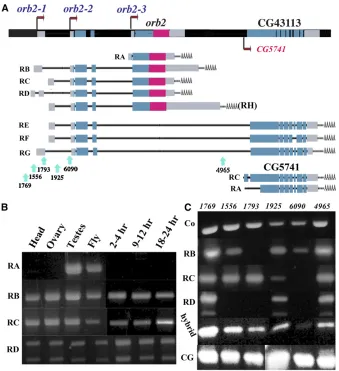
Theorb2gene is located in the cytogenetic interval 66E4 on the left arm of the third chromosome (Hoskinset al.2000). According to FlyBase, theorb2locus is predicted to produce two sets of transcripts and proteins (Figure 1A). One set of transcripts, theorb2RNAs, RA, RB, RC, and RD encode the Orb2 CPEB protein. The other set of predicted transcripts are theorb2-CG43113 hybrid RNAs, RE, RF, and RG, which encode a composite protein of unknown function.
The orb2 transcripts, RB, RC, and RD, are expected to encode the same 75-kDa CPEB protein and differ only in their 59-UTRs and/or 39-UTRs (Figure 1A). The differences in the UTRs are due to the use of two different promoters (orb2-1 and orb2-2), slightly different transcription start sites, alternative splicing, and polyadenylation. The fourth
orb2 transcript, RA, is expressed from a downstream pro-moter, orb2-3, that is close to the major orb2 exons and encodes a protein of 60 kDa. The 75-kDa and 60-kDa
Orb2 CPEB proteins share a common C-terminal region of 542 amino acids, but have different sequences at the N ter-minus (see Figure 1). The larger isoform has 162 unique amino acids at its N terminus, while the smaller has only nine unique amino acids. While the sequence in the N-terminal half of Orb2 is poorly conserved, it resembles Orb in that it is characterized by many polyglutamine and polyglycine repeats and is rich in histidine and serine. The sequence in the C-terminal half of the Orb2 protein is highly conserved especially for proteins within the same CPEB subgroup, and it contains the two 90-amino-acid RRM domains and a zinc finger domain.
The second set of predicted transcripts is the hybridorb2 -CG43113 RNAs, RE, RF, and RG (Figure 1). These RNAs are derived from the upstream orb2-1 and orb2-2 promoters, and share 59-UTR sequences and sequences encoding the first 162 amino acids with the larger orb2 RNAs. The re-mainder of the 1221-amino-acid CG43113 protein, which is derived from exons located downstream of the Orb2 coding sequences, bares no resemblance to the Orb2 CPEB proteins. Database searches reveal that this part of theorb2 -CG43113 gene is found in otherflies. However, it is a com-pletely independent transcription unit and does not include N-terminal protein coding sequences derived from the 75-kDa Orb2 isoform. In addition, as is in other Drosophilaspecies,
melanogasterhas an independent transcription unit, CG5741, which expresses the CG43113-specific protein coding sequen-ces. The CG5741 promoter is located downstream of theorb2
39-UTR and polyadenylation signals, and it generates five distinct mRNAs (two of which are shown in Figure 1) that differ in their splicing patterns. These mRNAs are predicted to encode proteins ranging from 887 to 1093 amino acids and overlap all of the hybridorb2-CG43113 sequences except for those derived from theorb2N terminus.
Orb2 is expressed throughout development in both the soma and germline
orb2 gene activity: We used RT–PCR with different primer combinations to examine the activity of theorb2promoters during development. The predicted orb2 transcripts (RB, RC, and RD) derived from the two upstream orb2 pro-moters,orb2-1andorb2-2, are detected in 2- to 4-hr, 9- to 12-hr, and 18- to 24-hr embryos, adultflies, heads, ovaries, and testes (Figure 1). In contrast, transcripts from the orb2-3 promoter, which generates theorb2-RA mRNA, are only observed in testes. We also found that transcripts containing the major orb2(not shown) and CG5741 (CG43113) (Fig-ure 1) specific protein coding exons are present in all stages and tissues examined. The relationship between orb2 and the hybridorb2-CG43113/CG5741 genes will be considered further in a subsequent section, while we focus below on the products specific to theorb2gene.
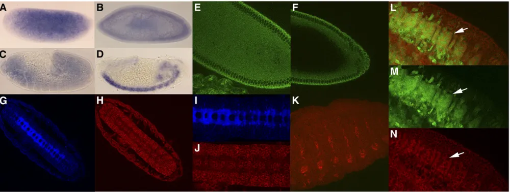
embryogenesis. We found that orb2transcripts are depos-ited maternally and are distributed uniformly throughout the embryo until the extended germband stage (Figure 2 A–C). In contrast, orb mRNA is not detected in Northern blots by 2–4 hr and is seen only in the pole cells by RNA
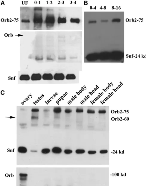
in situ(Lantzet al.1992). Whole mount staining with Orb2 monoclonal antibodies shows that the protein is distributed rather uniformly throughout the precellular blastoderm em-bryo (Figure 2E). However, there appears to be an apical concentration of the protein, just above the nuclei as the blastoderm cellularizes. Consistent with a maternal deposi-tion, a protein of the expected size (75 kDa) for the RB, RC, and RD transcripts is found in extracts from unfertilized eggs. A protein of the same size is found in precellular blas-toderm embryos (0–1, 1–2, and 2–3 hr) and early gastrula embryos as well (3–4 hr, Figure 3A). The 60-kDa protein predicted for the RA transcript is not detected at these stages and is only found much later in development (see below). In contrast to Orb2, Orb is not seen in Western blots of unfer-tilized eggs or in 0- to 4-hr embryos (Figure 3A).
Mid-to-late embryogenesis: During germband extension, theorb2message appears to turnover and there is a drop in
the in situhybridization signal in most regions of the em-bryo. Consistent with this decrease, the level of Orb2 protein also drops as can be seen in the Western blots of 0- to 4-hr and 4- to 8-hr embryo extracts (Figure 3B). Expression is upregulated in 8- to 16-hr embryos and there is an increase in the amount of Orb2 protein (Figure 3B). After germband retraction commences, the highest levels oforb2mRNA are found in the developing CNS (Figure 2D) and PNS, and there are lower levels elsewhere in the embryo. While it seems likely that mRNA seen at earlier stages is largely of maternal origin, much of the message present in the CNS and PNS in mid-to-late embryogenesis is likely due to de novo transcription.
Like the mRNA, the highest levels of Orb2 protein are in germband retracted embryos in the CNS (Figure 2, H and J) and PNS (Figure 2K). Interestingly, the protein in the CNS is concentrated in the cell bodies, while it is largely excluded from the longitudinal and commissural axon tracts. This is evident from the fact that there is little overlap between Orb2 (Figure 2, H and J) and BP102 (Figure 2, G and I), which is a marker for the longitudinal connectives and commissural axon tracts in the CNS. We also found evi-dence for Orb2 expression in the cells derived from specific
Figure 1Theorb2transcription unit. (A)orb2
locus is predicted to encode two distinct gene products. One is the CPEB homolog Orb2 with four predictedorb2mRNAs. RB, RC, and RD (and also RH, which has a larger 39-UTR) are transcribed from theorb2-1 andorb2-2 pro-moters and encode the same 701-amino-acid (75 kDa) Orb2 isoform. RA is transcribed from theorb2-3 promoter, and encodes a smaller, 550-amino-acid (60 kDa) isoform (see text for details). The other predictedorb2gene prod-ucts are hybridorb2-CG43113 mRNAs. The hy-brid mRNAs are generated by theorb2-1and
orb2-2promoters and share 59-UTR sequences
and sequences encoding the N-terminal 162 amino acids with theorb2transcripts generated by these two promoters (see text). CG5741 has its own promoter and produces at leastfive different RNA species. These mRNAs encode proteins ranging in size from 887 to 1093 amino acids, depending upon the splicing pat-tern and translation start codon. Also indicated are the locations of the transposon insertions. (B) RT–PCR of RNAs from heads, ovaries, testes, wholeflies, and from 2- to 4-hr, 9- to 12-hr, and 18- to 24-hr embryos. The PCR reactions were done with primers specific to theorb2
RNAs listed on the left. (C) RT–PCR of RNA from transposon insertions: orb21769, 1769;
orb21556, 1556; orb21793, 1793; orb21925,
1925; orb26090, 6090; and orb24965, 4965.
The PCR reactions were done with primers spe-cific to the RNAs listed on the left. Co, common toorb2RA, RB, RC, RD, and RH mRNAs; hybrid,
neuronal lineages. One such neuronal lineage is the Even-skipped (Eve)-positive neuroblast (NB)4-2 / ganglion mother cell (GMC)4-2a / RP2/sib lineage. In stage 11 embryos (Figure 4, A and B), Orb2 can be seen in Eve-positive GMC4-2a cells (see arrows; GMC4-2a is also known as GMC-1), while in stage 14–15 embryos (Figure 4, C and D) Orb2 is found in the Eve-positive RP2 cells (see arrows). Orb2 is also detected in the Eve-positive aCC/pCC neurons (see arrowhead).
Our antibody staining indicates that Orb2 is not restricted to the nervous system and lower levels of the protein can be detected in the ectoderm and mesoderm. For example, Fig-ure 2, L–N shows that Orb2 is expressed in at least a subset of the Twist-positive mesodermal cells. Note that the GFP marker seems to accumulate in both nucleus and cytoplasm of these mesodermal cells, while Orb2 is restricted to the cytoplasm (Figure 2N).
Larval stage: Western blots (Figure 3C) indicate that the amount oforb2is relatively low during the larval stage. The lowest levels are seen in thefirst and second instars, while the level begins to increase during the third instar (data not shown). To examine the distribution of Orb2 protein in the nervous system at this stage, we dissected the brain and ventral nerve cord. Figure 5, A–C shows the condensed/ retracted nerve cord and two successive images of a lobe from the brain of a third instar larva. As was observed in the embryo, Orb2 is found primarily in the cell bodies of neurons. While labeling of cell bodies of different neurons is evident in each focal plane, Orb2 appears to be largely excluded from axonal projections. Also, Orb2 is expressed at
high levels in only a subset of the neurons (see Figure 5, B and C). As in embryos it is largely restricted to cell bodies of neurons in the ventral nerve cord as well.
Adults: Orb2 levels increase substantially during the pupal stage and remain relatively high in adults (Figure 3C). To determine where Orb2 is expressed in adults, we probed Western blots of extracts from the heads, bodies, and gonads of males and females. For comparison, we also probed the same blots with Orb antibody. As shown in Figure 3C, the overall distribution of Orb2 differs substantially from that of Orb. There is little, if any, Orb in somatic tissues from the beginning of embryogenesis to the adult stage (Lantz et al.
1994) and as illustrated in thisfigure, it is not detected in the head or bodies of adult flies. In contrast, high levels of the 75-kDa Orb2 protein are present in the head and bodies of both sexes. There are also differences in the expression of the two CPEB proteins in the adult germline. Although both are found in male and female gonads, Orb2 is much more abundant in the testes than in the ovaries. Figure 3C shows that the 75-kDa Orb2 isoform is barely detected in the slightly overloaded sample from ovaries. In contrast, both the 75-kDa and 60-kDa Orb2 isoforms are readily observed in the greatly underloaded extract from testes. Note that while the 60-kDa RA Orb2 is present in testes, it is not found in extracts from heads or bodies of adult flies. This is con-sistent with our RT–PCR data, which indicates that the orb2-3promoter is only active in testes.
Orb2 expression in the adult CNS:Whole mount staining of dissected brains reveals that Orb2 is expressed throughout
the brain. However, it is also greatly enriched in certain specialized structures. This is shown in serial confocal sections in Figure 5. There is a high level of Orb2 present in the ellipsoid body (Figure 5, D–L), which begins as a dis-crete posterior structure (Figure 5, D–H), then becomes a ring-like structure (Figure 5, I–L, shown also in Figure 5, P and Q as line drawings). Moreover, two Orb2-positive discrete structures above the ellipsoid body can also be ob-served (marked by arrowheads in Figure 5, D–I); both of these are not part of the ellipsoid body, but just above it (see Figure 5, D–I). There is also another Orb2-positive structure (indicated by an arrow in Figure 5, D and E),
but visible only on the left side of the brain, suggesting that there might be asymmetry in the left–right Orb2 expression pattern in adult brains. It is possible that this novel structure is present only on the left side of the brain, as opposed to Orb2 expression being limited to only one of the two struc-tures. Orb2 is also found in the Fan-shaped body (Figure 5, L–O, R, and S). The staining pattern suggests that Orb2 is localized to the synaptic terminals in a fashion similar to the localization of Synaptotagmin (see Gazi et al.2009). These structures are part of the central complex within the central brain area and are believed to function as higher order pro-cessing sites for locomotion control (Strauss 2002). These structures are rich in axonal and dendritic terminals and have efferents from the protocerebral bridge. In this context it is interesting to note that the intracellular distribution of Orb2 in neurons of adult brains differs from that seen at earlier stages. Whereas most of the protein localized in the cell bodies of neurons at earlier stages, in the adult brain, the protein appears to be localized to synaptic terminals.
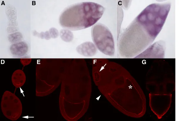
Orb2 accumulation in ovaries:Since the expression oforb
mRNA and protein in developing egg chambers has been extensively characterized, it was of interest to compare it with that oforb2.orbmRNA and protein arefirst detected in the newly formed 16-cell cysts in the germarium where both are concentrated primarily in the presumptive oocyte. Between stages 1 and 7,orbmRNA and protein are concen-trated in the region near the posterior pole of the oocyte. After the onset of vitellogenesis,orbmRNA is concentrated along the anterior margin of the oocyte, while Orb protein is localized around the entire oocyte cortex (Figure 6G). A very different expression pattern is observed fororb2. There is little orb2 mRNA in either the germarium or in early previtellogenic stage chambers (Figure 6, A and B). Midway through oogenesis, the level of orb2 mRNA begins to in-crease and it then remains high through at least stage 10; however,orb2differs fromorbin that the message is largely
Figure 3 Western blot analysis of Orb2 during development. (A) Orb2, Orb, and the loading control Snf in unfertilized eggs (UF) and in very early stages of embryogenesis (0- to 1-hr, 1- to 2-hr, 2- to 3-hr, and 3- to 4-hr embryos). The 75-kDa isoform is deposited maternally and like the Snf loading control, it is detected in unfertilized eggs and in fertilized pre-blastoderm and pre-blastoderm-stage embryos. The 60-kDa Orb2 isoform or Orb are not detected in these embryos. (B) Orb2 protein is present throughout embryogenesis. (C) Orb2 expression in larvae, pupae, and various adult tissues. The 75-kDa isoform of Orb2 is detected in larvae and pupae. In adultflies it is found in the heads, bodies, and gonads of both sexes. Note that very high levels of the 75-kDa isoform are found in male testes (see Snf loading control), while there is less in larvae and in ovaries. The 60-kDa isoform is found in testes, while it is not readily detected in adult heads or bodies. There may be a small amount of the 60-kDa isoform in pupae. (Bottom) Orb is not found outside of the germ-line. In this experiment, Orb is also not seen in the testes lane because this lane was intentionally underloaded because of the very high levels of Orb2 in the male gonad (compare levels of Snf, a loading control).
restricted to the nurse cells (Figure 6, B and C). The distri-bution of Orb2 protein also differs from that of Orb. While Orb is restricted to the germline, Orb2 is present in both germ cells and follicle cells (Figure 6D). In the germline nurse cells, it is most heavily concentrated in a ring around the nuclei (see arrows in Figure 6F) and in the cytoplasm of nurse cells close to the oocyte (see arrowhead in Figure 6F). Even though there is littleorb2mRNA in the oocyte, Orb2 protein is present and its localization pattern resembles that seen for Orb. In previtellogenic chambers, it forms a cap posterior to the oocyte nucleus (Figure 6D). At later stages, most of the Orb2 protein appears to be localized along the cortex of the oocyte just like Orb (compare Fig-ure 6, F and G).
Orb2 RNAi causes lethality
We used two different approaches to learn more about the biological functions of theorb2gene. In thefirst, we gener-ated a double-stranded RNA specific to a region of theorb2
mRNAs common to the 60-kDa and 75-kDa Orb2 isoforms, which does not contain the conserved CPEB RRM domains (seeMaterials and Methods). We used the Gal4-UAS system to express the orb2 dsRNA in a tissue-specific manner (Brand and Perrimon 1993). Flies carrying two independent
UAS-orb2RNAiinserts were crossed to different Gal4 lines to drive expression in distinct tissue-specific patterns. Since high levels of orb2 mRNA and protein are present in the
CNS and PNS, we reasoned that expression of orb2dsRNA using nervous system Gal4 drivers might have deleterious effects. However, there were no obvious effects on viability using theelav-GAL4 driver, which drives expression mostly in postmitotic neurons (Bergeret al.2007). While this could mean thatorb2has no essential function in thefly nervous system, it is also possible that the amount of dsRNA pro-duced by theelavdriver is not sufficient and/or the timing of its production is not appropriate to compromise a vitalorb2
function in the nervous system. This possibility is supported by the fact that lethal effects are observed for many other nervous system drivers. Altogether we tested 12 other Gal4 drivers that have been reported to be expressed in the CNS and/or PNS of embryos and/or larva and found that all had lethal effects when combined with UAS-orb2-RNAi trans-genes (Wardet al.2002). As shown inSupporting Informa-tion, Table S1, the severity of the lethal effects varied depending upon the driver and the UAS-orb2-RNAi insert. Further supporting the idea that orb2has a vital function during the development of the nervous system, we found that expression of orb2RNAi using a scabrous Gal4 driver also had lethal effects (not shown). scabrous is activated earlier in development than elav. It initially comes on in the late blastoderm stage in the neuroectoderm and is fur-ther upregulated in neuroblasts after they delaminate from neuroectoderm; however, it is not active in postmitotic neu-rons (Mlodzik et al.1990).
Piggybac insertions in the orb2 locus disrupt expression of orb2 gene products
In the second approach, we characterized a collection offive
piggybactransposons and oneP-element transposon inserted within or near the orb2/CG43113/CG5741 transcription units. Thepiggybactransposonorb21769is inserted upstream of the orb2-1 promoter, while the piggybac transposon
orb21556and theP-elementorb21793are inserted in thefirst (or second) exon for theorb2-1promoter. These three inser-tions should only affect transcripts expressed from theorb2-1
promoter. The piggybac transposons orb21925 and orb26090 are located in thefirst intron downstream of theorb2-2 pro-moter and could potentially affect transcripts from both this promoter and theorb2-1promoter. Thefinalpiggybac inser-tion,orb24965, is located in a large intron downstream of the
orb2protein coding sequences, but upstream of the protein coding sequences unique to CG43113/CG5741. This trans-poson would not be expected to have any effect on the expression of the variousorb2or CG5741 mRNAs, but could disrupt expression of the hybrid orb2-CG43113 transcripts originating from theorb2-1andorb2-2promoters. We used RT–PCR, Western blots, and whole mount antibody staining to examine the effects of these transposons on the expres-sion oforb2and CG43113/CG5741 gene products in adults. With respect to the different orb2 and hybrid orb2 -CG43113 transcripts, we used semiquantitative analysis and found that the piggybac 1769 insertion, which is up-stream of the orb2-1 promoter, had no apparent effect on any of theorb2or the hybridorb2-CG43113 RNAs (Figure 1) and the levels of the different RNAs were approximately equivalent to that in wild type (data not shown). Similarly, the orb24965transposon, which is inserted downstream of theorb2mRNA coding sequences, did not affect any of the
orb2 mRNAs. However, expression of the hybrid orb2 -CG43113 mRNAs originating from the orb2promoters up-stream of the insertion was substantially reduced in 4965
compared to the control (1769in the experiment shown in Figure 1). Significantly, when we used RT–PCR primers com-plementary to sequences located entirely within the CG5741 protein-coding region, we found that the level of the CG5741 RNAs in4965is close to that in the1769(or wild type) control. Moreover, a similar pattern is seen for some of the other transposon insertions: they have reduced levels of the hybridorb2-CG43113 RNAs generated by theorb2 pro-moters, but nevertheless have substantial amounts of the CG5741 specific RNAs. These results suggest that sequences critical for CG5741 promoter activity are located down-stream of the4965 transposon insertion and that this pro-moter is fully active in the4965allele and in all of the other transposon insertions. It would also appear that a substantial fraction of the CG5741/CG43113 transcripts are derived from the CG5741 promoter rather than from the upstream
orb2promoters.
When we used a primer set specific for the 75-kDaorb2
protein-coding sequence, only two transposons, 6090 and
1925, showed a significant reduction in the levels oforb2
mRNA. Both mutants also showed reduced amounts of the
orb2RB, RC, and RD transcripts when primer sets specific for these RNAs were used (Figure 1). The insertions in1556
and1793are upstream of the RCorb2-2promoter, and the levels of RC transcripts in both these alleles were similar to the control. On the other hand, both had reduced levels of theorb2RD and RB transcripts (Figure 1). As expected, we found that none of the transposon insertions had any effect on the expression of either the 60-kDa Orb2 RA protein or theorb2RA mRNA (data not shown). Consistent with our RT–PCR experiments, only two,6090and1925, had major
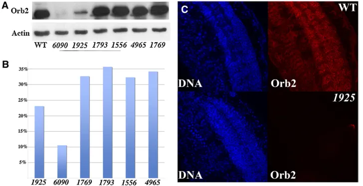
effects on the expression of the 75-kDa protein. As shown in the Western blot of adult heads in Figure 7A, the levels of the 75-kDa protein are markedly reduced in both mutants. By contrast, there is no obvious change in the expression of this protein in the remaining transposon alleles. We also examined the expression of Orb2 in whole mounts of
orb21925andorb6090embryos. As shown in Figure 7C, the expression of Orb2 in the embryonic CNS, PNS, and in other tissues of homozygous orb21925 embryos is reduced com-pared to wild type. Similar results were obtained for
orb26090.
Two of the piggybac insertions cause lethality
Since the RNAi knockdowns of orb2had lethal effects, we tested whether any of thepiggybac orP-element insertions also had lethal effects. As shown in Figure 7B,1769,1556,
1793, and4965flies are fully viable. In the case of1769, this is not surprising since this allele expresses wild-type levels of
orb2 RNA and protein. While there are reductions in the levels of several orb2 transcripts in 1556 and 1793, the reductions are either not sufficient to cause lethality or are not in tissues in whichorb2activity is needed for full viabil-ity. Similarly, the disruptions in the expression of the hybrid
orb2-CG43113 transcripts in 4965 showed no adverse effects on viability.
On other hand, lethal effects are observed for both6090
and1925. These results are consistent with studies of a null allele oforb2(orb2D) generated by homologous recombina-tion and described by Kelemanet al.(2007). For6090, only about one-third of the homozygous flies survive to adult-hood, while about two-thirds of the1925homozygotes sur-vive. While some mutant animals die during embryogenesis, most appear to die during the pupal stage. Arguing against potential background effects, we found that when6090flies are transtoDf(3L)ED4421, which uncovers theorb2gene, only10% (n= 351) of the expected number of thetrans -heterozygous flies survived to adulthood. Similar results were obtained for 1925. We also found that the lethality of 1925 and 6090 can be reverted by precise excision of
the piggybactransposons. Finally, we found that theorb26090 allele showed a reduction in life span (seeFigure S1). orb2 functions in asymmetric cell division in the CNS
Since Orb2 appears to specifically accumulate in the cell bodies but not in their axons during embryogenesis, we wondered whether it has any role in the specification of these neurons. To explore this question, we examined the specification of RP2 neurons, a well-studied neuronal lineage (reviewed in Bhat 1999; Gaziova and Bhat 2007), under conditions whereorb2activity is compromised. Dur-ing embryogenesis, each neuroblast (NB) undergoes a series of asymmetric divisions generating a self-renewing NB and a daughter GMC. Each GMC divides, producing two daugh-ter cells, which typically differentiate into two different neu-rons. The Eve-positive RP2 neuron arises from the NB4-2/ GMC4-2a lineage (Bhat and Schedl 1994; Buescher et al.
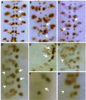
1998; Skeath and Doe 1998; Waiet al.1999). In wild-type embryos, GMC4-2a (also known as GMC-1) divides asym-metrically, producing two daughter neurons: RP2 and sib. The sib cell is smaller in size compared to RP2. While Eve is expressed in both daughter cells initially, it eventually dis-appears from the sib (Frasch et al.1987; Patel et al.1989; Fujiokaet al.2003). In wild-type stage 14 embryos, a single RP2 neuron is observed in each hemisegment (arrows in Figure 8A). However, if the asymmetric division of a GMC4-2a is disrupted, two cells with the same identity (RP2 or sib) can be formed, which can be determined by the Eve-staining pattern (Bhat and Schedl 1994; Bhatet al.
1995; Buescheret al.1998; Waiet al.1999).
As shown in Figure 8, A–D, in 11% of theorb21925and 25% of theorb26090embryos (Figure 9A), we observed RP2 neuron specification defects. In most instances (70%) the RP2 neurons are duplicated and there are two equal sized Eve-positive cells per hemisegment (arrows in Figure 8, B and C). However, we also observed hemisegments that lacked Eve-positive RP2 neurons (arrowhead in Figure 8B), an indication that such hemisegments had either no RP2 lineage formed or that the GMC divided into two sib
cells. These findings argue that orb2 is required for the proper elaboration of this lineage. Consistent with this con-clusion, RP2 specification defects were also observed when
orb2activity is knocked down by RNAi in the nervous sys-tem using Gal4 drivers such ascb43,scabrous(Figure 9A), andcb36.
orb2 may have a general role in asymmetric cell division
Many of the genes implicated in the asymmetric NB4 / GMC-1/RP2/sib cell division pathway are known to func-tion in the asymmetric division of other lineages (Ruiz Gomez and Bate 1997; Carmena et al. 1998; Paululat
et al. 1999; Tioet al.2001). To test whether orb2also has a more general role in asymmetric cell division, we exam-ined cell division patterns in the mesoderm. In each hemi-segment there are two Eve-expressing mesodermal lineages that undergo asymmetric divisions. In one of these lineages, the P2 cell divides asymmetrically to generate the founder of the Eve-positive pericardial cells (FEPCs) and FEPC sib cells. The FEPC daughter then divides symmetrically to form a pair of Eve-positive pericardial cells (EPCs) in each hemisegment (arrow in Figure 8D). In the other lineage, the P15 cell divides asymmetrically to produce the Eve-positive DA1 muscle founder cell FDA1 and the FDA1 sib cell. The
FDA1 daughter then generates the multinucleate DA1 mus-cle (arrowhead in Figure 8D).
As was observed for the RP2 lineage,orb2is required for asymmetric cell division in the P2/EPC and P15/DA1 lineages. In bothorb21925andorb26090mutant embryos, we observe abnormalities in these two lineages. Figure 8E shows a hemisegment in which the P2 cell divided symmet-rically to produce an extra Eve-positive EPC cell (arrow), while Figure 8F shows a hemisegment in which the P2 or FEPC asymmetric division was abnormal and no Eve-positive EPC cell was produced (arrow). Extra and missing Eve-positive cells are also seen in the P15/DA1 lineage. Figure 8G (arrowhead) shows a hemisegment in which P15 divided symmetrically to produce two DA1 muscles, while Figure 8H shows a hemisegment in which the DA1 muscle is absent (arrowhead).
Alignment of the mitotic spindle is disrupted in orb2 mutants
Mastushita-Saki et al. (2010) have shown that one of the mRNAs associated with Orb2 in the adult CNS encodes Dro-sophilaatypical protein kinase C (aPKC). Since this kinase is known to play a central role in asymmetric cell division, we wondered whether there were any effects on aPKC inorb2 Figure 8 Specification defects are observed in neuronal and mesodermal lineages in orb21925 and orb26090
mutant neuroblasts. In nondividing wild-type neuroblasts, aPKC is distributed around the cortex of the cell (Figure 10A, cell with arrowhead), while in neuroblasts about to undergo asymmetric division (Figure 10A, arrow) it concen-trates on the apical side of the cell. Orb2 is also in a cortical ring in nondividing neuroblasts (Figure 10A, arrowhead). However, the Orb2 ring does not appear to overlap with aPKC (Figure 10A, left). Instead, it is localized in a slightly more interior region of the cell. In dividing neuroblasts,
Orb2 differs from aPKC in that it disappears from the cortex and becomes largely unlocalized (Figure 10A, arrow). When wild-type neuroblasts divide, the mitotic spindle (arrowhead in Figure 10B) is oriented along the apical–basal axis. After division, the apical daughter cell that inherits the cap of aPKC (arrow in Figure 10B) retains neuroblast identity, while the basal daughter becomes a GMC. Inorb2mutants, aPKC can be detected in nondividing neuroblasts; however, it seems to be somewhat less tightly linked to the cortex than in wild type. In dividing mutant neuroblasts, the mi-totic spindle orientation is often randomized with respect to the apical–basal axis (arrowhead in Figure 10C). Interest-ingly, in these neuroblasts, the apical cap of aPKC is also missing. Thesefindings further support the conclusion that
orb2plays a role in asymmetric cell division and suggest that it may function by promoting the localized accumulation of aPKC.
Behavioral deficiencies in orb2 adults
In the adult CNS, high levels of Orb2 are found in the ellipsoid and fan shaped bodies. As these structures have been implicated in sensing distance, orientation, and walk-ing, we tested differentorb2alleles in behavioral assays that measure locomotion and activity of adultflies. These assays
Figure 9 RP2 lineage, locomotion, and activity defects in the transposon insertions. (A) Frequency of embryos with RP2 lineage defects whenorb2
function is compromised. With the exception of Gal4 39A/UAS cb43, .130 embryos were examined in each case. WT, wild type; 39A/sca,
UAS-39A/Gal4-sca; 39A/cb43, UAS-39A/Gal4-cb43; delQ, orb2delQ;
1925,orb21925;6090,orb26090;6090-1,orb26090-1; and4925,orb4925.
6090-1is a precise excision of the6090 piggybacinsertion. (B) Percent-age offlies that have recovered from vortex after 10 sec (blue) or 20 sec (red). Genotypes are as indicated. (C) Percentage offlies climbing a de-fined distance after being knocked down to the bottom of the vial in 10 sec. (D) Percentage offlies still standing after a 10-min heat shock.
Figure 10 Neuroblast cell division is altered inorb2mutants. (A) Distri-bution of aPKC (green) and Orb2 (red) in nondividing (arrowhead) and dividing (arrow) neuroblasts. Both aPKC and Orb2 are cortical in non-dividing neuroblasts, but do not colocalize. In non-dividing neuroblasts, aPKC concentrates apically, while Orb2 becomes diffusely distributed. (B) Mito-sis in WT and1925(orb21925) neuroblasts. In wild type, aPKC (red) is
were recovery from vortexing (bang assay) and recovery from knockdown (climbing assay). We also tested recovery from heat shock. As shown in Figure 9, B and C, the three
orb2 mutants, 1769, 1793, and 1556, that had no appar-ent effects on Orb2 protein expression in adult brains re-cover from vortexing and knockdown as rapidly as wild type. This is also true for anorb2mutant,orb2delQ, lacking the N-terminal glutamine-rich region, which has previously been implicated in learning and memory (Keleman et al.
2007). The4965allele, which disrupts the orb2 promoter-dependent CG43113 mRNAs, but not theorb2mRNAs, also resembles wild type. In contrast, 6090, and to a lesser extent 1925, take longer than wild type to recover from vortexing. Even more pronounced defects are evident in the knockdown experiment. Finally, we also found that
6090, but not1925, recovers more slowly from heat shock than wild type (Figure 9D).
Discussion
Sequence organization of Orb2 and the neighboring CG43113 transcription units
The sequence organization and coding properties of theorb2
transcription unit is quite complex. There are at least three
orb2promoters and they generate two quite different sets of mRNAs. One set of mRNAs, the orb2mRNAs, encode the CPEB protein Orb2, while the other set of mRNAs, the hy-bridorb2-CG43113 mRNAs, encode a conserved protein of unknown function (Figure 1).
Threeorb2mRNAs, RB, RC, and RD, are expressed from theorb2-1andorb2-2promoters and encode the same 75-kDa Orb2 isoform. The fourth mRNA, RA, is expressed from the
orb2-3 promoter and encodes a smaller 60-kDa protein. These two Orb2 isoforms share a common 542-amino-acid C-terminal region, which includes the two CPEB RRM domains and the zinc finger, but have unique N termini of 162 and 9 amino acids, respectively. The other set of tran-scripts is the hybrid orb2-CG43113 mRNAs. They are gen-erated by theorb2-1andorb2-2promoters and share several 59 exons with theorb2RB, RC, and RD mRNAs. These 59 exons include the translation start signal and the “unique” 162-amino-acid N terminus of the 75-kDa Orb2 isoform. However, instead of being spliced to the downstream sequences encoding Orb2, the CG43113 mRNAs are spliced to a conserved 1059-amino-acid open reading frame, which is located7 kb beyond the end oforb2. This downstream open reading frame is also part of an independent transcrip-tion unit, CG5741, which has its own promoter and gener-atesfive different mRNAs. These mRNAs encode variants of the same conserved open reading frame depending on their splicing pattern and the location of the AUG codon.
An important issue is the relationship between orb2,
orb2-CG43113, and CG5741. In most other fly species
orb2 and CG5741 (CG43113) appear to be distinct genes. For example, thepseudoobscuraCG43113 gene, GA19098, is
also located downstream (12 kb) of the orb2 (GA1909) open reading frame and like CG5741, it is predicted to have its own promoter. Our data would suggest that in spite of the fact that some of the CG571/CG43113 transcripts are de-rived fromorb2promoters, theorb2and CG5741 genes are probably distinct in melanogaster as well. In particular, we found that apiggybacinsertion,orb4965, located downstream of the orb2 open reading frame interrupts CG43113 tran-scripts emanating from the twoorb2promoters, and greatly reduces the level of the hybridorb2-CG43113 mRNAs. How-ever, there were no obvious reductions in the level of mRNA specific for the CG5741/CG43113 protein coding sequences. We also found that insertions in the upstreamorb2introns, which disrupt transcripts from the orb2-1and orb2-2 pro-moters, reduce the levels oforb2andorb2-CG43113 hybrid mRNAs but do not alter the level of mRNAs specific for the CG5741/CG43113 protein coding sequences.
Orb2 expression pattern is different from that of Orb during development
Drosophila has two CPEB genes, orb and orb2. While the expression of orbis restricted to the germline and its only essential functions are in the female ovary where it plays a key role in the development of the egg,orb2is expressed not only in the germline, but also in a wide range of somatic tissues in the embryo, larvae, and adult. Whileorbandorb2
mRNAs and proteins are deposited in the developing egg during oogenesis, their fate in embryos is quite different. With the exception of the orbgene products that are incor-porated into the germline pole cells, all maternalorbmRNAs and proteins turnover by the midblastula transition. In con-trast, maternally derivedorb2mRNA is translated in precel-lular blastoderm embryos, and high levels of the 75-kDa Orb2 protein are distributed throughout the early precellu-lar and celluprecellu-lar blastoderm embryo. After the onset of gas-trulation, there is a general reduction in the levels oforb2
message and protein, which likely reflects the turnover of the maternal gene products in the absence of significantde novosynthesis. However, once the embryonic CNS and PNS begin to differentiate,orbmRNA and the 75-kDa Orb2 pro-tein isoform are expressed at high levels in these tissues. The Orb2 protein in the embryonic CNS is found predominantly in the cell bodies and is largely excluded from axon tracts. Orb2 protein is also detected in other differentiating tissues like the mesoderm, though the levels are lower than in the CNS or PNS.
High levels of Orb2 persist in the nervous system during the larval stages, and as in the embryos, the protein is predominantly localized in cell bodies, and not in axons or dendrites. Interestingly, in the adult brain, Orb2 is found mostly in axon/dendritic terminals. Asorb2has been impli-cated in learning and memory (Kelemanet al.2007; Siet al.
assemble into large aggregates in response to synaptic stim-ulation when ectopically expressed in Aplysiasensory neu-rons (Si et al. 2010). Once formed, these large aggregates were proposed to have self-perpetuating properties that would contribute to long-term facilitation. It was argued that the special prion-like properties of the N-terminal do-main in the 60-kDa isoform was responsible for multimeri-zation in response to synaptic stimulation. In contrast, when the larger DrosophilaOrb2 75-kDa isoform was ectopically expressed in Aplysia neurons it was unable to form these self-propagating bodies either with or without stimulation (Siet al.2010). As this study seemed to directly implicate the Orb2 60-kDa isoform in long-term facilitation, it was of interest to examine its distribution in different fly tissues. Strikingly, there is little if any of the 60-kDa isoform in West-erns of adult heads, even when overloaded, and only the larger Orb2 75-kDa isoform seems to be expressed in this tissue. In contrast, both isoforms are found in testes, though even in this tissue the 60-kDa isoform is less abundant than the 75-kDa isoform. Thus, it seems quite unlikely that the Orb2 60-kDa isoform plays a central role in nervous system function in adult Drosophila. If the assembly of Orb2 into prion-like aggregates is critical for synaptic stimulation-de-pendent long-term facilitation, then the experiments of Si
et al.(2010) would imply that there must be special ancil-lary factors in the fly CNS, but not inAplysia sensory neu-rons, that are able to promote the assembly of the 75-kDa isoform into prion-like aggregates upon synaptic stimula-tion. Since all of the smaller isoforms are included in the larger isoform except for nine amino acids at the very N terminus, one could imagine that Orb2 75 kDa should also be capable of forming prion-like aggregates under certain condition(s).
Function of Orb2 during development
Though orb2function has been implicated in learning and memory (Kelemanet al.2007; Mastushita-Sakiet al.2010), what other functions it might have, if any, during the Dro-sophilalife cycle are largely unknown. In our work, we have attempted to identify some of these other activities using RNAi knockdowns and transposon insertion mutations. This task has been complicated by the fact that the orb2 locus generates mRNAs encoding not only the Orb2 CPEB protein (s) but also a hybrid Orb2-CG43113 protein.
While not entirely definitive, the properties of theorb2
transposon insertions argue that the observed phenotypes are due to an effect on orb2gene activity and not on the activity of either the hybrid orb2-CG43113 or the CG5741 gene. First, with the exception of 1769, which is inserted upstream of theorb2-1promoter, all of the transposon inser-tions reduce expression of the hybrid orb2-CG43113 mRNAs. However, only two of these, 1925and6090, have obvious phenotypic effects. While6090has little of the hy-bridorb2-CG43113 mRNA, the amount of hybrid mRNA in
1925 is close to that seen in the three other transposon insertions (1556,1793, and4965) that have no phenotype.
Second,4965differs from the otherorb2transposons in that it is downstream of the orb2 coding sequences. It has no effect on orb2 mRNAs, but does reduce expression of the hybrid mRNAs. However, it has none of the phenotypes ev-ident in the1925,6090, ororb2RNAi animals. Third, while all but one of the transposon insertions reduce expression of the hybrid orb2-CG43113 mRNAs, the total level of the CG5741 (CG43113) transcripts appears to be unaffected in all of the mutants. This is likely due to the fact that the bulk of the CG5741/CG43113 transcripts are derived from the CG5741 promoter and not the orb2 promoters. Since the level of the hybrid orb2-CG43113 mRNAs can be reduced without any phenotypic effects, this would argue that CG5741 may be able to substitute, perhaps even fully, for whatever function the hybrid orb2-CG43113 protein might have. The suggestion that the 1925 and 6090 phenotypes are attributable to orb2 rather than the hybrid orb2 -CG43113 gene product is supported by the fact that they are also observed inorb2RNAi experiments. Less than 10% of the1 kb double-stranded RNA expressed byorb2RNAi vector is from the common region and thus theorb2RNAi would be expected to have a much greater effect on orb2
than on the hybrid mRNAs.
We found that both theorb2RNAi knockdowns and the
1925and6090 piggybacinsertions reduce viability. As might be expected from the high levels oforb2mRNA and protein in the CNS/PNS, Gal4 drivers that were expressed in the nervous system generally reduced viability. Also consistent with important functions in the nervous system, surviving
1925 and 6090 adults exhibit behavioral defects in assays that measurefly locomotion and activity. One of thepiggybac
mutants was also found to have a reduced life span. More-over, the studies of Mastushita-Sakiet al. (2010) on poten-tial targets for orb2 regulation showed that mRNAs encoding factors involved in neuronal growth and synapse formation are associated with Orb2 in the adult brain.
One of the important nervous system functions appears to be in asymmetric cell division. In the embryonic CNS, Orb2 protein can be detected in the cell bodies of specific neurons such as the Eve-positive RP2 and aCC/pCC neurons. However, it appears to be needed prior to the formation of these neurons as we found that the proper elaboration of the NB4-2/GMC4-2a/RP2/sib lineage depends uponorb2
activity. There are defects in the specification of the RP2 and sib cells in two different orb2 piggybacalleles, and also in
The genes that have been implicated in asymmetric cell division fall into two general categories, those responsible for the asymmetry and those responsible for specifying the fate of the daughter cells. In the former category are genes like inscuteable, bazooka, and atypical protein kinase C
(Schaefer and Knoblich 2001; Bhaleraoet al.2005; Gaziova and Bhat 2007). Mutations in these genes disrupt the pro-cess of asymmetric cell division and give rise to two identical cells. In the latter category are genes like numb, the tran-scription factor prospero, and various components of the
Notch signaling pathway. Cells mutant in these genes still divide asymmetrically; however, though the daughter cells are unequal, they still assume the same fate. It seems likely that orb2 falls into the former category as the duplicated RP2 cells are equal in size. This is also observed for muta-tions ininscuteable, but not for mutations in theNotch path-way, where the two daughter cells are still unequal in size (Buescheret al.1998; Wai et al.1999).
Our analysis of neuroblast cell division in orb2mutants points to roles in localizing aPKC and orienting the mitotic spindle. In wild-type nondividing neuroblasts, aPKC is local-ized around the cortex. Orb2 is also concentrated around the cortex, but localized just inside the cortical layer that contains aPKC. When neuroblasts divide asymmetrically, aPKC relocalizes to the apical cortex, and during mitosis, the spindle is oriented along the apical–basal axis. At this point, Orb2 is distributed uniformly in the cytoplasm. In
orb2 mutants, aPKC is cortical in nondividing neuroblasts but appears to be somewhat more diffusely localized than in wild type. During cell division, aPKC does not concentrate at the apical cortex and the spindles are not properly aligned relative to the apical–basal axis. Since Orb2 is known to bind to apkc mRNA (Mastushita-Saki et al. 2010), a plausible mechanism is thatorb2helps promote the apical accumula-tion of aPKC by activating the localized translaaccumula-tion of apkc
mRNA.
While thesefindings point to a role in polarizing the cell during asymmetric cell division, the twoorb2mutants (and RNAi knockdowns) we have examined differ from mutations in genes like apkc and inscuteable in that only a relatively small percentage of the mutant embryos exhibit defects in the RP2 lineage. This could be due to the fact that we have not fully eliminated orb2activity. Alternatively,orb2might have only an ancillary role in generating asymmetry. Further studies will be required to determine howorb2fits into the asymmetry pathway and why the phenotypic effects oforb2
mutations are relatively modest.
Acknowledgments
We thank members of the Schedl laboratory and Bhat laboratory for discussion and advice on this project. We acknowledge J. Goodhouse for help with confocal micros-copy and Gordon Grey for fly food. We also acknowledge three undergraduates for their contributions to this work: Cindy Hodakoski, Mahala Burn, and Blessing Agunwamba.
This work was supported by a grant from National Institutes of Health (NIH) (GM056937) and, subsequently, by grants from NIH (GM043432) to P.S. and (GM080538) to K.B.
Literature Cited
Alarcon, J. M., R. Hodgman, M. Theis, Y. S. Huang, E. R. Kandel
et al., 2004 Selective modulation of some forms of schaffer collateral-CA1 synaptic plasticity in mice with a disruption of the CPEB-1 gene. Learn. Mem. 11: 318–327.
Bally-Cuif, L., W. J. Schatz, and R. K. Ho, 1998 Characterization of the zebrafish Orb/CPEB-related RNA binding protein and localization of maternal components in the zebrafish oocyte. Mech. Dev. 77: 31–47.
Berger, C., S. Renner, K. Luer, and G. M. Technau, 2007 The com-monly used marker ELAV is transiently expressed in neuroblasts and glial cells in the Drosophila embryonic CNS. Dev. Dyn. 236: 3562–3568.
Berger-Sweeney, J., N. R. Zearfoss, and J. D. Richter, 2006 Reduced extinction of hippocampal-dependent memo-ries in CPEB knockout mice. Learn. Mem. 13: 4–7.
Bhalerao, S., D. Berdnik, T. Torok, and J. A. Knoblich, 2005 Localization-dependent and -independent roles of numb contribute to cell-fate specification in Drosophila. Curr. Biol. 15: 1583–1590.
Bhat, K. M., 1999 Segment polarity genes in neuroblast formation and identity specification during Drosophila neurogenesis. Bio-essays 21: 472–485.
Bhat, K. M., and P. Schedl, 1994 The Drosophila miti-mere gene, a member of the POU family, is required for the specification of the RP2/sibling lineage during neurogenesis. Development 120: 1483–1501.
Bhat, K. M., S. J. Poole, and P. Schedl, 1995 The miti-mere and pdm1 genes collaborate during specification of the RP2/sib lin-eage in Drosophila neurogenesis. Mol. Cell. Biol. 15: 4052– 4063.
Brand, A. H., and N. Perrimon, 1993 Targeted gene expression as a means of altering cell fates and generating dominant pheno-types. Development 118: 401–415.
Buescher, M., S. L. Yeo, G. Udolph, M. Zavortink, X. Yang et al., 1998 Binary sibling neuronal cell fate decisions in the Dro-sophila embryonic central nervous system are nonstochastic and require inscuteable-mediated asymmetry of ganglion mother cells. Genes Dev. 12: 1858–1870.
Carmena, A., B. Murugasu-Oei, D. Menon, F. Jimenez, and W. Chia, 1998 Inscuteable and numb mediate asymmetric muscle pro-genitor cell divisions during Drosophila myogenesis. Genes Dev. 12: 304–315.
Christerson, L. B., and D. M. McKearin, 1994 orb is required for anteroposterior and dorsoventral patterning during Drosophila oogenesis. Genes Dev. 8: 614–628.
Costa, A., Y. Wang, T. C. Dockendorff, H. Erdjument-Bromage, P. Tempstet al., 2005 The Drosophila fragile X protein functions as a negative regulator in the orb autoregulatory pathway. Dev. Cell 8(3): 331–342.
Frasch, M., T. Hoey, C. Rushlow, H. Doyle, and M. Levine, 1987 Characterization and localization of the even-skipped protein of Drosophila. EMBO J. 6: 749–759.
Fujioka, M., B. C. Lear, M. Landgraf, G. L. Yusibova, J. Zhouet al., 2003 Even-skipped, acting as a repressor, regulates axonal projections in Drosophila. Development 130: 5385–5400. Gazi, M., B. V. Shyamala, and K. M. Bhat, 2009 A
Gaziova, I., and K. M. Bhat, 2007 Generating asymmetry with and without self-renewal in Recent Advances in Molecular Biology, edited by A. M. Coelho. Springer-Verlag, New York.
Hake, L. E., and J. D. Richter, 1994 CPEB is a specificity factor that mediates cytoplasmic polyadenylation during Xenopus oocyte maturation. Cell 79: 617–627.
Hoskins, R. A., C. R. Nelson, B. P. Berman, T. R. Laverty, R. A. George et al., 2000 A BAC-based physical map of the major autosomes of Drosophila melanogaster. Science 287: 2271– 2274.
Huang, Y. S., M. C. Kan, C. L. Lin, and J. D. Richter, 2006 CPEB3 and CPEB4 in neurons: analysis of RNA-binding specificity and translational control of AMPA receptor GluR2 mRNA. EMBO J. 25: 4865–4876.
Keleman, K., S. Kruttner, M. Alenius, and B. J. Dickson, 2007 Function of the Drosophila CPEB protein Orb2 in long-term courtship memory. Nat. Neurosci. 10: 1587–1593. Kikuno, R., T. Nagase, M. Nakayama, H. Koga, N. Okazaki et al.,
2004 HUGE: a database for human KIAA proteins, a 2004 up-date integrating HUGEppi and ROUGE. Nucleic Acids Res. 32: D502–D504.
Kurihara, Y., M. Tokuriki, R. Myojin, T. Hori, A. Kuroiwa et al., 2003 CPEB2, a novel putative translational regulator in mouse haploid germ cells. Biol. Reprod. 69: 261–268.
Lantz, V., L. Ambrosio, and P. Schedl, 1992 The Drosophila orb gene is predicted to encode sex-specific germline RNA-binding proteins and has localized transcripts in ovaries and early em-bryos. Development 115: 75–88.
Lantz, V., J. S. Chang, J. I. Horabin, D. Bopp, and P. Schedl, 1994 The Drosophila orb RNA-binding protein is required for the formation of the egg chamber and establishment of polarity. Genes Dev. 8: 598–613.
Luitjens, C., M. Gallegos, B. Kraemer, J. Kimble, and M. Wickens, 2000 CPEB proteins control two key steps in spermatogenesis in C. elegans. Genes Dev. 14: 2596–2609.
Mastushita-Saki, T., E. White-Grndley, J. Samuelson, C. Seidel, and K. Si, 2010 Drosophila Orb2 targets genes involved in neuro-nal growth, synapse formation and protein turnover. Proc. Natl. Acad. Sci. USA 107: 11987–11992.
Mendez, R., and J. D. Richter, 2001 Translational control by CPEB: a means to the end. Nat. Rev. Mol. Cell Biol. 2: 521–529. Minshall, N., J. Walker, M. Dale, and N. Standart, 1999 Dual roles of p82, the clam CPEB homolog, in cytoplasmic polyadenylation and translational masking. RNA 5: 27–38.
Mlodzik, M., N. E. Baker, and G. M. Rubin, 1990 Isolation and expression of scabrous, a gene regulating neurogenesis in Dro-sophila. Genes Dev. 4: 1848–1861.
Patel, N. H., B. Schafer, C. S. Goodman, and R. Holmgren, 1989 The role of segment polarity genes during Drosophila neurogenesis. Genes Dev. 3: 890–904.
Paululat, A., S. Breuer, and R. Renkawitz-Pohl, 1999 Determination and development of the larval muscle pattern in Drosophila melanogaster. Cell Tissue Res. 296: 151–160.
Ruiz Gomez, M., and M. Bate, 1997 Segregation of myogenic lineages in Drosophila requires numb. Development 124: 4857–4866.
Schaefer, M., and J. A. Knoblich, 2001 Protein localization during asymmetric cell division. Exp. Cell Res. 271: 66–74.
Si, K., M. Giustetto, A. Etkin, R. Hsu, A. M. Janisiewicz et al., 2003 A neuronal isoform of CPEB regulates local protein syn-thesis and stabilizes synapse-specific long-term facilitation in aplysia. Cell 115: 893–904.
Si, K., Y. B. Choi, E. White-Grindley, A. Majumdar, and E. R. Kandel, 2010 Aplysia CPEB can form prion-like multimers in sensory neurons that contribute to long-term facilitation. Cell 140: 421– 435.
Skeath, J. B., and C. Q. Doe, 1998 Sanpodo and Notch act in opposition to Numb to distinguish sibling neuron fates in the Drosophila CNS. Development 125: 1857–1865.
Strauss, R., 2002 The central complex and the genetic dissection of locomotor behaviour. Curr. Opin. Neurobiol. 12: 633–638. Tay, J., and J. D. Richter, 2001 Germ cell differentiation and
syn-aptonemal complex formation are disrupted in CPEB knockout mice. Dev. Cell 1: 201–213.
Theis, M., K. Si, and E. R. Kandel, 2003 Two previously unde-scribed members of the mouse CPEB family of genes and their inducible expression in the principal cell layers of the hippocam-pus. Proc. Natl. Acad. Sci. USA 100: 9602–9607.
Tio, M., G. Udolph, X. Yang, and W. Chia, 2001 cdc2 links the Drosophila cell cycle and asymmetric division machineries. Na-ture 409: 1063–1067.
Vogler, C., K. Spalek, A. Aerni, P. Demougin, A. Muller et al., 2009 CPEB3 is associated with human episodic memory. Front Behav Neurosci 3: 4.
Wai, P., B. Truong, and K. M. Bhat, 1999 Cell division genes pro-mote asymmetric interaction between Numb and Notch in the Drosophila CNS. Development 126: 2759–2770.
Walker, J., N. Minshall, L. Hake, J. Richter, and N. Standart, 1999 The clam 39UTR masking element-binding protein p82 is a member of the CPEB family. RNA 5: 14–26.
Ward, E. J., I. Thaipisuttikul, M. Terayama, R. L. French, S. M. Jacksonet al., 2002 GAL4 enhancer trap patterns during Dro-sophila development. Genesis 34: 46–50.
Wu, L., D. Wells, J. Tay, D. Mendis, M. A. Abbott et al., 1998 CPEB-mediated cytoplasmic polyadenylation and the regulation of experience-dependent translation of alpha-CaMKII mRNA at synapses. Neuron 21: 1129–1139.
GENETICS
Supporting Information
http://www.genetics.org/content/suppl/2011/09/07/genetics.110.123646.DC1
The
Drosophila
CPEB Protein Orb2 Has a Novel
Expression Pattern and Is Important for Asymmetric
Cell Division and Nervous System Function
Nathaniel Hafer, Shuwa Xu, Krishna Moorthi Bhat, and Paul Schedl
!
'&#!!
!"
+5!!%!!# !! *(4228($
!!# $$'&#!!!#"32,34&!%"$ !
" " '&#!!!!!"62&!*
#
# 0#
0
0
+; ",
#
#
0 #
0
0
+; ",
026)026 /5) 026)/5 53 07+45, 28)2 !$/5) /5)<%
28)< 07/ 0+3,
026)026 !$/8) 026)/8 80 28+75, 28)2 !$/8) /8)<%
28)< 368 55+44,
026)026 03) 026)03 36 /+/, 28)2 !$03) 03)<%
28)< 07/ /+/,
026)026 )1/ 026)<%
1/)< 046 54+71, 28)2 )1/
1/)<%
28)< 300 30+3/,
026)026 )18 026)<%
18)< 1/5 61+6/, 28)2 )18
18)<%
28)< 3/0 6+6,
026)026 )25 026)<%
25)< 106 /+/, 28)2 )25
25)<%
28)< 154 /+/,
'0#(#*1 3 & #)'0(&'0 ( # & #
"&