| FLYBOOK GENE EXPRESSION
Three-Dimensional Genome Organization and
Function in
Drosophila
Yuri B. Schwartz*,1and Giacomo Cavalli†,1 *Department of Molecular Biology, Umeå University, 901 87 Umeå, Sweden, andyHuman Genetics, Centre National de la Recherche Scientifique, UPR1142 and University of Montpellier, 34396 Montpellier Cedex 5, France
ABSTRACT Understanding how the metazoan genome is used during development and cell differentiation is one of the major challenges in the postgenomic era. Early studies inDrosophilasuggested that three-dimensional (3D) chromosome organization plays important regulatory roles in this process and recent technological advances started to reveal connections at the molecular level. Here we will consider general features of the architectural organization of the Drosophilagenome, providing historical perspective and insights from recent work. We will compare the linear and spatial segmentation of thefly genome and focus on the two key regulators of genome architecture: insulator components and Polycomb group proteins. With its unique set of genetic tools and a compact, well annotated genome,Drosophilais poised to remain a model system of choice for rapid progress in understanding principles of genome organization and to serve as a proving ground for development of 3D genome-engineering techniques.
KEYWORDSFlyBook; genome architecture; chromatin insulators; epigenetics
TABLE OF CONTENTS
Abstract 5
Introduction 5
Early Evidence for a Role of Chromosome Architecture in Fly Genome Function 6 Partitioning of theDrosophilaGenome into Domains with Discrete Chromatin Types 8 The Hierarchical Nature of Fly Genome Architectural Organization 9 Defining the Borders of Topological and Functional Chromosomal Domains 11 Polycomb Complexes: Linking Epigenetic Regulation with 3D Chromatin Organization 14
Conclusions/Perspectives 18
T
HE first metazoan whole genome sequence was com-pleted in Drosophila melanogaster only 16 years ago (Adams et al.2000). The 180-Mbfly genome, is packagedinto sex chromosomes, two large metacentric autosomes, and a smaller heterochromatic autosome (chromosome 4). Each of the large chromosomes has a DNA molecule of5 cm, but
Copyright © 2017 Schwartz and Cavalli doi: 10.1534/genetics.115.185132
Manuscript received July 20, 2016; accepted for publication October 15, 2016 Available freely online through the author-supported open access option.
This is an open-access article distributed under the terms of the Creative Commons Attribution 4.0 International License (http://creativecommons.org/licenses/by/4.0/), which permits unrestricted use, distribution, and reproduction in any medium, provided the original work is properly cited.
it has tofit into a nucleus of an average diameter of5mm. Therefore, chromosomes must be condensed thousands of times on the linear scale to fit into the nucleus. Impor-tantly, chromatin compaction must be achieved in a way that allows access to the machineries that carry out DNA-dependent processes, such as transcription, replication, recombination, and repair. This is achieved thanks to chro-matin folding into a hierarchy of structures, such as nucleo-somes, nucleosome fibers, chromosome domains, and chromosome territories (Cavalli and Misteli 2013). Recent data have suggested that this organization is an important contributor to the regulation of gene expression. In partic-ular, epigenomic maps of histone modifications and chroma-tin factors have shown that the genome is partitioned into domains that have a limited diversity in their chromatin composition (Filion et al. 2010; Kharchenko et al. 2011; Ho et al. 2014). Furthermore, analysis of genome-wide chromosome contact data by Hi-C technology showed that epigenomic domains correspond to physical domains of chromosome folding (Houet al.2012; Sextonet al.2012). These physical domains have also been identified in mam-mals and dubbed as topologically associating domains (TADs) (Dixonet al.2012; Noraet al.2012). They are also present in other animal species and, to some extent, they can also be found in yeast and plants (Grob et al. 2014; Hsieh et al. 2015), suggesting that they represent a con-served mode of chromosome organization (Ciabrelli and Cavalli 2015; Sexton and Cavalli 2015).
Research inDrosophilahas greatly contributed to under-standing the importance of three-dimensional (3D) genome organization for its function. Genetic evidence for long-range effects in the regulation of gene expression was linked to a role of heterochromatin in gene silencing (Cohen 1962). The discovery of the transvection phenomenon by Ed Lewis revealed that interchromosomal interactions may modulate gene expression (Lewis 1954). These interactions were later shown to mediate not only transcriptional acti-vation but also repression and to be mediated either by heterochromatin (Csink and Henikoff 1996; Dernburg et al.1996) or Polycomb components (Pirrotta and Rastelli 1994; Zink and Paro 1995; Bantignieset al.2003). Another class of chromatin components that affect gene expression in cis, and in trans, were dubbed as chromatin boundaries or insulators: regions of several hundred base pairs that are bound by a variety of components (Holdridge and Dorsett 1991; Kellum and Schedl 1991; Geyer and Corces 1992). Many of these findings were later shown to apply to other species of animals and plants, even though their detailed mo-lecular mechanisms differ to some extent. Below, we will de-scribe general features of the architectural organization of the
fly genome, providing historical background and insights from recent studies. We will then describe two main regulators of genome architecture, namely insulator components and Poly-comb group proteins. Finally, we will outline relevant open questions and provide perspectives into future directions that remain to be explored.
Early Evidence for a Role of Chromosome Architecture in Fly Genome Function
Although recent technologies suggest that 3D chromosome organization may have regulatory roles,Drosophilagenetics had indicated that this may be the case for many decades. First hints toward this came with the description of the phe-nomenon of position-effect variegation (Muller 1930). Ini-tially described for the whitegene, this phenomenon was later shown to extend to many other genes and to consist of a clonal gene silencing effect, which was found to depend on the proximity of the silenced gene to heterochromatin (Lewis 1945, 1950; Spofford 1959, 1967; Cohen 1962). Heterochromatin wasfirst discovered in microscopy prepa-rations by Emil Heitz in 1928, who defined it as a genetically inert part of the genome, which remains heavily condensed throughout the cell cycle (Heitz 1928). A plethora of later studies showed that heterochromatin is formed by large genomic domains rich in repetitive elements and is tran-scriptionally silent (Dejardin 2015). The variegated eye phenotype was of seminal importance in the chromatinfield, since it allowed the development of genetic screens for mod-ifiers of position-effect variegation (Reuter and Wolff 1981). These screens led to the identification of critical compo-nents of heterochromatin, such as Su(var)3-9 (Tschiersch et al.1994), and provided a genetic basis for the regulatory function of post-translational histone modifications. These earlyfindings showing that cytological proximity to hetero-chromatin induced variable degrees of gene silencing were later extended by many other works, making a strong case for long-range chromosomal effects in the regulation of gene expression (Lewis 1945, 1950; Cohen 1962). Heterochroma-tin formation was proposed to involve a large number of proteins, forming macromolecular complexes whose action would follow a mass-action law (Tartof et al. 1989). The relative concentration of the various components of hetero-chromatin would determine the extent to which it would silence the genes immediately adjacent to the pericentrome-ric regions, and the cell-to-cell variability in these compo-nents might explain the variable extent of silencing observed in position-effect variegation.
Position effects are not limited to heterochromatin, how-ever. The wide use of P-element-mediated transformation (Rubin and Spradling 1982), which results in semirandom integration of reporter constructs in theDrosophilagenome, was instrumental in studying these effects. Used for over two decades until the advent of site-specific integration tech-niques (Bischofet al.2007), it effectively sampled position effects at hundreds of thousands of genomic locations. A common observation from transgenic reporters carrying the
must exist to normally protect gene regulation from illegiti-mate effects of surrounding chromatin.
In addition to relatively short-range effects that involve genes and regulatory regions from the same genomic neigh-borhood, higher-order chromatin structures can have long-range effects on distant locations in the same or even different chromosomes. In Drosophila, a frequent case of long-range chromatin contacts that can result in gene regulation de-pends on the property of somatic homologous chromosome pairing. That homologous chromosomes can pair was sug-gested by microscopy study from the beginning of the 20th century, but genetic studies clearly substantiated the regula-tory nature of this phenomenon in the 1950s. Ed Lewis coined the term“transvection”in 1954 to indicate situations in which the phenotype of a given genotype can be altered solely by disruption of somatic (or meiotic) pairing. Origi-nally, Ed Lewis identified transvection at the bithorax complex (Lewis 1954). Independently, Madeleine Gans had identified another case of this phenomenon 1 year earlier, while studying the zeste locus and its regulatory effects on thewhitegene (Gans 1953). Later, many other cases of transvection were identified at other loci, including
decapentaplegic,eyes absent,vestigial, andyellow, and repre-senting cases of gene activation as well as repression (Pirrotta and Rastelli 1994; Wu and Morris 1999; Duncan 2002). In the case of activation, the typical case of transvection is when enhancers located on a chromosome carrying a mutation in their target promoter can activate the promoter of the same gene on the homologous chromosome (Morriset al.1999). In the case of silencing, the term pairing-sensitive silencing (PSS) is often used instead of transvection (Kassis et al. 1991). Pairing effects have been documented in the case of comb-mediated gene silencing and heterochromatin. Poly-comb proteins were originally identified as repressors of homeotic genes (Lewis 1978), although later they were shown to repress a large number of genes, many of which are involved in developmental patterning and in the regulation of cell pro-liferation (Grimaudet al.2006b; Schwartz and Pirrotta 2007; Schuettengruber and Cavalli 2009). They are targeted to chro-matin at specific regions called Polycomb response elements (PREs) (Entrevanet al.2016). When these PREs are inserted in transgenesflanking a reporter such as the mini-whitegene, they silence it in a variegated manner. Silencing is often en-hanced when the transgene is in a homozygous state, com-pared to the heterozygous condition (Pirrotta and Rastelli 1994; Zink and Paro 1995). In some cases, Polycomb-regulated transgenes inserted at different genomic locations also associate. This leads to stronger silencing and shows that trans-interactions are not restricted to homologous sites (Pal-Bhadraet al.1997; Mulleret al.1999; Bantignieset al. 2003). Another silencing system linked to chromosomal trans-interactions is heterochromatin. InDrosophila, similar to other organisms, the telomeric and centromeric regions of each chromosome areflanked by large blocks of repetitive sequences that assemble into heterochromatin. In particular, pericentromeric heterochromatin blocks can span over 10 Mb
of DNA. These blocks establishtrans-interactions, such that they form a cytologically visible structure called the chromo-center (Hiraokaet al.1993). One particular case of hetero-chromatin-mediated gene silencing is thebrownDominant(bwD) allele, in which a block of2 Mb of heterochromatin contain-ing the AAGAG satellite sequence is inserted in the codcontain-ing region of thebwgene. Strikingly, when thebwDallele is het-erozygous to a wild-type (WT) copy of bw, this copy is re-pressed by bwD. The repression involves a contact between the two alleles intrans, and the repositioning of the WT allele from its normal nuclear location toward centromeric hetero-chromatin (Csink and Henikoff 1996, 1998; Dernburg et al. 1996). Finally, in addition to transcriptional repressors or ac-tivators, insulator proteins also establish long-range contacts (Gerasimova et al.2000). In this case, the contacts seem to orchestrate genome architecture and, rather than directly in-ducing or repressing the specific contact loci, they seem to modulate gene expression by optimizing the spatial organiza-tion of the genome (Gomez-Diaz and Corces 2014).
From the early evidence described above, it became clear that chromatin and nuclear architecture must play an impor-tant role in regulating all aspects of genome function. Neverthe-less, thefield has progressed relatively slowly for decades, due to the paucity and the technical challenges of the methods to study the 3D architecture. The veryfirst interesting observa-tions came from the study of polytene chromosomes of the salivary gland cells. Polytene chromosomes have always been an invaluable asset forDrosophilaresearch. Initial studies using
first light, and then electron, microscopy allowed to partition theDrosophila melanogastergenome in 102 main cytological divisions, further divided into six subsections each, and even further in variable numbers of subdivisions. Systematicin situ hybridization of genomic libraries to polytene chromosomes allowed assignment of each gene to a cytological localization (Kafatoset al.1991; Hartlet al.1992). The development of protein immunostaining and simultaneous application of in situhybridization enabled localization of a protein of interest to specific gene loci, the approach that inspired contemporary chromatin profiling studies (Zink and Paro 1989; Clarket al. 1991; Stephenset al.2004; Dejardinet al.2005). Electron and confocal microscopy was also applied to salivary gland nuclei, allowing the reconstruction of the architecture of poly-tene chromosomes (Agard and Sedat 1983; Semeshinet al. 1985a,b, 1989). Although the information gained from these studies may not be easy to generalize because of the poly-ploid nature of salivary gland nuclei, this work stimulated the development of sophisticated microscopy tools to study dip-loid cells. The use offixed tissue as well asin vivotechniques tracking GFP-tagged chromatin components and individual genes identified many general principles ofDrosophila chro-matin organization and dynamics (Marshall et al. 1997; Gerasimova et al.2000; Harmon and Sedat 2005; Cheutin and Cavalli 2012).
techniques. Those allowed systematic mapping of multiple chromatin components and histone modifications (Schwartz et al.2006, 2012; Filionet al.2010; Negreet al.2010a; Kharchenkoet al.2011) and led to the development of methods to map 3D chromatin contacts in live cells and with high precision (Houet al.2012; Sextonet al.2012).
Partitioning of theDrosophilaGenome into Domains with Discrete Chromatin Types
The striking banding pattern ofDrosophilapolytene chromo-somes visually demonstrates that interphase chromochromo-somes are partitioned into stable chromatin domains (Zhimulev 1996). However, which chromatin features underlie the pat-tern? Could unique combinations of post-translationally modified histones or specific sets of nonhistone proteins
de-fine chromatin domains? First attempts to map components of the Polycomb repressive system by chromatin immunopre-cipitation (ChIP) coupled with hybridization of ChIP prod-ucts to high-resolution genomic tiling microarray suggested that this hypothesis is correct, at least to some extent (Negre et al.2006; Schwartzet al.2006; Tolhuiset al.2006). Thus, genes repressed by Polycomb mechanisms reside within broad domains enriched with histone H3 trimethylated at lysine 27 (H3K27me3). Embedded within H3K27me3 do-mains are one or several PREs, which appear as narrow high-affinity binding platforms for Polycomb proteins (Schwartzet al.2006). Although instructive, Polycomb-controlled chromatin domains cover only a small part of the genome. What about the rest? In the pioneering attempt to address this question, Filionet al.(2010) used DNA adenine methyltransferase identification (DamID) technology to map genome-wide distributions of 53Drosophilanonhistone chro-matin proteins representing some of the histone-modifying enzymes, proteins that bind specific histone modifications, general transcription machinery components, nucleosome remodelers, structural components of chromatin, and a set of sequence-specific transcription factors. In DamID, the bac-terial Dam is fused to a chromatin protein of interest and leaves a stable adenine-methylation mark at thein vivo in-teraction sites of the chromatin protein (van Steensel and Henikoff 2000). DamID has lower resolution compared to ChIP, but does not require large numbers of high-quality antibodies. Using principal component analysis (Jolliffe and Cadima 2016) of binding profiles of 53 chromatin proteins followed by hidden Markov modelfitting (Schuster-Bockler and Bateman 2007), Filionet al.(2010) were able to parti-tion the Drosophilagenome into domains of five principle chromatin types, which they color coded as blue, green, black, red, and yellow. In this classification, the blue chroma-tin corresponds to loci regulated by Polycomb proteins and the green chromatin corresponds to pericentromeric regions enriched in HP1 and Su(var)3-9. Even at such coarse-grained partitioning, the chromatin of transcriptionally active genes is represented by two distinct (red and yellow) states, sug-gesting that gene expression is accompanied by multiple
distinct chromatin remodeling processes. Finally, in this classification, the major part of the transcriptionally inactive genome was assigned to black chromatin, with poorly under-stood and possibly repressive properties.
Shortly after, followed a comprehensive analysis of thefly chromatin landscape by the large-scale model organism en-cyclopedia of DNA elements (modENCODE) project. This project produced detailed ChIP profiles of chromatin compo-nents and mapped Drosophila transcripts and small RNAs (modENCODE Consortiumet al.2010). With this information and a machine-learning approach similar to that of Filion et al. (2010), the genome of interphase Drosophila cells was partitioned into nine chromatin types, characterized by unique combinatorial patterns of 18 histone modifications (Kharchenkoet al.2011). In agreement with the“five-color” chromatin partitioning, more distinct chromatin types were associated with transcriptionally active genes. Thus, active transcription start sites (TSSs), exons, and introns of tran-scribed genes were each associated with distinct chromatin types. In addition, active genes on the X chromosome of male cells were associated with a specific chromatin state rich in histone H4 acetylated at lysine 16 (H4K16ac). The latter reflects the process of dosage compensation where expres-sion of genes on the single male X chromosome is upregu-lated roughly twofold (Lucchesi and Kuroda 2015). The nine-state model also distinguishes the two kinds of hetero-chromatin-like types of chromatin, which differ in the ex-tent of di- and trimethylation of lysine 9 of histone H3 (H3K9me2/me3). Similar to the five-color chromatin parti-tioning, a large fraction of the transcriptionally inactive ge-nome is assigned to a“void”chromatin type low in any of the measured histone modifications. More complex models that use probability of the presence or absence of individual his-tone modifications can partition the genome into even larger sets of chromatin types. For example, using the same data on combinatorial patterns of 18 histone modifications, the chro-matin was partitioned into 30 different types (Kharchenko et al.2011). Compared to nine-type partitioning, such afine division does not necessarily bring many new biological in-sights but can, for example, identify distinct chromatin sig-nature of transcriptional elongation in genes embedded with pericentric heterochromatin (Kharchenkoet al.2011; Riddle et al.2011). We should note that, regardless of the complex-ity, any general partitioning of the genome remains an approximation. For example, the exact positions of the
“boundaries”between distinct chromatin states depend on the parameters of computational algorithms and some fine chromatin features (i.e., composition of individual nucleo-somes within a more homogeneous neighborhood) may get averaged out during the analysis. Therefore, although any two genes or regulatory elements assigned to the same chro-matin type are likely to share many properties, their func-tional behavior may still be different.
To conclude, the distribution of post-translationally
domains with distinct chromatin types. At a chromosome scale view, we can see pericentric regions (sometimes called hetero-chromatin) and chromosome 4 embedded within chromatin domains rich in H3K9me2/me3 and HP1 and the rest of the genome (sometimes collectively referred to as euchromatin) represented by10- to 200-kb domains of black/void chro-matin alternating with similar-sized domains enriched in H3K27me3/Polycomb proteins or clusters of short domains with chromatin types characteristic of active genes. As we dis-cuss in the following section, the segmentation of the linear Drosophila genome into distinct chromatin types is in many ways connected to its architectural organization in 3D space.
The Hierarchical Nature of Fly Genome Architectural Organization
The utilization of Hi-C technology (Lieberman-Aidenet al.2009) to map in an unbiased manner genome-wide chromatin con-tacts has allowed for thefirst time to deduce basic underlying principles of genome folding in different species. In its original version, this method, applied at a shallow sequencing depth, allowed the identification of two main compartments, an active or A type, including large multimegabase-sized regions that are dense in active genes, and an inactive or B type, which includes similar sized regions with low levels of gene expression. When a variation of this method was applied to the fly genome and sequencing power was increased massively, in addition to active and inactive compartments, smaller domains with a size on the order of 100 kb on average were readily detected (Sextonet al. 2012). The distinguishing feature of these domains is that high levels of interaction are found among all fragments within each domain, whereas interdomain interactions have lower fre-quency, and sharp boundaries define the points at which inter-action frequencies change. These regions were therefore called physical domains. Increasing the sequencing depth allowed de-tection of similar regions, TADs, in mammalian genomes (Dixon et al.2012; Noraet al.2012). In contrast toDrosophila, the size of TADs in human and mice is on the order of 1 Mb on average. In both human andflies, inactive TADs roughly correspond to regions of strong attachment to the nuclear lamina [lamina-associated domains (LADs)], whereas active TADs are charac-terized by lower frequencies of lamina association (Pickersgill et al.2006; Guelenet al.2008; Peric-Hupkeset al.2010; Dixon et al.2012). Furthermore, TADs correlate even better with do-mains of a defined timing of DNA replication during the S phase of the cell cycle, with active TADs equivalent to early replicating domains and heterochromatic TADs equivalent to late replicat-ing domains (Rybaet al.2010; Popeet al.2014). This suggests that the architectural partitions of the genome correspond to their physical and functional organization. Recently, different variants of the Hi-C method have been applied in different species, both in the eukaryote and the prokaryote domains. Each of these variants has advantages and limitations, which should be carefully considered when designing experiments and interpreting their results, as reviewed and discussed else-where (Sati and Cavalli 2016). However, the existing work
shows that Hi-C is a powerful and robust method, which en-ables reliably detecting chromatin interactions even when pre-sent in only a few percent of the cells in the sample, as in the case of very long-distance interactions in the same (Sextonet al. 2012) or in different chromosomes (Schoenfelderet al.2015). In all cases, chromosomes do not fold as generic polymer struc-tures but instead they possess some kind of specific domain organization (Sexton and Cavalli 2015). Nevertheless, nema-tode and plant Hi-C data show that, although some domain structure exists, strongly demarcated TADs are lacking (Sexton and Cavalli 2015). In yeast, small physical domains exist of,10-kb average size, whereas in some bacteria, large domains in the megabase size range have been identified. A general rule for eukaryotic genomes seems to be that, when physical domains exist, they seem to scale with the average size of genes and of the genome. Species with larger genes and genomes tend to have larger individual TADs. Bacteria do not seem to follow this rule and, possibly, TADs are not only linked to gene function but also to other functions like genome replication and segregation (Badrinarayananet al. 2015; Marboutyet al.2015; Le and Laub 2016).
One of the main observations from mammalian Hi-C stud-ies is that the majority of TADs are invariant in different cell types and also strongly conserved in evolution (Dixonet al. 2012). Comparison of Hi-C profiles betweenfly embryos and Kc cells revealed a similar robustness offly TADs among cell types (Houet al.2012) and even between diploid and poly-tene tissue (Eagen et al. 2015), suggesting that these do-mains represent a chromosome organizational blueprint of most fly cells. But what defines these domains and what are the forces responsible for their formation? A striking ob-servation from the original Hi-C study is that there is a strong correspondence between TADs and epigenomic marks (Fig-ure 1). Typically, each TAD has a dominant type or combi-nation of epigenetic marks, corresponding to a specific functional demarcation. Inspection of this correspondence revealed four different types of TADs, including one active and three different inactive classes.
from the dynamics of chromatin motion. Indeed, chromatin moves with a specific speed inside the nucleus, which de-pends on chromatin type and position within the chromo-some (Heun et al. 2001; Cheutin and Cavalli 2012). It might thus be possible that, on average, active chromatin has faster dynamics and that chromatin contacts are shorter lived than in other types of chromatin. Of note, the agent used for capturing contacts, formaldehyde, has slow kinetics (tens of minutes of cross-linking are required in Hi-C proto-cols) compared to the kinetics of motion and the average residence time of many proteins on chromatin (Misteli 2001; Cheutinet al. 2003). Therefore cross-linking might be less efficient in this type of chromatin compared to more inactive chromatin types in which the average duration of contacts might be longer. More work is required to investigate this point (Gavrilovet al.2015).
In addition to active TADs, three types of inactive TADs have been identified. Thefirst corresponds to Polycomb re-pressed loci enriched in histone H3 trimethylated at lysine 27 (H3K27me3) (Houet al.2012; Sextonet al.2012). Poly-comb TADs represent10% of thefly genome and contain a large number of developmental genes, many of which encode transcription factors involved in patterning. These physical domains have a counterpart in microscopy, as antibody
stain-ing and GFP fusion protein detection had previously
mapped to a specific locus. This means that the one-third of the fly genome containing repeats is invisible to Hi-C. It would of course be important to analyze chromatin compo-sition and architecture of this portion as well, since it is likely to influence genome function in a major way. As discussed above, genes in the vicinity of large heterochromatic blocks, either on the linear scale or spatially, can be repressed by heterochromatic components. Since hundreds of full-length or defective transposons are inserted in thefly genome, it is possible that many of them might regulate genes that either reside in the vicinity or are associated in the 3D space of the nucleus. Indeed, the possibility of 3D organization for repet-itive regions beyond pericentric or telomeric repeats is sup-ported by careful analysis of embryonic Hi-C data. This analysis indicates that gene clusters encoding Piwi-interacting small RNAs form preferential contacts (Grob et al. 2014). It will be interesting to analyze whether these contacts have a regulatory value. Afinal type of repressive chromatin domain is defined as void or black chromatin. This encompasses a large portion (up to 50% of euchromatin) of the genome, characterized by low or no transcriptional activity and low or absent histone modifications of any kind (assuming that the full catalog of modifications is known) (Filionet al.2010; Sexton et al.2012; Hoet al.2014). The initial mapping of chromatin factors to this genomic portion did, however, iden-tify low levels of various chromatin components that are shared with Polycomb and, to a lesser extent, heterochroma-tin domains (Filionet al.2010; Hoet al.2014). This suggests the possibility that black chromatin may represent a passive inactive state which, upon selective recruitment of specific components depending on developmental cues, can switch into a Polycomb, a heterochromatic, or an active state. On the other hand, it is also likely that, when mapping in different cell types or developmental stages, genomic regions shift from black to active to accommodate changes in gene expres-sion. Evidence for these kinds of changes is, however, sparse and it will be important to address these issues in the future. One important feature of genome folding is that, in addition to domains, a yet higher-order level of chromatin folding involves interactions among TADs. In embryos, a clear tendency for TADs of the same kind to interact preferentially was detected (Sexton et al.2012), suggesting that direct protein–protein interactions among components decorating each of the types may be caus-ally linked to these long-distance interactions. Lower but dis-cernible interactions also exist between the three types of chromatin domains, forming a repressive compartment in the
fly genome, whereas active and any of the repressed domains segregate in clearly different nuclear compartments (Sexton et al.2012). This tendency is even exaggerated in mammalian genomes, where several adjacent TADs often behave like a sin-gle multimegabase-sized domain when considering very-long-range interactions (Lieberman-Aiden et al. 2009; Rao et al. 2014). These interactions involving large domains are probably at the base of the formation of the chromosome territories, which are detected by fluorescent in situDNA hybridization (DNA FISH) using whole chromosome probes. In such DNA
FISH experiments, each chromosome appears to occupy a dis-tinct portion of the nuclear space. In addition to these generic interactions, other much more specific interactions may occur between individual regulatory regions in the genome. First, a recent survey of interactions between a large set of embryonic enhancers and promoters identified specific interactions not only between each enhancer and its cognate neighboring gene promoter target, but also include very-long-range interactions with genes located hundreds of kilobases and several TADs away. Furthermore, these interactions are largely preset, before the time at which the target gene of each of these enhancers will start to be activated (Ghavi-Helmet al.2014). Similar observa-tions have been made when studying mammalian embryonic stem (ES) cell differentiation, suggesting that architectural or-ganization may be a critical requirement to set up regulatory landscapes at least for a subset of genes. Second, as discussed above, regulatory processes such as transvection can involve interactions not only incis, but also intrans, among different chromosomes. In some cases, these interactions have also been detected in microscopy (Ronshaugen and Levine 2004), but how widespread they are in the genome is not clear. Third, a specific type of long-range contacts may involve a subset of the sites that specify the borders of TADs. Thus, preferential inter-actions have been reported for subsets of TAD borders that contain binding sites for insulator factors in flies (Hou et al. 2012; Van Bortle et al. 2014), as well as for CTCF sites in humans (Rao et al. 2014). Understanding the role of these long-range contacts for regulation of specific genes is an impor-tant task for future research.
Defining the Borders of Topological and Functional Chromosomal Domains
Segmentation of thefly genome into linear and topological chromatin domains raises the question of whether special kinds of elements or structures define the transition from one domain to another. The first attempt to discover such elements nearly 30 years ago, was motivated by the question of what limits the extent of the polytene chromosome region decompacted upon activation of theHsp70genes after heat stress (so-called“heat-shock puff”). Using DNase I accessibil-ity assay, Udvardyet al.(1985) have foundspecialized chro-matin structures(scs) andscs9toflank theHsp70locus and proposed that those define the boundaries of the heat-shock puff. As would be expected from such boundary elements,scs
from the promoter by the insulator element. The two lines of research converged when, similarly togypsyinsulator,scsand scs9 were shown to block enhancer–promoter communica-tions (Kellum and Schedl 1992; Kuhnet al.2003) and the gypsy insulator was shown to protect a reporter gene from chromosomal position effects (Rosemanet al.1993). Another set of paradigmatic insulator elements was discovered while dissecting the regulation of the Drosophila bithorax gene cluster. The bithorax complex contains three genes Ubx,
abd-A, and Abd-B, which encode transcription factors that specify anterior–posterior identity of the last thoracic and the abdominal segments of the developingfly (Maeda and Karch 2006). The three genes are controlled by an300-kb region, which is divided by insulator elements into nine functionally independent regulatory units (Galloni et al. 1993; Karch et al. 1994; Mihaly et al.1997; Bargeset al. 2000; Bender and Hudson 2000; Bender and Lucas 2013; Savitskyet al.2016).
Insulator elements exert their function via associated chro-matin insulator proteins and much of what we know about
them was discovered in studies of the paradigmatic insulator elements described above. For example, the function ofgypsy insulator requires the Su(Hw), Mod(mdg4), and Cp190 pro-teins (Georgiev and Gerasimova 1989; Geyer and Corces 1992; Paiet al.2004) and the BEAF-32 and Dwg (also known as Zw5) proteins are integral components of thescs9andscs
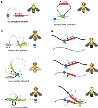
insulators (Zhaoet al.1995; Gaszneret al.1999). Overall, the known Drosophila insulator proteins can be divided into three groups based on their biochemical and functional prop-erties. Thefirst group contains sequence-specific DNA bind-ing proteins: Su(Hw), CTCF, BEAF-32, Ibf1, Ibf2, Pita, ZIPIC (also known as CG7928), Dwg, and GAF (the product of the Trithorax-like gene) (Geyer and Corces 1992; Zhao et al. 1995; Gaszneret al.1999; Moonet al.2005; Cuarteroet al. 2014; Maksimenkoet al.2015; Wolleet al.2015). The sec-ond group consists of the Cp190 protein and multiple protein isoforms encoded by themod(mdg4) gene (Dornet al.2001; Pai et al. 2004; Van Bortle et al. 2012). Both Cp190 and Mod(mdg4) appear to lack sequence-specific DNA bind-ing activity but can mediate homotypic and heterotypic Figure 2 Transgenic insulator assays. (A) Position-effect assay. Thewhitegene (red rectangle) confers red pigmentation tofly eyes. When integrated else-where in the genome,whiteis frequently repressed by neighboring repressor elements (blue pentagon, R) yielding flies with pale yellow eyes. When a transgene (here and below shown as bold line) carries insulators (green rectangles, inst) at its 59 and 39ends, thewhiteexpression becomes much more uniform between different insertion sites. (B) Enhancer blocking assay. One of the common en-hancer-blocking assays uses the yellow reporter gene (yellow rectangle), which controls dark pig-mentation of thefly. The expression ofyellowin wings, body, and bristles is controlled by distinct enhancer elements (gray circles marked with B, W, and Br, respectively). Whenflies lacking endog-enousyellowfunction are transformed with a copy of the WTyellow gene, their pigmentation fully restores (black wings, body, and bristles). In con-trast, whenyellow-deficientflies are transformed with a transgene that contains an insulator (green rectangle, inst) interposed between the upstream wing- and body-specific enhancers and theyellow
promoter, the transgenic insulator interacts with the nearest genomic insulator (blue rectangle, ins) to form a loop that blocks enhancer–promoter communication. Note that the interactions be-tween the promoter and the downstream bristle-specific enhancer are not impaired. This yields transgenicfiles that have yellow wings and body but black bristles. When two insulators are placed between the upstream enhancers and the pro-moter, they preferentially interact with each other and stimulate rather than inhibit enhancer– promoter interactions. Corresponding transgenic flies appear as WT. (C) An example of long-distancetrans-interactions enhanced by insulator elements. In transgenes containingwhitepaired with a PRE (blue pentagon), thewhitegene gets stochastically inactivated in embryonic precursor cells, which results inflies with variegated eye pigmen-tation. When two such transgenes, integrated in different nonhomologous chromosomes (illustrated as black and dashed lines), are combined, the variegation becomes much less pronounced or even disappears. Strikingly, when in addition to PREs the two transgenes also contain insulator elements,
thewhiterepression is greatly enhanced, often resulting inflies with completely white eyes. This suggests that insulator elements promote long-distance
protein–protein interactions via their BTB/POZ (Broad com-plex, Tramtrack, Bric-a-brac)/(Poxvirus and Zincfinger) do-mains. The third group contains biochemically diverse proteins: Elba1, Elba2, Elba3, Shep, and Rump, which were proposed to modulate the enhancer-blocking ability of insu-lator elements at specific stages of development or in a tissue-specific manner (Aokiet al.2012; Matzatet al. 2012; King et al.2014). Most of these proteins are specific to diptera, but at least one of them, CTCF, is widely conserved in evolution and, in mammals, acts as the main insulator protein (Herold et al.2012).
Insulator proteins bind chromatin in distinct combinations (Negreet al.2010b; Schwartzet al.2012), often as parts of multisubunit complexes (Paiet al.2004; Cuarteroet al.2014; Maksimenkoet al.2015). It appears that only sites cobinding certain combinations of insulator proteins can act as robust enhancer blockers (Schwartzet al.2012). This suggests that some insulator proteins may have functions unrelated to chromatin insulation. Indeed, genetic analyses indicate that sites bound by Su(Hw), but not any of the other known in-sulator proteins, act as transcriptional repressor elements (Schwartzet al.2012; Soshnevet al.2013).
How insulator elements block enhancer–promoter com-munications is not entirely clear. According to the most pop-ular hypothesis, insulator elements interact with each other and form chromatin loops, which compete with chromatin looping required for enhancer–promoter communication. Indeed there is ample evidence that pairs of insulator ele-ments interact. It wasfirst noted, that while the singlegypsy insulator placed between the enhancer and promoter of a transgenic reporter gene blocks their communication, the blocking activity is lost when the two gypsyinsulators are used in place of one (Cai and Shen 2001; Muravyovaet al. 2001). This“insulator bypass”effect is explained by assum-ing that a sassum-ingle transgenicgypsyinsulator interacts with the nearest genomic insulator to form a loop that obstructs the enhancer–promoter communication (Figure 2B). When two gypsyinsulators are placed between enhancer and promoter, they would preferentially interact with each other and form a loop, which would shorten the distance between enhancer and promoter, stimulating rather than inhibiting their inter-action (Cai and Shen 2001; Muravyova et al.2001). Pair-wise interactions are not limited togypsyinsulators. In fact, the majority of tested insulator elements appear to interact when paired up. However such interactions vary in strength and sometimes are detected only by more sensitive trans-genic assays based on stimulation of short-range enhancers (Kuhnet al.2003; Gruzdevaet al.2005; Kyrchanovaet al. 2007, 2008a,b; Fujioka et al.2016). The interactions are often directional and can happen between two distinct ele-ments (Kyrchanova et al.2008a; Fujiokaet al.2016). For specific subclasses of insulator elements, the looping inter-actions have been demonstrated at the molecular level (Blanton et al.2003; Comet et al.2011). However, more work is needed to understand the factors that define com-binations of distinct insulator elements that can interact.
Interactions between insulator elements are not limited to pairs contained within one transgenic construct (Figure 2C). Several elegant studies indicate that interaction between in-sulator elements can mediate the transvection between loci located hundreds of thousands of base pairs apart or even on different chromosomes (Kravchenkoet al.2005; Fujiokaet al. 2016). Likewise, insulators were shown to mediate long-distance interactions and enhance repression of reporter genes by PREs (Liet al.2011, 2013). In this view, the same kinds of interactions that lead to blocking enhancer– promoter communications also bring different genomic ele-ments together and juxtapose regulatory eleele-ments with target promoters. The 3D contacts facilitated by insulator elements are likely transient, implying that the role of insu-lators is to increase the probability of certain chromatin con-formations rather than generating a rigid loop structure.
How could the elements that facilitate transient looping contacts delimit the boundaries of combinatorial patterns of chromatin modifications (chromatin states)? One scenario that is easy to envision is when histone modifications are produced via looping interactions of a protein complex an-chored at a fixed chromatin site. For example, Polycomb complexes anchored at PREs (see below for details) loop out and trimethylate H3K27 at extended distances from their principal binding sites (Kahnet al.2006; Schwartzet al.2006). The“spreading”of H3K27me3 from PREs can be blocked by chromatin insulator elements due to the reduction of transient looping contacts between the PRE-anchored complexes and surrounding chromatin (Kahn et al. 2006; Cometet al. 2011). Similar to the enhancer blocking case, a pair of inter-acting insulator elements can be bypassed, leaving the stretch of chromatin between the insulators free of H3K27me3 with the high level of H3K27 methylation in chromatin further away from the insulator pair (Cometet al. 2011). Genomic studies indicate that insulator elements do restrict the spread-ing of H3K27me3 domains around Polycomb target genes (Bartkuhnet al.2009; Schwartzet al.2012). However, their contribution is most critical to prevent the methylation of the neighboring genes that are transcriptionally inactive (Schwartzet al.2012) because chromatin remodeling linked to transcriptional activity can, by itself, inhibit H3K27 methyl-ation. For example, histone H3 molecules methylated at lysine 4 or lysine 36 are poor substrates for histone methyltransferase activity of the PRC2 complexes (Schmitgeset al.2011; Yuan et al.2011; Voigtet al.2012). When histone modifications are produced in the immediate vicinity or by processive movement of an enzyme along the chromatin fiber, for instance by an enzyme linked to transcribing RNA polymerase, insulator ele-ments would have little effect on their spreading. It is, there-fore, not surprising that the boundaries of domains with distinct chromatin states (combinations of histone modifi ca-tions) show only limited overlap with the insulator protein binding sites (Kharchenkoet al.2011; Schwartzet al.2012). Thanks to the remarkable property to formtrans-interactions, insulator elements are naturally expected to play a role in partitioning theDrosophilagenome into TADs. Indeed, the partitioning of the bithorax complex cluster of homeotic genes into distinct TADs by theFubinsulator element repre-sents one such beautiful example (Bender and Lucas 2013; Savitskyet al.2016). However, what fraction of TADs is
de-fined by insulator elements remains an open question. Iron-ically, although the quest to define specific chromatin boundary elements started with the suggestion thatscsand scs9insulators delimit the extent of the decondensed chromatin domain of theHsp70locus, careful microscopy measurements showed that scsand scs9 reside well within and not at the borders of theHsp70puff (Kuhnet al.2004). Comparison of TADs defined by the Hi-C approach to genomic distributions of insulator proteins indicates that Cp190 and BEAF-32 fre-quently colocalize with TAD borders (Hou et al. 2012; Sextonet al.2012; Ulianovet al.2016). This can be taken to indicate that insulator proteins define the TAD limits.
How-ever, the interpretation is likely more complex. A large fraction of Cp190 and BEAF-32 binding sites are in the vicinity of TSSs of active genes (Busheyet al.2009; Schwartzet al.2012) and whether these Cp190 and/or BEAF-32 binding sites corre-spond to insulator elements is unknown. It is important to keep in mind that TAD borders are defined as points at which the frequencies of interactions between adjacent chromatin stretches change. Since transcriptionally active and inactive chromatin display distinct folding regimes (Sexton et al. 2012; Boettiger et al.2016), the transition between the two kinds of chromatin is likely to appear as TAD boundary. Con-sistently, combinations of histone modifications typical of ac-tive genes can predict the positions of many DrosophilaTAD borders (Ulianovet al.2016). It is therefore possible that over-representation of Cp190 and BEAF-32 at TAD borders simply reflects their bias toward active TSSs.
How many of the TAD borders depend on insulator ele-ments is not entirely clear. In principle, Hi-C profiles of TADs defined by looping interactions of two insulator elements should carry distinct signatures of enhanced contact frequen-cies between the insulator elements. Those would appear as brighter “dots”at the top of the corresponding TAD“ pyra-mids”(Figure 1). Indeed, such high-contact foci correspond-ing to enhanced contacts between some of the CTCF bindcorrespond-ing sites are clearly visible in the high-resolution contact map of the human genome (Raoet al.2014). For some reason, the foci of enhanced contacts are not as readily detectable in the DrosophilaHi-C maps (Houet al.2012; Sextonet al.2012; Eagenet al.2015; Ulianovet al.2016).
Future experiments that look at changes in genome topology in cells lacking key insulator proteins will tell us how many of the TAD borders depend on insulator elements. Until then, it is safe to conclude that a fraction of TAD borders is formed by insulator elements and these borders are essential for faithful regulation of genes with complex regulatory regions. The role of the insulator-based borders is likely most critical in cells where they separate TADs that cover genes with similar tran-scriptional states. For example, an insulator element placed between a TAD encompassing a Polycomb-repressed gene and a TAD with a transcriptionally inactive gene is needed to prevent the latter from being permanently repressed.
Polycomb Complexes: Linking Epigenetic Regulation with 3D Chromatin Organization
PcG proteins are a group of chromatin regulatory components that are able to modulate or silence the expression of euchro-matic genes. First discovered in 1947 by Pamela Lewis (Lewis 1947) and originally believed to be one of the homeotic (Hox) genes that specify the anterior–posterior body plan, the
disrupts the initiation of the correct segmental expression of Hox genes, mutations in Polycomb Group (PcG) genes initially show little or no phenotypes but, later, induce Hox gene de-repression (Struhl and Akam 1985). Screening for suppressors of PcG function led to the discovery of a set of genes that counteract PcG-mediated silencing, named trithorax-group (trxG) genes after the first member of the group (Ingham 1983; Kennison and Tamkun 1988; Tamkunet al.1992).
Analysis of polytene chromosomes showed that PcG proteins bind to Hox loci and to a variety of other loci in the genome (Zink and Paro 1989; Rastelliet al.1993), indicating that their functions may be more widespread than Hox gene regulation. Furthermore, cloning ofPcshowed that its protein possesses a short region, defined as chromo domain, very similar to that of the HP1 protein (Paro 1990; Paro and Hogness 1991). The genetically defined function in maintenance of gene silencing and the conservation of the chromo domain with HP1, which was involved in heterochromatin maintenance, suggested that PcG genes may somehow set up a memory of cell transcription states (Paro 1990). This memory function was later demon-strated using transgenes containing PcG binding sites (Cavalli and Paro 1998, 1999; Maurange and Paro 2002; Pouxet al. 2002; Bejarano and Milan 2009).
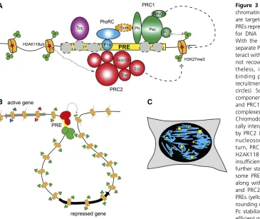
Indeed, PcG proteins were shown to bind to specific reg-ulatory elements of about 1000 bp. These polycomb response elements (PREs) could recruit PcG proteins also when they were inserted in transgenic constructs, where they could drive repression of reporter genes (Busturia and Bienz 1993; Fauvarque and Dura 1993; Simon et al. 1993; Chan et al. 1994; Zink and Paro 1995). The analysis of PRE-containing transgenes showed that PcG-mediated silencing has proper-ties similar to those of heterochromatin, such as a variegated silencing of the mini-whitereporter gene. However, two no-table differences are that higher temperatures enhance PcG-dependent silencing, whereas they reduce heterochromatin silencing, and that homologous pairing of PRE-containing transgenes induced increased silencing efficiency at a subset of the transgene insertion loci (Pirrotta and Rastelli 1994). Since the identification of genomic elements necessary and sufficient for PcG targeting, much effort has been dedicated to reveal how the targeting happens at the molecular level. The results of this large body of work were summarized in an excellent recent review (Kassis and Brown 2013). Briefly, PREs seem to represent collections of recognition sequences for multiple DNA binding adaptor proteins (Figure 3A). With an exception of Pleiohomeotic (Pho) or closely related Pleiohomeotic-like (Phol) proteins (Brownet al.1998, 2003), which form a distinct PhoRC complex (Klymenkoet al.2006), the other DNA binding proteins interact with the core PcG complexes too weakly to be recovered as stoichiometric com-ponents of the complexes. It was proposed that individual weak interactions of the DNA binding proteins combine to provide robust recruitment of Polycomb repressive complexes 1 and 2 (PRC1 and PRC2, see below).
At the beginning of the 1990s, laboratories studying mam-malian development became interested in this intriguing set of
proteins and they identified mammalian homologs offly PcG proteins (Pearceet al.1992; Ishidaet al.1993; Mulleret al. 1995). Strikingly, trxG genes were also shown to be conserved (Djabaliet al. 1992). Furthermore, homology of the twofly members of the PcGPscandSu(z)2to a murine protooncogene named Bmi-1 strongly suggested that, in addition to its re-quirement during development, the appropriate regulation of PcG function is also required to prevent the emergence of cancer. Since then, countless reports have corroborated this hypothesis (Koppens and van Lohuizen 2016), extended the link with cancer to trxG proteins (Schuettengruberet al.2011) and,finally, the appropriate regulation offly PcG components has also been demonstrated to prevent oncogenesis (Classen et al.2009; Martinezet al.2009; Sieverset al.2014). These
findings have raised the interest of the scientific community for PcG and trxG proteins considerably.
ubiquitylation and PRC2 recruitment has been called into question and further investigations are required to clarify the issue (Leeet al.2015; Pengellyet al.2015). Finally, it is important to realize that some Polycomb genes have one or more paralogs. Inflies, this is true for one PRC2 member,esc, which has a paralog calledescl(Wanget al.2006; Ohnoet al. 2008), but also for thephandPscloci (Grimaudet al.2006b; Schuettengruberet al.2007). Mammalian PcG components are even more redundant, with several paralogs for each of the subunits (Piunti and Shilatifard 2016). Detailed work in mammals has identified a whole series of PRC1-related com-plexes. Canonical PRC1 variants (cPRC1) are those that con-tain homologs to each of the original PRC1 components isolated inflies,i.e., PC, PH, PSC, and SCE, whereas in non-canonical PRC1 variants, PC and PH homologs are absent and the ncPRC1-specific RYBP proteins are present instead. Fur-thermore, specific types of PSC paralogs characterize specific ncPRC1 variants and other proteins are present in some of them (Gao et al. 2012). While most of the members of ncPRC1 are conserved in flies, whether they form similar complexes and their function is conserved is still unclear. More studies are required to address these questions.
In parallel to the biochemical characterization of PcG complexes, a large body of work has been dedicated to de-scribe and understand their genome-wide distribution. Initial
the PH component of PRC1 in larval development but not in embryos (Martinez et al. 2009). Indeed a recent study has shown that PcG binding is dynamic during development and that two types of PcG target genes exist: canonical targets car-rying PRC1 and PRC2 binding in the presence of the H3K27me3 mark, and a novel category, defined as neo-PRC1 genes, which include the Notch gene and are bound by PRC1 and PRC2 in the absence of its H3K27me3 mark (Loubiereet al.2016).
One intriguing observation that came from the comparison of genome-wide location with microscopy studies, done either by immunofluorescence or by analysis of GFP-fusion proteins of the PcG, is that PcG components stain as foci in the nucleus (Buchenauet al.1998; Grimaudet al.2006a; Terranovaet al. 2008; Cheutin and Cavalli 2012) (Figure 3C). Although any comparison between microscopic foci and genome-wide binding sites is difficult, the detection of a rather limited number of nuclear foci suggested the possibility that long-range contacts might exist between PcG-bound elements. This idea, also supported by the phenomenon of pairing sen-sitive silencing in PRE-containing transgenes, was directly tested by DNA FISH analysis of the 3D nuclear location of PRE-containing transgenes. Initially, transgenes containing one regulatory region called Fab-7, which contains a PRE
flanking a chromatin insulator, were shown to be able to
frequently contact the endogenousFab-7element even when inserted in a different chromosome (Bantignieset al.2003). The combination of FISH with immunostaining showed that this colocalization occurred at PcG foci and was dependent on PcG proteins, as well as on RNA interference components, although the molecular mechanism linking these proteins to PcG members could not be elucidated (Grimaud et al. 2006a). Later, other transgenes were shown to be capable of inducing contacts and the insulator elements flanking PREs were demonstrated to play a pivotal role in targeting 3D interactions (Li et al. 2011). The action of insulators seems to be neutral with regard to the regulatory outcome, i.e., insulators can drive associations in the 3D space of the cell nucleus of their target elements. If these elements are in a repressed state, their association is accompanied and possibly stabilized by PcG components, whereas if they are active, TrxG proteins assist their association to transcriptionally ac-tive regions of the nucleus (Liet al.2013).
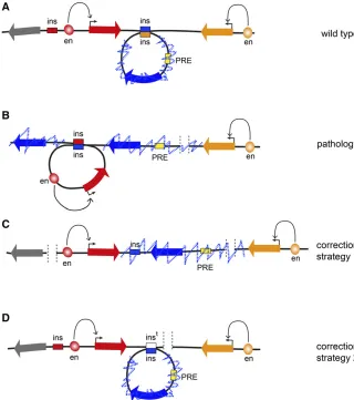
These data suggested that a cooperation between PcG pro-teins, TrxG propro-teins, and insulators may lead to higher-order organization of a large set of genomic loci. This idea is at least partly supported by several lines of evidence. First, the analysis of nuclear positioning of Hox genes has shown that the bithorax and the antennapedia complexes colocalize in nuclei in which Figure 4 Prospects of 3D genome engineering. (A) In a fictional WT locus, the transcription of the
Hox genes are corepressed (Bantignies et al. 2011). Micro-scopic colocalization signifies chromatin contacts, as shown by 4C studies both in embryos and in larval brains (Bantignieset al.2011; Tolhuiset al.2011). Furthermore, 4C and Hi-C studies identified a large network of contacts involv-ing PcG target loci (Bantignieset al.2011; Tolhuiset al.2011; Sextonet al.2012). Importantly, reducing the frequency of Hox gene contacts by mutating regulatory elements in the bithorax complex, induced the derepression of Hox genes in the anten-napedia complex, which is located 10 Mb away (Bantignies et al.2011). This result suggests that clustering of PcG target genes may stabilize silencing. Furthermore, recent evidence also suggests that 3D architecture may also stabilize recruit-ment of Polycomb proteins (Schuettengruber et al. 2014; Entrevanet al.2016). It is noteworthy that contacts between genomic regions bound by PcG components are conserved in mammalian cells (Schoenfelderet al.2015; Vieux-Rochaset al. 2015), suggesting that the 3D aspect of PcG biology is also a conserved feature. Obviously, it is of great importance to un-derstand the mechanisms regulating clustering of PcG sites in space. Initial work suggests that the PH subunit of PRC1 may be important for this process, both inflies and in mammals (Isono et al.2013; Waniet al.2016). Clearly, much more work has to be done to understand the mechanisms of PcG-mediated gene contacts, those that govern delimitation of PcG chromatin spreading in theflanking genome, and the interplay between PcG, TrxG components, and other chromosomal proteins to regulate nuclear organization of their target genes.
Conclusions/Perspectives
For decadesDrosophilahas been a pioneering model system to identify and describe the connection between 3D organization of the genome and its function. Some important aspects of this organization, for instance, the compartmentalization of the genome parts to specific subnuclear locations, such as the nu-clear lamina or the nucleolus, were not discussed in this review due space limitations. Nevertheless, the examples provided here clearly show that the genome cannot be reduced to a string of DNA nucleotides and that all levels of higher-order genome organization, from the nucleosome to chromatin fi -bers, chromosomal domains, chromosome territories, and chromosome localization within nuclear space, must be con-sidered. A great amount of work is still required to dissect the links between each of these organizational levels and various aspects of genome function. Part of this work will likely in-volve the analysis of chromosome architecture of specific cell types or developmental stages, similar to the recent analysis of the spatial regulation of the bithorax complex along the anteroposterior body plan during embryogenesis (Bowman et al.2014). To this aim, the development of low- or single-cell techniques is needed, since most of these studies do not allow isolation of large amount of homogeneous cells.
As studies that connect disruptions of TADs to patholog-ical rewiring of enhancer–promoter interactions and herita-ble malformations in human patients started to emerge
(Lupianezet al.2015), there is mounting pressure to under-stand the basic rules that govern genome folding. Knowing these rules will advance our ability to interpret consequences of deletions, duplications, inversions, and translocations that are found within normal human population. Many of these structural variations are linked to predisposition to disease. In cases when a variation changes gene dosage the link is easier to explain. In contrast, the consequences of inversions or rearrangements that affect noncoding DNA are much more difficult to predict unless we know how they may impact genomic interactions. Once the principles that govern ge-nome architecture are charted, we will be in position to cor-rect some of the 3D aberrations using advances from the burgeoning field of precision genome editing (Figure 4). We have no doubt thatDrosophilawill continue to be instru-mental in our quest to achieve this goal.
Acknowledgments
The authors thank Mikhail Savitsky for thefly cartoons used in Figure 2. Research in the G.C. laboratory was supported by grants from the European Horizon 2020 (H2020) MuG project under grant agreement 676556, Centre National de la Recherche Scientifique, the European Network of Excel-lence EpiGeneSys, Agence Nationale de la Recherche (EpiDevoMath), Fondation pour la Recherche Médicale (DEI20151234396), Institut National de la Santé et de la Recherche Médicale/Plan Cancer Epigenetics and Cancer Program (MM&TT), the Laboratory of Excellence Epi-GenMed, and Fondation ARC pour la Recherche sur le Can-cer. Research in the Y.B.S. laboratory was supported by grants from the Swedish Research Council, Cancerfonden, the European Network of Excellence EpiGeneSys, the Knut and Alice Wallenberg Foundation, Kempestiftelserna, and Umeå University Insamlingsstiftelsen.
Literature Cited
Adams, M. D., S. E. Celniker, R. A. Holt, C. A. Evans, J. D. Gocayne et al., 2000 The genome sequence of Drosophila melanogaster. Science 287: 2185–2195.
Agard, D. A., and J. W. Sedat, 1983 Three-dimensional architec-ture of a polytene nucleus. Naarchitec-ture 302: 676–681.
Alkema, M. J., M. Bronk, E. Verhoeven, A. Otte, L. J. van’t Veer et al., 1997 Identification of Bmi1-interacting proteins as con-stituents of a multimeric mammalian polycomb complex. Genes Dev. 11: 226–240.
Aoki, T., A. Sarkeshik, J. Yates, and P. Schedl, 2012 Elba, a novel developmentally regulated chromatin boundary factor is a hetero-tripartite DNA binding complex. eLife 1: e00171.
Badrinarayanan, A., T. B. Le, and M. T. Laub, 2015 Bacterial chromosome organization and segregation. Annu. Rev. Cell Dev. Biol. 31: 171–199.
Bantignies, F., C. Grimaud, S. Lavrov, M. Gabut, and G. Cavalli, 2003 Inheritance of Polycomb-dependent chromosomal inter-actions in Drosophila. Genes Dev. 17: 2406–2420.
Barges, S., J. Mihaly, M. Galloni, K. Hagstrom, M. Muller et al., 2000 The Fab-8 boundary defines the distal limit of the bi-thorax complex iab-7 domain and insulates iab-7 from initiation elements and a PRE in the adjacent iab-8 domain. Development 127: 779–790.
Bartkuhn, M., T. Straub, M. Herold, M. Herrmann, C. Rathkeet al., 2009 Active promoters and insulators are marked by the cen-trosomal protein 190. EMBO J. 28: 877–888.
Bejarano, F., and M. Milan, 2009 Genetic and epigenetic mecha-nisms regulating hedgehog expression in the Drosophila wing. Dev. Biol. 327: 508–515.
Bender, W., and A. Hudson, 2000 P element homing to the Dro-sophila bithorax complex. Development 127: 3981–3992. Bender, W., and M. Lucas, 2013 The border between the
ultra-bithorax and abdominal-A regulatory domains in theDrosophila bithorax complex. Genetics 193: 1135–1147.
Bischof, J., R. K. Maeda, M. Hediger, F. Karch, and K. Basler, 2007 An optimized transgenesis system for Drosophila using germ-line-specific phiC31 integrases. Proc. Natl. Acad. Sci. USA 104: 3312–3317.
Blackledge, N. P., N. R. Rose, and R. J. Klose, 2015 Targeting Polycomb systems to regulate gene expression: modifications to a complex story. Nat. Rev. Mol. Cell Biol. 16: 643–649 Blanton, J., M. Gaszner, and P. Schedl, 2003 Protein:protein
in-teractions and the pairing of boundary elements in vivo. Genes Dev. 17: 664–675.
Boettiger, A. N., B. Bintu, J. R. Moffitt, S. Wang, B. J. Beliveauet al., 2016a Super-resolution imaging reveals distinct chromatin folding for different epigenetic states. Nature 529: 418–422. Bonchuk, A., S. Denisov, P. Georgiev, and O. Maksimenko,
2011 Drosophila BTB/POZ domains of “ttk group” can form multimers and selectively interact with each other. J. Mol. Biol. 412: 423–436.
Bonchuk, A., O. Maksimenko, O. Kyrchanova, T. Ivlieva, V. Mogila et al., 2015 Functional role of dimerization and CP190 inter-acting domains of CTCF protein in Drosophila melanogaster. BMC Biol. 13: 63.
Bowman, S. K., A. M. Deaton, H. Domingues, P. I. Wang, R. I. Sadreyev et al., 2014 H3K27 modifications define segmental regulatory domains in the Drosophila bithorax complex. eLife 3: e02833.
Boyer, L. A., K. Plath, J. Zeitlinger, T. Brambrink, L. A. Medeiros et al., 2006 Polycomb complexes repress developmental regu-lators in murine embryonic stem cells. Nature 441: 349–353. Brown, J. L., D. Mucci, M. Whiteley, M. L. Dirksen, and J. A. Kassis,
1998 The Drosophila polycomb group gene pleiohomeotic en-codes a DNA binding protein with homology to the transcription factor YY1. Mol. Cell 1: 1057–1064.
Brown, J. L., C. Fritsch, J. Mueller, and J. A. Kassis, 2003 The Drosophila pho-like gene encodes a YY1-related DNA binding protein that is redundant with pleiohomeotic in homeotic gene silencing. Development 130: 285–294.
Buchenau, P., J. Hodgson, H. Strutt, and D. J. Arndt-Jovin, 1998 The distribution of polycomb-group proteins during cell division and development in Drosophila embryos: impact on models for silencing. J. Cell Biol. 141: 469–481.
Bushey, A. M., E. Ramos, and V. G. Corces, 2009 Three subclasses of a Drosophila insulator show distinct and cell type-specific genomic distributions. Genes Dev. 23: 1338–1350.
Busturia, A., and M. Bienz, 1993 Silencers in abdominal-B, a ho-meotic Drosophila gene. EMBO J. 12: 1415–1425.
Cai, H. N., and P. Shen, 2001 Effects of cis arrangement of chro-matin insulators on enhancer-blocking activity. Science 291: 493–495.
Cao, R., Y. I. Tsukada, and Y. Zhang, 2005 Role of Bmi-1 and Ring1A in H2A ubiquitylation and Hox gene silencing. Mol. Cell 20: 845–854.
Cao, R., L. Wang, H. Wang, L. Xia, H. Erdjument-Bromage et al., 2002 Role of histone H3 lysine 27 methylation in polycomb-group silencing. Science 298: 1039–1043.
Cavalli, G., and R. Paro, 1998 The Drosophila Fab-7 chromosomal element conveys epigenetic inheritance during mitosis and mei-osis. Cell 93: 505–518.
Cavalli, G., and R. Paro, 1999 Epigenetic inheritance of active chromatin after removal of the main transactivator. Science 286: 955–958.
Cavalli, G., and T. Misteli, 2013 Functional implications of ge-nome topology. Nat. Struct. Mol. Biol. 20: 290–299.
Chan, C. S., L. Rastelli, and V. Pirrotta, 1994 A polycomb response element in the Ubx gene that determines an epigenetically in-herited state of repression. EMBO J. 13: 2553–2564.
Cheutin, T., and G. Cavalli, 2012 Progressive polycomb assem-bly on H3K27me3 compartments generates polycomb bodies with developmentally regulated motion. PLoS Genet. 8: e1002465.
Cheutin, T., and G. Cavalli, 2014 Polycomb silencing: from linear chromatin domains to 3D chromosome folding. Curr. Opin. Genet. Dev. 25: 30–37.
Cheutin, T., A. J. McNairn, T. Jenuwein, D. M. Gilbert, P. B. Singh et al., 2003 Maintenance of stable heterochromatin domains by dynamic HP1 binding. Science 299: 721–725.
Ciabrelli, F., and G. Cavalli, 2015 Chromatin-driven behavior of topologically associating domains. J. Mol. Biol. 427: 608–625. Clark, R. F., C. R. Wagner, C. A. Craig, and S. C. Elgin, 1991 Distribution
of chromosomal proteins in polytene chromosomes of Drosophila. Methods Cell Biol. 35: 203–227.
Classen, A. K., B. D. Bunker, K. F. Harvey, T. Vaccari, and D. Bilder, 2009 A tumor suppressor activity of Drosophila Polycomb genes mediated by JAK-STAT signaling. Nat. Genet. 41: 1150– 1155.
Cohen, J., 1962 Position-effect variegation at several closely linked loci in Drosophila melanogaster. Genetics 47: 647–659. Comet, I., B. Schuettengruber, T. Sexton, and G. Cavalli, 2011 A
chromatin insulator driving three-dimensional Polycomb re-sponse element (PRE) contacts and Polycomb association with the chromatinfiber. Proc. Natl. Acad. Sci. USA 108: 2294–2299. Csink, A. K., and S. Henikoff, 1996 Genetic modification of het-erochromatic association and nuclear organization in Drosoph-ila. Nature 381: 529–531.
Csink, A. K., and S. Henikoff, 1998 Large-scale chromosomal movements during interphase progression in Drosophila. J. Cell Biol. 143: 13–22.
Cuartero, S., U. Fresan, O. Reina, E. Planet, and M. L. Espinas, 2014 Ibf1 and Ibf2 are novel CP190-interacting proteins re-quired for insulator function. EMBO J. 33: 637–647.
Czermin, B., R. Melfi, D. McCabe, V. Seitz, A. Imhof et al., 2002 Drosophila enhancer of Zeste/ESC complexes have a histone H3 methyltransferase activity that marks chromosomal Polycomb sites. Cell 111: 185–196.
de Napoles, M., J. E. Mermoud, R. Wakao, Y. A. Tang, M. Endoh et al., 2004 Polycomb group proteins Ring1A/B link ubiquity-lation of histone H2A to heritable gene silencing and X inacti-vation. Dev. Cell 7: 663–676.
Dejardin, J., 2015 Switching between epigenetic states at pericen-tromeric heterochromatin. Trends Genet. 31: 661–672. Dejardin, J., A. Rappailles, O. Cuvier, C. Grimaud, M. Decoville
et al., 2005 Recruitment of Drosophila Polycomb group pro-teins to chromatin by DSP1. Nature 434: 533–538.
Dernburg, A. F., K. W. Broman, J. C. Fung, W. F. Marshall, J. Philips et al., 1996 Perturbation of nuclear architecture by long-dis-tance chromosome interactions. Cell 85: 745–759.