Abstract
Thomas, Gracie A. Expression of Virulence Genes in Salmonella. (Under the direction on Dr. Stephen J. Libby)
A role for slyA in Salmonella pathogenesis has been clearly demonstrated. Originally, slyA was identified as a gene that encoded a cryptic hemolysin. When cloned into E. coli,
slyA was shown to be required for the cytolytic death of nucleated cells and the lysis of red
blood cells. However, upon further investigation, it was discovered that expression of the hemolysin is directly regulated by slyA and is, therefore, not a function of the gene itself. It is
previously reported that SlyA is required for the intracellular survival of the bacteria within host macrophages and that slyA mutant strains of S. typhimurium are severely attenuated for
virulence in the murine model. In addition, Daniels et al. showed that SlyA is required for the destruction of murine M-cells but not for invasion. Further studies show that slyA may be required for resistance to oxidative stress and, more recently, that slyA may act as a regulator
of virulence genes involved in the systemic phase of infection, but not the enteric phase. Based on sequence homology with other transcriptional regulatory proteins of other Gram-negative bacteria, it was first suggested by Ludwig et al. that SlyA may act as a
regulatory protein, affecting expression of both E. coli and Salmonella genes. In view of this, SlyA was initially grouped with MarR and EmrR (MprA) of E. coli, Hpr of Bacillus subtilis,
and PecS of Erwinia chrysanthemi. However, it is now apparent that SlyA is more distantly related to these regulatory proteins than previously reported. SlyA is currently classified as a member of a unique family of low molecular weight transcriptional regulatory proteins that
SPI-4 is an approximate 25kb sequence insertion located at centisome 92 on the Salmonella chromosomal map. This pathogenicity island is flanked on either side by the ssb
and yciB genes in both S. typhimurium and S. typhi. Originally, SPI-4 was identified via a
transposon insertion as a chromosomal sequence required for survival inside of murine macrophages. Wong et al. originally claimed that SPI-4 contained 18 ORFs designated A-R.
The recently completed annotation of both S. typhimurium LT2 and S.typhi genomes shows that only 6 open reading frames exist. Ahmer et al. demonstrated SPI-4 regulation by SirA, a transcriptional regulator of SPI-1 (invasion locus). Experiments in our laboratory have
shown that the beta-galactosidase activity of a SPI4-K::MudJ insertion (SL3277) in a slyA mutant background is reduced 8-fold, suggesting that SPI-4 is regulated by slyA. We recently
discovered that the predicted SPI4-K protein has significant homology to a class of autotransporter toxins found in pathogenic E.coli, Bordetella, Neisseria, and Shigella species, referred to as SPATE toxins. Members of this family are serine proteases possessing a
characteristic GDSGS motif with a central catalytic Ser residue. We have constructed and purified a His-tagged SPI4-K protein fragment based on he earlier annotation, and used this protein to obtain a polyclonal antibody. In this study, we examined the protein expression
characteristics of SPI4-K in Salmonella typhimurium and other Salmonellae. Here, we confirm through immunoblot analysis that SPI4-K is a large molecular weight protein that
appears to be secreted from the intracellular compartment.
In an attempt to further clarify the role of both SlyA and SPI4-K in Salmonella pathogenesis, polyclonal antibodies were purified to these proteins. Here, we show that the
Expression of Virulence Genes in Salmonella
by
Gracie A. Thomas
A thesis submitted to the Graduate Faculty of North Carolina State University
in partial fulfillment of the requirements for the Degree of
Master of Science
MICROBIOLOGY
Raleigh
2002
APPROVED BY:
________________________________ _________________________________ Amy M. Grunden Geraldine Luginbuhl
________________________________ Chair of Advisory Committee
Biography
1. Graduated from Eau Gallie High School Melbourne, FL
05/1994
2. Graduated from University of Florida Gainesville, FL
05/1998
Degree: BS Microbiology and Cell Science 3. Graduated from North Carolina State University
Raleigh, NC 12/2002
Acknowledgments
I would like to thank my parents for all of their support and positive encouragement they have given me over the years. I give a special thanks to my mentor, Dr. Stephen J. Libby, for showing me the ropes and always being patient. I offer my gratitude to
Thomas A. Halsey, Michael J. Stewart, Elizabeth Boutt, and Kara Main for their helpful discussions and participation in the project. In addition, I would like to thank the other
Table of Contents
List of Tables v
List of Figures vi
Salmonella Pathogenesis Review 1 Disease Causing Mechanisms of Salmonella 1 Surviving the Oxidative Burst within Phagocytes 2 The Contribution of Salmonella Pathogenicity Islands to Virulence 5 The Salmonella Virulence Plasmid 10 Role of slyA in Salmonella Pathogenesis 11 Chapter 1: Purification of a polyclonal antibody for the detection of SlyA protein expression 13
Introduction 13
Materials and Methods 14
Results 19
Discussion 22
Chapter 2: Purification of a polyclonal antibody for the detection of SPI4-K protein expression 35
Introduction 35
Materials and Methods 37
Results 42
Discussion 45
List of Tables
Chapter 1: Purification of a polyclonal antibody for the detection of SlyA protein expression
Table 1: Bacterial strains and plasmids used 34
Chapter 2: Purification of a polyclonal antibody for the detection of SPI4-K protein expression
List of Figures
Chapter 1: Purification of a polyclonal antibody for the detection of SlyA protein expression
Figure 1: Schematic of SlyA His-tag antibody production 28 Figure 2: SDS-PAGE gel of His-tag protein purification steps 29 Figure 3: Western blot analysis of SlyA stationary phase protein expression 30 Figure 4: Western blot analysis of SlyA stationary phase protein expression 31 Figure 5: Western blot analysis of SlyA stationary phase protein expression 32 Figure 6: Growth curve expression of SlyA 33 Chapter 2: Purification of a polyclonal antibody for the detection of SPI4-K protein expression
Figure 1: Map of STM4261 and location of His-tagged protein construction 51 Figure 2: Schematic of SPI4-K His-tag antibody production 52 Figure 3: SDS-PAGE gel of His-tag protein purification steps 53 Figure 4: Western blot analysis of SPI4-K total protein expression 54 Figure 5: Western blot anlaysis of SPI4-K total protein expression 55 Figure 6: Western blot analysis of secreted SPI4-K protein 56
Salmonella Pathogenesis Review
Disease Causing Mechanisms of Salmonella
Salmonella is a Gram-negative facultative intracellular pathogen that is able to
infect and cause disease in humans and domestic animals. The World Health
Organization reports that there are 2213 known serotypes of Salmonella to date, the
majority of which are capable of causing disease in humans. The serotypes are
categorized into seven taxonomic groups based on the variation in their biotypes. Groups
I, II, IIIa, IIIb, IV, and VI are all members of S. enterica, while group V is composed of
S. bongori. Salmonella infection in humans usually results from consumption of
contaminated food and water. Symptoms following infection depend on the serotype and
range from mild self-limiting enteritits where infection remains localized in the gut, to
acute systemic salmonellosis where bacteria enter the bloodstream and disseminate to
organs such as the spleen and liver.
For Salmonella to cause systemic disease, it must first survive the gastric acidity of the
stomach, invade the intestinal epithelia, and resist host phagocytes. Because Salmonella
is unable to survive at a pH lower than 4.5, a high oral concentration is required to cause
infection. In the itys mouse model, the infectious dose is 106 organisms. The site of infection is primarily initiated at M-cells of the Peyer’s Patches in the small intestine
(67). M-cells are specialized epithelial cells that constantly sample antigenic material in
the lumen and present it to the immune system. The interaction between Salmonella and
M cells facilitates host colonization. Salmonella gains entry into M-cells during
transcytosis and enter the follicle dome of the lamina propia. The follicle dome houses
host macrophages and lymphocytes that are constantly assessing the environment for
bacteria. In some cases, the invading bacteria will be phagocytosed and killed by the
professional phagocytes. Sometimes, however, virulent strains can invade and take up
residence inside of host macrophages. The latter scenario leads to severe systemic
infection by way of matriculating host macrophages that carry Salmonella to organs of
the body such as the spleen and liver (41). In order to cause systemic infection, a
sufficient number of Salmonella must transverse the M cells and survive within the
patrolling phagocytes.
Surviving the Oxidative Burst within Phagocytes
The innate immune system of the host plays a vital role in resistance to
Salmonella infection, as do a variety of other bacterial, viral, and parasitic pathogens.
Professional phagocytes become activated when Salmonella is opsonized or tagged by
complement factors and/or antibodies for recognition. Efforts have focused on
understanding the ability of Salmonella to resist both dependent and
oxygen-independent antimicrobial activity of host phagocytic cells. Oxygen-oxygen-independent killing
mechanisms by host phagocytes include bioactive peptides, alteration of phagosomal
ionic concentration or other nutrient limitation, and reduced pH (42). The oxidative
mechanisms are the focus of this review and are discussed below.
The oxidative burst is the release of oxygen-dependent molecules with antimicrobial
properties. The effects of the oxidative burst are most often detrimental to the bacterial
is extremely detrimental and is linked to a disorder in humans known as chronic
granulomatous disease (GCD) (9). A deficiency in one of the protein components of the
oxidase renders it non-functional and, as a result, humans are susceptible to repeated
fungal and bacterial infections (90, 91, 104).
Intense effort has been directed at understanding the molecular mechanisms by which
Salmonella is able to survive and replicate in host macrophages. Salmonella survival and
replication in host phagocytic cells is required for the manifestation of virulence (39, 40,
42-44). Genes that have been shown to be required for Salmonella survival in host
macrophages and virulence include slyA, phoP, purE, aroA, rpoE, SPI2, sspJ, mig-14,
pagC, and recA, among many others (15, 19, 20, 29, 36, 61).
Until recently, the molecular mechanisms by which Salmonella is able to
withstand reactive oxygen species produced by the respiratory burst of phagocytes were
poorly understood. The respiratory burst of phagocytes following ingestion of bacteria
results in the production of superoxide from molecular oxygen (4). The cytochrome b558
portion of the NADPH phagocyte oxidase (phox) associates with the phagosomal
membrane following phagocytosis to form an active complex with the cytoplasmic
subunits p47 and p67. This enzyme produces superoxide radical ·O2- that can dismutate to form hydrogen peroxide (H2O2) near the ingested microbe. These two reactive oxygen species (ROS) can also react with nitric oxide (NO) to produce peroxynitrite (ONOO-), a reactive nitrogen species (RNS). Hydrogen peroxide can react with Fe(II) (Fenton
reaction) to form hydroxyl radical (·OH). Thus, the production of superoxide radical by
phagocytes can give rise to a variety of toxic and highly reactive antimicrobial
genetic and biochemical factors are used by Salmonella to resist or avoid
phagocyte-derived oxidative antimicrobial products (15, 27-30, 37, 76). Several lines of evidence
have demonstrated the importance of the phagocyte respiratory burst in resistance to
Salmonella. Salmonella mutants defective in the DNA repair genes recA and recBC are
able to survive and replicate in murine macrophages lacking the ability to generate a
respiratory burst, but are killed in burst-competent macrophages (15). Mice that have a
defined mutation in the gp91 subunit of the NADPH oxidase (phox knockouts) have
increased susceptibility to Salmonella infection (29), and macrophages from these mice
are unable to control Salmonella replication. An S. typhimurium sodA mutant lacking
cytoplasmic Mn-superoxide dismutase remains virulent in mice and is able to replicate in
macrophages (100). However, the importance of the periplasmic Cu,Zn-SOD for
resistance to reactive oxygen and nitrogen compounds has recently been demonstrated
(33). S. typhimuriumsodC mutants are attenuated for virulence in mice and are unable to
replicate in respiratory burst-competent macrophages. Restoration of virulence and
macrophage growth can be observed in gp91phox-/- mice or following inhibition of inducible nitric oxide synthase (iNOS). Recently, studies (21, 66) have demonstrated that
the Salmonella Pathogenicity island 2 genes (SPI2) are required for survival in host
phagocytes. Vazquez-Torres (101) subsequently showed that the SPI2 genes interfere
with proper trafficking of NADPH oxidase-containing vesicles to phagosomes harboring
Salmonella, even though the ability of these macrophages to generate a respiratory burst
was not affected. There was an approximate 10-fold reduction of co-localized
phagosome-associated NADPH oxidase in wild type Salmonella-infected macrophages as
products from the SPI2 locus affect intracellular trafficking events to reduce delivery of
the NADPH oxidase to the Salmonella-containing vacuole.
The ability of E.coli and Salmonella typhimurium to withstand oxidative stress has
been shown to involve more than 60 genes (38). S. typhimurium has been utilized as a
model pathogen for in vivo resistance to the oxidative environment of host phagocytes It
is interesting that many genetic loci required to resist oxidative stresses in vitro are not
essential for Salmonella virulence. Only slyA, sodC, rpoE, recA, and recBC have clearly
been demonstrated to mediate resistance to reactive oxygen species in vitro and play
essential roles in survival within macrophages and virulence in mice in vivo (5, 14, 16,
98).
The Contribution of Salmonella Pathogenicity Islands to Virulence
Salmonella diverged from its most closely related counterpart, E. coli,
approximately 100 million years ago (87). Evidence of this divergence is indicated by
large, conserved clusters of virulence genes located in the chromosomal sequence of
pathogenic Salmonella spp. that are not present in the analogous chromosomal regions of
non-pathogenic spp. such as E. coli K12. These extended regions of unique DNA
sequences are referred to as SPIs (Salmonella pathogenicity islands) (12). In general, the
GC content of pathogenicity islands is much lower than that of the remaining
chromosomal DNA sequence. Presumably, pathogenicity islands were attained through
horizontal transfer from phage or plasmid of an undetermined source. Other bacterial
pathogens such as E. coli, Shigella, Yersinia, Helicobacter pylori, Vibrio cholera, and
88), although none of these bacteria have more than two islands. Thus far, five
pathogenicity islands have been identified in S. typhimurium and S. typhi (52, 104, 105),
suggesting that a set of extremely complex systems for virulence exist. No matter what
the species, however, each of these gene clusters uniquely contributes to the virulence of
the pathogen they are associated with and its ability to cause disease.
The initial step leading to the divergence of Salmonella from E. coli is believed to
be the acquisition of SPI-1 through phage or plasmid-mediated horizontal transfer. The
first pathogenicity island was identified as a 40 kb chromosomal DNA sequence, absent
from the E. coli K-12 genome, which codes for genes required for Salmonella invasion
(47, 80). SPI-1 is found in both S. enterica and S. bongori isolates and is located at
centisome 63 on the Salmonella chromosome map and contains the inv/spa gene cluster.
In all, over 29 genes in SPI-1 are involved in the assembly of a type III secretion
apparatus vital for entry into host epithelial cells (14-16)(24). The SPI-1 type III
secretion system is composed of needle-like complexes that span the inner and outer
membranes. The needle structures facilitate the transport of virulence factors from the
bacterium into the cytosol of the host cell. Galan et al., showed that if mutations are
introduced in the inv/spa gene cluster, virulence is not attenuated when given
intraperitoneally but is avirulent when administered orally (47). Clearly, this
demonstrates a role for SPI-1 that is associated with the intestinal phase of disease and
not systemic. In addition to host epithelial cell invasion, SPI-1 is also proposed to be
involved in the recruitment of neutrophils (48)and intestinal fluid secretion at the site of
The SPI-2 gene cluster is present at centisome 30.7 on the Salmonella
chromosome and is an approximate 40 kb insertion absent from non-pathogenic bacteria
(93). SPI-2 is only found in S. enterica isolates which suggests a more recent acquisition
than that of SPI-1 (32, 85). Sequence analysis of the SPI-2 locus has identified 44 ORFs
and functional analysis has revealed that only a 25 kb portion of the 40 kb insertion is
required for virulence (59). The 25 kb region contains genes that encode the
two-component regulatory system SsrAB and a type III secretion apparatus. SPI-1 and SPI-2
are the only Salmonella pathogenicity islands that code for type III secretion systems.
However, SPI-2 encodes a type III secretion system functionally distinct from the SPI-1
system in that it is required for growth inside of host epithelial cells and macrophages
(65)(23)(86). Shea et al. was the first to show that SPI-2 was required for virulence in
a mouse model (93). Whether administered orally or intraperitoneally, SPI-2 mutants are
severely attenuated for virulence, suggesting a role in systemic infection as they are
unable to proliferate in the spleen or liver (92). In addition, the SPI-2 genes facilitate the
evasion of Salmonella from intracellular killing. Vazquez-Torres et al. reveal that SPI-2
may interfere with the trafficking of the phagocyte NADPH oxidase to Salmonella
-containing vacuoles, even though the ability to generate a respiratory burst was not
affected (101). Because there is no physical contact between the two, the components of
the respiratory burst are unable to effectively inhibit Salmonella infection. Presumably
SPI-2 genes contribute to a reduction in oxidative stress and conceivably to increased
oxidative damage to host tissues.
SPI-3 was first identified by Blanc-Potard et al. and described as a17 kb region
inside macrophages (10). The mtgCB operon lies within this virulence cluster and is
required for both intramacrophage survival and growth in Mg2+-limited conditions. Low magnesium conditions are found in the phagosomal compartment where nutrient
availability is scarce (10, 51). Interestingly, the mtgCB operon is transcriptionally
regulated by the two-component regulatory system PhoP/PhoQ (94). The PhoP/PhoQ
system is a regulator of Salmonella pathogenesis and facilitates the metabolic adjustment
at low Mg2+ conditions (50, 94).
The fourth pathogenicity island to be identified was SPI-4, an approximate 25 kb
sequence located at centisome 92 on the Salmonella chromosomal map located between
the ssb and yciB genes in both S. typhimurium and S. typhi (104). Originally, SPI-4 was
identified via a transposon insertion as a chromosomal sequence required for survival
inside of murine macrophages (6, 40). Wong et al. originally claimed that SPI-4
contained 18 ORFs designated A-R (104). The recently completed annotation of both S.
typhimurium LT2 and S.typhi genomes shows that only 6 open reading frames with a
single open reading frame (STM4261) proposed to encode a 660 kD protein (~16 kB
gene) exist. Ahmer et al. (1) demonstrated SPI-4 regulation by SirA, a transcriptional
regulator of SPI-1 (invasion locus). Allen et al. were the first to show that the genes on
SPI-4 play a role in the invasion of cultured epithelial cells (3). This finding is
significant because the ability to invade was predominantly attributed to SPI-1 in the past.
However, Murray et al. showed that when the entire nucleotide sequence that codes for
SPI-1 was deleted, the ability of Salmonella to invade was not completely eliminated
on SPI-4. Clearly, further studies are needed to work out the contribution of SPI-4 to
Salmonella pathogenesis.
SPI-5 was initially identified in S. dublin at centisome 25 on the chromosome (68,
105). Like SPI-4, the fifth pathogenicity island is found only in Salmonella spp. SPI-5
was absent from other enteropathogenic bacteria such as Shigella sonnei, EPEC, and
Yersinia pseudotuberculosis when assayed through Southern blot analysis(105).
Sequencing of SPI-5 revealed the presence of five novel genes: pipA, pipB, pipC, pipD,
and orfX. pipA, pipB,and pipD mutants showed diminished fluid/chloride secretion and
poor inflammatory responses when assayed in the ileal ligated loop model. Therefore,
SPI-5 is most likely involved in the enteric phase of the disease. Interestingly, sopB, a
gene present on SPI-5 that codes for a protein required for fluid secretion and neutrophil
recruitment, is actually translocated by the SPI-1 inv/spa secretion system (46, 48, 60,
105). This would suggest an example of an exceedingly intricate network of cross-talk
between genes located on different pathogenicity islands.
The contribution of the pathogenicity islands to Salmonella pathogenesis can be
divided into three stages (55). The initiation of invasion of the small intestine comprises
the first stage. Through the involvement of invasion genes located on SPI-1 and SPI-5,
the bacteria attaches to the epithelial cell layer of the intestinal lumen. SPI-2 genes are
incorporated in the second stage and abet in the penetration of the host epithelial layer.
The final stage comprises replication inside of macrophages and infection of deep tissues.
Genes located on SPI-2, SPI-3, SPI-4, and the Salmonella virulence plasmid (spv) are
involved. The acquisition of unique virulence clusters has unquestionably led to the
The Salmonella Virulence Plasmid
The majority of Salmonella strains possess plasmids that carry virulence factors
associated with the ability to cause systemic disease with the notable exception of S. typhi
(56). Most isolates belonging to Salmonella enterica serovar Typhimurium carry a 90 kb
virulence plasmid with a conserved 8 kb region harboring the spv genes (45, 53). The
spvRABCD locus contains five genes that contribute to an increased rate of bacterial
growth during the systemic phase of infection, primarily inside of macrophages, and
influences the interaction of Salmonella with host cells (57). The exact molecular
functions of spvA, spvB, spvC, and spvD remain somewhat elusive. Work by Spink et al.
suggests that SpvA is a negative regulator of the spv operon (95), while SpvD is most
likely secreted (35). Recent work by Matsui et al. suggests that SpvB and SpvC alone
may directly mediate the virulent attributes of the Salmonella virulence plasmid (77). It
has been shown that spvB encodes an ADP-ribosylating enzyme that is essential for
virulence in the murine model (73). Although such enzymes have previously been
described in extracellular bacterial pathogenesis (cholera and diphtheria toxin), the
finding is novel for intracellular Salmonella pathogenesis. SpvR, the spv regulatory
locus, is the best characterized of the five gene products produced by the operon. The
expression of spvABCD relies on both the SpvR positive activator protein and RpoS
(stationary phase sigma factor) (54). The virulence plasmid of S. typhimurium harbors
other interesting loci required for virulence in addition to the spv operon. For instance,
the plasmid-encoded fimbriae region (pef) is involved in the attachment of Salmonella to
epithelial cells of the intestine (7). TlpA is a repressor protein that autoregulates its own
Rck is an outermembrane protein found only in S. typhimurium that is coded for on the
virulence plasmid. This protein mediates resistance to host complement factors and is
involved in intestinal epithelial cell invasion (22). Clearly, the Salmonella virulence
plasmid is a principal element of Salmonella pathogenesis.
The Role of slyA in Salmonella Pathogenesis
slyA was originally identified as a gene that encoded a cryptic hemolysin (74).
When cloned into E. coli, slyA was shown to be required for the cytolytic death of
nucleated cells and the lysis of red blood cells (74). However, upon further investigation,
it was discovered that expression of the hemolysin is directly regulated by slyA and is,
therefore, not a function of the gene itself. Instead, the hemolytic activity is actually
conferred by the gene clyA (also referred to as sheA or hlyE)in E. coli (76, 88). Based on
sequence homology with other transcriptional regulatory proteins of other gram-negative
bacteria at that time (31), it was suggested by Ludwig et al. that SlyA may act as a
regulatory protein, affecting expression of both E. coli and Salmonella genes (75).
Independent studies have demonstrated that S. typhimurium slyA mutants are profoundly
attenuated for virulence in mice by intravenous, intraperitoneal, and oral routes of
administration (26, 74). Analysis of Salmonella invasion in the murine model shows that
while slyA is required for survival in Peyer’s patches and the destruction of M-cells, it is
not required for adherence to and invasion of M-cells (26). In addition, slyA mutants of
S. typhimurium are unable to effectively replicate within the reticuloendothelial system of
the host (74). Buchmeier et al. showed that slyA mutants of S. typhimurium were
found within the anti-microbial environment of macrophages suggesting a role for SlyA
in oxidative stress resistance (14). While the murine model is used widely to examine
typhoidal or systemic disease mechanisms, there is no convenient model for studying the
enteric phase of disease. In view of this, Watson et al. performed infection studies in
calves and showed that slyA mutants of S. tyhpimurium were not appreciably attenuated
for virulence in cattle signifying a role for slyA in systemic, but not enteric infection
(103). SlyA belongs to an eclectic family of transcriptional regulatory proteins (96).
RovA of Yersinia pestis and Yersinia tuberculosis, Rap of Serratia marcescens, and Hor
of Erwinia carotovora are some of the proteins that have recently been grouped with
SlyA (82, 89, 99), while SlyA is more distantly related than previously reported to MarR
and EmrR (MprA)of E. coli, Hpr of Bacillus subtilis, and PecS of Erwinia chrysanthemi
(31). Future studies that examine the nature of slyA as a transcriptional regulator of
virulence genes should reveal more about the nature of SlyA and its regulatory
mechanisms.
Chapter 1: Purification of a Polyclonal Antibody for the Detection of SlyA Protein Expression
Introduction
Salmonella acquired infection remains a serious worldwide threat to public health,
with illness ranging from minor gastroenteritis (S. typhimurium)to acute systemic disease
(S. typhi). Salmonella enterica serovar Typhimurium is the most frequent origin of
salmonellosis, with nearly all of the 2000 serotypes of the genus Salmonella able to cause
disease in humans. Because the Center for Disease Control estimates that 1.4 million
cases of the food-born illness occur annually in the United States with 1,000 deaths
resulting from severe salmonellosis infections, further investigation into the mechanisms
of disease caused by this pathogen are warranted.
A role for slyA in Salmonella pathogenesis has been clearly demonstrated. Originally,
slyA was identified as a gene that encoded a cryptic hemolysin (74). When cloned into E.
coli, slyA was shown to be required for the cytolytic death of nucleated cells and the lysis
of red blood cells (74). However, upon further investigation, it was discovered that
expression of the hemolysin is directly regulated by slyA and is, therefore, not a function
of the gene itself. It is previously reported that SlyA is required for the intracellular
survival of the bacteria within host macrophages and that slyA mutant strains of S.
typhimurium are severely attenuated for virulence in the murine model (74). In addition,
Daniels et al. showed that SlyA is required for the destruction of murine M-cells but not
oxidative stress (13) and, more recently, that slyA may act as a regulator of virulence
genes involved in the systemic phase of infection, but not the enteric phase (103).
Based on sequence homology with other transcriptional regulatory proteins of other
gram-negative bacteria (31), it was first suggested by Ludwig et al. that SlyA may act as
a regulatory protein, affecting expression of both E. coli and Salmonella genes (75). In
view of this, SlyA was initially grouped with MarR and EmrR (MprA) of E. coli, Hpr of
Bacillus subtilis, and PecS of Erwinia chrysanthemi (31). However, it is now apparent
that SlyA is more distantly related to these regulatory proteins than previously reported.
SlyA is currently classified as a member of an unique family of low molecular weight
transcriptional regulatory proteins that include RovA of Yersinia pestis and Yersinia
tuberculosis, Rap of Serratia marcescens, and Hor of Erwinia carotovora (82, 89, 96,
99).
In an attempt to further clarify the role of SlyA in Salmonella pathogenesis, a
polyclonal antibody was raised to His-tagged SlyA. We used this antibody investigate
the intracellular expression levels of SlyA within the Salmonellae and in other pathogenic
bacteria. Here, we show that the anti-His-tagged SlyA polyclonal antibody reacted with
several low molecular weight proteins. In addition, we have found that SlyA may be
expressed in early to mid logarithmic phase and not in stationary phase.
Materials and Methods
Bacterial strains, plasmids and media. Bacterial strains used in this study are
supplemented with kanamycin (50 µg/ml), penicillin (200 µg/ml), and chlorampehncol
(25 µg/ml ) when required.
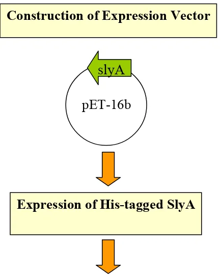
Construction of Expression Vector. A DNA fragment from Salmonella typhimurium
ATCC 14028s was derived by PCR amplification and cloned into the expression vector
pET-16b (Novagen) in the same reading frame as the 10xHis affinity tag with the tag
placed at the N-terminal end. An oligonucleotide primer was constructed that spanned
the ATG start codon to create a NdeI site. The reverse primer was made past the stop
codon of the slyA fragment. These primers were used to amplify a 469 bp fragment using
S. typhimurium ATCC 14028s as a template under the following conditions: denature at
95ºC for 5 min, 94ºC—30 sec—50ºC—30 sec—72ºC—1 min with vent polymerase
(NEB, MA). The PCR fragment was gel purified and cloned into the EcoRV site of pSK
(Stratagene, CA). Colonies containing inserted DNA were digested with NdeI and
BamHI and the fragments were purified. The vector, pET-16b was restricted with NdeI
and BamHI and ligated with the NdeI/BamHI slyA fragment. Following transformation
into DH5α E. coli, DNA was purified from random colonies and restricted with
NdeI/BamHI to check for the insert.
Expression of Recombinant Proteins. pET-16b:slyA was transformed in the E. coli
strain BL21(DE3) pLysS for expression analysis studies. This strain harbors a lambda
lysogen with an inducible bacterial T-7 RNA polymerase. The plasmid pLysS encodes
To purify His-tagged SlyA, a 1 ml freezer stock of pET-16b:slyA was added to 1 L of
LB media supplemented with 100 µg/ml ampicillin (SIGMA, MO) in a 2 L flask. Cells
were grown vigorously with agitation (250rpm) at 37ºC until log phase was reached (a
cell density of A600 = 0.6-0.8). Protein expression was induced with the addition of up to 5mM IPTG (RPI Corp., IL). Cells were harvested by centrifugation for 20 min at 4ºC at
10,000g after additional agitation (250rpm) at 37ºC for 4 hours to ensure ample
expression. The cell pellet was frozen at -80ºC until protein preparation. The cell pellet
was resuspended in lysis buffer (50 mM NaH2PO4, pH 8.0; 300 mM NaCl; 10 mM imidazole) followed by disruption on ice with a microtip sonicator ( Heat
Systems-Ultrasonics, Inc. Cell Disrupter model W-370) with 15 second bursts followed by 1
minute rests on ice until low viscosity was achieved. The lysate was centrifuged at
10,000g at 4ºC for 30 min to spin down the cellular debris.
Purification of recombinant Protein Using Nickel Nitrilotriacetic acid Agarose
under Native Conditions. Column chromatography was performed at room
temperature using Ni-NTA Agarose coupled to Sepharose CL-6B (Qiagen, CA). This
system facilitates a generous binding potential of 5-10 mg of 10xHis-tagged protein per
ml of resin. The column was assembled according to the manufacturer’s instructions
(Bio-Rad, CA). The column was packed with Ni-NTA slurry until the desired bed size
was achieved and equilibrated with 5 column volumes of lysis buffer. The lysate was
added to the column and washed with 10 column volumes of wash buffer (50 mM
NaCl; 500 mM imidazole). All fractions were analyzed by SDS-PAGE analysis on a
12% denaturing polyacrylamide gel. Eluted fractions containing a significant amount of
SlyA protein (determined by SDS-PAGE) were pooled in dialysis tubing (10,000 M.W.
cut-off) and concentrated using PEG (polyethylene glycol 7,000-9,000 MW). Proteins
were dialyzed overnight in phosphate buffered saline to remove any remaining imidazole
and prepare the antigen for immunization.
Production of goat anti-SlyA antibody. Prior to the initial challenge, goat serum was
collected from the test animal (provided by Dr. Glenn Songer at the University of
Arizona, Department of Veterinary Science and Microbiology) and analyzed for a lack of
cross-reaction with SlyA through immunoblot analysis. A booster injection was given 2
weeks and 6 weeks after the first injection. Antiserum was collected 4 weeks and 8
weeks after the initial injection. The primary and secondary bleeds were both analyzed
for SlyA specificity via recognition with a horseradish peroxidase (HRP)-conjugated
anti-goat whole molecule antibody (SIGMA, MO) through Western blot analysis. Production
of anti-His tagged SlyA antibody is described in Figure 1.
Western blot analysis. Bacteria from 10ml of an overnight culture were grown for 16 to
17 hours in LB medium and supplemented with appropriate antibiotics. A 1:1000
dilution of overnight culture in 25 ml LB was grown to fresh stationary phase (O.D.
2.5-3.0), harvested by centrifugation at 6,000 rpm for 10 minutes, washed several times with
phosphate buffered saline, and lysed by the addition of 125 µl of B-Per (Pierce, IL) and
lysate was centrifuged at 14,000 rpm for 10 minutes at 4ºC and the supernatant was
collected. Protein concentration was determined using the Bio-Rad protein assay, using
bovine serum albumin as the standard. Two volumes of sample buffer (2.9 ml deionized
water, 1.0 ml 0.5 M Tris-Hcl, pH 6.8, 2.0 ml glycerol, 1.6 ml 10% w/v SDS, 0.1 ml 1.0%
bromophenol blue, 0.4 ml beta-mercaptoethanol were added to one volume of the protein
fraction. Protein fractions were boiled for 5 minutes and clarified by centrifugation at
14,000 rpm for 3 minutes. Equal amounts of total cell protein were loaded onto a 12%
denaturing polyacrylamide gel. In some instrances the polyacrylamide gel was cut so that
only proteins in the low molecular weight range were transferred. Immunoblotting was
performed and the separated proteins were transferred onto a nitrocellulose membrane.
The proteins were probed using either polyclonal goat or rabbit anti-SlyA serum and
detection of bound antibodies was achieved by the addition of horseradish peroxidase
(HRP)-conjugated monoclonal anti-goat IgG (SIGMA, MO).
Growth Phase Analysis of wild-type S. typhimurium.
Bacteria from 10 ml of an overnight culture of wild-type S. typhimurium were
grown for 16 to 17 hours in LB medium. A 1:1000 dilution of overnight culture in 25 ml
LB was grown to fresh stationary phase (O.D. 3.0), with samples taken throughout the
growth cycle (O.D. 0.308, 0.508, 0.702, 0.940, 1.53, 1.88, 2.29, 2.97) along with an
aliquot from the original overinight culture. Cells were harvested by centrifugation at
6,000 rpm for 10 minutes, washed several times with phosphate buffered saline, and
lysed by the addition of 75 µl of B-Per (Pierce, IL) and 25µl of Dnase with vortexing at
rpm for 10 minutes at 4ºC and the supernatant was collected. Protein concentration was
determined using the Bio-Rad protein assay, using bovine serum albumin as the standard.
Two volumes of sample buffer (2.9 ml deionized water, 1.0 ml 0.5 M Tris-Hcl, pH 6.8,
2.0 ml glycerol, 1.6 ml 10% w/v SDS, 0.1 ml 1.0% bromophenol blue, 0.4 ml BME) were
added to one volume of the protein fraction. Protein fractions were boiled for 5 minutes
and clarified by centrifugation at 14,000 rpm for 3 minutes. Equal amounts of total cell
protein were loaded onto a 12% denaturing polyacrylamide gel. Immunoblotting was
performed and the separated proteins were transferred onto a nitrocellulose membrane.
The proteins were probed using polyclonal goat anti-SlyA serum and detection of bound
antibodies was achieved by the addition of horseradish peroxidase (HRP)-conjugated
monoclonal anti-goat IgG (SIGMA, MO).
Results
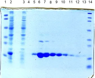
Expression and Purification of His-tagged SlyA protein fragment. Figure 1 shows
the results of the His-tag SlyA purification system used in this study. The protein was
expressed well, as it was the dominant band in the total protein fraction (Fig. 1, lane 2).
A comparison of the total protein with the flow through showed that the Ni-NTA
chromatography column worked effectively to bind the His-tag on SlyA (Fig. 1, lanes 2
and 3), since the protein was absent in the flow through fraction. Proteins were eluted off
the column with a high molar concentration of imidazole (500 mM) to increase the yield
of protein per 1 ml fraction. Most of the protein became unbound within elution fractions
6x His-tag is 21 kD and this coincides with the size of the protein visualized on the gel.
This purification procedure provided SlyA sufficiently pure enough to be used for the
preparation of a polyclonal antibody.
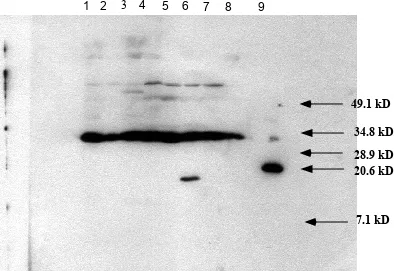
The SlyA polyclonal antibody reacts with a small molecular weight protein in the pSX34::PBAD-slyA strain.
Equal amounts of total protein derived from fresh stationary phase were separated
by 12% SDS-PAGE, transferred to a nitrocellulose membrane, reacted with the SlyA
polyclonal antibody, and detected using chemiluminescence. The anti-his-tagged SlyA
antiserum reacts with the 21 kD 6x His-tagged SlyA protein (Fig. 2, Lane 9) An
immunoreaction is detected in only one strain. A small molecular weight protein running
at approximately 17 kD (Fig. 2, Lane 6) is observed in SL2571 (pSX34::PBAD-slyA). No immunogenic reaction was observed in the other strains: SL2757 (wild-type S.
typhimurium ATCC 14028s) (Fig. 2, Lane 1), SL3129 (pCR2.1:slyA DH5α) (Fig. 2, Lane
2), SL2317 (pSL101:slyA S. typhimurium) (Fig. 2, Lane 3), SL2774 (E. coli 0157:H7)
(Fig. 2, Lane 4), SL2570 (pSX34::PBAD) (Fig. 2, Lane 5), SL2818 (MG1655) (Fig. 2, Lane 7), and SL3343 (slyA::km S. typhimurium ) (Fig. 2, Lane 8).
The SlyA polyclonal antibody reacts with two small molecular weight proteins.
Fig. 3 reveals that the anti-His-tagged SlyA polyclonal antibody reacts with two
small molecular weight proteins. Equal amounts of total protein derived from fresh
stationary phase were separated by 12% SDS-PAGE, the gel was cut, proteins in the low
antibody reacted with the 6xHis-tagged SlyA protein (Fig. 3, Lane 1). Two small
molecular weight proteins were also observed along with an interesting doublet banding
pattern. A single protein in the 17 kD SlyA molecular weight range was detected in
SL2570 (pSX34::PBAD) (Fig. 3, Lane 3), SL3343 (slyA::km S. typhimurium) (Fig. 3, Lane
4), and SL3129 (pCR2.1:slyA DH5α) (Fig. 3, Lane 6). Two distinct proteins in the SlyA molecular weight range were visualized SL2571 (pSX34::PBAD-slyA) (Fig. 3, Lane 2), SL3203 (S. dublin) (Fig. 3, Lane 5), SL2818 (MG1655) (Fig. 3, Lane 7), and SL2757
(wild-type S. typhimurium ATCC 14028s) (Fig. 3, Lane 8).
The aforementioned experiment described in Fig. 4 was repeated with the addition
of two MarR expressing strains, SL3298 (pWSK:marR (MG1655) in pEP185.2::slyA)
and SL3291 (pWSK129:marR E. coli DH5α). These strains were added to check for cross-reactivity of the antibody. Data in Fig. 4 reveals a doublet banding pattern that is
identical to the one described previously. Two distinct proteins in the SlyA molecular
weight range were visualized in SL2757 (wild-type S. typhimurium ATCC14028s) (Fig.
4, Lane 1), SL2818 (MG1655) (Fig. 4, Lane 2), SL3203 (S. dublin) (Fig. 4, Lane 4), and
SL2571 (pSX34:PBAD-slyA) (Fig. 4, Lane 7). Again, a single protein in the 17 kD SlyA
molecular weight range was detected in SL3129 (pCR2.1:slyA DH5α) (Fig. 4, Lane 3), SL3203 (S. dublin ) (Fig. 4, Lane 4), and SL2570 (pSX34:PBAD) (Fig. 4, Lane 6). The MarR expressing strains pWSK:marR (MG1655) in pEP185.2::slyA (Fig. 4, Lane 8) and
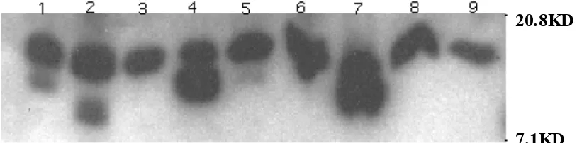
Analysis of Growth Phase Proteins.
Bacterial cells were collected at various times throughout the growth cycle of SL2757
(wild-type S. typhimurium ATCC 14028s). The optical densities at which cells were
collected along with the results are depicted in Fig. 6, Lanes 1-9. A strong immunogenic
reaction was detected in early log phase at O.D. 0.308 (Fig. 6, Lane 2). A weaker
reaction was detected at mid log phase at O.D. 0.508 (Fig. 6, Lane 3). SlyA was not
detected in any other phase of the growth cycle. The reaction appeared as a doublet
pattern, possibly denoting two small molecular weight proteins that ran in the low
molecular weight range. The approximate 21 kD His-tagged SlyA protein (Fig. 6, Lane
10) was used as a reference point.
Discussion
This report describes the expression system and purification method for the
production of a polyclonal antibody for the detection of SlyA protein. SlyA had
previously been identified only by Daniels et al. through immunoblot analysis when
induced on a high copy plasmid and in stationary phase wild-type S. typhimurium., albeit
this immunoreaction was very poor (26). Therefore, we thought it would be interesting to
develop a polyclonal antibody of our own to investigate SlyA expression in the
Salmonellae.
Expression of SlyA
The pET system is the prevailing and most powerful system developed forthe expression
because a 10xHis affinity tag could be engineered in the same open reading frame as
SlyA. In doing so, we could readily purify the protein through His-tagged column
chromatography. In this case, the 6x His-tag was engineered onto the N-terminal end of
the protein. The expression vector contained an ampicillin antibiotic marker for selection
and a thrombin cleavage site that followed the N-terminal his-tag sequence. The system
was under the control of an inducible T7-lac promoter. The gene plasmid construct used
to prepare the His-tagged SlyA protein is shown in Fig. 5.
Up to 5 mM IPTG was added when bacteria reached log phase (O.D. 0.6-0.8) and cells
were grown with agitation for another 4 hours to ensure ample protein expression. The
outcome of the SlyA purification steps is shown in Fig. 1. After a 4 hour induction, the
cell lysate reveals that SlyA was expressed well, as it was the prevailing band in the total
protein fraction (Fig. 1, Lane 2).
Purification of SlyA
Column chromatography was performed at room temperature using Ni-NTA Agarose
coupled to Sepharose CL-6B (Qiagen, CA). The high affinity of Ni-NTA matrix for
biomolecules containing a 6x histidine tag supports a generous binding potential of 5-10
mg of 6xHis-tagged protein per ml of resin and minimal non-specific binding.
After 4 hours of induction, cells were harvested and the cell pellet was lysed by the
addition of lysis buffer, sonication, and the intrinsic pLysS activity of the plasmid. The
cellular debris was separated from the supernatant by centrifugation, and the supernatant
protein was eluted off the column by the addition of a high molar concentration of
imidazole (500 mM).
A comparison of the cell lysate after the induction (Fig. 1, Lane 2) with the flow
through fraction (Fig. 1, Lane 3) demonstrated that the matrix worked effectively to bind
the His-tag on the protein, since there was no His-tagged protein visible in the flow
through fraction. The maximal binding capacity of the affinity column, therefore, was
not exceeded.
The molecular weight of His-tagged SlyA is estimated to be approximately 21 kD.
This estimation corresponds with the size of the eluted protein fractions visualized by
SDS-PAGE post-induction (Fig. 1, Lanes 4-13). Eluted fractions containing a significant
amount of SlyA protein were pooled together in dialysis tubing and concentrated to the
mg/ml range. Dialysis in phosphate buffered saline overnight was performed to remove
any remaining imidazole and prepare the antigen for immunization. The Ni-NTA
purification procedure provided SlyA sufficiently pure enough to be used for the
preparation of a polyclonal antibody.
The polyclonal antibody reacts with more than one protein in 17 kD molecular weight range.
Western blot analysis was performed with cells grown to fresh stationary phase.
The proteins were transferred to nitrocellulose and the entire membrane was probed with
the anti-His-tagged SlyA polyclonal antibody. The detection of SlyA was visualized only
and not in SL2757 (wild-type S. typhimurium ATCC 14028s), SL2317 (pSL101:slyA S.
typhimurium), SL2774 (E. coli 0157:H7), and SL2818 (MG1655) could be attributed to
the levels of SlyA produced within the cell. Studies to determine how many copies per
cell of SlyA are produced have yet to be performed. The slyA mutant strain SL3343 does
not detect the presence of SlyA, nor does SL2570 (pSX34:PBAD). Although, it should be noted that the expression levels for SL2757 (wild-type S. typhimurium ATCC 14028s)
and SL2570 (pSX34:PBAD) should be similar since SL2570 is in a S. typhimurium
background. We can not explain why an immunoreaction was not detected in the SlyA
expressing strains pCR2.1:slyA or pSL101:slyA S. typhimurium.
Cross-reaction is commonly seen with polyclonal antibodies. Fig. 2 shows that an
immunogenic reaction with several other proteins was detected, specifically a protein at
approximately 35 kD. The possibility that this band represents a SlyA dimer is quickly
discounted by the fact that the reaction is detected in the slyA mutant strain, SL3343 .
Furthermore, the denaturing conditions used in the protein preparation and the
SDS-PAGE gel itself would most likely reduce non-specific interaction.
If SlyA is made at very low levels then the amount of protein transferred to the
membrane for detection by chemiluminescence would be minute. Moreover, because of
the strong affinity of the polyclonal antibody for non-SlyA proteins, we hypothesized that
the amount of antibody needed to detect the low levels of SlyA protein may not be
accessible. Therefore, only proteins in the low molecular weight range were transferred
to the membrane. In this manner, there would be additional antibody available to bind
The results are shown in Fig. 3. Indeed, the antibody detected two proteins in the low
molecular weight range. We observed an interesting banding pattern. Two distinct bands
were observed in SL2571 (pSX34:PBAD-slyA), SL 3203 (S. dublin), SL2818 (MG1655), and SL2757 (wild-type S. typhimurium ATCC 14028s). Only one upper band was
detected in SL2570 (pSX34:PBAD), SL3343 (slyA::km S. typhimurium), SL3129
(pCR2.1:slyA),SL3298 (pWSK:marR (MG1655) in pEP185.2::slyA) (Fig. 4, Lane 8), and
SL3291(pWSK129:marRE. coli DH5α). We proposed that the bottom band represented
SlyA, since it was detected in strains that contained slyA—with the unexplainable
exception again, SL3129 (pCR2.1:slyA DH5α). We proposed that the top band might be
MarR or some other low molecular weight protein that shares homologous regions with
SlyA(31, 82, 89, 96, 99). Construction of a marR mutant might provide some insight as
to whether or not the top band is indeed MarR.
SlyA may be expressed in early to mid log phase.
Previous work by Buchmeier et al. suggested that SlyA expression is induced
during stationary phase (14). This finding was based on the observation that slyA mutant
cells exhibited changes in protein expression patterns when compared to S. typhimurium
14028s during stationary phase, but not log phase. Daniels et al. also showed that the
production of SlyA in S. typhimurium 14028s was barely detectable through immunoblot
analysis in stationary phase, but not detectable at all in log phase (26). For these reasons,
we conducted all of our SlyA expression studies with cells that were grown to stationary
regions of the gel to the membrane, long exposure times) we thought it would be
interesting to examine the expression patterns of SlyA through the growth cycle to verify
the previous findings.
In fact, recent experiments in our laboratory suggest that perhaps SlyA is expressed in
early to mid log phase and not in stationary phase (Fig. 6). This novel finding would
explain the difficulty in detecting SlyA expression natively within the Salmonellae. The
evidence presented here is quite compelling. Obviously, further experiments neede to be
Construction of Expression Vector
Expression of His-tagged SlyA
Purification of His-tagged SlyA through Nickel column chromatography
FIG. 1. Schematic of SlyA His-tag antibody production..
pET-16b
slyA
FIG 2.
SDS-PAGE gel of His-tag protein purification
steps. Lane 1, molecular weight markers, 103, 77, 50,
34.3, 28.8, and 20.7 kD; Lane 2, cell lysate after a 4-h
induction; Lane 3, flow through; Lanes 4-13, 500 mM
imidazole elutions; Lane 14, molecular weight markers,
209, 124, 80, 49.1, 34.8, 28.9, 20.6, and 7.1 kD.
FIG. 3.
Western blot analysis of SlyA stationary phase
protein expression. Lane 1, SL2757; Lane 2, SL3129;
Lane 3, SL2317; Lane 4, SL2774; Lane 5, SL2570; Lane
6, SL2571; Lane 7, SL2818; Lane 8, SL3343; Lane 9,
His-tagged SlyA protein.
1 2 3 4 5 6 7 8 9
1 2 3 4 5 6 7 8
FIG. 4.
Western blot analysis of SlyA stationary
phaseprotein expression. Lane 1, His-tagged SlyA
protein; Lane 2, SL2571; Lane 3, SL2570; Lane 4,
SL3343; Lane 5, SL3203; Lane 6, SL3129; Lane 7,
SL2818; Lane 8, SL2757.
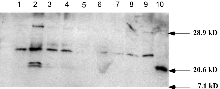
20.6 kD
FIG. 5. Western blot analysis of SlyA stationary phase protein expression. Lane 1, SL2757; Lane 2, SL2818; Lane 3, SL3129; Lan e 4, SL3203; Lane 5, SL3343; Lane 6, SL2570; Lane 7, SL2571; Lan e 8, SL3298; Lane 9, SL3291 .
20.8KD
1 2 3 4 5 6 7 8 9 10
FIG. 6.
Growth curve expression of SlyA.
Lane 1, overnight stationary phase cells; Lane
2, O.D. 0.308; Lane 3, O.D. 0.508; Lane 4,
O.D. 0.702; Lane 5, O.D. 0.940; Lane 6, O.D.
1.52; Lane 7, O.D. 1.88; Lane 8, O.D. 2.27;
Lane 9, O.D. 2.97, Lane 10, His-tagged SlyA
protein.
20.6 kD
TABLE 1. Strains and plasmids used
Strain Relevant features Source
S. typhimurium 14028s (SL2757)
Wild type ATCC
SL3129 pCR2.1:slyA DH5α Lab stock
SL2317 PSL101:slyA S. typhimurium Lab stock
SL2774 E. coli 0157:H7 Lab stock
SL3203 S. dublin Lab stock
SL2570 pSX34:PBAD Lab stock
SL2571 PSX34: PBAD-slyA Lab stock
SL2818 MG1655 Lab stock
SL3343 slyA::km S. typhimurium Lab stock SL3298 PWSK129:marR(MG1655) in
pEP185.2::slyA S. typhimurium
Lab stock
SL3291 PWSK129::marR DH5α Lab stock
E. coli DH5α endA1 hsdR17(rK-mK+)supE44 thi-1
recA1 pyrA(Nalr) relA1∆ (lacIAA-argF)U169 deoR[∆80dlac∆ (lacZ)M15]
Invitrogen
BL21(DE3)pLysS F-ompT hsdSB (rB-mB-) gal dcm (DE3)
pLysS (CmR)
Novagen
Plasmids
pET16b His-tag vector Novagen
Chapter 2: Purification of a Polyclonal Antibody for the Detection of SPI4-K Protein Expression
Introduction
Salmonella acquired infection remains a serious worldwide threat to public health,
with illness ranging from minor gastroenteritis (S. typhimurium)to acute systemic disease
(S. typhi). Salmonella enterica serovar Typhimurium is the most frequent origin of
salmonellosis, with nearly all of the 2000 serotypes of the genus Salmonella able to cause
disease in humans. Because the Center for Disease Control estimates that 1.4 million
cases of the food-born illness occur annually in the United States with 1,000 deaths
resulting from severe salmonellosis infections, further investigation into the mechanisms
of disease caused by this pathogen are warranted.
Salmonella diverged from its most closely related counterpart, E. coli,
approximately 100 million years ago (87). Evidence of this divergence is indicated by
large, conserved clusters of virulence genes located in the chromosomal sequence of
pathogenic Salmonella spp. that are not present in the analogous chromosomal regions of
non-pathogenic spp. such as E. coli K12. These extended regions of unique DNA
sequences are referred to as Salmonella pathogenicity islands (12). Other bacterial
pathogens such as E. coli, Shigella, Yersinia, Helicobacter pylori, Vibrio cholera, and
Pseudomonas syringae possess pathogenicity islands as well (11, 17) (18) (34) (70, 72,
88), although none of these bacteria have more than two islands. Thus far, five
pathogenicity islands have been identified in Salmonella typhimurium (52, 104, 105),
suggesting that a set of extremely complex systems for virulence exist. SPIs are required
implicated in invasion (47, 80) as well as intracellular survival within macrophage (6,
40).
SPI-4 is an approximate 25 kb sequence insertion located at centisome 92 on the
Salmonella chromosomal map. This pathogenicity island is flanked on either side by the
ssb and yciB genes in both S. typhimurium and S. typhi (104). Originally, SPI-4 was
identified via a transposon insertion as a chromosomal sequence required for survival
inside of murine macrophages (6, 40). Unfortunately, the amount of information available
about SPI-4 is modest. Wong et al. originally claimed that SPI-4 contained 18 ORFs
designated A-R (104). The recently completed annotation of both S. typhimurium LT2
and S.typhi genomes shows that only 6 open reading frames exist. Ahmer et al. (1)
demonstrated SPI-4 regulation by SirA, a transcriptional regulator of SPI-1 (invasion
locus). Experiments in our laboratory have shown that the beta-galactosidase activity of
a SPI4-K::MudJ insertion (SL3277) in a slyA mutant background is reduced 8-fold,
suggesting that SPI-4 is regulated by slyA. Allen et al. were the first to show that the
genes on SPI-4 play a role in the invasion of cultured epithelial cells (3). This finding is
significant because the ability to invade was predominantly attributed to SPI-1 in the past.
However, Murray et al. showed that when the entire nucleotide sequence that codes for
SPI-1 was deleted, the ability of Salmonella to invade was not completely eliminated
(81). Perhaps the residual invasive properties of Salmonella are due to the genes encoded
on SPI-4.
In collaboration with Dr. Brian Ahmer, our laboratory has isolated MudJ transposon
insertions localized to an open reading frame designated SPI4-K. This ORF was initially
part of the larger STM4261 ORF proposed to encode a 660 kD protein (~16 kB gene).
We recently discovered that the predicted SPI4-K protein has significant homology to a
class of autotransporter toxins found in pathogenic E.coli, Bordetella, Neisseria, and
Shigella species, referred to as SPATE toxins (2, 8, 49, 58, 61-64, 79, 83, 84, 97, 102).
Members of this family are serine proteases possessing a characteristic GDSGS motif
with a central catalytic Ser residue. We have constructed and purified a His-tagged
SPI4-K protein fragment based on the earlier annotation, and used this protein to obtain a
polyclonal antibody. In this study, we examined the protein expression characteristics of
SPI4-K in Salmonella typhimurium and other Salmonellae. We present preliminary
evidence through Western blotting that SPI4-K is a large molecular weight protein that
appears to be secreted.
Materials and Methods
Bacterial strains and plasmids. Bacterial strains used in this study are described in
Table 1. All strains were grown in Luria-Bertani (LB) and were supplemented with
kanamycin (50 µg/ml) or chloramphenicol (25 µg/ml) when required.
Construction of Expression Vector. A DNA fragment from Salmonella typhimurium
ATCC 14028s was derived by PCR amplification and cloned into the expression vector
pET-16b (Novagen) in the same reading frame as the 6x His affinity tag with the tag
placed at the N-terminal end. An oligonucleotide primer was constructed that spanned the
of the SPI4-k fragment. These primers were used to amplify a 900 bp fragment using S.
typhimurium ATCC 14028s as a template under the following conditions: denature at
95ºC for 5 min, 94ºC—30 sec—50ºC—30 sec—72ºC—1 min with pfu for 40 cycles.
The PCR fragment was gel purified and cloned into the EcoRV site of pSK (Stragene,
CA). Colonies containing inserted DNA were digested with NdeI and BamHI and the
fragments were purified. The vector, pET-16b was restricted with NdeI and BamHI and
ligated with the NdeI/BamHI SPI4-k fragment. Following transformation into DH5α E.
coli, DNA was purified from random colonies and restricted with NdeI/BamHI to check
for the 900 bp insert.
Expression of Recombinant Proteins. pET-16b:SPI4-k was transformed in the E. coli
strain BL21(DE3) pLysS for expression analysis studies. This strain harbors a lambda
lysogen with an inducible bacterial T-7 RNA polymerase. The plasmid pLysS encodes
T-7 Lysozyme and is used to repress transcription from the T-7 promoter.
To purify His-tagged SPI4-K, 1 ml freezer stocks were added to 1 L of LB media
supplemented with 100 µg/ml ampicillin (SIGMA, MO) in a 2 L flask. Cells were grown
vigorously with agitation (250 rpm) at 37ºC until log phase was reached (a cell density of
A600 = 0.6-0.8). Protein expression was induced with the addition of up to 5mM IPTG
(RPI Corp., IL). Cells were harvested by centrifugation for 20 min at 4ºC at 10,000g
after additional agitation (250 rpm) at 37ºC for 4 hours to ensure ample expression. The
cell pellet was frozen at -80ºC until protein preparation. The cell pellet was resuspended
in lysis buffer (50 mM NaH2PO4, pH 8.0; 300 mM NaCl; 10 mM imidazole) followed by
model W-370) with 15 second bursts followed by 1 minute rests on ice until low
viscosity was achieved. The lysate was centrifuged at 10,000g at 4ºC for 30 min to
remove the cellular debris. Construction of His-tagged SPI4-K is described in Figure 1.
Purification of Recombinant Protein Using Nickel Nitrilotriacetic acid Agarose
under Native Conditions. Column chromatography was performed at room temperature
using Ni-NTA Agarose coupled to Sepharose CL-6B (Qiagen, CA). This system
facilitates a generous binding potential of 5-10 mg of 6x His-tagged protein per ml of
resin. The column was assembled according to the manufacturer’s instructions (Bio-Rad,
CA). The column was packed with Ni-NTA slurry until the desired bed size was
achieved and equilibrated with 5 column volumes of lysis buffer. The lysate was added
to the column and washed with 10 column volumes of wash buffer (50 mM NaH2PO4, pH
8.0; 300 mM NaCl; 20 mM imidazole). His-tagged protein was eluted off the column in 1
ml fractions with elution buffer (50 mM NaH2PO4, pH 8.0; 300 mM NaCl; 500 mM
imidazole). All fractions were collected for SDS-PAGE analysis and electrophoresed on
a pre-cast 12% denaturing polyacrylamide gel (Bio-Rad). Eluted fractions that contained
a significant amount of protein (determined by SDS-PAGE) were pooled in dialysis
tubing (10,000 M.W. cut-off) and concentrated using PEG (polyethylene glycol
7,000-9,000 MW). Proteins were dialyzed overnight in phosphate buffered saline (PBS) to
remove any remaining imidazole and prepare the antigen for immunization.
SDS-PAGE Analysis of His-tag Purification. Sodium dodecyl sulfate-polyacrylamide
analyzed. 100 µl of 2X laemmli buffer (3.0 ml deionized water, 1.0 ml 1.0 M Tris-HCl,
pH 6.8, 1.6 ml glycerol, 1.6 ml 20% w/v SDS, 0.4 ml 0.5% bromophenol blue ) was
added to each 100 µl protein fraction collected at each step of the column
chromatography process. Protein fractions were boiled for 5 minutes and clarified by
centrifugation at 14,000 rpm for 3 minutes. Protein fractions were loaded in 10 µl
increments on a pre-cast 12% SDS-PAGE denaturing gel (Bio-Rad, CA) and visualized
by Coomassie brilliant Blue staining.
Production of Rabbit Anti-SPI-4 Antibody. Antibody production was performed in a
rabbit by Scantibodies, Ramon, CA. Prior to the initial challenge, serum was collected
from the test animal and analyzed for a lack of cross-reaction with SPI4-K through
immunoblot analysis. A booster injection was given 4 weeks and 8 weeks after the first
injection. Antiserum was collected 1 week, 5 weeks, and 9 weeks after the initial
injection. The primary and secondary bleeds were both analyzed for SPI4-K
specificity via recognition with HRP-conjugated monoclonal anti-rabbit IgG (SIGMA,
MO) through Western blot analysis. Production of anti-His tagged SPI4-K antibody is
described in figure 2.
Western blot analysis of total cell protein. Bacteria from 10ml of an overnight culture
were grown for 16 to 17 hours in LB medium and supplemented with appropriate
antibiotics. A 1:1000 dilution of overnight culture in 25 ml LB was grown to fresh
stationary phase (O.D. 2.5-3.0), harvested by centrifugation at 6,000 rpm for 10 minutes,
(Pierce) and 75µl of DNase with vortexing at 5 minute intervals for a total of 15 minutes
on ice. The lysate was centrifuged at 14,000 rpm for 10 minutes at 4ºC and the
supernatant was collected. Protein concentration was determined using the Pierce BCA
protein assay, using bovine serum albumin as the standard. Two volumes of sample
buffer (2.9 ml deionized water, 1.0 ml 0.5 M Tris-HCl, pH 6.8, 2.0 ml glycerol, 1.6 ml
10% w/v SDS, 0.1 ml 1.0% bromophenol blue, 0.4 ml BME) were added to one volume
of the protein fraction. Protein fractions were boiled for 5 minutes and clarified by
centrifugation at 14,000 rpm for 3 minutes. Equal amounts of total cell protein were
loaded onto a 12% denaturing polyacrylamide gel and separated by sodium dodecyl
sulfate-polyacrylamide gel electrophoresis (SDS-PAGE). The separated proteins were
transferred onto a nitrocellulose membrane. The proteins were probed using a polyclonal
rabbit anti-SPI4-K serum and detection of bound antibodies was achieved by the addition
of horseradish peroxidase (HRP)-conjugated monoclonal anti-rabbit IgG (SIGMA, MO).
Immunoblotting was performed with a 1:500 dilution of the primary antibody and
1:10,000 of the secondary antibody.
Western blot analysis of secreted protein. Bacteria from 10 ml of an overnight culture
were grown for 16 to 17 hours in LB medium and supplemented with appropriate
antibiotics. A 1:1000 dilution of overnight culture in 50 ml LB was grown to fresh
stationary phase (O.D. 2.5-3.0) and the cell pellet harvested by centrifugation at 6,000
rpm for 10 minutes. The supernatant was centrifuged at 12,000 rpm for 30 minutes at
4ºC several times, filter sterilized, supplemented with a 10% volume of 100% TCA