ISSN(Online): 2320-9801
ISSN (Print): 2320-9798
I
nternational
J
ournal of
I
nnovative
R
esearch in
C
omputer
and
C
ommunication
E
ngineering
(A High Impact Factor, Monthly, Peer Reviewed Journal) Website: www.ijircce.com
Vol. 5, Issue 10, October 2017
A Prototype Model on CBMIR Using Canny
Edge Detection and Neural Network for Visual
Search
Dr. Jasmine Samraj1, Jeevitha K.2
Associate Professor, Department of Computer Science, Quaid-E-Millath Government College for Women, Chennai,
Tamil Nadu, India1
Research Scholar, Department of Computer Science, Quaid-E-Millath Government College for Women, Chennai,
Tamil Nadu, India 2
ABSTRACT: The advancement in the area of medical imaging method has lead industries to conceptualize a complete
automated system for the medical procedures, diagnosis, treatment, and prediction. Content-Based Image Retrieval (CBIR) is a process to retrieve a stored image from the database by supplying an image as a query instead of text. The success of such system largely depends upon the robustness, accuracy, and speed of the retrieval systems. CBIR system is valuable in medical systems as it provides retrieval of the images from the large dataset based on similarities. This paper proposes a model for Content-Based Medical Image Retrieval (CBMIR) using Canny Edge Detection and Neural Network for visual search on hand radiography. The initial Stage of Image Retrieval namely pre-processing is used in order to get a better image; it eliminates the noise present in the query image. Edge detection by Canny method is performed on the hand radiography. The Canny Edge detector finds linear and continuous Edges. A rule for comparing the images using Neural Network is defined for retrieving images that match the given query from a large database. This Comparison is done by Convolutional Neural Network (CNN), Probabilistic Neural Network (PNN) and Extreme Learning Machine (ELM). CNN is a deep Neural Network. It is based on Multilayer feed-forward Neural Network. PNN is also a multilayer feed-forward Neural Network and classifies faster than the CNN. Euclidean distance is used for calculating the distance between each pair of Region Of Interest (ROI) for similarity measurement. The Proposed technique allows users to retrieve similar query image from the database, higher retrieval performance can be achieved and also compares CNN and PNN classifiers on hand radiography images.
KEYWORDS: CBIR, CBMIR, Canny Edge Detection, CNN, PNN, ELM.
I. INTRODUCTION
ISSN(Online): 2320-9801
ISSN (Print): 2320-9798
I
nternational
J
ournal of
I
nnovative
R
esearch in
C
omputer
and
C
ommunication
E
ngineering
(A High Impact Factor, Monthly, Peer Reviewed Journal) Website: www.ijircce.com
Vol. 5, Issue 10, October 2017
Similarity Matching. These four stages are consecutively performed between training and query medical images for extracting the most relevant images.
Pre-processing - Pre-Processing refers to the initial steps in Image Processing that are applied to the raw
image in order to remove noise in an image.
Feature Extraction - Feature Extraction is the process of extracting information from images based on
color, shape, texture.
Classification - Image Classification is one of the significant steps in image retrieval method because it
saves more time while searching the images from the large database. Classification is identification of different regions of the images by which the retrieval efficiency of the system will be improved.
Similarity Matching – A similarity measure plays an important role in image retrieval. Similarity
measurement is one of the key issues in content-based image retrieval. The Similar images from the database can be retrieved by using Similarity Measure.
A. Content-Based Image Medical Image Retrieval
Content-Based Medical Image Retrieval (CBMIR) has become a major necessity with the growing technological advancements. In the Medical Field, images and especially digital images are produced in ever-increasing quantities and used for diagnosis and therapy. Medical image plays an important role in surgical planning, medical training, and patient diagnosis. Content-based access to medical images is following the medical decision making has been designed that would ease the administration of medical data and scenarios for the combination of content-based access methods into Picture Archiving and Communication Systems (PACS) [2]. Every day large volumes of different types of Medical images such as dental, endoscopy, skull, MRI, ultrasound, radiology are produced in various hospitals as well as various medical centres.
Medical image retrieval systems are special from regular image retrieval in several ways. Medical image retrieval has many significant applications, especially in medical diagnosis, Education, and research fields. The retrieval of digital medical images can also be done in the form text-based and content-based techniques. Medical image retrieval for diagnostic purposes is significant because the chronological images of different patients in medical centres have valuable information for the upcoming diagnosis with a system which retrieves similar cases, makes more accurate diagnosis and to decide on appropriate treatment.
II. RELATED WORK
ISSN(Online): 2320-9801
ISSN (Print): 2320-9798
I
nternational
J
ournal of
I
nnovative
R
esearch in
C
omputer
and
C
ommunication
E
ngineering
(A High Impact Factor, Monthly, Peer Reviewed Journal) Website: www.ijircce.com
Vol. 5, Issue 10, October 2017
III.PROPOSED WORK
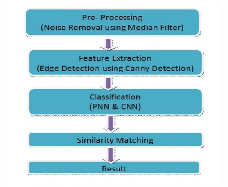
The Proposed Methodology comprises the general phases of CBIR such as Pre-processing, feature Extraction based on edge, classification and Similarity Metric analysis. The purpose of this work was to improve the accuracy of a CBMIR application by allowing the system to retrieve the similar images.
Fig. 1. Architecture of the Proposed Work CBMIR PNN – CNN
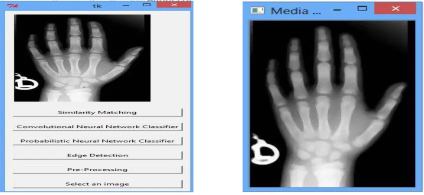
The input query image is taken for the process of retrieval of the similar image from a lot of medical images in the database.Medical Image, left hand radiography was taken as input to the system.
A.NOISE REMOVAL USING MEDIAN FILTER:
Pre-processing is performed in order to remove the noise in the medical images. Most of the X-ray images are passed through many sources. Therefore, noise should be removed before extracting the features. The Proposed Methodology uses the Median Filter for removing Noise. The median filter is a nonlinear filter and it has widely used in digital image processing because of its good edge keeping characteristics and reducing impulse noise ability. The median filter is a rank-order filter. Its noise-reducing effects depend on the size and shape of the filtering mask. Median Filter is used for noise removing because it removes the noise as well as preserved the edge information in images. When Noise affects a point in a grayscale image, the result is called salt & pepper noise. The Median Filter is very useful for removing the Salt & Pepper Noise. The median filtering output is
g(x,y) = med { f (x- i , y- j), i, j £ w } eq. (1)
Where f (x, y), g (x, y) are the original image and the output image respectively, W is the two-dimensional
mask: the mask size is n x n(where n is commonly odd) such as 3 X 3, 5 X 5, etc. Because the median filter is a nonlinear filter, its mathematical analysis is relatively complex for the image with random noise. For an image with zero mean noise under normal distribution, the noise variance of the median filter approximately.
eq. (2)
1
σ²i π
4 n f 2 ( n ) n +π/2 – 1 2
ISSN(Online): 2320-9801
ISSN (Print): 2320-9798
I
nternational
J
ournal of
I
nnovative
R
esearch in
C
omputer
and
C
ommunication
E
ngineering
(A High Impact Factor, Monthly, Peer Reviewed Journal) Website: www.ijircce.com
Vol. 5, Issue 10, October 2017
Where σ²is input noise power (the variance), n is the size of the median filtering mask, f (n) is the function of the noise density. And the noise variance of the average filtering is
σ²0 = 1 σ²i eq.(3)
n
Comparing the equation (2) and (3), the median filtering effects depend on two things: the size of the mask, and the distribution of the noise. The median filtering performance of random noise reduction is better than the average filtering performance, but to the impulse noise, especially narrow pulses are farther apart and for the pulse width less than n/2, the median filter is very effective.
B. EDGE-FEATURE DETECTION USING CANNY METHOD:
Feature Extraction is used to extract inherent features in images such as shape, colour, and texture. It helps to diagnose and determine the accurate segmentation of images. Feature extraction can be done in three levels, thepixel, the local and the global. The simplest visual image feature extraction are based on pixel. Local features are extracted from sub-images from the original image. Global features are extracted to describe the whole image in an average.
Color: Color is the common features extracted in CBIR, because of the simplicity in extracting the color information from images.
Texture: Texture is the most important feature of an image. It is used to represent visual patterns in images
and to determine and specify how they are spatially defined. The process of identifying something in specific textures in an image is a variation. The brightness of pairs of pixels are calculated as the degree of contrast, regularity,
coarseness, and directionality which may be estimated.
Shape: Shape feature extraction normally includes the extraction of edges, corners, and curvature scale space and chain codes. Shape contains the most absorbing visual information for easy understanding. It does not define the shape of an image, but it defines the shape of a particular area.
For detecting edges Canny Edge Detection Algorithm is used and for connecting the edges to form the shape of the image. Canny edge detection is a multi-step calculation that can recognize edges with noise covered up in the meantime. The Canny edge indicator was created in 1986 by John F. Shrewd. It is still broadly utilized today was one
of the default edge locators in Image Processing.The Canny edge detector is based on computing the squared gradient
magnitude [7]. Local maxima of the gradient extent that are more than some threshold are then recognized as edges. The Canny edge detector is an edge detection operator that uses a multi-stage algorithm to detect an extensive range of edges in images. The Canny aim was to realize the optimal edge detection algorithm. In this situation, an "optimal" edge detector means
Good detection – It should mark as many real edges in the image as probable.
Good localization – Edges marked should be as close as possible to the edge in the actual image.
Minimal response – A given edge in the image should only be marked once, and where possible, image sound should not create copied edges.
The Canny edge location calculation can be separated into 5 stages:
Step 1: Smooth the image utilizing a Gaussian/Median filter to evacuate high recurrence noise.
eq. (4)
where G σ is calculated as
eq. (5) Step 2: Compute the gradient intensity representations of the image.
and
ISSN(Online): 2320-9801
ISSN (Print): 2320-9798
I
nternational
J
ournal of
I
nnovative
R
esearch in
C
omputer
and
C
ommunication
E
ngineering
(A High Impact Factor, Monthly, Peer Reviewed Journal) Website: www.ijircce.com
Vol. 5, Issue 10, October 2017
Where M is the Threshold, M can be calculated as
Where is so chosen that all edge elements are kept while most of the noise is suppressed.
Step 3: Apply non-maximum suppression to remove “false” responses to edge detection. Step 4: Apply thresholding using a lower and upper boundary on the gradient values.
Step 5: Track edges using hysteresis by suppressing weak edges that are not connected to strong edges.
C. CONVOLUTIONAL NEURAL NETWORK:
Convolutional Neural Network (CNN, or ConvNet) is a class of deep, feed-forward artificial neural network that have successfully been applied to analysing visual imagery. Convolutional Neural Networks (ConvNets or CNNs) are a gathering of Neural Networks that are very effective in fields such as image recognition and classification. The Deep learning algorithm of CNN in the multilayer feed forward network introduced the outstanding neural network structures [8], it is broadly utilized as a part of extraordinary scientific fields. Ordinarily, a neural framework contains three levels:
(i) an input level;
(ii) A middle or hidden level; (iii) An output level.
To calculate the gradient of classification ofthe input to the l-th layer and the hidden layer of the image is
calculated as
δ(l)=((W(l))Tδ(l+1))∙f′(z(l)) eq.(7)
And the output level gradients are
∇W(l)J(W,bas;x,y)=δ(l+1)(a(l))T
∇b(l)J(W,b;x,y)=δ(l+1). eq. (8)
Each output map is given its own multiplicative bias β and an additive bias b of the image.
D. PROBABILISTIC NEURAL NETWORK
A Probabilistic Neural Network (PNN) is a feed-forward neural network, which was got by the Bayesian network and a statistical algorithm called Kernel Fisher Discriminant analysis. It was presented by D.F. Specht in the mid-1990s. In a PNN, the operations are organized into a multilayered feed forward network with four layers:
Input layer
Hidden layer
Pattern layer/Summation layer
Output layer
Input Layer: The input vector, denoted as p, its dimension is R×1.
Radial Basis Layer: In Radial Basis Layer, the vector distances between input vector p and the weight vector made of each row of weight matrix W are calculated. Here, the vector distance is defined as the dot product between two vectors. Assume the dimension of W is Q×R. The dot product between p and the i-th row of W produces the i-th element of the distance vector ||W−p||, whose dimension is Q×1. The minus symbol, “−”, indicates that it is the distance between vectors. Then, the bias vector b is combined with ||W−p|| by an element-by-element multiplication. The result is denoted as n =||W−p||·∗p. The transfer function in PNN has built into a distance criterion with respect to a center. In this paper, we define it as
radbas(n) = e− n2eq. (9)
Each element of n is substituted into the above formula, and produces corresponding element of a, the output vector of Radial Basis Layer. We can represent the i-th element of a as
ai = radbas(||Wi −p||·∗bi) eq.(10)
Where Wi is the vector made of the I-th line of W and bi is the I-th component of predisposition vector b.
ISSN(Online): 2320-9801
ISSN (Print): 2320-9798
I
nternational
J
ournal of
I
nnovative
R
esearch in
C
omputer
and
C
ommunication
E
ngineering
(A High Impact Factor, Monthly, Peer Reviewed Journal) Website: www.ijircce.com
Vol. 5, Issue 10, October 2017
is effective to noise examples. The most important advantage of PNN is that training is easy and instantaneous. Weights are not “trained” but assigned. Existing weights will never be exchanged however, just new vectors are embedded into weight grids when preparing. So it can be utilized in real-time. Since the training and running technique can be implemented by matrix manipulation, the speed of PNN is very quick. The execution of the PNN classifier was evaluated in terms of training, performance and classification accuracies. Probabilistic Neural Network gives fast and accurate classification and is a promising tool for classification.
F.SIMILARITY MEASUREMENT
Measures can be evaluated using distance functions, and it is important to determine the most suitable function for each type of ROI. Euclidean distance is used to evaluate distances in n dimensional vector spaces. The Euclidian method is used to calculate the distances between the query image and extracted images. The Euclidian method is utilized to calculate the distances between the query image and extracted images. Euclidean distance is the square root of the sum of squared differences between corresponding elements of the two vectors. The distance between vectors X and Y is characterized as
IV.RESULTS
The proposed CBIR Based image retrieval System for Medical image CBMIR PNN-CNN, are implemented with the left hand Wrist X-ray samples collected from the IRMA database. The radiographs were classified and scanned in an X-ray format with 256 x 260 pixel size. The Proposed Work is implemented in Python and the Packages are installed in Python to work with images. The Installed Packages are PIL, OpenCV, Numpy, and matplotlib. Neural Network classifiers are applied in the Proposed Model CBMIR PNN-CNN. Convolutional Neural Network (CNN) and Probabilistic Neural Network (PNN) are used to classify the ROI of the images. PNN provides the fast and Accurate to classify the ROI in Medical Images. The estimation results of PNN are compared with those of CNN and ELM. ELM based calculations are carried from the existing method. To demonstrate the merits of the presented approach on a more definite and tangible basis, the accuracy of PNN model estimation is compared with the accuracy of estimation of the CNN and ELM methods, which served as a benchmark. PNN approach has much faster learning speed compared to CNN and ELM.
ISSN(Online): 2320-9801
ISSN (Print): 2320-9798
I
nternational
J
ournal of
I
nnovative
R
esearch in
C
omputer
and
C
ommunication
E
ngineering
(A High Impact Factor, Monthly, Peer Reviewed Journal) Website: www.ijircce.com
Vol. 5, Issue 10, October 2017
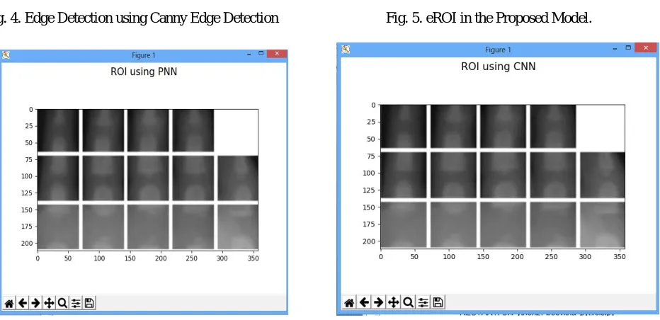
Fig. 4. Edge Detection using Canny Edge Detection Fig. 5. eROI in the Proposed Model.
Fig.6.ROI Matching using PNN Fig.7. ROI Matching using CNN
ISSN(Online): 2320-9801
ISSN (Print): 2320-9798
I
nternational
J
ournal of
I
nnovative
R
esearch in
C
omputer
and
C
ommunication
E
ngineering
(A High Impact Factor, Monthly, Peer Reviewed Journal) Website: www.ijircce.com
Vol. 5, Issue 10, October 2017
Fig.8. Results and Analysis of the Proposed CBMIR PNN-CNN Model
V. CONCLUSION AND FUTURE WORK
In this Proposed Work CBMIR PNN-CNN, An efficient system is designed to extract the features of images and classify using Probabilistic Neural Network for Content Based Image Retrieval for Medical Images. The query image can be read from the database and then eliminate the noise using median filter. Median filtering based pre-processing step was carried to de-noise the image, for feature extraction uses shape extraction & texture extraction. Edge Detection uses the canny Edge Detection Method to detect the selection of medical image edges and the feature that contains Region of Interest (ROI) processing, Choice of ROI – Choosing ROI based on quality, density, size, shape, smoothness, thickness of border, etc., and the Probabilistic Neural Network (PNN) Classifier and Convolutional Neural Network (CNN) is used to classify the ROI. PNN is often used in classification problems and for similarity measure the proposed model uses the Euclidean Distance, When Implemented the Proposed System can easily retrieve a similar images from the PNN classifier.Performance of the PNN classifier was evaluated in terms of training performance and classification accuracies. Probabilistic Neural Network gives fast and accurate classification and is a promising tool for
classification of the X-ray of left hand wrist images. The main disadvantage of CNN is high computational cost and
also CNN use to need a lot of training data, the speed of PNN is very fast when compared to other Artificial Neural Network Convolutional neural network (CNN). As image collections grow in size of the proposed system may take a lot of time and eventually reduce the query retrieval process. Increasing the speed and the user interaction with the image retrieval systems can be done as future work. The Future Work will also involve the Bone Age Assessment accuracy by expanding the database of images. The Future Work will also be focused on extending the system to work for different Medical images like Brain, Liver, Breast, Ear, Spinal Cord etc. and to be implemented over the World Wide Web.
REFERENCES
1. Sameer antani et al “Content- based image retrieval for large biomedical archives“, 2004.
2. A. Grace selvarani and Dr. S. Annadurai, “Content Based Retrieval for Medical images using Generic Fouric descriptor”..
3. Pietka E, Gretych A PosipiechS,Cao F, Huang H K , And Gilsanz V. “ Computer Assisted bone age assessment: Image Pre-processing and epiphyseal/metaphyseal ROI Extraction “,IEEE Transaction on Medical Imaging, 20(8). PP. 715-729. 2001
4. ManjulaGururaj H, Dr. G. S. Nagaraja (2016),” Content-Based Image Retrieval: Bone Age Assessment” , International Journal of Science and Research pp. 2319-7064
5. Thangam P. and Mahendiran T.V (2015). “Tetrolets – Based System For Automatic Skeletal Bone Age Assessment”, International Journal Of Engineering Research & Science (IJOER). Pp. 19-31
6. Mansourvar M, Shamshirband S, Raj R G, Gunalan R, and Mazinani I. (2015). “An Automated System for skeletal maturity assessment by Extreme Learning Machines”, PloS one. 10(9). Pp. e0138493.
7. Geng Xing, Chen ken, Hu Xiaoguang “An improved Canny edge detection algorithm for color image” IEEE TRANSATION, 2012 978-1-4673-0311-8/12/$31.00 ©2012 IEEE
8. Tajbakhsh N, Shin JY, Gurudu SR, Hurst RT, Kendall CB, Gotway MB, et al. Convolutional neural networks for medical image analysis: full training or fine tuning? IEEE Trans Med Imaging. 2016;35: 1299–1312.
9. J. Du, D. Huang, X. Wang, and X. Gu, “Shape recognition based on radial basis probabilistic neural network and application to plant species identification,” in Proceedings of 2015 International Symposium of Neural Networks, ser. LNCS 3497. Springer, 2015.
88.90 86.90 88.40 88.90 91.8
91.20
88.40 90.80 91.60 92.89
93.80
93.80 94.70 95.80 94.50
10.00 20.00 30.00 40.00 50.00 60.00 70.00 80.00 90.00 100.00
RECALL % PRECISION % SENSITIVITY % SPECIFICITY % ACCURACY %
ELM
CNN