ABSTRACT
BOWEN, KRISTEN BENNETT. Activity-based protein profiling and labeling of bacterial monooxygenase enzymes. (Under the direction of Dr. Michael R. Hyman).
The role bacterial monooxygenase enzymes play in the degradation of polluting substances has long been recognized. While much is known about how these enzymes contribute to contaminant biodegradation, there are currently no effective approaches that enable estimation of biodegradation rates from biomarkers associated with monooxygenase abundance. The research in this dissertation explores the use of activity-based protein profiling (ABPP) as a method for identifying and quantifying bacterial monooxygenases. Here, the foundation of ABPP involves the use of bi-functional mechanism-based inactivators. These probes possess one functional group that inactivates the target enzyme through covalent modification. Following inactivation, the other functional group can be conjugated with a reporter molecule using a copper-catalyzed alkyne/azide cycloaddition (CuAAC) reaction. This group can enable the affinity purification of monooxygenase components and their identification through proteomic analyses. Alternatively, the use of fluorescent reporters enables detection of labeled polypeptides in SDS-PAGE or quantification of fluorescence using flow cytometry.
AlexaFluor 647-azide (a fluorescent reporter group) using CuAAC reactions and then analyzed by SDS-PAGE. Subsequent scanning showed labeling of a 28-kDa membrane-associated polypeptide, which self-aggregated when samples were heated in the presence of β-mercaptoethanol and SDS. Labeling was not seen when cells were pre-treated with
inhibitors such as allythiourea or acetylene or after pre-treatment with n-terminal alkynes, rather than terminal diynes. The membrane fraction of 17OD pre-treated cells conjugated with biotin, streptavidin purified and tryptically digested were analyzed by LC-MS. This experiment, and an in-gel digestion and MALDI-TOF/TOF analysis, revealed that the 28-kDa polypeptide was from AmoA, a subunit of AMO.
Lastly, we examined activity-based labeling in Mycobacterium sphagni ENV482 and Mycobacterium vaccae JOB5. A ~60-kDa band, consistent with the molecular weight of the α chain of the hydroxylase component of a soluble diiron monooxygenase (SDIMO), was
observed when alkane-grown cells were treated with 17OD, reacted with AlexaFluor 647-azide, and then analyzed by SDS-PAGE. An examination of labeling intensity and the rate of propylene oxide generation for JOB5 cells grown on C2-C8 n-alkanes suggested that fluorescent labeling may be useful in predicting activity levels for this enzyme. Mass spectral analysis in two studies suggested that an SDIMO was detected in ethane-grown cells of M. sphagni ENV482. A shotgun proteomic analysis showed that subunits of an SDIMO are highly expressed in JOB5 cells grown on propane, n-butane, isobutane, and n-pentane.
Activity-based protein profiling and labeling of bacterial monooxygenase enzymes
by
Kristen Bennett Bowen
A dissertation submitted to the Graduate Faculty of North Carolina State University
in partial fulfillment of the requirements for the Degree of
Doctor of Philosophy
Microbiology
Raleigh, North Carolina 2017
APPROVED BY:
_______________________________ _______________________________
Dr. Michael Hyman Dr. Reza Ghiladi
Committee Chair
_______________________________ _______________________________
DEDICATION
BIOGRAPHY
ACKNOWLEDGMENTS
Success is never possible without the help of others. For that reason, I would like to first thank Dr. Michael Hyman for allowing me to come into his lab and for teaching me about what these bacteria can do! Thank you for your guidance, encouragement, and patience throughout this journey. I have learned so much from you and am incredibly grateful to you for all of your help.
I would also like to thank my committee members, Drs. Reza Ghiladi, Amy Grunden, and Eric Miller. Thank you so much for believing in me even when I didn’t believe in
myself! Your encouragement, insightful comments, and willingness to help me whenever I’ve needed it have allowed me to grow so much as a scientist.
There have been several people in the Hyman lab who I’ve enjoyed working with over the years. I’d like to thank Christy Smith for her friendship and for answering all of my incessant questions. She has taught me the ins and outs of the Hyman lab, and I could not have completed all of my work in the lab without her help! I’d also like to thank Dr. Samanthi Kottegoda, Dr. Elizabeth Waligora, Weijue Chen, Amie McElroy, and Alejandra Oyarzu for making working in the lab enjoyable. Thank you all for your friendship and help when I’ve needed it! I’d also like to thank all of the students that I’ve met during my time here. Thanks for the friendly smiles and encouraging words!
all of our data and helping me make sense of it. We couldn’t have done several parts of this project without you!
I’ve had many, many questions throughout my time at NC State that the members of plant and microbial biology program office have been kind enough to help me with. Thank you to Cindy Whitehead, Vicki Lemaster, Sue Vitello, Catherine Freeman, and Dwayne Barnes for all of your help!
TABLE OF CONTENTS
LIST OF TABLES………ix
LIST OF FIGURES………x
LIST OF ABBREVIATIONS………..xii
CHAPTER 1: Literature Review………..….1
1. Bacterial monooxygenases………....2
1.1 Background and Significance………..2
1.2 Ammonia monooxygenase………..7
1.2a Function, Structure, and Distribution of Ammonia Monooxygenase………..8
1.2b Substrates and Inhibitors of Ammonia Monooxygenase in N. europaea…..10
1.3 Alkane Hydroxylase………...11
1.3a Function, Structure, and Distribution of Alkane Hydroxylase………..12
1.3b Substrates and Inhibitors of Alkane Hydroxylase in Pseudomonas putida, GPo1………...………..15
1.4 Methane monooxygenase………...16
1.4a Function, Structure, and Distribution of Methane Monooxygenase……….17
1.4b Substrates and Inhibitors of Methane Monooxygenase………....19
1.5 Activity-based protein profiling………19
1.5a Background and Significance………...19
1.6 Mechanism-based inactivators………...21
1.6a Acetylene………...…...22
1.6b 1,7-Octadiyne………...23
1.6c Other Inhibitors………24
1.7 Copper-Catalyzed Azide-Alkyne Cycloaddition Reactions………..26
1.7a CuAAC Reagents and Their Roles………..27
1.8 Applications for the Analysis of ABPP Reactions………28
1.8b Mass Spectrometry-Based Detection………...29
1.8c Emerging ABPP Platforms………..32
References………..36
CHAPTER 2: Activity-based protein profiling of ammonia monooxygenase in Nitrosomonas europaea………..50
Abstract………...51
Introduction……….53
Materials and Methods………57
Results……….64
Discussion………...71
Figures and Tables………..79
References………...87
CHAPTER 3: Activity-based fluorescent labeling of DCPK-induced and n-alkane-grown cells of Pseudomonas putida GPo1 and other alkane hydroxylase-expressing pseudomonads...……….93
Abstract………...94
Introduction………...96
Materials and Methods………..…..99
Results………...…108
Discussion……….114
Figures and Tables………121
References……….129
Abstract……….134
Introduction………...136
Materials and Methods………..140
Results………...150
Discussion……….156
Figures and Tables………162
References……….171
CONCLUDING REMARKS………..176
APPENDICES………..182
APPENDIX A: Permission statement for reprinting article in Chapter 2……….183
Chapter 2 Supplemental Material………...184
LIST OF TABLES
CHAPTER 1: Literature review
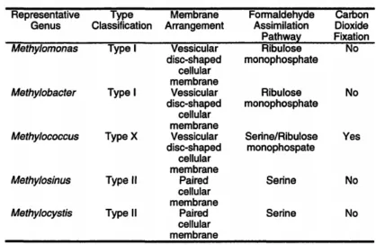
Table 1-1: Classification of monooxygenase/hydroxylase enzymes………4 Table 1-2: Distribution of alkB and CYP153 genes in microbial genomes…………..14 Table 1-3: Categorization of methanotrophic bacteria………..…..16
CHAPTER 2: Activity-based protein profiling of ammonia monooxygenase in Nitrosomonas europaea
Table 2-1: Quantitative proteomic analysis of probe (17OD) labeling of crude
membrane fractions N. europaea………...86 Table 2-S1: Complete quantitative mass spectral analysis of peptide fragments detected from on-bead digestion of crude membrane fractions of N. europaea cells
pretreated with (Plus) and without (Minus)……….195 CHAPTER 4: Activity-based labeling of monooxygenases in Mycobacterium sphagni ENV482 and Mycobacterium vaccae JOB5
Table 4-1: Growth substrate range for M. sphagni ENV482………162 Table 4-2: Proteomic analysis by PNNL of crude membrane fractions from
M. sphagni………167 Table 4-3: Proteomic analysis of crude membrane fractions from M. sphagni
conducted in-house at North Carolina State University………..168 Table 4-4: Estimation of signal fluorescence from an SDS-PAGE gel for JOB5 on various n-alkanes using Image Studio Lite software………...170 Table 4-S1: Complete results from the on-bead trypsin digestion and LC-MS/MS analysis of M. sphagni conducted by PNNL………...198 Table 4-S2: Complete results from the on-bead trypsin digestion and LC-MS/MS analysis of M. sphagni conducted in-house at North Carolina State
LIST OF FIGURES
CHAPTER 1: Literature Review
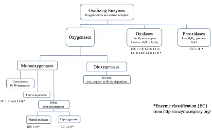
Figure 1-1: Classification of oxidoreductase enzymes………...3
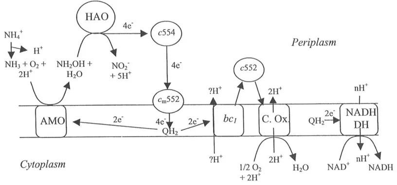
Figure 1-2: Flow of electrons through N. europaea……….…9
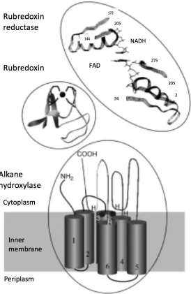
Figure 1-3: The structure of membrane-bound AlkB-like alkane hydroxylase systems………..…..13
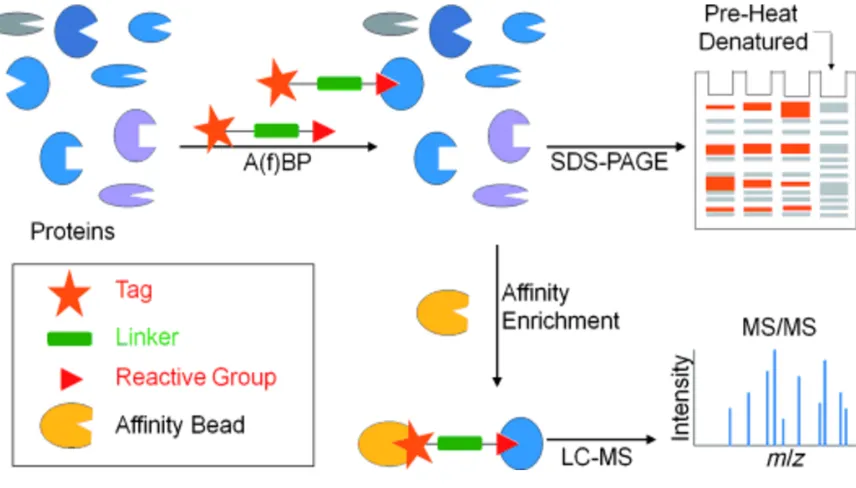
Figure 1-4: Schematic diagram of activity-based protein profiling………21
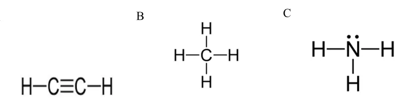
Figure 1-5: Substrates and their structural similarity to acetylene……….23
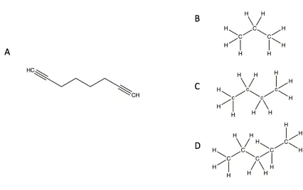
Figure 1-6: Substrates and their structural similarity to 1,7-octadiyne………...24
Figure 1-7: The structure of two additional mechanism-based inactivators…………...25
Figure 1-8: Copper(I)-catalyzed azide-alkyne cycloaddition mechanism………..27
Figure 1-9: Schematic of the process of in-gel fluorescent scanning (IGFS)………….29
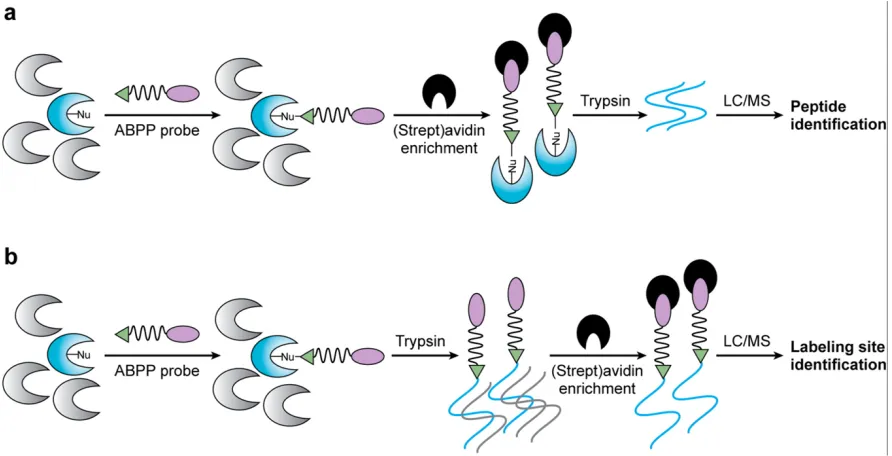
Figure 1-10: The process of ABBP-MudPIT processing and active-site peptide profiling………...32
Figure 1-11: The process of tandem-orthogonal proteolysis (TOP)ABPP……….33
Figure 1-12: The use of antibody microarrays for the analysis of ABPP experiments……….…………35
CHAPTER 2: Activity-based protein profiling of ammonia monooxygenase in Nitrosomonas europaea Figure 2-1: Schematic of the activity-based protein profiling (ABPP) method for detection of ammonia monooxygenase (AMO)………...………...…79
Figure 2-2: Time courses of O2 uptake as determined in an O2 electrode apparatus …80 Figure 2-3: Semilog plots of the percent remaining NH4+-dependent O2 uptake activity of N. europaea……….……..81
Figure 2-4: Effect of 17OD pre-treatment and protein synthesis inhibitors on NH4+- dependent NO2- production in N. europaea washed cells………...82
Figure 2-5: Effect of cell lysing and omission of CuAAC reagents on subsequent polypeptide labeling in IR scanned SDS-PAGE gels……….83
Figure 2-6: Effects of catalytic activation, cell fractionation, and thermal and reductant treatments on labeling of the 28-kDa polypeptide.……….…84
Figure 2-7: IR fluorescence associated with the 28-kDa polypeptide after pre-treatment of cells with individual n-alkynes and diynes.………....85
Figure 2-S1: Effect of incubation time on CuAAC reaction-dependent fluorescent labeling of the 28-kDa polypeptide in N. europaea……….190
dependent fluorescent labeling of the 28-kDa polypeptide in N. europaea………192 Figure 2-S4: Effect of CuSO4 concentration and Cu2+
:THPTA molar ration on CuAAC reaction-dependent fluorescent labeling of the 28-kDa polypeptide in
N. europaea...193 Figure 2-S5: Polypeptides from AmoA detected following in-gel digestion and mass spectral analysis of fluorescent 28-kDa polypeptide………...…194
CHAPTER 3: Activity-based labeling of alkane hydroxylase in Pseudomonas putida strain GPo1
Figure 3-1. Variations of the activity-based protein profiling method for detecting
target enzymes……….121 Figure 3-2: Polypeptide labeling and MTBE oxidation rates of GPo1 and GPo12
dextrose-grown cells during a DCPK induction time course………...122 Figure 3-3: Effects of DCPK induction and omission of click chemistry reagents on polypeptide labeling in GPo1 and GPo12………123 Figure 3-4: Inactivation of alkane hydroxylase activity by individual n-alkynes and diynes………...…124 Figure 3-5: Location of polypeptide labeling in fractionated and whole GPo1 cells...125 Figure 3-6: Polypeptide labeling and MTBE oxidation in GPo1 and other
Pseudomonads………..126 Figure 3-7: Flow-cytometry analysis of CuAAC reactions conducted with GPo1 and GPo12 with and without added 17OD, DCPK, and each CuAAC reagent……….127 Figure 3-8: Relationship of the fluorescence of dextrose grown GPo1 DCPK-induced cells and the rate of MTBE oxidation to TBA……….128 CHAPTER 4: Activity-based labeling of monooxygenases in Mycobacterium sphagni ENV482 and Mycobacterium vaccae JOB5
LIST OF ABBREVIATIONS
17OD 1,7-octadiyne
ABPP activity-based protein profiling AMO ammonia monooxygenase AOB ammonia-oxidizing bacteria BSM basal salts medium
CuAAC copper-catalyzed azide-alkyne cycloaddition EDB ethylene dibromide (1,2-dibromoethane) GC gas chromatography
HAO hydroxylamine dehydrogenase IR infrared
MTBE methyl tertiary-butyl ether
LC/MS-MS liquid chromatography/tandem mass spectrometry pMMO particulate methane monooxygenase
TBA tertiary butyl alcohol THF tetrahydrofuran
SDS sodium dodecyl sulfate
SDS-PAGE sodium dodecyl sulfate-polyacrylamide gel electrophoresis SDIMO soluble diiorn methane monooxygenase
CHAPTER 1
1) Bacterial monooxygenases 1.1 Background and Significance
Oxidoreductase enzymes catalyze biochemical reactions in which electrons are transferred from one molecule to another in redox reactions (29, 104). The majority of these enzymes require cofactors for their catalytic activity, but some possess aromatic residues in their active sites that drive their activity (34, 39, 65, 82, 104). Oxidoreductase enzymes can be divided into 22 different subclasses (EC 1.x.x.x), however a more general classification system divides these enzymes into 4 subgroups: oxidases, peroxidases,
oxygenases/hydroxylases, and dehydrogenases/reductases (147).
Figure 1-1: Classification of oxidoreductase enzymes.
(Adapted from Reference: Burton, S. G.; Oxidizing Enzymes as Biocatalysts, Trends in Biotech., 2003. 21, 543-549.)
pterin-dependent monooxygenases, monooxygenases that depend on other types of cofactors, and cofactor-independent monooxygenases (7, 104). Some of these classes contain specific enzymes that are useful in the degradation of recalcitrant compounds, and understanding their activity and which organisms possess them could help in determining how they might be better utilized.
Table 1-1: Classification of monooxygenase/hydroxylase enzymes. FAD: flavin adenine dinucleotide, FMN: flavin mononucleotide
One family of monooxygenase enzymes are the cytochrome P450 enzymes - heme-containing oxygenases that are perhaps the best characterized of the monooxygenase
completely absent from some Archaea. They also appear to be abundant in eukaryotes (22, 77, 104). These enzymes, which are soluble in prokaryotes and membrane-bound in eukaryotes, also bind and activate molecular oxygen, catalyzing its insertion into various substrates (90, 100, 108). The sub-classification of cytochrome P450 enzymes depends on their electron transport system. Class I systems, which can be found in many bacteria, consist of 3 separate components: the heme domain, a ferredoxin, and an NADH-dependent, FAD-containing ferredoxin reductase. Class II cytochrome P450 enzymes are membrane-bound, two-component systems containing a heme-domain and a reductase unit. Similarly, class III systems contain the same components, except that both domains are relegated to a single polypeptide chain, which increases the reaction rate and promotes electron transport. Class III systems can be soluble or membrane bound. Lastly, class IV cytochrome P450s have the same components as those of class I, except that the domains are contained in a single
polypeptide chain, similar to class III cytochrome P450s. Cytochrome P450 enzymes are able to perform epoxidation, hydroxylation, dehydration, heteroatom-dealkylation and -oxidation, dehydrogenation, dehalogenation, oxidative deamination, dehydration, and reduction
reactions (104).
Another small class of monooxygenases includes those that are copper-dependent and require copper ions to catalyze hydroxylation reactions. One of these, ammonia
monooxygenase, is described in more detail in later sections. Copper-dependent
Pterin-dependent monooxygenases hydroxylate the amino acids tyrosine, tryptophan, and phenylalanine at their aromatic ring (104).
Other types of monooxygenases do not contain heme, flavin, or copper as a cofactor (7). One of these, aclacinomycin 10-hydroxylase (a methyltransferase homolog discovered in Streptomyces purpurascens), was found to utilize S-adenosyl-L-methionine as a cofactor (66). The ortho-nitrophenol 2-monooxygenase from Alcaligenes sp., NyZ215 is the only other known example of these other cofactor-dependent monooxygenases. Similarly, there exists only a few examples of monooxygenase enzymes that do not require any cofactors. Several of these require only molecular oxygen for activity, and they often utilize the substrate as a reducing agent (39,104). The tetraceneomycin F1 monooxygenase from Streptomyces glauscens is one such example (104, 114).
Flavin-dependent monooxygenases utilize either flavin adenine dinucleotide (FAD) or flavin mononucleotide (FMN), which is either tightly bound to the enzyme (prosthetic group) or functions as a substrate (coenzyme). A few internal versions of flavin-dependent monooxygenases have been found, but most are external enzymes that require NADH or NADPH to provide reducing potential for the supply of electrons to the substrate. They are plentiful in prokaryotic genomes and are thought to have spread through horizontal gene transfer. In general, flavin-dependent monooxygenases have been found to catalyze Baeyer-Villiger oxidations, halogenations, and epoxidation reactions (76,104). The external versions of this enzyme are classified into six subclasses, based on their amino acid sequence
The non-heme iron-dependent monooxygenases require two iron atoms, utilized as cofactors, to catalyze their monooxygenase activity (104). Often, they are referred to as bacterial multicomponent monooxygenases because they consist of three components, which include a monooxygenase, a reductase and a small regulatory protein. They catalyze
hydroxylation and epoxidation reactions (76, 104, 135). Members of this family include soluble methane monooxygenase, alkene monooxygenase, phenol hydroxylase, alkane hydroxylase, and four-component alkene/aromatic monooxygenases (40, 76, 104, 107, 129, 130, 141).
The study of soluble diiron monooxygenases (SDIMOs) has increased in recent years and has particularly been aimed at expanding the substrate range of bacteria that contain these enzymes to include various hazardous hydrocarbons, such as trichloroethylene and benzene (76). Many of these compounds are widely distributed in the biosphere due to anthropogenic activities (7). There are three classes of alkane-oxidizing bacteria. These are organisms that grow on methane (methanotrophs), those that grow best on gaseous alkanes, and those that grow optimally on liquid alkanes (46, 47). While much is understood about the hydrocarbon degradation mechanisms of these organisms, the need for detecting and linking activity measurements of these alkane-oxidizing enzymes in environmental samples is great and has generally gone unmet with the current line of detection and activity-based assays.
1.2 Ammonia monooxygenase
ammonia (NH3) to nitrite (NO2-) and nitrate (NO3-). Ammonia-oxidizing bacterium, typified by Nitrosomonas, are an important component of activated wastewater sludge in treatment bioreactors (153). This autotrophic bacterium obtains all of its energy for growth from the oxidation of ammonia to nitrite, which is in part catalyzed by the enzyme ammonia monooxygenase (57, 63).
1.2a Function, Structure, and Distribution of Ammonia Monooxygenase Ammonia monooxygenase catalyzes the initial oxidation of ammonia to
Figure 1-2: Flow of electrons through N. europaea. AMO, ammonia
monooxygenase; HAO, hydroxylamine oxidoreductase; bc1, Complex III; C. Ox., cytochrome oxidase; NADH DH, NADH dehydrogenase; c, cytochrome; QH2, quinol. (Reference: Arp, D. J. and Stein, L. Y.; Metabolism of Inorganic N Compounds by Ammonia-Oxidizing Bacteria, Crit. Rev Biochem. Molec. Biol. 2003. 38, 471-495.)
Since ammonia monooxygenase has not been extensively purified with activity, current insight into the structure of ammonia monooxygenase has been gathered from studies of intact bacteria, subcellular fractions, genomic data, partially purified ammonia
monooxygenase is thought to be copper-containing since it is sensitive to copper chelating agents such as allylthiourea, and non-heme iron was discovered to be present in electron paramagnetic resonance measurements (35, 41).
PCR-based assays targeting amoA have revealed that amoA-like sequences from Nitrosomonas spp. and Nitrospira spp. were most abundant in terrestrial samples (71, 111). The amoA of Nitrosomonas spp. can be detected in freshwater, (hyper)saline lakes, estuaries, sewage sludge, sediments, and flooded soil, while Nitrospira spp. are abundant in different types of soils. amoA is also distributed amongst various ammonia-oxidizing archaea. It has been detected in archaea from soils, sediments, coral, and wastewater treatment plants (16). Unlike their bacterial counterparts, these organisms have also been isolated in samples from hot springs (71). Understanding the distribution of these enzymes and what environmental conditions influence their performance is paramount to understanding how they can be utilized in biodegradation processes.
1.2b Substrates and Inhibitors of Ammonia Monooxygenase in Nitrosomonas europaea Nitrosomonas europaea is perhaps the most widely studied ammonia-oxidizing bacterium, and its complete genome has been sequenced (24). While ammonia is the only physiologically relevant substrate for this enzyme, whole cells of N. europaea have also been found to oxidize a wide range hydrocarbons through the activity of this enzyme. Among many others, these include straight-chain hydrocarbons (≤C8), straight-chain n-alkenes (<C5), methanol, benzene, phenol, cyclohexane, bromocarbons, and carbon monoxide (61, 62).
and substrates such as ethylene and methane that inhibit ammonia oxidation by binding to AMO as competitive alternate substrates (15, 57, 61, 62, 59, 83).
Another group of AMO inhibitors are suicide substrates or mechanism-based inactivators. The majority of the currently known inactivators of AMO are alkynes. These compounds are thought to be catalytically activated by the AMO to reactive intermediates that then covalently bind to the enzyme’s active site. However, covalent modification of AMO has only, thus far, been established for acetylene and propargylamine. In the case of acetylene, covalent modification of AMO has been established using 14C2H2, while the site of action of propargylamine was established using a fluorescent derivative of this compound (57, 91). The lack of further study is most likely due to the fact that radiolabeled forms of these inactivators are either difficult to obtain commercially or their use is impractical under certain conditions (33, 45).
1.3 Alkane Hydroxylase
1.3a Function, Structure, and Distribution of Alkane Hydroxylase
Figure 1-3: The structure of membrane-bound AlkB-like alkane hydroxylase systems. (Adapted from Reference: van Beilen, J. B., Li, Z., Duetz, W. A., Smits, T. H. M., and Witholt, B.; Diversity of alkane hydroxylase systems in the environment, Oil Gas Sci. Technol. 58:427-440.
the plasmid-located alk genes (119). Some bacteria have been found to possess multiple alkane hydroxylase enzymes, and this redundancy is believed to expand the alkane growth substrate range of these strains (96). A recent study of bacterial genomes and metagenomes from terrestrial, freshwater, and marine environments found hundreds of diverse alkB genes
in different distribution patterns. None were detected in archaeal genomes (96). It has been
proposed that these organisms are able to thrive in these environments due to hydrocarbon
seeps from anthropogenic and natural sources (140). The study also found that only
Proteobacteria, Actinobacteria, Bacteroidetes, and Spirochaetes possessed alkB genes, while
cytochrome P450-encoding genes (e.g. CYP153) were only found in the Proteobacteria and
Actinobacteria phyla systems are generally capable of hydroxylating n-alkanes with chain lengths of C5 up to C16, but some species possessing this enzyme have been discovered to hydroxylate n-alkanes up to C32 (96, 121, 130) (Table 1-2).
Table 1-2: Distribution of alkB and CYP153 genes in microbial genomes.
1.3b Substrates and Inhibitors of Alkane Hydroxylase in Pseudomonas putida, GPo1. The best characterized alkane hydroxylase system is that of Pseudomonas putida, GPo1. In this strain, the OCT-plasmid-encoded alk system enables this organism to grow on linear n-alkanes C3-C12 (5, 49, 51, 68, 110, 120). This alk system is also capable of
hydroxylating branched alkanes and alicyclic and alkyl aromatic compounds, epoxidizing terminal olefins, oxidizing alcohols to aldehydes and thioethers to sulphoxides, and demethylating branched methyl ethers such as methyl tertiary-butyl ether (MTBE) (6).
Mycobacterium vaccae, JOB5 is also thought to express an alkane-inducible alkane hydroxylase enzyme system. This versatile hydrocarbon-oxidizing bacterium can grow on straight chain n-alkanes ranging from C2 to C40, certain alkenes, aromatics, and long chain (C10 to C18) monohalogenated alkanes, (98, 133). Current evidence suggests that this bacterium possesses a short-alkane monooxygenase system (SCAM) that initiates the catabolism of capable of short chain n-alkanes (<C8) (58). This enzyme is also thought to enable this strain to cometabolically oxidize a wide variety of environmental pollutants after growth on propane. These pollutant compounds include aromatics (propylbenzene, o-xylene, benzene, ethylbenzene, and styrene), chlorinated hydrocarbons (1,2-dichloroethylene,
1.4 Methane Monooxygenase
Methanotrophic bacteria are aerobic microorganisms that obtain their carbon and energy for growth from the metabolism of methane. These microorganisms are of
considerable interest as they not only have the ability to consume important greenhouse gas but they can also degrade a wide range of environmental contaminants through the activity of methane monooxygenase (52, 70, 99, 152). Methanotrophs can be classified into three
different groups depending on phylogeny, carbon dioxide fixation, chemotaxonomy, internal membrane ultrastructure, what pathway is used for formaldehyde assimilation, and other biochemical features (127). Type I, II, and type X methanotrophs exist (Table 1-3). Table 1-3: Categorization of methanotrophic bacteria.
Type I methanotrophs belong to the γ-subdivision of Proteobacteria and family Methylococcaceae of which there are six validated genera: Methylomonas, Methylobacter,
Methylomicrobium, Methylosphaera, Methylosarcina, and Methylohalobius. Type I
methanotrophs have lamellar stacks of intracytoplasmic membranes (ICM) and assimilate
formaldehyde produced from the oxidation of methane or methanol via the ribulose
monophosphate (RuMP) pathway. Most members of this class require higher levels of copper for growth than do the members of type II and type X. The amount of copper needed for growth determines which form of methane monooxygenase, soluble or particulate, is produced (50, 127).
Type II methanotrophs belong to the α-subdivision of the Proteobacteria and family Methylocystaceae. There are 4 validated genera that are classified as type II methanotrophs: Methylosinus, Methylocystis, Methylocella, and Methylocapsa, which have ICM located on the cell periphery and assimilate formaldehyde through the serine pathway (127).
In contrast, the intermediate methanotrophs of type X follow the RuMP pathway to assimilate formaldehyde like type I, but also possess low levels of the enzymes for the serine pathway like type II methanotrophs. They are thermophilic and thermotolerant, and as such, a higher temperature is required for optimal growth compared to the ideal temperatures for types I and type II (50, 127).
1.4a Function, Structure, and Distribution of Methane Monooxygenase
passes electrons to a specialized cytochrome cL acceptor, which in turn is oxidized by cytochrome cH. Much of the reducing power for the metabolism of methane is derived from the oxidation of formaldehyde (127).
Soluble methane monooxygenase (sMMO) is produced in methanotrophs when the copper concentration is less than 0.89 mM of copper per gram (dry weight) of cells. It utilizes NADH as an electron donor and has a lower affinity for methane than does the particulate form of methane monooxygenase (pMMO) (9, 50, 76, 126). sMMO is encoded by a six-gene operon: mmoXYZ encodes the α (60.6-kDa), β (45-kDa), and γ (19.8-kDa) subunits of the hydroxylase, mmoC encodes the reductase (38.6-kDa), mmoB encodes the regulatory or coupling protein (38.6-kDa), and mmoD encodes orfY (12-kDa), which may play a role in the assembly of the enzyme’s diiron center (50).
pMMO is expressed by methanotrophs when copper concentrations exceed 0.85 mM to 1mM of copper per gram (dry weight) of cells. In methanotrophs that can produce sMMO, membrane proteins associated with pMMO appear, growth yields increase, and sMMO activity is lost if the copper concentration of the growth medium is increased (50). The structural genes for pMMO are organized in a three-gene operon, pmoCAB, which encodes three polypeptides of 23- (ʏ subunit), 45- (α subunit), 27- (β subunit) kDa, respectively. Many of the methanotrophs studied thus far contain duplicate copies of this operon. Although it is widely accepted that pMMO is a copper-containing enzyme, there is ongoing debate about the number and type of metal centers and the physiological nature of the electron donor (53). It also has been found to share a high sequence identity with genes that encode
Both type I and type II methanotrophs possess sMMO. All known methanotrophs possess pMMO (50, 127). These organisms typically reside in swamps, muds, soils from meadows and deciduous woods, rice paddies, ponds, oceans, sewage sludge, and rivers where they serve as sinks for atmospheric methane (73, 87).
1.4b Substrates and Inhibitors of Methane Monooxygenase
Methane monooxygenase has a broad substrate range including carbon monoxide, substituted methanes, alkanes, cycloalkanes, alkenes, haloalkenes, halogenated aliphatics, aromatic and alicyclic hydrocarbons, and diverse ethers (52, 70, 81, 92, 144).
Several inhibitors of methane monooxygenase have been confirmed. These include, but are not limited to phenylacetylene, methylfluoride and acetylene. (83, 99, 109). The particulate form of methane monooxygenase and ammonia monooxygenase are thought to be evolutionarily related, and compounds such as allythiourea and acetylene have been shown to inhibit the activities of both enzymes (56). The inhibitor range for both of these enzymes may be similar as well.
1.5) Activity-Based Protein Profiling 1.5a Background and Significance
modification states, protein microarrays to discern proteome-wide activities, and yeast-two hybrid assays for global mapping of protein interactions (27, 41, 112, 122, 123, 148). Despite the successes of these methods, these technologies only provide limited information about the functional states of these proteins in their native proteomes (148). Expression levels of enzymes often do not correlate with their activity because many are regulated by post-translational modification, protein-protein interactions, and by endogenous small molecules or protein attenuators. For example, in attempting to quantify mRNA or protein levels, misinterpretation of the roles of the enzymes may occur because it is the overall active population that is important for enzyme function (143).
Activity-based protein profiling (ABPP) has been used extensively in eukaryotic systems to identify and quantify active enzymes in complex mixtures. ABPP has also been used to study various aspects of microorganisms, including enzyme function in microbial pathogens, protein redox dynamics, and antibiotic resistance (112). Currently, probes have been developed for several enzyme classes, including major classes of proteases, kinases, phosphatases, glycosidases, and oxidoreductases, amongst others. These probes can
selectively label active enzymes but not their inactive precursor or forms that are bound with inhibitors (14, 27, 37, 115, 134). In this way, inhibition or induction of active isoforms of the target enzyme can be tracked by ABPP, while classical methods for enzyme quantification, such as transcriptomics and global proteomics, fail to link enzyme abundance with functional activity (13, 27, 123, 143).
inactivates the target, active enzyme. The remaining functional group is typically an azide or alkyne group that can be conjugated, in vitro, with a reporter group using a copper-catalyzed cycloaddition (click) reaction (123, 143). The reporter group can be used for infrared
detection of labeled polypeptides or for purification purposes. Affinity enrichment steps allow for analysis of polypeptides through LC-MS/MS, which can be used to assign specific sites of probe labeling to enriched proteins from complex proteomes (13, 27) (Figure 1-4).
Figure 1-4: Schematic diagram of activity-based protein profiling.
(Reference: Yang, P., Lui, K.; Activity-based protein profiling: Recent Developments in Probe Development and Applications, ChemBioChem, 2015. 16: 712-724.)
1.6 Mechanism-Based Inactivators
particular target enzyme (27, 117, 138). As probes in activity-based protein profiling, they report on function by reactions dependent upon the catalytic mechanism of the target enzyme (4, 42, 112). Often referred to as suicide substrates, they have specific target enzymes which participate in their own destruction by catalyzing the unmasking of a latent functional group. This functional group irreversibly inactivates the enzyme without leaving the active site (19, 117, 136).
There are several characteristics a compound must possess to be considered a good candidate as a mechanism-based inactivator. These characteristics have been discussed by several authors, but the general consensus has settled on seven distinguishing features: 1) inhibition of enzyme activity must be time and concentration dependent; 2) the inactivation kinetics must be saturable; 3) natural substrates must protect against inactivation; 4) the stoichiometry of inactivation must be less than or equal to 1:1 with the enzyme; 5)
inactivation requires the target enzyme to be catalytically active; 6) inactivation must occur prior to the release of the active species from the enzyme, and finally, 7) inactivation must be irreversible (1, 26, 117, 136). Both acetylene and 1,7-octadiyne have been evaluated as mechanism-based inactivators for various types of monooxygenase enzymes but not as probes for activity-based protein profiling (60, 119).
1.6a Acetylene
Methylococcus capsulatus (Bath), the mechanism of inactivation by acetylene has been deduced. High-resolution MALDI-TOF MS of intact pMMO complexes has shown that the enzyme oxidizes acetylene to a ketene intermediate, which forms an acetylation adduct with the transmembrane portion, the PmoC subunit, of pMMO (106). Likewise, a ketene
intermediate is generated during inhibition of ammonia monooxygenase (AMO) activity in Nitrosomonas europaea by acetylene. The ketene has been shown to bind amino acid H191, which is thought to reside in or nearby the active site of this enzyme (44). Recovery of ammonia monooxygenase activity after exposure to acetylene can only occur through de novo protein synthesis (60). This proposed mechanism of inactivation has also been confirmed for cytochrome P450 2B6 and could be the same or similar for other monooxygenase enzymes (38).
Figure 1-5: Substrates and their structural similarity to acetylene.
(A) Acetylene, a mechanism-based inactivator, is similar in structure to (B) methane, a substrate of pMMO and (C) ammonia, a substrate of AMO
1.6b 1,7-Octadiyne
The use of 1,7-octadiyne as a mechanism based inactivator of bacterial enzymes has been studied far less than acetylene (Figure 1-6). 1,7-octadiyne has been shown to inhibit the
oxidation of MTBE to TBA by the alkane hydroxylase of Pseudomonas putida, GPo1, a non-heme iron monooxygenase (88, 119).
Figure 1-6: Substrates and their structural similarity to 1,7-octadiyne.
(A) 1,7-octadiyne, a mechanism-based inactivator, is similar in structure to (B) propane, (C) n-butane, and (D) n-pentane, which are some of the normal substrates for AlkB in
Pseudomonas putida, GPo1. 1.6c Other Inhibitors
Allyl sulfide was found to be another mechanism-based inactivator of ammonia monooxygenase activity in Nitrosomonas europaea (72) (Figure 1-7A). Its mechanism of inactivation is proposed to be like that of acetylene, but in studies of microsomal P-450 2E1 monooxygenase, it was concluded that allyl sulfide is not a general inhibitor of
monooxygenases (20).
Ethylene oxide has also been evaluated as a mechanism-based inactivator of various bacterial monooxygenases (Figure 1-7B). It irreversibly inhibits butane monooxygenase activity in Pseudomonas butanovora in a time- and concentration-dependent manner. However, ethylene oxide does not inhibit the activity of other bacterial monooxygenases, including ammonia monooxygenase in Nitrosomonas europaea, toluene-2-monooxygenase in Burkholderia cepacia G4 and alkane monooxygenase in Pseudomonas oleovorans. Of these, only alkane monooxygenase in Nocardioides sp. CF8 and M. vaccae JOBS exhibited ethylene oxide sensitivity (125).
1.7 Copper-Catalyzed Azide-Alkyne Cycloaddition Reactions
Figure 1-8. Copper(I)-catalyzed azide-alkyne cycloaddition mechanism.
(Reference: Liang, L. and Astruc D.; The copper(I)-catalyzed alkyne-azide cycloaddition (CuAAC) “click” reaction and its applications. An overview., Coordination Chemistry Reviews, 255: 2933-2945.
1.7a CuAAC Reagents and Their Roles
Copper(I)-catalyzed azide-alkyne reactions require a copper catalyst, usually CuSO4, which is prepared with an appropriate chelating ligand. It must be maintained in the Cu(I) oxidation state (57, 79). Due to the instability of copper in air, large excesses of copper and ligand are necessary for efficient reactions, and these large concentrations may cause protein damage or precipitation, as well as residual metal remaining after purification. To remedy this difficulty, an in situ reducing agent can be used. Typically, this reagent is sodium
ascorbate, which reduces copper(II) to copper(I) and reduces dissolved oxygen. However, the copper-generated reactive oxygen species dehydroascorbate and other ascorbate byproducts can react with lysine amine and arginine guanidine groups, leading to covalent modification and potential aggregation of proteins (57).
stabilize copper(I). It also scavenges the reaction byproducts dehydroascorbate and reactive oxygen species, preventing them from modifying amino acids. A 5:1 ratio of ligand:Cu is recommended. At a ratio of 1:1 or higher, the CuAAC reaction proceeds to completion in less than 10 minutes (57).
The addition of aminoguanidine also prevents the aggregation of polypeptides, presumably by preventing the crosslinking of protein subunits. Its addition, however, has been found not to increase the rate of CuAAC reactions (57).
Probes for activity-based protein profiling must possess a final element to allow for target enzyme characterization. A reporter tag, which can be modified by click chemistry, allows for visualization of target proteins after the CuAAC reaction has proceeded. Reporter tags that have previously been used consist of small organic fluorophores (less than 1-kDa), biotin, and other types of alkynes or azides. The type of tag selected dictates the experimental options for the downstream analysis of ABPP labeling experiments, and the analytical
platforms for ABPP are ever increasing (4, 27, 41, 42).
1.8 Applications for the Analysis of ABPP Reactions
Once appropriate probes have been found, there are a variety of different analytical platforms to choose from. Each platform has its advantages and disadvantages, and these must be carefully evaluated. Additionally, the type of probe-reporter tag system chosen may put limitations on the analytical platforms that can be utilized (27). Selection of the
1.8a Gel-Based Detection
One of the first analytical platforms for activity-based protein profiling were gel-based methods for the detection of enzyme activities. For this approach, proteomes that have been treated with a specific probe are separated by one- or two-dimensional polyacrylamide gel electrophoresis. Labeled enzymes can then be visualized by fluorescent scanning of the gel if fluorescent probes are used or by avidin blotting if biotinylated probes are used (Figure 1-9) (27, 41, 67).
Figure 1-9. Schematic of the process of in-gel fluorescent scanning (IGFS).
Reference: Sieber, S. and Cravatt, B. Analytical platforms for activity-based protein profiling - exploiting the versatility of chemistry for functional proteomics. Chem. Commun., 2006. 2311–2319.
In-gel fluorescence scanning is attractive because of its simplicity, robustness,
comparative analysis of many proteomes at once. This level of throughput exceeds most other methods (27).
The drawback of gel-based methods is that a low resolving power makes it difficult to detect classes of proteins that might be of importance. These include membrane-associated proteins and proteins that are present in low abundance. Even when detection with gel-based methods is possible, this method does not allow for designations of molecular identity nor does it help to designate sites of probe modification. However, newer “gel free” methods for quantitative proteomics have somewhat alleviated these issues (41, 67, 115).
1.8b Mass Spectrometry-Based Detection
To improve upon the lack of resolution that comes with gel-based methods, “gel free” methods were developed (27, 41, 54 67). These approaches involve liquid chromatography-mass spectrometry (LC-MS), and coupling this technology with activity-based protein profiling has enabled successes in identifying conserved catalytic residues in enzyme active sites and has provided a means for screening selective inhibitors (13). The LC-MS
approaches either seek to analyze the protein targets of probes or they analyze probe-labeled peptides derived from these targets (27, 41, 54, 67).
Methods that involve analyzing peptides derived from probe-labeled targets consist of first biotinylating probes that have been used to label proteomes. Next, direct incubation with (strept)avidin beads enriches the labeled proteome. On-bead trypsin-digestion is then
The enzyme activities in two or more proteomes can also be determined using semi-quantitative parameters like spectral counting when this method is employed (27, 41, 67, 115).
If trypsin digestion of a probe-labeled proteome precedes (strep)avidin enrichment, identification of the labeling site in probe-labeled peptides of enzyme targets is possible. This method is called active-site peptide profiling (2, 27, 115). Importantly, this method allows the resolution of probe-labeled enzymes that have the same molecular mass and tend to co-migrate in SDS-PAGE analyses. Active site-peptide profiling also facilitates the
characterization of low abundance targets of ABPP probes (Figure 1-10b) (115).
Figure 1-10. The process of (a) ABBP-MudPIT processing and (b) active-site peptide profiling.
Reference: Cravatt, B., Wright, A., and Kozarich, J. Activity-based protein profiling: from enzyme chemistry to proteomic chemistry, 2008. An Rev. Biochem. 77: 383-414.
1.8c Emerging ABPP Platforms
Trypsin and TEV protease digests are then analyzed in LC-MS/MS experiments, which characterize probe-labeled proteins and sites of probe modification (27, 115, 122) (Figure 1-11). It is anticipated that this method could be useful to perform high-content comparative proteomic studies in vivo (115).
Figure 1-11. Process of tandem-orthogonal proteolysis (TOP)-ABPP.
Reference: Cravatt, B., Wright, A., and Kozarich, J. Activity-based protein profiling: from enzyme chemistry to proteomic chemistry, 2008. An Rev. Biochem. 77: 383-414.
residues in the active sites of enzymes, which allows proteomes to be analyzed at the level of probe-modified active site peptides following trypsin digestion (115). To perform this
method, proteomes are treated with fluorescent probes, digested with trypsin, and then the resulting peptides are enriched with anti-fluorophore antibody resins. These enriched
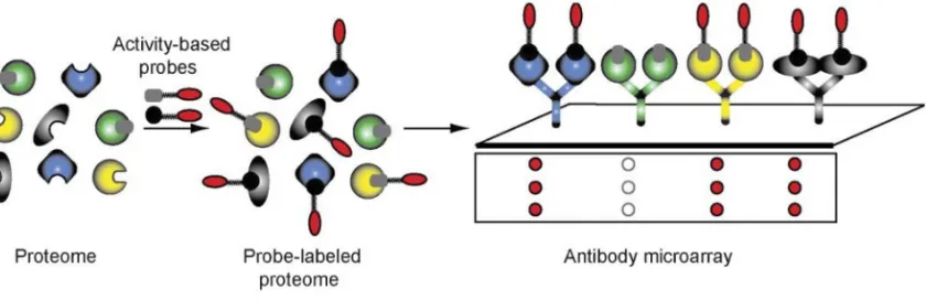
peptides are then eluted and analyzed by CE-LIF. Minimal amounts of sample are consumed, run times are short, and the method has a far greater separation resolution compared to 1D-SDS-PAGE. On the other hand, the identity of enzyme targets after employing this method can only be obtained through complementary LC-MS experiments where individual CE peaks are assigned to specific enzyme targets, and this process may be slow (27, 97, 115). The ABPP microarray also has a low sample demand and high-throughput/high resolution analysis. It was developed for the comparison of protein expression and modification state. Antibodies, fixed to glass slides, are used as capture tools for specific enzyme targets of ABPP probes, which have been pre-incubated together in solution. Once the probes are bound to their specific antibodies on the microarray, they are directly detected by fluorescence scanning (Figure 1-12). This method shows improved sensitivity compared to gel-based methods, and minimal amounts of sample are required for analysis. Since many antibodies may be arrayed in parallel on a single slide, this method allows for high
Figure 1-12. The use of antibody microarrays for the analysis of ABPP experiments. Reference: Sieber, S. and Cravatt, B. Analytical platforms for activity-based protein profiling - exploiting the versatility of chemistry for functional proteomics. Chem. Commun., 2006. 2311–2319.
REFERENCES
1. Abeles RH, Maycock AL. 1976. Suicide enzyme inactivators. Acc. Chem. Res. 9:313-319.
2. Adam GC, Burbaum J, Kozarich JW, Patricelli MP. 2004. Mapping enzyme active sites in complex proteomes. J. Am. Chem. Soc. 126:1363-1368.
3. Adams MA, Jia Z. 2005. Structural and biochemical evidence for an enzymatic quinone redox cycle in Escherichia coli: identification of a novel quinol
monooxygenase. J. Biol. Chem. 280: 8358–8363.
4. Agard NJ, Baskin JM, Prescher JA, Anderson L, Bertozzi CR. 2006. A
comparative study of bioorthogonal reactions with azides. ACS Chem. Biol. 1:644-648.
5. Alonso H, Kleinfeld O, Yeheskel A, Ong PC, Liu YC, Stok JE, De Voss JJ, Roujeinikova A. 2014. Structural and mechanistic insight into alkane hydroxylation by Pseudomonas putida AlkB. Biochem. J. 460:283-293.
6. Alonso H, Roujeinikova A. 2012. Characterization and two-dimensional crystallization of membrane component alkB of the medium-chain alkane hydroxylase system from Pseudomonas putida GPo1. Appl. Environ. Microbiol. 78:7946-7953.
7. Arora PK, Srivastava A, Singh VP. 2010. Application of monooxygenase in dehalogenation, desulphurization, denitrification, and hydroxylation of aromatic compounds. J. Biorem. Biodeg. 112:1-8.
8. Arp DJ, Stein LY. 2003. Metabolism of inorganic N compounds by ammonia-oxidizing bacteria. Crit. Rev. Biochem. Mol. Biol. 38:471-495.
9. Ayala M, Torres E. 2004. Enzymatic activation of alkanes: constraints and prospective. Appl. Catal. A-Gen. 272:1-13.
10.Babu JP, Brown LR. 1984. New type of oxygenase involved in the metabolism of propane and isobutane. Appl. Environ. Microbiol. 48:260-264.
12.Baldwin TO, Ziegler MM. 1992. The biochemistry and molecular biology of
bacterial bioluminescence. Chemistry and Biochemistry of Flavoenzymes. CRC Press, Boca Raton, FL.
13.Barglow KT, Cravatt BF. 2007. Activity-based protein profiling for the functional annotation of enzymes. Nat. Methods. 4:822-827.
14.Baskin JM, Bertozzi CR. 2007. Bioorthogonal click chemistry: covalent labeling in living systems. QSAR Comb. Sci. 26:1111-1323.
15.Bédard C, Knowles R. 1989. Physiology, biochemistry, and specific inhibitors of CH4, NH4+, and CO oxidation by methanotrophs and nitrifiers. Microbiol. Rev. 53:68-84.
16.Beman JM, Roberts KJ, Wegley L, Rohwer F, Francis CA. 2007. Distribution and diversity of archaeal ammonia monooxygenase genes associated with corals. Appl. Environ. Microbiol. 73:5642-5647.
17.Binnerup SJ, Svenning MM, Hestnes AG, Wartiainen I. 2005. Methane Oxidizing Bacteria as Environmental Indicators. Norden Council of Ministers, Copenhagen, Denmark.
18.Bisswanger H. 2014. Enzyme assays. Perspect. Sci. 1:41-55.
19.Blobaum AL. 2006. Mechanism-based inactivation and reversibility: is there a new trend in the inactivation of cytochrome p450 enzymes? Drug Metab. Dispos. 34:1-7. 20.Brady JF, Ishizaki H, Fukuto JM, Lin MC, Fadel A, Gapac JM, Yang CS. 1991.
Inhibition of cytochrome P-450 by diallyl sulfide and its metabolites. Chem. Res. Toxicol. 6:642-647.
21.Burback BL, Perry JJ. 1993. Biodegradation and biotransformation of groundwater pollutant mixtures by Mycobacterium vaccae. Appl. Environ. Microbiol. 59:1025-1029.
22.Burton SG. 2003. Oxidizing enzymes as biocatalysts. Trends Biotechnol. 21:543-549.
24.Chain P, Lamerdin J, Regala W, Lao V, Land M, Hauser L, Hooper A, Klotz M, Norton J, Sayavedra-Soto L, Arciero D, Hommes N, Arp D. 2003. Complete genome sequence of the ammonia-oxidizing bacterium and obligate
chemolithoautotroph Nitrosomonas europaea. J Bacteriol. 185:2759-2773.
25.Choi DW, Kunz RC, Boyd ES, Semrau JD, Antholine WE, Han JI, Zahn JA, Boyd JM, de la Mora AM, DiSpirito AA. 2003. The membrane-associated methane monooxygenase (pMMO) and pMMO-NADH:quinone oxioreductase complex from Methylococcus capsulatus Bath. J. Bacteriol. 185:5755-5764.
26.Copeland RA. 2013. Evaluation of Enzyme Inhibitors in Drug Discovery: A Guide for Medicinal Chemists and Pharmacologists, John Wiley and Sons, Inc., Hoboken, NJ.
27.Cravatt BF, Wright AT, Kozarich JW. 2008. Activity-based protein profiling: from enzyme chemistry to proteomic chemistry. Annu. Rev. Biochem. 77:383-414.
28.Das N, Chandran P. 2011. Microbial degradation of petroleum hydrocarbon contaminants: an overview. Biotech. Res. Int. 2011:1-13.
29.Diaper JP, Tither K, Edwards C. 1992. Rapid assessment of bacterial viability by flow cytometry. Appl. Microbiol. Biotechnol. 38: 268-272.
30.Dixon, M, Webb EC. 1979. Enzymes. Academic Press, Cambridge, MA. 31.Doughty DM, Sayavedra-Soto LA, Arp DJ, Bottomley PJ. 2006. Product
repression of alkane monooxygenase expression in Pseudomonas butanovora. J. Bacteriol. 188:2586-2592.
32.Dinamarca MA, Ruiz-Manzana A, Rojo F. 2002. Inactivation of cytochrome o ubiquinol oxidase relieves catabolic repression of the Pseudomonas putida GPo1alkane degradation pathway. J. Bacteriol. 184:3785-3793.
33.Ebie Y, Miura H, Noda N, Matsumura M, Tsuneda S, Hirata A, Inamori Y. 2002. Detection and quantification of expression of amoA by competitive reverse transcription-PCR. Water Sci. Technol. 46:281-288.
34.Ellis EM. 2002. Microbial aldo-keto reductases. FEMS Microbiol. Lett. 216:123-131. 35.Ensign SA, Hyman MR, Arp DJ. In vitro activation of ammonia monooxygenase
36.Entsch B, van Berkel WJH. 1995. Flavoprotein structure and mechanism: structure and mechanism of p-hydroxybenzoate hydroxylase. FASEB J. 9:476–483.
37.Evans MJ, Saghatelian A, Sorensen EJ, Cravatt BF. 2005. Target discovery in small-molecule cell-based screens by in situ proteome reactivity profiling. Nat. Biotechnol. 23:1303-1307.
38.Fan PW, Gu C, Marsh SA, Stevens JC. 2003. Mechanism-based inactivation of cytochrome P450 2B6 by a novel terminal acetylene inhibitor. Drug Metab. Dispos. 31:28-36.
39.Fetzner S. 2002. Oxygenases without requirement for cofactors or metal ions. Appl. Microbiol. Biotechnol. 60:243-257.
40.Gallagher SC, Cammark R, Dalton H. 1997. Alkene monooxygenase from
Nocardia corallina B-276 is a member of the class of dinuclear iron proteins capable of stereospecific epoxygenation reactions. Eur. J. Biochem. 247:635–641.
41.Galmozzi A, Dominguez E, Cravatt BF, Saez E. 2014. Application of activity-based protein profiling to study enzyme function in adipocytes. Methods Enzymol. 538: 151-169.
42.Giepmans BN, Adams SR, Ellisman MH, Tsien RY. 2006. The fluorescent toolbox for assessing protein location and function. Science. 312:217-224.
43.Gilch S, Meyer O, Schmidt I. 2009. A soluble form of ammonia monooxygenase in Nitrosomonas europaea. Biol. Chem. 390:863-873.
44.Gilch S, Vogel M, Lorenz MW, Meyer O, Schmidt I. 2009. Interaction of the mechanism-based inactivator acetylene with ammonia monooxygenase of Nitrosomonas europaea. Microbiology. 155:279-284.
45.Glanemann C, Loos A, Gorret N, Willis LB, O’Brien XM, Lessard PA, Sinskey AJ. 2003. Disparity between changes in mRNA abundance and enzyme activity in Corynebacterium glutamicum: implications for DNA microarray analysis. Appl. Microbiol. Biotechnol. 61:61-68.
46.Hamamura N, Storfa RT, Semprini L, Arp DJ. 1999. Diversity in butane
47.Hamamura N, Yeager CM, Arp DJ. 2001. Two distinct monooxygenases for alkane oxidation in Nocardioides sp. strain CF8. Appl. Environ. Microbiol. 67:4992-4998.
48.Hand S, Wang B, Chu KH. 2015. Biodegradation of 1,4-dioxane: effects of enzyme inducers and trichloroethylene. Sci. Total Environ. 1520:154-159.
49.Hanson JR, Ackerman CE, Scow KM. 1999. Biodegradation of methyl tert-butyl ether by a bacterial pure culture. Appl. Environ. Microbiol. 65:4788-4792.
50.Hanson RS, Hanson TE. 1996. Methanotrophic bacteria. Microbiol. Rev. 60:439-471.
51.Hatzinger PB, McClay K, Vainberg S, Tugusheva M, Condee CW, Steffan RJ. 2001. Biodegradation of methyl tert-butyl ether by a pure bacterial culture. Appl. Environ. Microbiol. 67:5601-5607.
52.Hatzinger PB, Streger SH, Begley JF. Enhancing aerobic biodegradation of 1,2-dibromoethane in groundwater using ethane or propane and inorganic nutrients. J Contam. Hydrol. 172:61-70.
53.Hazen, T. C. 2010. Cometabolic bioremediation. Handbook of Hydrocarbon and Lipid Microbiology, Springer-Verlag, Berlin, Heidleberg, p. 2506-2514.
54.Heal WP, Dang TH, Tate EW. 2011. Activity-based probes: discovering new biology and new drug targets. Chem Soc Rev. 40: 246-257.
55.Hollocher, TC. 1981. Oxidation of ammonia by Nitrosomonas europaea: definitive 18O-tracer evidence that hydroxylamine formation involves a monooxygenase. J Biol. Chem. 256:10834-10836.
56.Holmes AJ, Costello A, Lidstrom ME, Murrell JC. 1995. Evidence that particulate methane monooxygenase and ammonia monooxygenase may be evolutionarily related. FEMS Microbiol. Lett. 132:203-208.
57.Hong V, Presolski SI, Ma C, Finn, MG. 2009. Analysis and optimization of copper-catalyzed azide-alkyne cycloaddition for bioconjugation. Angew. Chem. Int. Ed. Engl. 48:9879-9883.
59.Huijbers MM, Montersino S, Westphal AH, Tischler D, van Berkel WJ. 2014. Flavin dependent monooxygenases. Arch. Biochem. Biophys. 544:2-17.
60.Hyman MR, Arp DJ. 1992. 14C2H2- and 14CO2-labeling studies of the de novo synthesis of polypeptides by Nitrosomonas europaea during recovery from acetylene and light inactivation of ammonia monooxygenase. J. Biol. Chem. 267:1534-1545. 61.Hyman MR, Murton IB, Arp DJ. 1988. Interaction of ammonia monooxygenase
from Nitrosomonas europaea with alkanes, alkenes, and alkynes. Appl. Environ. Microbiol. 54:3187-3190.
62.Hyman MR, Wood PM. 1984. Ethylene oxidation by Nitrosomonas europaea. Arch. Microbiol. 137:155-158.
63. Hyman MR, Wood PM. 1985. Suicidal inactivation and labelling of ammonia mono-oxygenase by acetylene. Biochem. J. 227:719–725.
64.Iida T, Sumita T, Ohta A, Takagi M. 2000. The cytochrome P450ALK multigene family of an n-alkane assimilating yeast, Yarrowia lipolytica: cloning and
characterization of genes coding for new CYP52 family members. Yeast. 16:1077-1087.
65.Imhoff RD, Power NP, Borrok MJ, Tipton PA. 2003. General base catalysis in the urate oxidase reaction: evidence for a novel Thr-Lys catalytic diad. Biochemistry. 42:4094-4100.
66.Jansson A, Niemi J, Lindqvist Y, Mantsala P, Schneider G. Crystal structure of aclacinomycin-10-hydroxylase, an S-adenosyl-L-methinonine-dependent
methyltransferase homolog involved in anthracycline biosynthesis in Streptomyces purpurascens. J. Mol. Biol. 334:269-280.
67.Jessani N, Neissen S, Wei BQ, Nicolau M, Humphrey M, Ji Y, Han W, Noh DY, Yates JR, Jeffrey SS, Cravatt BF. 2005. A streamlined platform for high-content functional proteomics of primary human specimens. Nat Methods. 2: 691-697. 68.Johnson EL, Hyman MR. 2006. Propane and n-butane oxidation by Pseudomonas
putida GPo1. Appl. Environ. Microbiol. 72:950-952.
70.Jiang H, Chen Y, Jiang PX, Zhang C, Smith TJ, Murrell JC, Xing XH. 2010. Methanotrophs: multifunctional bacteria with promising applications in
environmental bioengineering. Biochem. Eng. J. 49:277-288.
71.Jiang H, Huang L, Deng Y, Wang S, Zhou Y, Liu L, Dong H. 2014. Latitudinal distribution of ammonia-oxidizing bacteria and archaea in the agricultural soils of eastern China. Appl. Environ. Microbiol. 80:5593-5602.
72.Juliette LY, Hyman MR, Arp DJ. 1993. Mechanism-based inactivation of ammonia monooxygenase in Nitrosomonas europaea by allylsulfide. Appl. Environ. Microbiol. 59:3728-3735.
73.Kazda J. 1980. Mycobacterium sphagni sp. nov. Int. J Syst. Evol. Micr. 30:77-81. 74.Keller RM, Wuthrich K, Debrunner PG. 1972. Proton magnetic resonance reveals
high-spin iron (II) in ferrous cytochrome P450cam from Pseudomonas putida. PNAS. 69:2073-2075.
75.Klotz MG, Alzerreca J, Norton JM. 1997. A gene encoding a membrane protein exists upstream of the amoA/amoB genes in ammonia oxidizing bacteria: a third member of the amo operon? FEMS Microbiol. Lett. 150:65-73.
76.Leahy JG, Batchelor PJ, Morcomb SM. 2003. Evolution of the soluble diiron monooxygenases. FEMS Microbiol. Rev. 27:449-479.
77.Lewis DFV, Wiseman A. 2005. A selective review of bacterial forms of cytochrome P450 enzymes. Enzyme Microb. Tech. 36:377-384.
78.Levin EY, Levenberg B, Kaufman S. 1960. The enzymatic conversion of 3,4-dihydroxyphenylethylamine to norepinephrine. J. Biol. Chem. 235:2080-2086. 79.Liang L, Astruc D. 2011. The copper(I)-catalyzed azide-alkyne cycloaddition (CuAAC) “click” reaction and its applications, an overview. Coor. Chem. Rev. 255:2933-2945.
80.Link AJ, Tirrell DA. 2003. Cell surface labeling of Escherichia coli via copper(I)-catalyzed [3+2] cycloaddition. J. Am. Chem. Soc. 125:11164-11165.
81.Lipscomb JD. 1994. Biochemistry of the soluble methane monooxygenase. Annu. Rev. Microbiol. 48:371-399.
83.Lontoh S, DiSpirito AA, Krema CL, Whittaker MR, Hooper AB, Semrau JD. 2000. Differential inhibition in vivo of ammonia monooxygenase, soluble methane monooxygenase and membrane-associated methane monooxygenase by
phenylacetylene. Environ. Microbiol. 5:485-494.
84.Maeng JH, Sakai Y, Tani Y, Kato N. 1996. Isolation and characterization of a novel oxygenase that catalyzes the first step on n-alkane oxidation in Acinetobacter sp. strain M-1. J. Bacteriol. 178:3695-3700.
85.Maier T, Forster HH, Asperger O, Hahn U. 2001. Molecular characterization of the 56-kDa CYP153 from Acinetobacter sp. EB104. Biochem. Biophys. Res. Commun. 286:652-658.
86.Mahendra S, Alvarez-Cohen L. 2006. Kinetics of 1,4-dioxane biodegradation by monooxygenase-expressing bacteria. Environ. Sci. Technol. 40:5435-5442.
87.Mancinelli, RL. 1995. The regulation of methane oxidation in soil. Annu. Rev. Microbiol. 49: 581-605.
88.May SW, Schwartz RD, Abbott BJ, Zaborsky OR. 1975. Structural effects on the reactivity of substrates and inhibitors in the epoxidation system of Pseudomonas oleovorans. Biochim. Biophys. Acta. 403:245-255.
89.McGuirl MA, Dooley DM. 1999. Copper-containing oxidases. Curr. Opin. Chem. Biol. 3:138-144.
90.McLean KJ, Sabri M, Marshall KR, Lawson RJ, Lewis DG, Clift D, Balding PR, Dunford AJ, Warman AJ, McVey JP, Quinn AM, Sutcliffe MJ, Scrutton NS, Munro AW. 2005. Biodiversity of cytochrome P450 redox systems. Biochem. Soc. Trans. 33:796-801.
91.McTavish H, Fuchs JA, Hooper AB. 1993. Sequence of the gene coding for ammonia monooxygenase in Nitrosomonas europaea. J. Bacteriol. 175:2436-2444. 92.Merkx M, Lippard SJ. 2001. Why OrfY? Characterization of MMOD, a
long-overlooked component of the soluble methane monooxygenase from Methylococcus capsulatus (Bath). J Biol. Chem. 277:5858-5865.
94.Murrell JC, Gilbert B, McDonald IR. 2000. Molecular biology and regulation of methane monooxygenase. Arch. Microbiol. 173:325–332.
95.Nagatsu T, Levitt M, Udenfriend S. 1964. Tyrosine hydroxylase: the initial step in norepinephrine biosynthesis. J. Biol. Chem. 239:2910-2917.
96.Nie Y, Chi CQ, Fang H, Liang JL, Lu SL, Lai GL, Tang YQ, Wu XL. 2014. Diverse alkane hydroxylase genes in microorganisms and environments. Sci. Rep. 4:1-11.
97.Okerberg ES, Wu J, Zhang B, Samii B, Blackford K, Wiin, DT, Shreder KR, Burbaum JJ, Patricelli MP. 2005. High-resolution functional proteomics by active-site peptide profiling. PNAS. 102:4996-5001.
98.Ooyama J, Foster, JW. 1965. Bacterial oxidation of cycloparaffinic hydrocarbons. A. Van. Leeuw. J Microb. 31:45-65.
99.Oremland RS, Culbertson CW. 1992. Importance of methane-oxidizing bacteria in the methane budget as revealed by the use of a specific inhibitor. Nature. 356:421-423.
100. Ortiz de Montellano PR. 2005. Cytochrome P450: Structure, Mechanism, and Biochemistry. Springer Publishing Co. New York City, NY.
101. Padda RS, Pandey KK, Kaul S, Nair VD, Jain RK, Basu SK, Chakrabarti T. 2001. A novel gene encoding a 54-kDa polypeptide is essential for butane utilization by Pseudomonas sp. IMT37. Microbiology. 147:2479-2491.
102. Pandey KK Mayilraj S, Chakrabarti T. 2002. Pseudomonas indica sp. nov., a novel butane-utilizing species. Int. J. Syst. Evol. Microbiol. 52:1559-1567.
103. Patakva P, Linhova M, Vykydalova, Branska B, Rychtera M, Melzoch K. 2014. Use of fluorescent staining and flow cytometry for monitoring physiological
changes in solventogenic bacteria. Anaerobe 29: 113-117.
104. Pazmino DET, Winkler M, Glieder A, Fraaije MW. 2010. Monooxygenases as biocatalysts: classification, mechanistic aspects and biotechnological applications. J. Biotechnol. 146:9-24.
105. Peixoto RS, Vermelho AB, Rosado AS. 2011. Petroleum-degrading enzymes: bioremediation and new prospects. Enz. Res. 475193:1-7.
Han CC, Chen CH, Li MS, Yu SS, Chan SI. 2015. Inactivation of the particulate methane monooxygenase (pMMO) in Methylococcus capsulatus (Bath) by acetylene. Biochim. Biophs. Acta. 1854:1842-1852.
107. Pikus JD, Studts JM, Achim C, Kauffmann KE, Münch E, Steffan RJ, McClay K, Fox BG. 1996. Recombinant toluene-4-monooxygenase: catalytic and Mössbauer studies of the purified diiron and Rieske components of a four-protein complex. Biochemistry. 35:9106–9119.
108. Porter TD, Coon MJ. 1991. Cytochrome P-450. Multiplicity of isoforms substrates, and catalytic and regulatory mechanisms. J. Biol. Chem. 266:13469-13472.
109. Prior SD, Dalton H. 1985. Acetylene as a suicide substrate and active site probe for methane monooxygenase from Methylococcus capsulatus (Bath). FEMS Microbiol. Lett. 29:105-109.
110. Rojo F. 2005. Specificity at the end of the tunnel: understanding substrate length discrimination by the AlkB alkane hydroxylase. J. Bacteriol. 187:19-22.
111. Rotthauwe JH, Witzel KP, Liesack W. 1997. The ammonia monooxygenase structural gene amoA as a functional marker: molecular fine-scale analysis of natural ammonia-oxidizing populations. Appl. Environ. Microbiol. 63:4704-4712.
112. Sadler NC, Wright AT. 2015. Activity-based protein profiling of microbes. Curr. Opin. Chem. Biol. 24:139-144.
113. Schomburg D, Stephan D. 1994. Class 1.13-1.97: Oxidoreductases. Enzyme Handbook 8. Springer Publishing Co. New York City, NY.
114. Shen B, Hutchinson CR. 1993. Tetracenomycin F1 monooxygenase: oxidation of a naphthacenone to a naphthacenequinone in the biosynthesis of a tetracenomycin C in Streptomyces glaucescens. Biochemistry. 32:6656-6663.
115. Sieber SA, Cravatt BF. 2006. Analytical platforms for activity-based protein profiling-exploiting the versatility of chemistry for functional proteomics. Chem. Comm. 22:2311-319.
116. Sieber SA, Mondala TS, Head SR, Cravatt BF. 2004. Microarray platform for profiling enzyme activities in complex proteomes. J. Am. Chem. Soc. 126:15640- 15641.
118. Sluis MK, Sayavedra-Soto LA, Arp DJ. 2002. Molecular analysis of the soluble butane monooxygenase from Pseudomonas butanovora. Microbiology. 148:3617- 3629.
119. Smith CA, Hyman MR. 2004. Oxidation of methyl tert-butyl ether by alkane hydroxylase in dicyclopropylketone-induced and n-octane-grown Pseudomonas putida GPo1. Appl. Environ. Microbiol. 70:4544-4550.
120. Smith CA, O’Reilly KT, Hyman MR. Cometabolism of methyl tertiary butyl ether and gaseous n-alkanes by Pseudomonas mendocina KR-1 grown on C5 to C8 n- alkanes. Appl. Environ. Microbiol. 69:7385-7394.
121. Smits TH, Balada SB, Witholt B, van Beilen JB. 2002. Functional analysis of alkane hydroxylases from gram-negative and gram-positive bacteria. J. Bacteriol. 184:1733-1742.
122. Speers AE, Cravatt BF. 2005. A tandem orthogonal proteolysis strategy for high- content chemical proteomics. J. Am. Chem. Soc. 127:10018-10019.
123. Speers AE, Cravatt BF. 2009. Activity-based protein profiling (ABPP) and click chemistry (CC)-ABPP by MudPIt mass spectrometry. Curr. Protoc. Chem. Biol. 124. Staijen IE, Marcionelli R, Witholt B. 1999. The PalkBFGHJKL promoter is under carbon catabolite repression control in Pseudomonas oleovorans but not in Escherichia coli alk+recombinants. J. Bacteriol. 181:1610-1616.