_____________________________________________________________________________________________________
www.sciencedomain.org
Role of Bone Marrow-Derived Mesenchymal Stem
Cells Differentiate into β Cells in the Pancreatic
Microenvironment
Chun-Yan Deng
1, Ya-Li Yang
1, Feng Gao
1, Hui Qi
1and Fu-Rong Li
1*1The Key Laboratory of Stem Cell and Cellular Therapy, The Second Clinical Medical College
(Shenzhen People’s Hospital), Jinan University, Shenzhen 518020, China.
Authors’ contributions
This work was carried out in collaboration between all authors. Author CYD wrote the protocol and the first draft of the manuscript. Author YLY managed the experimental process and analyzed data. Author FG managed the literature searches and the experimental process. Author HQ designed research and contributed to interpretation of data. Author F-RL contributed to research design; revising the paper critically and approval of the submitted and final versions. All authors read and approved the final manuscript.
Article Information
DOI: 10.9734/BJMMR/2015/16600 Editor(s): (1)E. Umit BAGRIACIK, Department of Immunology, Gazi University, Turkey. (2)Rui Yu, Environmental Sciences and Engineering, Gillings School of Global Public Health, The University of North Carolina at Chapel Hill, USA.
Reviewers: (1)Sanjay Mishra, School of Biotechnology, IFTM University, India.
(2)Anonymous, Urology and Nephrology Center, Egypt. (3)Anonymous, Roger Williams Hospital, USA. (4)Anonymous, Tianjin University of Science and Technology, China. Complete Peer review History:http://www.sciencedomain.org/review-history.php?iid=1117&id=12&aid=9085
Received 8th February 2015 Accepted 16th April 2015 Published 2nd May 2015
ABSTRACT
Aims: Mesenchymal stem cells (MSCs) can differentiate into multiple cell types including insulin-producing cells. However, these cells usually cannot be directed to efficiently differentiate into β cells in vitro. The present study aimed to explore whether the pancreatic microenvironment could induce bone marrow-derived (BM)-MSCs to differentiate into β cells to compensate for insufficient β-cells.
Methodology: We directly transplanted male enhanced green fluorescence protein (EGFP)-expressing BM-MSCs into the pancreas of female diabetic Sprague-Dawley rats by multi-point injection.
Results: BM-MSCs could restore serum insulin and C-peptide levels and reverse hyperglycemia by intra-pancreatic transplantation. BM-MSCs from male donors could differentiate into pancreatic stem/progenitor cells and β cells under female pancreas micro-environment. Neogenesis islets derived from BM-MSCs were verified in pancreatic tissue by histology and the expression of genes related to β cell gene biomarker was determined by RT-PCR and quantitative real time-PCR. Y-chromosome SRY and PDX-1 mRNA have expressed simultaneously in neogenesis β cells. Polyploidy and aneuploid DNA were not observed.
Conclusion: This study showed that transplanted BM-MSCs did not fuse with pancreatic cells and could contribute to repair, paracrine and differentiation into new islet β cells in the pancreatic microenvironment.
Keywords: Mesenchymal stem cells; transplantation; differentiation; pancreatic microenvironment; insulin-producing cells.
1. INTRODUCTION
Functional β cell replacement is a promising approach to treat type 1 diabetes [1]. However, the scarcity of transplantable donor islets and allograft rejection had historically hampered further development [2]. Currently, numerous cells including embryonic stem cells [3], pancreatic stem cells [4], hepatic oval cells [5] and bone marrow-derived mesenchymal stem cells (BM-MSCs) [6] have been reported to differentiate into insulin-producing cells (IPCs), which provide new cell sources for β cell transplantation. Among these cells, BM-MSCs are easily obtained and not associated with ethical concerns. Therefore, BM-MSCs may be an important cell source for the treatment of diabetes.
BM-MSCs have exhibited a surprising capacity to differentiate into various ectoderm, mesoderm and endoderm-derived cells [7], and contribute to the formation of tissues. It has been shown that BM-MSCs can differentiate into IPCs in vitro [8,9], though their expression profiles are dissimilar with human islet-mesenchymal stem cells [10]. After diabetes or pancreatic injury occurs, BM-MSCs migrate into injured pancreas and differentiate into corresponding cells that participate in tissue repair [11]. It has been shown that BM-MSCs also differentiate into β cells after transplantation [12]. Recent researchers have shown that stem cell differentiation is controlled by extracellular cues from the environment and by intrinsic genetic programs within stem cells [13,14]. These evidence shows that the microenvironment plays an important role in the survival and differentiation of stem cells. However, some studies question the trans-differentiation of BM-MSCs, and demonstrate that BM-MSCs may fuse with other cells to show corresponding biological characteristics [15,16]. Therefore, in
the pancreatic microenvironment, whether BM-MSCs fuse with β cells or differentiate into β cells via trans-differentiation or other pathways should be clarified. It is an interesting but puzzling question and, as a bottleneck, limits the clinical application of BM-MSCs to treat diabetes. We directly transplanted male enhanced green fluorescence protein (EGFP)-expressing BM-MSCs into the pancreas of female diabetic Sprague-Dawley rats by multi-point injection. Surprisingly, the glucose level gradually decreased and was approximately normal after 6 weeks. BM-MSCs-derived new islets were verified in pancreatic tissue by histology. The co-expression of Y-chromosome SRY and insulin genes were observed in new islets. This study showed that transplanted BM-MSCs did not fuse with pancreatic cells and could contribute to functional new islets in the pancreatic microenvironment.
2. METHODOLOGY
2.1 Animals
Sprague–Dawley (SD) rats were purchased from the Experimental Animal Center of Guangdong Province and housed in pathogen-free rooms at the Animal Center of the Second Clinical Medical College, Ji’nan University. All animal experiments were approved by the institutional ethics committee and conducted in accordance with institutional guidelines for animal care and use.
2.2 Isolation, Culture and Identification of BM-MSCs
CA) and identification was performed by adipogenic and osteogenic induction as described elsewhere [18].
2.3 Preparation of EGFP-BM-MSCs
Passage 3 BM-MSCs were seeded at 1×10⁶ cells/ml into a 96 well plate, and AdC-MV-EGFP adenoviral vectors with a multiplicity of infection of 100, 200, 400 and 800 particles / cell (n = 4) in serum-free Dulbecco’s modified Eagle’s medium (DMEM) were used to transfect BM-MSCs. Green fluorescence was visualized using an inverted fluorescence microscope (Nikon, Tokyo, Japan) 48 h after transfection. EGFP-expressing BM-MSC clones formed in DMEM containing 400×106 g/L G418 after 12 days. Flow cytometry was used to detect green fluorescence of the
EGFP-expressing BM-MSCs prior to
transplantation.
2.4 Preparation of Diabetic Rats
Female 8-week-old SD rats, weighting 200 g, were injected with 45 mg/kg streptozotocin (STZ; Sigma-Aldrich, St Louis, MO) via the tail vein. Blood was drawn from the tail vein and blood glucose was monitored by a Roche Accu Check III (Roche, Basel, Switzerland). Non-fasting blood glucose of ≥300 mg/dl for 3 consecutive days was considered as the onset of diabetes.
2.5 EGFP-BM-MSCs Transplantation
Forty diabetic female SD rats were randomly divided into two groups. The BM-MSCs transplantation group (n = 30) were injected with a EGFP-BM-MSCs suspension (5×10⁷ cells, 0.2 mL), prepared from male SD rats, at 10 distinct pancreatic sites by celiotomy. Diabetic controls (n = 10) and normal female SD rats (normal controls, n = 10) were injected with 0.2 mL saline in place of the cell suspension. All procedures were performed under sterile conditions. Experimental design scheme (Supplementary Fig. 1).
2.6 Detection of Blood Glucose, Insulin and C-peptide, and Glucose Tolerance Testing
Blood glucose was consecutively measured every day in the first week following transplantation. And then, it had been monitored every three days until 84 days after transplantation. 10 rats in each group were randomly selected for blood glucose monitoring at 4 pm after fasting 12 hours. The non-fasting
blood glucose level had been determined with a Roche Accu Check III. Glucose tolerance test was performed at 28, 56 and 84 days after transplantation. After fasting for 12 h, rats were injected with 2 g/kg glucose. Then, blood samples were collected from the tail vein at 0 (before the glucose load), 30, 60, 120 and 180 mins after the glucose load for the glucose assay. Rat enzyme-linked immunosorbent assay kits (Mercodia, Uppsala, Sweden) were used to measure serum insulin and C-peptide levels at 0, 2, 4, 6, 8, 10 and 12 weeks after transplantation.
2.7 Histological and Morphometry
Analyses
The pancreases were removed at 84 days after transplantation and fixed in 10% neutral-buffered formalin. Pancreatic tissues were embedded in paraffin wax and cut into 5μm of sections and stained with hematoxylin and eosin (HE). Islet diameters (20 islets from each group) were measured using Image J software (Hema GSM, Version 4.10; HM304, China). Islet diameter was compared using the Bonferroni / Dunn test.
2.8 Laser Capture Microdissection
Rats were anesthetized at 1, 2, 3, 4, 8 and 12 weeks after transplantation (n = 3 per time point) and pancreatic tissue with green fluorescence was collected with an in vivo imaging system (IVIS 200, Xenogen Inc, USA). Tissues were consecutively cut into 8-μm frozen sections and mounted on slides (Leica, Hanbao, Germany). Laser capture microdissection (Leica Microsystems Inc, Germany) was performed to collect EGFP-expressing cells.
2.9 Reverse Transcription (RT)
Total RNA was extracted with Trizol (GIBCO, USA) according to the manufacturer’s instructions. The concentration and purity of extracted RNA were determined with a UV spectrophotometer (Eppendorf, Hanbao, Germany), and the quality of RNA was detected by 1% agarose gel electrophoresis. RT was performed with a RevertAid™First Strand cDNA Synthesis Kit, according to the manufacturer’s instructions.
2.10 RT-PCR and Quantitative Real-time PCR
PCR was conducted according to the Platinum PCR protocol. The total reaction volume of 50 μl contained 2 μl cDNA, and β-actin served as an internal reference. Then, 1.5% agarose gel electrophoresis was performed with 5 μl PCR product followed by observation with a gel imaging system. Primers are listed in Table 1. Quantitative real-time PCR was performed in a 50 μl volume consisting of 2 μl cDNA. PCR conditions were 40 cycles of denaturation (93°C for 45s), annealing (55°C for 60s) and extension (72°C for 45s). A preheating step at 93°C for 3 mins and a final extension step consisting of 10 mins at 72°C were also carried out. PCR was performed in triplicates and β-actin served as an internal reference. Data analysis of was conducted with Light Cycler Software 4.05. The K value was obtained as follows: K = copiestarget
gene / copiesβ-actin. The broken line graph was
delineated using the time-point as the x-coordinate and corresponding K value as the y-coordinate, and the expression of corresponding genes was determined.
2.11 Immunofluorescence
Pancreatic tissue with green fluorescence was collected with an in vivo imaging system 84 days after transplantation. Tissue was consecutively cut into frozen sections, and then dehydrated in ethanol and fixed in 4% paraformaldehyde for 20 mins. Block was performed with 10% bovine serum albumin for 20 mins, and sections were incubated with an anti-insulin antibody at 4°C overnight. After washing, sections were treated with a Texas Red-conjugated secondary antibody
(Abcam, UK) for 1 h at room temperature in the dark. Nuclei were stained with 4',6-diamidino- 2-phenylindole (DAPI) for 10 mins and observed under a confocal microscopy.
2.12 Fluorescence in Situ Hybridiza-tion (FISH)
Sections were treated with protease K for 15 mins and pre-hybridization solution at 37°C for 4 h. FISH was performed according to manufacturer’s instructions (Cambio, Cambridge, UK). Probe sequences were Cy3-conjugated
SRY probe-5'-ATAGTGTGTAGGTTGTTG
TCCCATTGCAGC -3' and fluorescein
isothiocyanate (FITC)-conjugated insulin probe 5’-CTCCACCAGGTGA GGACCACAAAGGTG-3’ (Boster Biotech, China). Nuclei were stained with DAPI for 10 mins followed by observation under a fluorescence microscope (Nikon, Japan).
2.13 DNA Ploidy Analysis
Five SD rats were anesthetized 84 days after transplantation, and pancreatic tissue with green fluorescence was collected with an in vivo
imaging system. Pancreatic tissues from five normal rats were also collected. Tissues were cut into pieces and digested with collagenase IV followed by filtration through a 200 µm nylon mesh. A single cell suspension was prepared, and DNA staining was performed according to the manufacturer’s instructions (BD, San Diego, CA). DNA ploidy was detected with a flow cytometer and then analyzed with software.
Table 1. List of primer information for RT-PCR
Gene Forward primer Reverse primer Product
size(bp) Tm (°C)
Nkx2.2 a AGCCTGCCCCTTAAGAGTCC CAGTCCGTGCAGGGAGTATTG 393 56
Nkx2.2 b CAGCAGCAGCAACCCCTAC AAGAGCACTCGGCGCTTCC 204 58
PDX-1a TTTCCCGAATGGAACCGAG GCGTGAGCTTTGGTGGATTT 262 56
PDX-1b ATGAAATCCACCAAAGCTCAC AGTTCAACATCACTGCCAGCT 190 56
Nestin a TCGCTAGGGTCTGTGGATGAG CTACGTTCCACTTGCCCAGAG 500 58
Nestin b CTACGTTCCACTTGCCCAGAG GCTGCTTACCACTTTGCCCTCTAT 84 55
Ngn3 a GTGCTCAGTTCCAATTCCACC GGAGCTTCCTCGATGTCCCT 241 55
Ngn3 b CTATTCTTTTGCGCCGGTAGA CTCACGGGTCACTTGGACAGT 73 55
Pax-4 ab TGCGACCCTGTGACATCTCA CCCTTGGGTTCCAAGACTCC 216 53
Insulin a TGTGAACCAACACCTGTG CGTCTAGTTGCAGTAGT 262 56
Insulin b GCAGCCTTTGTGAACCAACA TTCCCCGCACACTAGGTAGAGA 69 60
Glucagon a TGAAGACCATTTACTTTGTGGCT TGGTGGCAAGATTGTCCAGAAT 492 57
Glucagon b CCCAAGATTTTGTGCAGTGGTT CAGCATGTCTCTCAAATTCATCGT 80 55
β-actin a GTAAAGACCTCTATGCCAACA GGACTCATCGTACTCCTGCT 300 58
β-actin b ACCACACCTTCTACAATGAGC GGTACGACCAGAGGCATACA 185 56
2.14 Statistical Analysis
Statistical analysis was performed with SPSS 10.0, and data were expressed as the mean±SD.
Comparisons between two means were
conducted with an independent t test, and comparisons among multiple means were conducted with one-way analysis of variance. A value of P<0.05 was considered statistically significant.
3. RESULTS
3.1 Isolation, Differentiation and Labeling of BM-MSCs
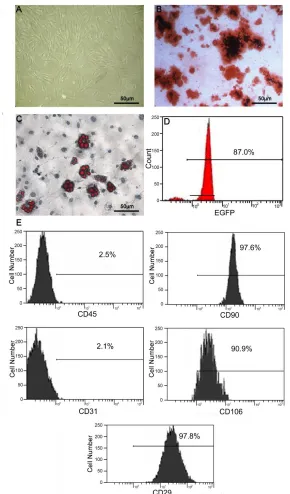
The separated and purified BMMSCs were shaped as elongated spindle, showing a vortex-like growth (Fig. 1A). Flow cytometry results showed that, BM-MSCs surface markers CD29 (98.8±3.4%), CD90 (98.4±3.8%) and CD106 (92.2±5.9%) were positive, while CD45 (3.0±3.1%) and CD31 (2.6±2.7%) were negative. Cell morphology and culture characteristics confirmed that the isolated and purified cells were rat BM-MSCs. For the differentiation assay, osteogenic medium induced mineral deposits as shown by alizarin red staining (Fig. 1B). After 28 days of adipogenesis induction, oil red-O-positive adipogenic cells were observed as indicated by red fat granules (Fig. 1C). EGFP fluorescence was strong 48 h after transfection. The rate of
EGFP expression was 87% prior to
transplantation (Fig. 1D).
3.2 Effect of Blood Glucose, Serum
Insulin and C-peptide after
Transplantation
Blood glucose levels from peripheral blood of rats were measured. BM-MSCs group rats caused an obvious decrease during 18 to 35 days after BM-MSCs transplantation (P<0.05). Although blood glucose were not restored normal rats levels after 35 days, blood glucose in rats of BM-MSCs group was significantly decreased compared with those of diabetic group (Fig. 2A). The levels of insulin and C-peptide were markedly elevated at 14 days after BM-MSCs group (P<0.01), compared with those of diabetic group which remained at a relatively high level (Fig. 2B, C). Glucose tolerance testing of normal controls showed the blood glucose level significantly increased 30 mins after glucose administration and peaked at 60 mins, followed by a decrease to the fasting level after 120 mins
(Figs. 3A, 3B, 3C). Glucose tolerance improved in rats that received BM-MSCs group during all the time after transplantation. The blood glucose level of recipient rats almost reached a normal level at 56 and 84 days after transplantation.
3.3 Morphology and Diameter of Islets
HE staining showed that no lymphocytes infiltrated into islets and the exocrine gland, suggesting immune rejection did not occur after BM-MSC group (Fig. 4A). Several residual islets with fibrosis were observed in diabetic group rats (Fig. 4B). Normal islets were observed in normal group rats (Fig. 4C). The number of islets was markedly increased in BM-MSCs group compared with that in diabetic group (P<0.05) (Fig. 4D), and the morphology and number of islets were similar to those in normal group. However, islet size was smaller than those in normal group rats (P<0.05) (Fig. 4E).
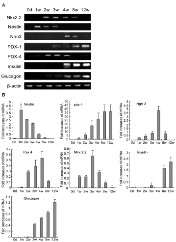
3.4 β Cell-related Gene Expression
In the pancreatic environment, the expression of genes related to β cell development in differentiated BM-MSCs was determined by RT-PCR (Fig. 5A) and quantitative real time-RT-PCR (Fig. 5B). Expression of seven genes associated with β cell development was extremely low or undetectable prior to transplantation. One to two weeks after transplantation, expression of Pdx-1, Ngn3, Nestin, Pax-4 and Nkx 2.2 was increased. Three weeks after transplantation, expression of Nkx2.2 and Ngn3 was up-regulated, and the K value was increased, which almost reached the maximal level. Four weeks after transplantation, Pax-4 expression reached a maximum. Eight weeks after transplantation, the expression of the
above genes was down-regulated and
accompanied by a decreased K value, except the expression of Pdx-1, insulin and glucagon gradually increased. Twelve weeks after transplantation, Nkx 2.2 expression was markedly decreased; expression of Ngn 3 and Pax-4 was undetectable. Pdx-1, insulin and glucagon expression reached a maximum. Nestin expression was up-regulated 1 week after transplantation, then down-regulated and was undetectable 12 weeks after transplantation.
3.5 Immunohistochemistry
co-expressed EGFP and insulin. Co-expression of EGFP and insulin was identified in a majority of cells (Fig. 6A). These results indicated that
BM-MSCs differentiated into β cells in a pancreatic environment.
Fig. 1. Characterization of isolated BM-MSCs. (A) Appearance of MSCs at passage 3. (B). Oil red staining of lipid droplet in adipocytes induced from MSCs. (C). Alizarin red staining of osteoblasts induced from MSCs. (D). The percent of EGFP-expressing MSCs. (Scale bar = 50 μm). (E) Flow cytometry analysis demonstrated a homogenous MSC polulation. As expected, MSCs were negative for reactivity to antigens CD31 and CD45, but positive for reactivity to
Fig. 2. Effects of BM-MSCs on blood glucose, insulin and C peptide level on STZ-induced diabetic SD rats. (A) Blood glucose levels in BM-MSC-transplanted diabetic SD rats decreased significantly compared with those of untreated diabetic SD rats after 18 days (P<0.05,P<0.001). (B) * P<0.05, **P<0.01, BM-MSCs-transplanted group vs diabetic group. (C) * P<0.05, **P<0.01,
Fig. 3. Blood glucose levels during glucose tolerance testing. (A) The curves of glucose tolerance testing 28 days after BM-MSCs transplantation. (B) Glucose tolerance testing after 56
Fig. 4. Pancreatic fresh frozen sections of BM-MSCs transplanted group were HE-stained. (A) BM-MSCs transplantation group.(B) diabetic group and (C) normal group. (Magnification ×400;
Scale bar = 50 μm) (D) The number of islets from each non-serial section of pancreas (three sections from each SD rat, mean±SEM). *P<0.05 by the Bonferroni-Dunn test. (E) Comparison
of islet size from transplantation group and normal group (20 islets from each group were measured for their diameter, mean±SEM). *P<0.05 by the Bonferroni-Dunn test. The analyses
were made after 84 days of transplantation
3.6 FISH
In pancreatic tissues with green fluorescence, Co-expressing SRY and insulin mRNA were observed, suggesting that BM-MSCs from male donors survived in pancreatic environment and differentiated into β cells (Fig. 6B).
3.7 DNA Ploidy Analysis of Transplanted EGFP-BM-MSCs
In pancreatic tissues from normal rats, G0/G1 phase accounted for 92.8±6.86% of cells and were diploid. The DNA content of these cells was 68±2.9%. S and G2 / M phases was 1.7±0.4% and 5.5±1.8%, respectively. S+G2/M phase accounted for 7.2±2.02% of cells and remained proliferation. The DNA content of these cells was 128 and tetraploid at 84 days after transplantation. The G1 phase of EGFP expressing cells was 89.8±13.56% and was diploid. The DNA content of these cells was 68±2.9%. S and G2/M phases were 2.1±0.62% and 8.1±2.12%, respectively. S+G2/M phase accounted for 10.2±2.46% and remained in the proliferation phase. The DNA content of these cells was 136±7.6%, and cells were tetraploid. Furthermore, cells were not polyploid or aneuploid.
4. DISCUSSION
Fig. 5. Expression of genes associated with beta-cell development after BM-MSCs transplantation was assessed by RT-PCR (A) and quantitative real-time PCR (B). Day 0 was measured prior to MSCs transplantation. Weeks 1, 2, 3, 4, 8 and 12 were detected after
Fig. 6. Immunofluorescence and FISH analysis in neo-islet cells of the BM-MSCs transplantation group. (A) Immunofluorescent localization of insulin and EGFP co-expression
in islet cells of recipient rats. The anti-insulin antibody was detected with a Texas Red-conjugated secondary antibody. Insulin and EGFP-positive cells are observed in islet sections.
Scale bar = 50 μm. (B) FISH analysis of the BM-MSCs-transplanted group. Nuclei were stained with DAPI (Blue). Hybridization signals of SRY (red) and insulin (green) were detected in the islet cells. Merged is image of DAPI, SRY and insulin. C. MSCs DNA Ploidy analysised by FCM.
Control: Normal pancreatic tissue cells, DNA analysis showed diploid. MSCs transplantation: BM-MSCs transplantation, cell cycle analysis of EGFP-positive cells revealed that transplanted
Studies have shown that the induced differentiation of stem cells is controlled by their intrinsic genetic program and surrounding microenvironment, which determines the fate of adult stem cells differentiation. Therefore, an efficient in vivo induction program must meet two conditions: (1) the gene expression and signal control for normal development of pancreatic β-cell; (2) the micro-environment for normal development of pancreatic β-cells. However, it is very difficult in vitro. The interaction between stem cells and their supportive microenvironment is critical for their maintenance, function and survival [25]. Two studies discussed the issue. One study indicated that pancreatic extract could stimulate mesenchymal stem cells (MSC) differentiating into insulin-producing cells and increase insulin secretion [26]. Wang et al. [27] reconstructed ‘personalized’ islets by using adult stem cells combined with microfabrication technology. In the present study, we transplanted male enhanced green fluorescence protein (EGFP)-expressing BM-MSCs into the pancreas of female diabetic SD rats by direct injection. It showed that transplantation of MSCs into diabetic rats improved glycemic control and restored normo-glycemia 35 days after transplantation. Insulin and C-peptide were significantly elevated compared to non-transplanted controls. Number of islets was increased in transplanted pancreases compared to controls. Expression of genes associated with beta-cell development increased after MSCs transplantation. We found some newly formed islets that were morphologically similar to normal islets in rats’ pancreas of transplantation group. More importantly, some of them expressed insulin, as well as EGFP, indicating some of the neo-islet cells were derived from transplanted BM-MSCs. 56 days after transplantation EGFP was still expressed in the plasma, providing useful trace evidence of our transplanted BM-MSCs location, existence and differentiation in diabetic pancreas. We presume that some transplanted MSCs contribute to β-cells under a pancreatic microenvironment. Theoretically, this method could overcome the use of inducer and low differentiation rates in vitro.
After BM-MSCs transplantation, cell cycle analysis of EGFP-positive cells revealed that transplanted cells were diploid and a few cells were tetraploid. None of the cells were polyploid or aneuploid, which excluded the possibility of cell fusion. Co-expression of EGFP and insulin was observed, but only with regular microscopy, not with confocal microscopy. Expression of
insulin was found in MSC-derived cells, as shown by FISH for SRY and insulin mRNA. On the other hand, although cells co-expressing EGFP and insulin were present in STZ-damaged pancreatic tissue, their low frequency suggested that the cells did not, in themselves, functionally rescue the recipients. It is very important that BM-MSCs activate the “self-repair” of endogenous stem cell, and provide critical factors for tissue regeneration [28], since complex and multiplex factors are involved in pancreatic regeneration. The rapidity and continuity of the pancreatic regeneration process suggests that endogenous pancreatic stem cells (PSCs) may mediate the restorative process through endothelial, mesenchymal and endodermal interactions [29], because PSCs which can self-renew and differentiate into pancreatic islets are found to exist around the acini, ducts, islets and etc [30,31]. However, we could only do some related reasoning and the molecular mechanisms of how MSCs play the role of lowering blood sugar in vivo are still not clear. Further tests are needed to find direct evidence.
5. CONCLUSION
The authors transplanted BM-MSCs derived from male SD rats into diabetic female SD rats and showed that MSCs survived, and expressed insulin as well as genes associated with beta-cell development. Glycemic control was improved and reached normo-glycemia after 35 days in several animals. BM-MSCs-derived new islets were verified in pancreatic tissue of transplantation group by histology. Co-expression of EGFP and insulin was observed and expression of insulin was found in BM-MSCs -derived cells, as shown by FISH for SRY and insulin mRNA. These results show that BM-MSCs can differentiate into beta cell in the pancreatic microenvironment without cell fusion.
CONSENT
All authors declare that ‘written informed consent was obtained from the patient (or other approved parties) for publication of this case report and accompanying images.
ETHICAL APPROVAL
standards laid down in the 1964 Declaration of Helsinki.
COMPETING INTERESTS
Authors have declared that no competing interests exist.
REFERENCES
1. Shapiro AM, Lakey JR, Ryan EA, Korbutt GS, Toth E, Warnock GL, et al. Islet transplantation in seven patients with type 1 diabetes mellitus using a glucocorticoid-free immunosuppressive regimen. N Engl J Med. 2000;343:230-238.
2. Ichii H, Ricordi C. Current status of islet cell transplantation. J Hepatobiliary Pancreat Surg. 2009;16:101-112.
3. Kroon E, Martinson LA, Kadoya K, Bang AG, Kelly OG, Eliazer S, et al. Pancreatic endoderm derived from human embryonic stem cells generates glucose-responsive insulin-secreting cells in vivo. Nat Biotechnol. 2008;26:443-452.
4. Ramiya VK, Maraist M, Arfors KE, Schatz DA, Peck AB, Cornelius JG. Reversal of insulin-dependent diabetes using islets generated In vitro from pancreatic stem cells. Nat Med. 2000;6:278-82.
5. Yang L, Li S, Hatch H, Ahrens K, Cornelius JG, Petersen BE, et al. In vitro trans-differentiation of adult hepatic stem cells into pancreatic endocrine hormone-producing cells.Proc Natl Acad Sci USA. 2002;99:8078-8083.
6. Xie QP, Huang H, Xu B, Dong X, Gao SL, Zhang B, et al. Human bone marrow mesenchymal stem cells differentiate into insulin-producing cells upon micro environmental manipulation in vitro. Differentiation. 2009;77:483-491.
7. Jiang J, Lv Z, Gu Y, Li J, Xu L, Xu W, et al. Adult rat mesenchymal stem cells differentiate into neuronal-like phenotype and express a variety of neuro-regulatory molecules in vitro. Neurosci Res. 2010; 66:46-52.
8. Tang DQ, Cao LZ, Burkhardt BR, Xia CQ, Litherland SA, Atkinson MA, et al. In vivo
and in vitro characterization of insulin-producing cells obtained from murine bone marrow. Diabetes. 2004;53:1721-1732. 9. Ianus A, Holz GG, Theise ND, Hussain MA.
In vivo derivation of glucose-competent pancreatic endocrine cells from bone marrow without evidence of cell fusion. J
Clin Invest. 2003;111:843-850.
10. Zanini C, Bruno S, Mandili G, Baci D, Cerutti F, Cenacchi G, et al. Differentiation of mesenchymal stem cells derived from pancreatic islets and bone marrow into islet-like cell phenotype. PLoS One. 2011; 6:e28175.
11. Oh SH, Muzzonigro TM, Bae SH, LaPlante JM, Hatch HM, Petersen BE. Adult bone marrow-derived cells trans-differentiating into insulin-producing cells for the treatment of type I diabetes. Lab Invest. 2004;84:607-617.
12. Ai C, Todorov I, Slovak ML, Digiusto D, Forman SJ, Shih CC. Human marrow-derived mesodermal progenitor cells generate insulin-secreting islet-like clusters
In vivo. Stem Cells Dev. 2007;16:757-770. 13. Putnam AJ. The Instructive Role of the
Vasculature in Stem Cell Niches. Biomater Sci. 2014;2:1562-1573.
14. Gregory CA, Ylostalo J, Prockop DJ. Adult bone marrow stem / progenitor cells (MSCs) are preconditioned by micro environmental niches in culture: A two-stage hypothesis for regulation of MSC fate. Sci STKE. 2005;2005:pe37.
15. Taneera J, Rosengren A, Renstrom E, Nygren JM, Serup P, Rorsman P, et al. Failure of transplanted bone marrow cells to adopt a pancreatic beta-cell fate. Diabetes. 2006;55:290-6.
16. Choi JB, Uchino H, Azuma K, Iwashita N, Tanaka Y, Mochizuki H, et al. Little evidence of transdifferentiation of bone marrow-derived cells into pancreatic beta cells. Diabetologia. 2003;46:1366-74. 17. Li L, Li F, Qi H, Feng G, Yuan K, Deng H,
et al. Coexpression of Pdx1 and betacellulin in mesenchymal stem cells could promote the differentiation of nestin-positive epithelium-like progenitors and pancreatic islet-like spheroids. Stem Cells Dev. 2008;17:815-23.
18. Peister A, Mellad JA, Larson BL, Hall BM, Gibson LF, Prockop DJ. Adult stem cells from bone marrow (MSCs) isolated from different strains o f inbred mice vary in surface epitopes, rates of proliferation, and differentiation potential. Blood. 2004;103: 1662-8.
19. Hess D, Li L, Martin M, Sakano S, Hill D, Strutt B, et al. Bone marrow-derived stem cells initiate pancreatic regeneration. Nat Biotechnol. 2003;21:763-70.
cells cooperate with bone marrow cells in therapy of diabetes. Stem Cells. 2008;26: 244-53.
21. Terada N, Hamazaki T, Oka M, Hoki M, Mastalerz DM, Nakano Y, et al. Bone marrow cells adopt the phenotype of other cells by spontaneous cell fusion. Nature. 2002;416:542-5.
22. Wang X, Willenbring H, Akkari Y, Torimaru Y, Foster M, Al-Dhalimy M, et al. Cell fusion is the principal source of bone-marrow-derived hepatocytes. Nature. 2003; 422:897-901.
23. Wada MR, Inagawa-Ogashiwa M, Shimizu S, Yasumoto S, Hashimoto N. Generation of different fates from multipotent muscle stem cells. Development. 2002;129:2987-95.
24. Alison MR, Poulsom R, Otto WR, Vig P, Brittan M, Direkze NC, et al. Plastic adult stem cells: will they graduate from the school of hard knocks? J Cell Sci. 2003; 116:599-603.
25. Zarrabi M, Mousavi SH, Abroun S, Sadeghi B. Potential uses for cord blood mesenchymal stem cells. Cell J. 2014;15: 274-81.
26. Xie H, Wang Y, Zhang H, Qi H, Zhou H, Li FR. Role of injured pancreatic extract
promotes bone marrow-derived
mesenchymal stem cells efficiently differentiate into insulin-producing cells. PLoS One. 2013;8:e76056..
27. Wang J, Song LJ, Gerber DA, Fair JH, Rice L, LaPaglia M, et al. A model utilizing adult murine stem cells for creation of personalized islets for transplantation. Transplant Proc. 2004;36:1188-90.
28. Attali M, Stetsyuk V, Basmaciogullari A, Aiello V, Zanta-Boussif MA, Duvillie B, et al. Control of beta-cell differentiation by the pancreatic mesenchyme. Diabetes. 2007; 56:1248-58.
29. Edsbagge J, Johansson JK, Esni F, Luo Y, Radice GL, Semb H. Vascular function and
sphingosine-1-phosphate regulate
development of the dorsal pancreatic mesenchyme. Development. 2005;132: 1085-92.
30. Wang YJ, Bailey JM, Rovira M, Leach SD. Sphere-forming assays for assessment of benign and malignant pancreatic stem cells. Methods Mol Biol. 2013;980:281-90. 31. Teng C, Guo Y, Zhang H, Zhang H, Ding M,
Deng H. Identification and characterization of label-retaining cells in mouse pancreas. Differentiation. 2007;75:702-12.
© 2015 Deng et al.; This is an Open Access article distributed under the terms of the Creative Commons Attribution License
(http://creativecommons.org/licenses/by/4.0), which permits unrestricted use, distribution, and reproduction in any medium,
provided the original work is properly cited.
Peer-review history:
The peer review history for this paper can be accessed here: