_____________________________________________________________________________________________________
*Corresponding author: E-mail: [email protected];
www.sciencedomain.org
Validation of CK-19 and HBME-1 Expression Pattern
in FNACs to Supplement Morphological Evaluation
for Preoperative Diagnosis of Thyroid Lesions
Shanone C. Pereira
1, V. T. Jissa
2, Jestin K. George
1, Shaji Thomas
2and K. Sujathan
1*1Divisions of Cancer Research and Surgical Oncology, Regional Cancer Centre, Post Box No: 2417,
Medical College PO, Thiruvananthapuram, Kerala, India. 2
Achutha Menon Centre for Health Science Studies, Sree Chitra Tirunal Institute of Medical Sciences and Technology, Thiruvananthapuram, Kerala, India.
Authors’ contributions
This work was carried out in collaboration between all authors. Author SCP carried out the experimental work, and wrote the first draft of the manuscript and the literature searches. Author JKG managed to perform a portion of the experimental work in the absence of author SCP. Statistical analyses of the study were carried out by authors VTJ and SCP. Author ST was responsible for recruiting the patients and providing tissue samples and author KS designed the study, wrote the protocol, supervised the experimental process, and evaluated the results, analysis and interpretation of data. All authors read and approved the final manuscript.
Article Information
DOI: 10.9734/BJMMR/2016/23061 Editor(s): (1) E. Umit Bagriacik, Department of Immunology, Gazi University, Turkey. Reviewers: (1) Anonymous, Paris 7 University, France. (2)Padmavathi Devi Chaganti, Guntur Medical College, Guntur, India. (3)Talia Missen Tremori, State University of Sao Paulo, Brazil. Complete Peer review History:http://sciencedomain.org/review-history/12998
Received 12th November 2015 Accepted 30th December 2015 Published 16th January 2016
ABSTRACT
Aim: Fine Needle Aspiration Cytology is the cost effective, quick and minimally invasive method for the initial evaluation of thyroid nodule. But it poses a diagnostic challenge in differentiating benign follicular adenoma from follicular carcinoma and follicular variant of papillary carcinoma as they have similar cytological appearance. Present study focused on the identification of a biological marker for the differential diagnosis of thyroid malignancy in indeterminate cases.
Place and Duration of the Study: Division of cancer Research, Regional Cancer Centre, Kerala, India, between August 2009 to September 2014.
Methodology: Immunohistochemistry was performed using Ret (Rearranged during transfection), Hector Battifora Mesothelial Cell Antigen-1 (HBME-1), Cytokeratin-19 (CK-19), Keratan sulphate (KS), Thyroid peroxidase (TPO), Estrogen receptor (ER) and Progesterone receptor (PR) on cell block prepared from FNA material and corresponding tissue sections on 153 samples. Western blot analysis of ER and PR was performed in surgically excised fresh tissue specimens.
Results: The present study found that HBME-1 is highly significant (P < .001) for the differential diagnosis with a diagnostic accuracy of 95.96%. Ret immunostaining may serve as good indicator of PTC whereas its sensitivity is very low in other lesions. The diagnostic accuracy of CK-19 was 90.91%. Intense positive staining of TPO was noted in majority of follicular epithelial cells of benign lesions (42.42%). Females had an increased prevalence in our study population so we examined the estrogen and progesterone receptors status in thyroid lesions. The specificity of KS, ER and PR are very low for the differentiation of adenoma from carcinomas of the thyroid.
Conclusion: Present study found that the combination of two markers may give a more accurate way in the differentiation of thyroid nodules in problematic cases. So we suggest morphological evaluation along with immunocytochemistry of HBME-1 and CK19 can help the differential diagnosis of thyroid lesions in FNAs.
Keywords: Biomarkers; HBME-1; CK-19; RET; KS; TPO; thyroid malignancy.
1. INTRODUCTION
Thyroid cancer forms the largest entity in a group of endocrine tumors with an increased incidence rate worldwide in the last three decades [1]. Thyroid tumors often present as palpable thyroid nodules, but majority of them are benign [2]. The primary modality for the preoperative diagnosis of thyroid nodule is Fine Needle Aspiration Cytology (FNAC). Classical papillary thyroid carcinomas (PTC) and anaplastic thyroid carcinomas (ATC) can easily be diagnosed by morphological evaluation itself. But benign follicular adenoma (FTA) cannot be distinguished from follicular carcinoma (FTC) and follicular variant of papillary carcinoma (FVPC) because of their similar cytological appearance [3]. Most experienced pathologist may encounter a diagnostic challenge and cytologically interpreted these follicular patterned lesions as Follicular neoplasm which may results in diagnostic dilemma for the clinicians. To overcome this problem, it is important to include an ancillary technique that can improve the standard morphologic assessment both in surgical specimens and in cytological samples [4]. Currently several potential biomarkers are being investigated in conjunction with FNACs to allow the precise diagnosis of indeterminate cases of the thyroid. Among them receptor signalling molecule RET (Rearranged during transfection) is found to be a promising one. In the pathogenesis of thyroid cancer, ret/PTC oncogene activate effectors that signal along the mitogen activated protein kinase (MAPK) pathway and it is an unique feature of papillary
nodules and in cells blocks obtained in FNA to see whether these protein expression either singly or in combination can be used as a diagnostic marker for the differentiation of PTC, FTC, FVPC from FTA. In our study population females had an increased prevalence with a male to female ratio of 1:3.So we evaluated ER and PR status of these samples. In order to confirm the immunoexpressions we performed western blot analysis of ER and PR in different lesions of thyroid.
2. MATERIALS AND METHODS
2.1 Tissue Specimens and FNACs
153 samples of thyroid from both cell blocks of FNA material of biopsy proved lesions and the corresponding tissue block from patients who had undergone surgery without any prior treatment for thyroid nodules were the study subjects. Written informed consent was obtained from all the patients and the work protocol was approved by the Institutional Review Board as well as Ethical Committee of Regional Cancer Centre (RCC), Thiruvananthapuram, Kerala. The samples included 81 papillary carcinoma, 2 follicular carcinoma, 29 follicular variant of papillary carcinoma, 28 follicular adenoma and 13 thyroiditis. Oncocytic tumors couldn’t be included in the study as we couldn’t get sufficient number of satisfactory samples and also immunocytochemistry do not seems to have much relevance in identification of these tumors. FNA was performed in the Division of Pathology, RCC by using 24 gauge needle and 5 ml syringe. The aspirates were immediately fixed in acetic acid alcohol formalin (AAF) and centrifuged at 10,000 rpm for 10 minutes and kept at room temperature overnight in slanting position. The cell button were wrapped in lens paper and processed for histopathology [7]. The information regarding patient was collected from medical records. Standard haematoxylin and eosin (H&E) staining were performed and the tissue sections were evaluated for the presence of tumor cells. Tissue sections with minimum five groups of cells were selected for immunohistochemistry. Surgically excised fresh materials obtained from Division of Surgical Oncology, RCC were used for western blot analysis.
2.2 Immunohistochemistry
5 µm thickness formalin fixed paraffin embedded (FFPE) tissue sections were mounted onto APES
(3-aminopropyl triethoxysilane) coated slides. After standard protocol of de-paraffinization and rehydration in descending grades of alcohol, heat induced antigen retrieval was performed using 10 mM citrate buffer (pH 6.0) for two consecutive cycles of 10 and 5 minutes each at high temperature. Endogenous peroxidase activity was blocked by using blocking buffer and rinsed in TBS (Tris-buffered saline) twice. Then the slides were incubated with protein block. Mouse monoclonal antibody against Ret, HBME-1, CK-19, TPO and KS were purchased from Santa Cruz Biotechnology Inc, USA. Antibodies against Estrogen and progesterone receptors were obtained from Novocastra Laboratories Ltd, UK. The primary antibodies were used at a dilution of 1:150 for Ret, HBME-1, CK-19, TPO and KS, and 1:40 for ER and PR. Novo Link Polymer Detection system (Novocastra) was used as secondary system. The reaction was visualized using DAB (3,3'-Diaminobenzidine) as chromogen followed by counter staining with haematoxylin, the slides were dehydrated, cleared and mounted in DPX. Positive controls were included in every batch as per the manufactures instruction. Negative controls were incubated with TBS in the place of Primary antibody.
2.3 Protein Isolation and Western Blotting
Tissue lysate was prepared with lysis buffer containing 1M Tris (pH7.4), 0.5M EDTA, triton X-100, 5M NaCl, glycerol and PMSF. Sample lysate was incubated in ice for 1 hour and centrifuged at 12,000 rpm for 20 min at 40C and quantified using Bradford assay kit (Sigma, USA). Protein was separated on 12% SDS-PAGE (sodium dodecyl sulphate- polyacrylamide gel electrophoresis) and blotted onto nitrocellulose membrane. The blots were incubated with mouse monoclonal antibody against ER and PR. After washing with TBST, the membrane was subjected to react with biotinylated anti-mouse immunoglobulin for 30min. Immunoreactivity were developed with enhanced chemiluminescence (ECL) kit (Proteo Qwest, Sigma, USA) and analysed in FluorChem-M multiplex fluorescent imaging system.
3. RESULTS
Age distribution of the study population revealed that majority of the participants was in the age group 31- 40 years. 75% of the participants were women with a male to female ratio of 1:3. The mean age of papillary carcinoma as well as follicular variant of papillary carcinoma was 40 years. Post-operative histopathology diagnosis of patients who were diagnosed as Follicular lesion/ follicular neoplasm in aspiration cytology revealed that 33.3% patients to had papillary carcinoma, 39.3% patients had follicular variant of papillary carcinoma, 12.1% had follicular adenoma and 15.1% patients had thyroiditis. The sensitivity of aspiration cytology for the differential diagnosis of PTC and FVPC was 71.67% and 41.03% respectively and specificity
was 80.39% and 72.46% respectively (Fig. 1). The diagnostic accuracy of PTC was 75.68%, whereas it varied drastically in other lesions of the thyroid. The sensitivity of the aspiration cytology was considerably low in the differentiation of FVPC, FTA and thyroiditis when compared with PTC.
A strong positive staining of Ret oncoprotein was noted mainly in the cytoplasm of papillary carcinoma in both cytology cell block (Fig. 2A) as well as in tissue sections with mean H-score of 152.50 and 169.51 respectively. In the case of follicular carcinoma intense expression of Ret oncoprotein was observed. 58 out of 61 follicular variant of papillary carcinoma showed moderate
to intense positive staining for this protein (Fig. 2B). Majority of the FTA showed focal
expression of this protein with a mean H-score of 24.67 in cytology samples and 42.14 in histology sections. Only 2 out of 13 samples of thyroiditis expressed Ret, but the staining was confined to less than 30% of cells. In some cases intense expression of this protein was found in macrophages and hurthle cells associated with thyroiditis.
Ret was found to be highly significant in the differential diagnosis of PTC from FVPC (P < .001) in both histology and cytology
specimens. Ret exhibited a moderate positive correlation with HBME-1 (r=.433, P < .001), CK-19(r=.605 P < .001) and KS (r=.597, P < .001) in histology specimens. The sensitivity of Ret for differentiating adenoma from carcinoma was 70.2% and 75.4% in cytology specimens and its surgical counterparts. The diagnostic accuracy of this protein (74.75%) in cytology specimen was low when compared to other markers.
Fig. 1. Diagnostic accuracy of cytology reports by taking histopathology as gold standard
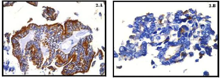
Fig. 2. Immunostaining of Ret in papillary thyroid carcinoma and in its variant A. FNA cell block of PTC showing strong and diffuse positivity of Ret (40X)
B. Weak immunopositivity of Ret in FVPC in cytology cell block (40X)
HBME-1 was positive in 96.2% of PTC (Fig. 3A) and 89.6% of FVPC. The staining pattern was membrane and cytoplasmic. The mean H-score of PTC and FVPC was 246.83 and 205.83 in cytology call block and histology sections respectively. Most cases of thyroiditis were mild and 3.5% cases of FTA were moderate positive for this protein (Fig. 3B). Normal thyroid follicular cells were negative. HBME-1 was found to have a diagnostic accuracy of 95.96% for the differential diagnosis of adenoma from carcinoma. Moreover, HBME-1 was found to be highly significant for differentiating FVPC from FTA (P < .001) in both cytology and histology counterparts. This protein showed a moderate positive correlation with the expression pattern of CK-19 (r=.600, P < .001) and KS (r=.603, P < .001) in FNA cell block. Unlike HBME-1, CK-19 showed an intense expression in all 77 (95%) and 74 (91.35%) PTC in both histology and cytology cell block. The staining pattern was predominantly cytoplasmic. Moderate to intense immunoreactivity was noted in FVPC with a mean H-score of 210 in cytology cell block (Fig. 4A). Only focal expression of this protein was observed in follicular adenoma (Fig. 4B) with mean H- score of 69.64. The staining intensity was very weak and diffuse in thyroiditis. CK-19 was found to be highly significant (P < .001) for the differentiation of thyroid neoplasm. The diagnostic accuracy of CK-19 for the differentiation of adenoma from carcinoma was 90.91% and 94.93% in cytology and histology samples respectively.
Keratan sulphate imuunostaining was evenly distributed along the cytoplasm and membrane of papillary carcinoma (Fig. 5) and in its variants. Very mild staining of KS was noted in follicular thyroid adenoma both in cytology cell block and
in histology sections. Only 15.38% of cytology samples of thyroiditis were positive for this protein. KS was found to have a key role in the differentiation of adenomas from carcinomas of the thyroid (P < .001), but the specificity of KS as a diagnostic marker for the differentiation of thyroid lesions was low (66.6%) when compared with HBME-1 and CK-19 in FNA cell block. The diagnostic accuracy of KS in the differentiation of FVPC from adenoma in cytology cell block and histology specimen was 84.62% and 84.21% respectively. Percentage of positivity and mean H-score of HBME-1, CK-19, KS and Ret immunostaining in Cytology cell block and corresponding surgical specimen in different lesions is summarized in Table 1.
Moderate expression of TPO protein was noted in 85% of PTC with a mean H-score of 165 in cytology cell block (Table 2). Intense positive staining of TPO was noted in majority of follicular epithelial cells from benign lesions (42.42%) (Fig. 6), while more than 45% of tumor cells of FVPC were negative for this protein. The diagnostic accuracy of TPO was only 71% in the differential diagnosis of thyroid malignancies. Correlation of immunostaining of each marker in cytology specimens and its corresponding histology counterparts is summarized in Table 3.
participants. None of the FTA or thyroiditis was positive for this protein. The sensitivity of ER is very low in differentiating follicular lesions. The diagnostic accuracy of this protein for the differential diagnosis of thyroid lesion in cytology
cell block and histology samples were 28% and 40.35% respectively. PR showed positivity only in 27% of PTC, whereas 82% of FVPC (Fig. 8) showed immunoreactivity for this protein. 12 out of 28 FTA were immunodecorated with
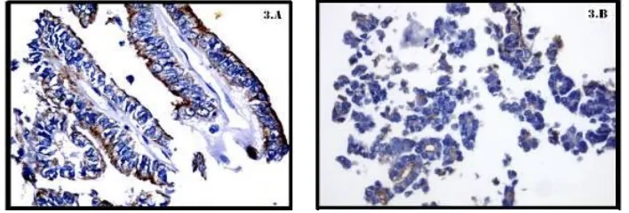
Fig. 3.Immunostaining of HBME-1 in papillary thyroid carcinoma and follicular thyroid adenoma
A. PTC showing strong and diffuse positivity of HBME-1 in FNA cell block (40X) B. HBME-1 was negative for FTA in cell block obtained from FNA (40X)
Fig. 4. Varying levels of immunostaining of CK-19 protein in different lesion of thyroid A. Intense immuno localisation of CK-19 in cytology section of FVPC (40X)
B. CK-19 showed very weak expression in FTA (40X)
Fig. 5. Immunostaining of KS in PTC Fig. 6. Immunostaining of TPO in FTA
Table 1. Percentage of positivity and mean H-score of HBME-1, CK-19, KS and Ret immunostaining in cytology cell block and corresponding surgical specimen in different
lesions of thyroid
Lesion (n)
Cytology cell block Histology sections Cytology cell block Histology sections
HBME-1 CK-19
Positive cases (%)
Mean Positive cases (%)
Mean Positive cases (%)
Mean Positive cases (%)
Mean
PTC (81) 78 (96.29) 246.83 78 (96.2) 261.11 74 (91.35) 240.00 77 (95) 257.78
FVPC (29) 27 (93.10) 205.83 26 (89.6) 206.55 24 (82.75) 197.92 27 (93.1) 210.00
FTA (28) 0 (0) 42.67 1 (3.5) 48.21 0 (0) 69.33 1 (3.5) 69.64
Thyroiditis (13) 0 (0) 40 0 (0) 48.46 2 (15.38) 61.25 4 (30.7) 78.46
KS Ret
PTC (81) 77 (95.06) 227.50 77 (95) 242.22 69 (85.18) 152.50 70 (86) 169.51
FVPC (29) 28 (95.55) 193.75 28 (96.5) 188.28 10 (34.48) 80.00 13 (44) 88.28
FTA (28) 9 (32.14) 42.67 8 (28.5) 53.57 0 (0) 24.67 0 (0) 42.14
Thyroiditis (13) 2 (15.38) 70 4 (30.7) 85.38 2 (15.38) 50 1 (7.6) 50.00
Table 2. Percentage of positivity and mean H-score of TPO immunostaining in cytology cell block and corresponding surgical specimen in different lesions of thyroid
Lesion (n) TPO
Cytology cell block Histology sections Positive cases (%) Mean Positive cases (%) Mean
PTC (81) 68 (83.95) 165.00 70 (86) 178.89
FVPC (29) 16 (55.17) 107.08 16 (55) 107.93
FTA (28) 13 (46.42) 102.67 17 (60.7) 121.07
Thyroiditis (13) 10 (76.92) 211.25 12 (92.3) 234.62
this protein with moderate intensity. The sensitivity of PR for the differential diagnosis of thyroid malignancy in histology cell block was 61.7% with diagnostic accuracy of 67.24%. Whereas the sensitivity (37.5%) and diagnostic accuracy (48%) of PR in cytology cell block was very low (Fig. 7).
Western blot analysis showed similar pattern of expression of ER in the case of FVPC and its adjacent normal epithelium (Fig. 9). Intense expression of PR was observed in FVPC when compared with normal epithelium, whereas papillary carcinoma specimens were negative for both proteins.
Table 3. Correlation of immunostaining of each marker in cytology specimens and its corresponding histology counterparts
HBME-1 cytology
CK-19 cytology
KS cytology Ret cytology TPO cytology
HBME-1 histology .919** .604** .622** .406** .051
.000 .000 .000 .000 .592
153 153 153 153 153
CK 19 histology .692** .774** .580** .510** .175
.000 .000 .000 .000 .066
153 153 153 153 153
KS histology .649** .592** .867** .585** .051
.000 .000 .000 .000 .597
153 153 153 153 153
Ret histology .489** .489** .554** .816** .201*
.000 .000 .000 .000 .035
153 153 153 153 153
TPO histology -.024 .091 .150 .210* .486**
.804 .341 .117 .027 .000
153 153 153 153 153
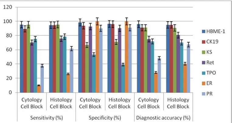
Fig. 7. Sensitivity, specificity and diagnostic accuracy of HBME-1, CK-19, KS, Ret, TPO, ER and PR staining in surgical specimens and corresponding cytology cell block
Fig. 8. Immunostaining of PR in FVPC
Intense immuno expression of PR in histology section of FVPC (40X)
Fig. 9. Western blot of ER and PR
Western blot analysis of ER and PR in FVPC and adjacent normal epithelium
4. DISCUSSION
For the workup of thyroid nodules, fine needle aspiration and subsequent cytopathological analysis are crucial steps. But in certain pathologic situations cytopathological report is ambiguous and surgery is the only option to establish a definite diagnosis in the differentiation of thyroid nodules. The reported accuracy of FNAC in the diagnosis of thyroid lesions ranges from 70% to 97% with false negative rates ranging from 1 to 6% [8]. The diagnostic accuracy of our study series also varies from 61% to 76% for the differentiation of thyroid nodules, whereas the negative predictive value
ranges from 47% to 71%. Hence it is important to incorporate an ancillary technique which could improve the standard morphologic assessment both in surgical specimens and in cytological samples.
Ret rearrangement in FNAC specimen by immunohistochemistry as a good diagnostic tool for the differentiation of thyroid nodules [10,11]. Cheung et al. demonstrated immunoexpression of Ret in 78% of PTC, 63% of FVPC, 57% of Hurthle cell carcinoma and all the benign nodule were negative for this protein [12]. Our study also documented a higher immunoexpression of this protein in PTC (85%) when compared to other lesions. Saleh et al. [13] suggested that Ret protein is significantly more expressed in malignant tumors than in benign lesions. 18 out of 20 cases of PTC they examined showed intense staining of Ret protein. 91.6% and 72.7% of cases of FVPC and FTC showed Ret expression but only 45.6% of staining was observed in follicular adenoma. Present study also documented a similar observation. In the case of adenomas the Ret protein expression was very low or negligible. But in the case of thyroiditis we got a moderate expression in focal
areas and the mean H-score was higher when compared to follicular adenoma. Ret
immunochemistry is useful for the differentiation of PTC from adenomas but it has limited value in the differentiation of other lesion due to its intense expression in macrophages and hurthle cells.
Studies suggested that HBME-1 is useful in differentiating follicular derived malignant tumors of thyroid in surgical samples. Whereas Mokhari et al. [14] suggested that negative staining for HBME-1 does not rule out malignancy. Saggiorato and co-workers evaluated HBME-1 expression in different lesions of thyroid and suggested that this protein shows a higher accuracy in the differentiation of follicular lesion when compared with other markers [15]. Our study also obtained a similar result. The diagnostic accuracy of HBME-1 was 95.96% and seems to be highly significant in the differential diagnosis of thyroid malignancies. CK-19 has been reported to express strongly and diffusely in PTC, but it is heterogeneously expressed in follicular carcinoma and absent or focally expressed in follicular adenoma in both cytological and surgical samples of thyroid tumors [16,17]. Sahoo et al. [18] reported that CK 19 immunoreactivity is not specific for PTC, although extend and intensity of staining are considerably greater in PTC than in follicular adenoma. The reported sensitivity of CK 91 is 92% and specificity is 97%, while using as a single marker in thyroid malignancies [19]. Cochand-Priollet et al. [20] have reported that immunocytochemistry of thyroid FNAs with
suggested that the effect of estrogen on thyroid gland may be an indirect mechanism. Current study was not able to detect greater expression of ER and PR in thyroid tumors than in normal tissue. This arise the question on the significance of expression of ER and PR in the differential diagnosis of thyroid nodules. Chen et al. [26] has reported that immunohistochemical evaluation of ERα and PR expression in patients with papillary carcinoma may aid in the prediction of the prognosis. The current study couldn’t find such an observation.
5. CONCLUSION
The present study has validated HBME-1 and CK-19 as highly significant markers for the differential diagnosis of thyroid malignancies. The combination of HBME-1 and CK-19 were able to provide a good diagnostic accuracy (> 95%) with high sensitivity and specificity which can be followed in the cytopathology practice. Ret immunostaining may serve as good indicator of PTC whereas its sensitivity and negative predictive value is very low in other lesions.
CONSENT
All authors declare that “written informed consent” was obtained from the patients (or other approved parties) for participation in the study of this publication.
ETHICAL APPROVAL
The authors hereby declare that the work protocol has been approved by the institution ethical committee, HCE No. 6/2009.
ACKNOWLEDGEMENTS
This work was supported by grants from Department of Biotechnology, Government of India.
COMPETING INTERESTS
Authors have declared that no competing interests exist.
REFERENCES
1. Pellegriti G, Frasca F, Regalbuto C, Squatrito S, Vigneri R. Worldwide increasing incidence of thyroid cancer: Update on epidemiology and risk factors. J Cancer Epidemiol; 2013.
Available:http://dx.doi.org/10.1155/2013/96 5212
2. Mazzaferri EL, Kloos RT. Current approaches to primary therapy for papillary and follicular thyroid cancer. J Clin Endocrinol Metab. 2001;86:1447-63. 3. Freitas BC, Cerutti JM. Genetic markers
differentiating follicular thyroid carcinoma from benign lesions. Mol Cell Endocrinol. 2010;321:77-85.
4. Fischer S, Asa SL. Application of immunohistochemistry to thyroid
neoplasms. Arch Pathol Lab Med. 2008;132:359-72.
5. Cahill S, Symth P, Finn SP, Denning K, Flavin R, O’Regan EM, Li J, Potratz A, Guenther SM, Henfrey R, O’Leary JJ, Sheils O. Effect of ret/PTC 1 rearrangement on transcription and post-transcriptional regulation in a papillary thyroid carcinoma model. Mol Cancer. 2006;5:70.
DOI: 10.1186/1476-4598-5-70
6. Segev DL, Umbricht C, Zeiger MA. Molecular pathogenesis of thyroid cancer. Surg Oncol. 2003;12:69-90.
7. Sujathan K, Pillai KR, Kannan S, Chandralekha B, Mathew A, Nair MK. Cytodiagnosis of serous effusions: A combined approach to morphological features in Pap and MGG stained smears and a modified cellblock technique. J Cytol. 2000;17(2):89-95.
8. Greenspan FS. The role of fine needle aspiration biopsy in the management of palpable thyroid nodules. Am J Clin Pathol. 1997;108:26-30.
9. Patel KN, Singh B. Genetic considerations in thyroid cancer. Cancer Control. 2006;13(2):111-18.
10. Salvatore G, Giannini R, Faviana P, Caleo A, Migliaccio I, Fagin JA, Nikiforov YE, Troncone G, Palombini L, Basolo F, Santoro. Analysis of BRAF point mutation and RET/PTC rearrangement refines the fine needle aspiration diagnosis of papillary thyroid carcinoma. J Clin Endocrinol Metab. 2004;89:5175-80. 11. Sapio MR, Posca D, Raggioli A, Guerra A,
Marotta V, Motta M, Limone PP, Troncone G, Caleo A, Rossi G, Fenzi G, Vitale M. Detection of RET/PTC, TRK and BRAF
mutations in preoperative diagnosis of thyroid nodules with intermediate
12. Cheung CC, Ezzat S, Freeman JL, Rosen IB, Asa SL. Immunohistochemical diagnosis of papillary thyroid carcinoma. Mod Pathol. 2001;14(4):338-42.
13. Saleh HA, Jin B, Barnwell J, Alzohaili O. Utility of immunohistochemical markers in differentiating benign from malignant follicularderived thyroid nodules. Diagn Pathol. 2010;5:9.
14. Mokhtari M, Sadeghi M, Talebi A. Monoclonal antibody HBME-1 usefulness in differentiation of benign neoplasm and differentiated thyroid carcinoma. Acta Medica Iranica, 2005;43(2):85-88.
15. Saggiorato E, De Pompa R, Volante M, Cappia S, Arecco F, Dei Tos AP, Orlandi F and Papotti M. Characterization of thyroid ‘follicular neoplasms’ in fine needle aspiration cytological specimens using a panel of immunohistochemical markers: a proposal for clinical application. Endocr Relat Cancer. 2005;12:305-17.
16. Beesley MF, McLaren KM. Cytokeratin 19 and galectin-3 immunohistochemistry in
the differential diagnosis of solitary thyroid nodules. Histipathology. 2002;
41(3):236-43.
17. Fonseca E, Nesland JM, Hoie J, Sobrinho-Simoes M.a Pattern of expression of intermediate cytokeratin filaments in the thyroid gland: an immunohistochemical study of simple and stratified epithelial- type cytokeratins. Virchows Arch. 1997;420:239-45.
18. Sahoo S, Hoda SA, Rosai J, DeLellis RA. Cytokeratin 19 Immunoreactivity in the Diagnosis of Papillary Thyroid Carcinoma: A note of caution. Am J Clin Pathol. 2001;116:696-702.
19. Nasser SM, Pitman MB, Pilch BZ, Faguin WC. Fine-needle aspiration biopsy of papillary thyroid carcinoma: diagnostic utility of cytokeratin19 immunostaining. Cancer. 2000;90(5):307-11.
20. Cochand-Priollet B, Dahan H,
Laloi-Michelin M, Polivka M, Saada M, Herman P, Guillausseau PJ, Hamzi L, Poté
N, Sarfati E, Wassef M, Combe H, Raulic-Raimond D, Chedin P, Medeau V, Casanova D, Kania R. Immunocyto-chemistry with cytokeratin 19 and anti-human mesothelial cell antibody (HBME1) increases the diagnostic accuracy of thyroid fine-needle aspirations: Preliminary report of 150 liquid-based fine-needle aspirations with histological control. Thyroid. 2011;21(10):1067-73.
DOI: 10.1089/thy.2011.0014
21. Savin S, Cevejic D, Isic T, Paunovic I, Tatic S, Havelka M. The efficacy of the thyroid peroxidase marker for distinguishing follicular thyroid carcinoma
from follicular adenoma. Exp Oncol. 2006;28(1):70-74.
22. De Micco C, Savchenko V, Giorgi R, Sebag F, Henry JF. Utility of malignancy markers in fine needle aspiration cytology of thyroid nodules: Comparision of Hector Battifora mesothelial antigen-1, thyroid peroxidase and dipeptidyl
aminopepetidase IV. Br J Cancer. 2008;98(4):818-823.
23. Bhargava S, Bansal R, Elhence P, Pandey S, MAkkar N. Cyto-Histological correlation of thyroid lesions with estrogen and progesterone receptor status on neoplastic lesions. J Clin Diagn Res. 2012;6(5):811-15.
24. Kumar A, Klinge CM, Goldstein RE. Estradiol induced proliferation of papillary and follicular thyroid cancer cells is mediated by estrogen receptors alpha and beta. Int J Oncol. 2010;36:1067-80. 25. Arain SA, Shah MH, Meo SA, Jamal Q.
Estrogen receptirs in human thyroid gland: An immunohistochemical study. Saudi Med J. 2003;174-78.
26. Chen D, Qi W, Zhang P, Guan H, Wang L. Expression of the estrogen receptor α, progesterone receptor and epidermal .growth factor receptor in papillary thyroid carcinoma tissues. Oncol Lett. 2015;10(1):317-320.
_________________________________________________________________________________ © 2016 Pereira et al.; This is an Open Access article distributed under the terms of the Creative Commons Attribution License
(http://creativecommons.org/licenses/by/4.0), which permits unrestricted use, distribution, and reproduction in any medium,
provided the original work is properly cited.
Peer-review history: