_____________________________________________________________________________________________________
*Corresponding author: E-mail: stroke.ynt@tut.by;
www.sciencedomain.org
Parameters of Systemic Inflammatory Response in
Patients with Passing Infringements of Brain Blood
Circulation
Natallia V. Halinouskaya
1*, Maria N. Starodubtseva
1, Evgenii V. Voropaev
1,
Nadzeya M. Halubykh
1, Sergey V. Samsonau
2and Vasiliy B. Smychek
31
Gomel State Medical University,Belarus. 2
Princeton International School of Mathematics and Science, USA. 3
National Science and Practice Centre of Medical Assessment and Rehabilitation, Belarus.
Authors’ contributions
This work was carried out in collaboration between all authors. Authors NVH and MNS designed the study, interpreted the data and wrote the first draft of the manuscript. Author NVH carried out the patient recruitment and data collection. Authors EVV and NMH performed all the laboratory research. Authors VBSand EVV managed the study. Authors MNSand SVS conducted a statistical analysis of the data. All authors read and approved the final manuscript.
Article Information
DOI: 10.9734/BJMMR/2016/24901 Editor(s): (1) Ricardo Forastiero, Professor of Physiology and Internal Medicine, Haematology, Favaloro University,
Argentina. Reviewers: (1) Cheung-Ter Ong, Chia-Yi Christian Hospital, Taiwan. (2)Sthefano Atique Gabriel, Pontifical Catholic University of Rio de Janeiro, Brazil. (3)K. O. Kragha, University of Louisville, Louisville, KY, USA. Complete Peer review History:http://sciencedomain.org/review-history/14153
Received 7th February 2016 Accepted 8th April 2016 Published 14th April 2016
ABSTRACT
Aims: The paper aims at revealing the difference in the change of Systemic Inflammation (SI)
parameters in blood plasma of patients with Passing Infringements of Brain Blood Circulation (PIBBC) and stroke during 10-day acute period.
Study Design: Prospective cohort study.
Place and Duration of Study: Department of Neurology and Neurosurgery of the Gomel State
Medical University, Stroke Unit of the Gomel Regional Veterans Hospital, between May 2013 and March 2015.
Methodology: This study included 108 patients (35 men, 73 women; age range is 46-90 years)
and 20 volunteers over 45 years (11 men, 9 women, mean age is 54.3±1.6 years). We compared
the parameters of systemic inflammation (blood plasma concentrations of interleukins (IL-6, IL-8, IL-10), tumor necrosis factor α (TNFα), C-reactive protein (CRP) and stable metabolites of nitric oxide (nitrite- and nitrate-ions, NOx) in patients with PIBBC including transient ischemic attack (TIA)
and cerebral hypertensive crisis (CHC), patients with lacunar stroke (LS) on the first and tenth day of patient staying in the hospital and in volunteers of a control group.
Results: On the first day, the IL-6 and CRP levels for PIBBC and LS groups was found to be
significantly higher than the corresponding parameters for a control group. An increase in IL-6 level during the 10-day therapeutic period was observed only in blood plasma of the LS group persons. The TNF-α level was about zero level for all the studied groups except for the LS group on the tenth day. On the first day, for the patients with LS the IL-8 level was significantly decreased comparing to that for the control group and PIBBC groups and the NOx level was significantly lower
than that for CHC group. Both parameters increased in values during the 10-day therapeutic period. No difference between the parameters (NOx and IL-8) for PIBBC and control groups was
detected on the first day. Coefficient of reactivity characterizing the systemic inflammation level in patients of all the studied groups was nonzero level and it increased in the LS patients during the therapeutic period.
Conclusion: The obtained data showed an important role a number of inflammation markers
(NOx, IL-6, IL-8, TNF-α, CRP) in the pathogenesis of different stroke episodes. Absence of specific neuroimaging changes in TIA and CHC group and nonzero coefficient of reactivity (kR) suggests
that systemic inflammation is not a result of the focal brain ischemia but its intrinsic cause. The differences in the concentration of IL-8, and NOx in the blood plasma of patients with CHC and LS
on their admission to the hospital will help to identify sanogenetic reserves in the case of PIBBC.
Keywords:Transient ischemic attack; cerebral hypertensive crisis; lacunar stroke; systemic inflammation.
1. INTRODUCTION
Stroke is the second leading cause of death for people in Europe and first cause of the disability in age after 60 years [1,2]. Passing infringements of brain blood circulation (PIBBC) including transient ischemic attack (TIA) and cerebral hypertensive crisis (CHC) often precede the stroke [1,3]. TIA patients constitute a high risk group for subsequent stroke with an early risk of 4-8% and 30% 1 month and 5 years after cerebrovascular event respectively [1,3]. Diagnosis of PIBBC is mainly based on clinical criteria only [1,4]. Biochemical parameters ("biomarkers") that characterize TIA and CHC pathological mechanisms are able to help clarify the diagnosis, improve the understanding of therapeutic strategies and estimation of the risk for stroke after PIBBC.
Biomarkers of the aseptic inflammation are found in the blood, other body fluids or tissues long before clinical symptoms of blood circulation disruption. Therefore, panels of biomarkers are used for the analysis of complex inflammatory processes. In study by Gusev et al. [5], the degree of systemic inflammation (SI) was characterized by reactivity coefficient (CR) calculated using five parameters (concentrations of interleukins 6 (IL-6), 8 (IL-8) and 10 (IL-10),
tumor necrosis factor α (TNF-α) and C-reactive protein (CRP). In our work, this panel of biomarkers was used for the SI study in patients with different PIBBC forms and stroke.
outcome of heart attack [6,16]. One of important roles of IL-10 is to inhibit excessive production of pro-inflammatory cytokines such as IL-1β, IL-6, IL-8, IL-12 and TNF-α [17]. According to an alternative hypothesis, the increase of IL-6, IL-10 and CRP levels can be an early predictor of infectious complications after heart attack [16]. However, Kumar D. et al. did not find a link between IL-6 and IL-10 levels in the acute period of the stroke in immunosuppressive patients [18]. Additionally to antiinflammatory effect, IL-10 has an antiapoptotic effect by way of suppressing the antigen presenting cell activity [19]. IL-10 stimulates the production of monocyte soluble forms of TNF-α receptor antagonist and IL-1 [19]. IL-10 level is correlated to the prognosis of functional recovery after acute cerebral ischemia [20]. IL-8 is known to be a proinflammatory cytokine in acute and chronic inflammatory processes. Its main effects are the induction of chemotaxis of neutrophils, eosinophils, basophils and monocytes as well as stimulation of angiogenesis [20]. IL-8 production increases under the action of TNF-α and IL-1, and under the oxidative stress condition [20]. TNF-α is a proinflammatory cytokine that level increases during the first 1-3 hours after the stroke [21,22]. The second peak of TNF-α evaluation is observed after next 24-36 hours [23,24]. Performing pleiotropic functions and having a proinflammatory potential, TNF-α mediates the tissue protection to hypoxic injury [25,26]. TNF-α
plays also important role of in angiogenesis [27] and increase of the necrosis volume after heart attack [21]. The high-sensitivity C-reactive protein [8,28-35] (CRP) is an acute phase inflammatory marker. The synthesis of the protein is regulated by IL-1 and IL-6. CRP is responsible for nonspecific autoimmune response [36]. Baseline CRP level less than 0.5 mg/L is used as a prognostic indicator in cardiovascular pathology and re-stroke, particularly in conjunction with high level of troponin, fibrinogen and cholesterol [36]. A diagnostic [31] and prognostic [9,37,38,39] significance of CRP level determination for the estimation of the risk of myocardial infarction and stroke in TIA patients was demonstrated in literature [32,33,34,37,40]. However, other researchers had shown the low effectiveness of the determination of only CRP level for prognosis of stroke [30,40,41,42,43,44,45]. In 2005, the National Heart Association (USA) was not officially recommended the routine screening CRP test for diagnosis and prognosis of stroke and TIA [44]. Further researches are needed to clarify CRP significance [45,46,47]. Nitric oxide
(NO) plays an important role in a wide variety of physiological processes including vascular relaxation and inflammation. Excessive production of reactive oxygen species (ROS) including NO are associated with ischemic stroke and other neurodegenerative diseases [48]. A relationship between CRP blood level and NO production [49] in stroke and in PIBBC [46] was shown. To assess NO production in tissues and liquids of an organism is broadly used the measuring of the concentration of the NO stable end products, nitrite and nitrate (NOx).
The paper aims at revealing the difference in the change of SI parameters in blood plasma of patients with passing infringements of brain blood circulation (PIBBC) and stroke during 10-day acute period.
2. MATERIALS AND METHODS
Study design was the prospective cohort study. The 1st and 10th days for the analysis were chosen because the patients involved in our study had to stay in the hospital during 10 days according to the Clinical Protocols of diagnosis and treatment of neurological diseases in the Republic of Belarus. Clinical Protocol of the Republic of Belarus for the Management of Patients with Stroke and Transient Ischemic Attacks includes: Monitoring and correction of the blood pressure (Captoprili, Enalaprili), antiplatelet drugs (Acetylsalicylic acid, Dipyridamole), correction hypercholesterolemia, dyslipidemia (atorvastatin), neuroprotection (Emoxipine, Piracetam, Mildronat, Glycine).
2.1 Patients
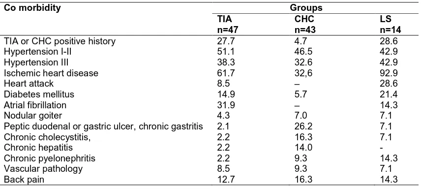
Table 1. Major co morbidities for PIBBC and stroke patients (in %)
Co morbidity Groups
TIA n=47
CHC n=43
LS n=14
TIA or CHC positive history 27.7 4.7 28.6 Hypertension I-II 51.1 46.5 42.9 Hypertension III 38.3 32.6 42.9 Ischemic heart disease 61.7 32,6 92.9
Heart attack 8.5 – 28.6
Diabetes mellitus 14.9 5.7 21.4 Atrial fibrillation 31.9 – 14.3
Nodular goiter 4.3 7.0 7.1
Peptic duodenal or gastric ulcer, chronic gastritis 2.1 26.2 7.1 Chronic cholecystitis, 2.2 16.3 7.1 Chronic hepatitis 2.2 14.0 - Chronic pyelonephritis 2.2 9.3 14.3 Vascular pathology 8.5 9.3 7.1
Back pain 12.7 16.3 14.3
TIA subjects had the ABCD2 score of 4.9; 4-6 (Mdn; 0.25-0.75). In patients with CHC we observed hemiparesis (14%), hypoesthesia (37.2%), instability in the Romberg position (74.1%), asymmetry of tendon reflexes (25.6%). The neurologic deficit in LS patients was objectified using the American National Institute of Health stroke scale (NIHSS). For LS patients the NIHSS score was 5; 3-7 on the 1st day and 2; 1-3 on the 10th day. The list of major comorbidities for PIBBC and stroke patients is presented in Table 1. Among the comorbidities in all groups prevailed hypertension, coronary heart disease and diabetes.
For the peoples of the control group somatic diseases were not identified. Exclusion criteria included severe neurological deficit (score>10 according to NIHSS), hemorrhagic stroke or subarachnoid bleeding, persons with the acute phase of chronic diseases. Catamnesis after the discharge of 22 patients from the hospital was formed on the base of telephone surveys and personal conversations. In period of 3 months after the patient discharge two persons had developed re-TIA (9%) and five persons had myocardial infarction (21.7%).
2.2 Biochemical Analysis
Venous blood samples were drawn at 8:00 AM on the 1st and 10th day of patient hospital staying. The same time conditions were fulfilled for the persons of the control group. Plasma levels of IL-6, IL-8, IL-10, TNF-α and CRPs were measured using a solid-phase chemiluminescent immunometric assay (Immunoassay Analyzer
AMF M/ 340, a panel of reagents from "Vector-Best, Russia"). The upper limits of the normal levels are 5 pg/dL (IL-6), 10 pg/dl (IL-8), 5 pg/dl (IL-10), 8 pg/ml (TNF-α), 8 mg/l (CRP). Coefficient of reactivity (kR) was calculated
according to the tables from the Gusev’s work [5]. Plasma concentration of NOx (nitrate and
nitrite ions) was measured using Griess reagent.
2.3 Statistical Analysis
Data are presented as median (Mdn), lower and upper quartiles (interquartile range (IQR): 0.25-0.75). Statistical analysis was performed using software “STATISTICA 7.0”. U-Mann-Whitney and Wilcoxon analysis of variance was used to establish the difference between groups. To determine the prognostic value of the SI parameters the method of non-linear logistic regression was used.
3. RESULTS
The ranges of the SI parameters in healthy persons of old age were: 0 pg / mL (0.00-0.49 pg / ml) for IL-6; 6.50 pg / ml (5.29-7.72 pg / ml) for IL-8; 0 pg / ml for IL-10; 0 pg / ml for TNF-α; 4.8 mg / l (2.3-7.7 mg / l) for CRP [50]. The ranges of NOx level for healthy persons of old age were
estimated in our early work study using a sample with size of 206 (mean age was 57±2 years) [51]. It was 17.1 µM (11.8-26.1 µM).
The plasma levels of the studied parameters (IL-6, IL-8, CRP, NOx) and their change during
and LS are shown in Fig. 1. The concentrations two other cytokines (IL-10 and TNF-α) for all patient groups were below the test-sensitivity (Table 2). The only exception was the level of TNF-α for LS patients on the 10th day of patient staying in the hospital. The Fig. 1 presents the parameters obtained on admission (axis x) versus the corresponding parameters obtained on the discharge of the patient from the hospital (axis y). Chosen type of data presentation is convenient to the easy fixing of the increase (rose area) and decreases (white area) in the parameters during the therapeutic course as well the variation of the parameters among the studied groups.
As shown in the Fig. 1A, the IL-6 level for PIBBC and LS groups was significantly higher than that for control group (p < 0.01 for all the ischemic groups). However, there is no significant difference between the IL-6 plasma concentration for CHC and TIA groups. U-Mann-Whitney analysis showed that the IL-6 and CRP levels in LS patients were significantly increased during the 10-day therapeutic period (p=0.004, p=0.008) (Figs. 1A and 1B). The IL-6 level in LS patients was higher than in TIA patients (p=0.048) on admission (Fig. 1A).
Fig. 1B shows that the CRP level for TIA group was significantly higher than that for control group both on admission and on discharge (p=0.011, p=0.006) of the patients from the hospital and its interquartile range was similar to that for the LS group.
A significant decrease in the concentration of IL-8 (Fig. 1C) was detected in the plasma of patients with LS versus the parameter for TIA and CHC patients (p=0.002, p=0.006) on admission. After the therapeutic course, the IL-8
level in the plasma of LS patients remained lower than that in CHC patients.
No significant differences were found in IL-8 and NOx levels between both PIBBC groups and
control group (Figs. 1C and 1D). NOx plasma
concentrations increased during the therapeutic course in patients with LS and TIA. The significant difference was established between the NOx levels for CHC and LS groups on
admission only (p=0.049).
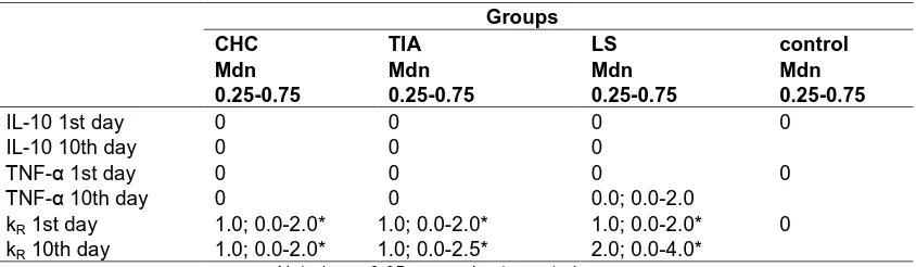
Parameter kR for LS and PIBBC patients was
above zero on admission (Table 2). For LS patient on the 10th day after their admission we found an increase of both statistical parameters for kR: the median (Mdn) and interquartile range
(0.25-0.75). The increase in the interquartile range was fixed also for parameter kR of TIA
patients on their discharge from the hospital.
Using the method of non-linear logistic regression we revealed that the IL-6 level on the 10th day was a significant predictor of re-TIA (OR = 0,86; χ2 = 6,5, p = 0.011). The IL-8 level on the 1st day (OR = 0,44; χ2 = 4,5, p = 0.033) and on the 10th day (OR = 0,2; χ2 = 2,7, p = 0.09) could be also considered as a predictor of re-TIA in 3-month follow-up. Among other laboratory parameters the blood plasma fibrinogen (OR = 0,28; χ2 = 4,6, p = 0.032) and blood plasma glucose levels on admission (OR = 0,51; χ2 = 5,3, p = 0.021) were of interest as prognostic factors.
4. DISCUSSION
Ischemic stroke is recently revealed to link to the phenomena of the endothelial dysfunction and endothelial activation [49,52,53]. Endothelial
Table 2. Plasma concentrations of IL-10,TNF-αααα and the coefficient of reactivity for the studied
groups depending on the day of analysis
Groups
CHC TIA LS control
Mdn 0.25-0.75
Mdn 0.25-0.75
Mdn 0.25-0.75
Mdn 0.25-0.75
IL-10 1st day 0 0 0 0
IL-10 10th day 0 0 0
ТNF-α 1st day 0 0 0 0
ТNF-α 10th day 0 0 0.0; 0.0-2.0
kR 1st day 1.0; 0.0-2.0* 1.0; 0.0-2.0* 1.0; 0.0-2.0* 0
kR 10th day 1.0; 0.0-2.0* 1.0; 0.0-2.5* 2.0; 0.0-4.0*
A
C
Fig. 1. The concentrations of IL
patients with LS (black circles), TIA (gray circles), CHC (white circles) and control subjects group (white diamonds) on admission in (axis x) and on discharge from (axis y) the stroke unit. The data for each axis are represente
the figure area into two parts: Area
the studied parameter values
dysfunction is determined as the state of the endothelium with the decreased synthesis, release and activity of endothelium
[53]. Endothelial activation is a proinflammatory and procoagulant state of the endothelial cells characterized by an increase in th
with leukocytes and caused by proinflammatory cytokines, hypercholesterolemia, turbulent blood flows and others [54]. These two phenomena are tightly bound through the mechanisms with participation of NO, cytokines and other factors
0
2
4
6
8
10
0
2
4
6
[I
L
-6
]
2,
p
g
/d
l
[IL-6]
1, pg
IL-6
1
3
5
7
9
11
1
3
5
7
9
[I
L
-8
]
2,
p
g
/d
l
[IL-8]
1, pg/dl
IL-8
B
D
The concentrations of IL-6 (A), CRP (B), IL-8 (C) and NOx (D) in the blood plasma in
patients with LS (black circles), TIA (gray circles), CHC (white circles) and control subjects group (white diamonds) on admission in (axis x) and on discharge from (axis y) the stroke unit. The data for each axis are represented as Mdn and 0.25-0.75 IQR. The bisectrix divides
Area (in rose) of an increase and area (in white) of a decrease of the studied parameter values after 10-day therapeutic course
is determined as the state of the endothelium with the decreased synthesis, release and activity of endothelium-derived NO . Endothelial activation is a proinflammatory and procoagulant state of the endothelial cells characterized by an increase in their interaction with leukocytes and caused by proinflammatory cytokines, hypercholesterolemia, turbulent blood . These two phenomena are tightly bound through the mechanisms with participation of NO, cytokines and other factors
including inflammatory mediators. Endothelial cell activation can lead to endothelial dysfunction by inhibiting endothelial NO-synthase and decreasing NO bioavailability through increasing in reactive oxygen species’ production
data have showed the difference in the endothelial cell behavior (the endothelial dysfunction and activation) related to LS and PIBBc types. The fact that LS was associated by us to low levels of NO metabolites (NO
could be evidence of the endothelial cell
8
10
, pg/dl
1
10
100
1
10
[C
R
P
]
2,
g
/d
l
[CRP]
1, g/dl
CRP
9 11
dl
5
15
25
35
45
5
15
25 35 45
[N
O
x]
2,
,
m
k
m
o
l/
l
[NO
x]
1, mkmol/
NO
x(D) in the blood plasma in patients with LS (black circles), TIA (gray circles), CHC (white circles) and control subjects
group (white diamonds) on admission in (axis x) and on discharge from (axis y) the stroke 0.75 IQR. The bisectrix divides (in rose) of an increase and area (in white) of a decrease of
ng inflammatory mediators. Endothelial cell activation can lead to endothelial dysfunction synthase and decreasing NO bioavailability through increasing in reactive oxygen species’ production [54]. Our erence in the endothelial cell behavior (the endothelial dysfunction and activation) related to LS and PIBBc types. The fact that LS was associated by us to low levels of NO metabolites (NOx) and IL-8
could be evidence of the endothelial cell
100
dysfunction in patients with LS on their admission of the hospital. In considered case, there was the decrease in both the production and bioavailability of NO in endothelial cells as well the release of IL-8 that could be also produced by endothelial cell as shown in the work by Rakesh et al. [55]. NO is a well-known important factor of vasodilatation and vasoconstriction[48]. IL-8 influences survival and proliferation of endothelial cells and regulates angiogenesis [20,56]. According to our data, the 10-day standard therapeutic course improves the NOx
and IL-8 levels of LS patients. On the other side, at the discharge from the hospital, LS patients had high levels of IL-6 and TNF-α that resulted in high value of coefficient kR characterizing the
systemic inflammation degree. The state of TIA and CHC patients can be associated with the endothelial activation, inflammation and oxidative stress in tissues of the brain [46]. That is proved by high CRP, IL-6 levels and the value of kR.
Moreover, TIA patient state differs from CHC patient state due to the difference in the origin of cerebrovascular accidents. TIA is related to ischemic pathology that makes TIA similar to LS in some extent.
5. CONCLUSION
Thus, our data have shown an important role a number of inflammation markers (NOx, IL-6, IL-8,
TNF-α, CRP) in the pathogenesis of different stroke episodes. Absence of specific neuroimaging changes in TIA and CHC group and nonzero coefficient of reactivity (kR)
suggests that systemic inflammation is not a result of the focal brain ischemia but its intrinsic cause. The differences in the concentration of IL-8, and NOx in the blood plasma of patients with
CHC and LS on their admission to the hospital will help to identify sanogenetic reserves in the case of PIBBC. The observed increase of the
ТNF-α and IL-6 levels in the plasma of LS patients after the course of standard therapy revealed a new side of the problem.
CONSENT
All authors declare that the written consent was obtained from the patients and the persons included in control group for publication of this case report and accompanying images.
ETHICAL APPROVAL
The study was approved by the Ethics committee of Gomel State Medical University.
COMPETING INTERESTS
Authors have declared that no competing interests exist.
REFERENCES
1. Jauch EC, Saver JL, Adams HP, et al. Guidelines for the early management of patients with acute ischemic stroke: A guideline for healthcare professionals from the American Heart Association. Stroke. 2013;44(3):870-947.
2. Lager KE, Mistri AK, Khunti K, et al. Interventions for improving modifiable risk factor control in the secondary prevention of stroke. Cochrane Database Syst. Rev. 2014;2.
DOI: 10.1002/14651858.CD009103.pub2 3. Lichachou SA, Astapenka AV, Bialausky
NN. Transient ischemic attack: Etiology, pathogenesis, classification, clinic, diagnostics. Med News. 2003;10:31-37. 4. Sorensen AG, Ay H. Transient ischemic
attack definition, diagnosis, and risk stratification. Neuroimaging Clin. N. Am. 2011;21(2):303-313.
5. Gusev EY, Yurchenko LN, Chereshnev VA. Methodology of research in systemic inflammation. Cytok Inflam. 2008;7(1): 15-23.
6. Whiteley W, Wardlaw J, Dennis M, et al. The use of blood biomarkers to predict poor outcome after acute transient ischemic attack or ischemic stroke. Stroke. 2012;43(1):86-91.
7. Zenga L, Wangb Y, Liua J. Proinflammatory cytokine network in peripheral inflammation response to cerebral ischemia. Neurosci. Lett. 2013; 548:4-9.
8. Welsh P, Lowe GD, Chalmers J, et al. Associations of proinfammatory cytokines with the risk of recurrent stroke. Stroke. 2008;39:2226-2230.
9. Vila N. Castillo J, Dávalos A, et al. Proinflammatory cytokines and early neurological worsening in ischemic stroke. Stroke. 2000;31(10):2325-2329.
10. Kang DW, Yoo SH, Chun S, et al. Inflammatory and hemostatic biomarkers associated with early recurrent ischemic lesions in acute ischemic stroke. Stroke. 2009:40:1653-1658.
clinical trial. TOAST. Trial of Org. 10172 in acute stroke treatment. Stroke. 1993; 24(1):35-41.
12. Smychok VB, Halinouskaya. The level of C-reactive protein in patients with passing infringements of brain blood circulation. Neurol Neurosurg Eastern Europe. 2012; 2(14):92-94.
13. Kelly PJ, Akijian L, Browne T, et al. Plasma interleukin-6 and C-reactive protein are associated with acute diffusion hyperintensity after transient ischemic attack. Stroke. 2014;45:AWP351.
14. Worthmann H, Tryc AB, Dirks M, et al. Lipopolysaccharide binding protein, interleukin-10, interleukin-6 and C-reactive protein blood levels in acute ischemic stroke patients with post-stroke infection. 2015;14:2-9.
15. Cojocaru IM, Cojocaru M, Tanasescu R, et al. Expression of IL-6 activity in patients with acute ischemic stroke. Rom. J. Intern. Med. 2009;47(4):393-396.
16. Tang YH, Vital S, Granger DN. Role of IL-6 in a novel murine model of transient ischemic attack. FASEB J. 2014; 28(S1):853.
17. Rodriguez-Yañez M, Castillo J. Role of inflammatory markers in brain ischemia. Curr Opin Neurol. 2008;21:353-357. 18. Kumar D, Rasool R, Masoodi KZ, et al.
Stroke-induced immune depression-a randomized case control study in Kashmiri population of North India. J. Stroke. Cerebrovasc. Dis. 2014;23(8):2041-2046. 19. Chasovskih NY, Ryazantseva NV,
Kaigorodova EV, et al. State of JNK and p38 MAP-kinase system in blood mononuclear leucocytes during inflammation. Med. Immunol. 2011;6: 515-522.
20. Gubarev YD, Sheremet AO, The role of immune system in patogenesis of the acute and chronic ischemic damages of the brain. Sci Gazette. 2009;4(59):47-52. 21. Kim SJ, Moon GJ, Bang OY. Biomarkers
for stroke. J Stroke. 2013;15(1):26-36. 22. Jin R, Liu L, Zhang S, et al. Role of
inflammation and its mediators in acute ischemic stroke. J Cardiovasc Transl Res. 2013;6(5):834-851.
23. Hallenbeck MJ. The many faces of tumor necrosis factor in stroke. Nat Med. 2002;8:1363-1368.
24. Barone FC, Arvin B, White RF, et al. Tumor necrosis factor-alpha. A mediator of
focal ischemic brain injury. Stroke. 1997; 28:1233-1244.
25. Meistrell ME, Botchkina GI, Wang H, et al. Tumor necrosis factor is a brain damaging cytokine in cerebral ischemia. Shock. 1997;8:341-348.
26. Ginis I, Jaiswal R, Klimanis D, et al. TNF-alpha-induced tolerance to ischemic injury involves differential control of NF-kappa B transactivation: The role of NF-kappaB association with p300 adaptor. J Cereb Blood Flow Metab. 2002;22:142-152. 27. Lee CC, Liu KJ, Chen LL, et al. Tumor
necrosis factor-α, interleukin-8 and interleukin-6 are involved in vascular endothelial cell capillary tube and network formation induced by tumor-associated macrophages. J Cancer Mol. 2006;2(4): 155-160.
28. Tohgi H, Konno S, Takahashi S, et al. Activated coagulation/fibrinolysis system and platelet function in acute thrombotic stroke patients with increased C-reactive protein levels. Thromb Res. 2000;100(5): 373-379.
29. Krupinski J, Turu MM, Slevin M, et al. Carotid plaque, stroke pathogenesis, and CRP: Treatment of ischemic stroke. Curr Cardiol Rep. 2008;10(1):25-30.
30. Arenillas JF, et al. C-reactive protein predicts further ischemic events in first-ever transient ischemic attack or stroke patients with intracranial large-artery occlusive disease. Stroke. 2003;34: 2463-2468.
31. Woodward M, Lowe GD, Campbell DJ. Associations of inflammatory and hemostatic variables with the risk of recurrent stroke. Stroke. 2005;36: 2143-2147.
32. Park CS, Ihm SH, Yoo KD, et al. Relation between C-reactive protein, homocysteine levels, brinogen, and lipoprotein levels and leukocyte and platelet counts, and 10-year risk for cardiovascular disease among healthy adults in the USA. Am J Cardiol. 2010;105:1284-1288.
33. Rajbhandari R, Gajurel B, Dhungana K, et al. C reactive protein in acute ischemic stroke patients in Tribhuvan University Teaching Hospital. Nepal J Neurosci. 2014;11:4-9.
35. Berezin AE, Lisovaya OA. Predictive value of C-reactive protein in hypertensive patients after suffering a cerebral ischemic stroke. Ukr Card J. 2014;2:36-42.
36. Black S, Kushner I, Samols D. C-reactive Protein. J Biol Chem. 2004;47(279): 48487-48490.
37. Rajeshwar K, Kaul S, Hazzani A, et al. C-reactive protein and nitric oxide levels in ischemic stroke and its subtypes: Correlation with clinical outcome. Inflammation. 2012;35(3):978-984.
38. Rist PM, Buring JE, Kase CS, et al. Biomarkers and functional outcomes from ischemic cerebral events in women: A prospective cohort study. Eur J Neurol. 2013;20(2):375-381.
39. Purroy F, Montaner J, Molina CA, et al. C-reactive protein predicts further ischemic events in transient ischemic attack patients. Acta Neurol Scand. 2007;115(1): 60-66.
40. Zhan X, et al. Transient ischemic attacks characterized by RNA profiles in blood. Neurology. 2011;77(19):1718-1724. 41. Vinychuk CM, Cherenko TM, Merkulan OL.
Method of predicting repeated ischemic stroke. Bulletin. 2010;21:1-2.
42. Kuwashiro Т, Sugimori H, Ago T, et al. Predictive role of C reactive protein in stroke recurrence after cardioembolic stroke: The Fukuoka Stroke Registry. BMJ. 2013;3:1-7.
43. Llombart V, García-Berrocoso T, Bustamante A, et al. Cardioembolic stroke diagnosis using blood biomarkers. Current Cardiology. 2013;9:340-352.
44. Di Napoli M, Schwaninger M, Cappelli R, et al. Evaluation of C-reactive protein measurement for assessing the risk and prognosis in ischemic stroke: A statement for health care professionals from the CRP Pooling Project members. Stroke. 2005; 36(6):1316-1329.
45. Du Clos TW. Function of C-reactive protein. Annals Med. 2000;32(4):274–278. 46. Starodubtseva MN, Halinouskaya NV,
Halubykh NM, et al. Nitric oxide and interleukin-6 production in patients with transient cerebral microcirculatory disturbances. Am J Clin Neurol Neuros. 2015;1(2):86-91.
47. Gontschar IA, Stepanova JI, Prudyvus IS, Kamyschnikov VS, editor. Biochemical predictors and markers of ischemic stroke (in Russian). Minsk: Belarusian Medical Academy of Postgraduate Education; 2013.
48. Szabo C, Ischiropoulos H, Radi R. Peroxynitrite: Biochemistry, patho-physiology and development of therapeutics. Nat. Rev. Drug Discov. 2007; 6(8):662-680.
49. Rajeshwar K, Kaul S, Hazzani A, et al. C-reactive protein and nitric oxide levels in ischemic stroke and its subtypes: Correlation with clinical outcome. Inflammation. 2012;35(3):978-984.
50. Halinouskaya NV, Shitikava MG, Blue Halubykhet, et al. Determination of reference intervals for the parameters describing systemic inflammatory response in the older age group. Modern problems of radiation medicine: From theory to practice. 2015;77.
51. Starodubtseva MN, Voropaev EV, Petrenyov DR, et al. Nitrite and nitrate ion distribution in blood of men from Gomel region depending on oncomarker concentration (prostate specific antigen, cancer antigen 19-9, сarcinoembryonic antigen and alpha fetoprotein). Problems of Health and Ecology (Belarus). 2015; 4:102-107.
52. Rajendran P, Rengarajan T, Thangavel J, et aI. The vascular endothelium and human diseases. Int J Biol Sci. 2013;9(10): 1057-1069.
53. James K. Liao. Linking endothelial dysfunction with endothelial cell activation. J Clin Invest. 2013;123(2): 540–541.
54. Sara P, Ruiz A, Anilkumar N, et al. Reactive oxygen species and endothelial activation. Antioxidants and Redox Signaling. 2008;10(6):1089-1100.
55. Wolff B, Burns AR, Middleton J, et al. Endothelial cell "memory" of inflammatory stimulation: Human venular endothelial cells store interleukin 8 in Weibel-Palade bodies. J. Exp. Med. 1998;(9):1757–62. 56. Rakesh D, Rakesh K, Li SA, et al. IL-8
APPENDIX
Table 1. The concentrations of IL-6, CRP, IL-8 and NOx in the blood plasma in patients with LS,
TIA, CHC and control subjects group (C) on admission in (index 1, axis x) and on discharge from (index 2, axis y) the stroke unit. The data for each axis are represented as Mdn and the
lower (-) and upper (+) limits of IQR
For figure A-D
For figure A
[IL-6]1 [IL -6]2 ∆∆∆∆x- ∆∆∆∆x+ ∆∆∆∆y- ∆∆∆∆y+
C 0 0 0 1.3 0 1.3
LS 1.58 5.08 0.7 1 4.9 4.25 TIA 0.81 0.49 0.73 1.05 0.49 1.85 CHC 0.7 0.89 0.46 1.43 0.65 2.44
For figure B
[CRP]1 [CRP]2 ∆∆∆∆x- ∆∆∆∆x+ ∆∆∆∆y- ∆∆∆∆y+
C 6.4 6.4 3.2 8.3 3.2 8.3 LS 16 21.6 6.31 40.2 5.9 123.2 TIA 14.98 17.4 6.5 55.2 9.6 49.6 CHC 13.1 10.53 6.2 30 5.7 34.6
For figure C
[IL -8]1 [IL -8]2 ∆∆∆∆x- ∆∆∆∆x+ ∆∆∆∆y- ∆∆∆∆y+
C 5.1 5.1 0.49 1.57 0.49 1.57 LS 3.53 5.75 2.1 0.42 3.69 0.53 TIA 5.81 6.04 1.06 2.77 2.92 1.56 CHC 5.81 7.07 0.35 0.69 0.65 4.02
For figure D
[NOx]1 [NOx]2 ∆∆∆∆x- ∆∆∆∆x+ ∆∆∆∆y- ∆∆∆∆y+
C 20.35 20.35 6.93 10.17 6.83 10.17 LS 14.56 22.14 5.1 5.92 4.37 27.82 TIA 20.52 26.92 4.96 9.38 11.5 13.81 CHC 24.53 21.85 9.06 8.83 5.03 8.71 _________________________________________________________________________________
© 2016 Halinouskaya et al.; This is an Open Access article distributed under the terms of the Creative Commons Attribution License (http://creativecommons.org/licenses/by/4.0), which permits unrestricted use, distribution, and reproduction in any medium, provided the original work is properly cited.
Peer-review history: