SCIENCEDOMAINinternational
www.sciencedomain.org
An
In Vitro
Study of Micro Leakage of Different
Types of Composites with Respect to Their
Matrix Compositions
FouadKadim Wahab
1, Inas Talib Abu-Tabra
2and Wala Majid Amin
3*1Restorative Sciences, Department of Conservative dentistry, Faculty of Dentistry, University
of Jordan,Jordan. 2Faculty of Dentistry, University of Jordan,Jordan. 3Prosthodontics and Biomaterials, Department of Prosthodontics, Faculty of Dentistry,
University of Jordan,Jordan.
Authors’ contributions This work was carried out in collaboration between all authors.Author FW designed the study, wrote the protocol, managed the literature searches and wrote the first draft of the manuscript. Author ITAT made the experimental work. Author WMA managed the analyses of the study, performed the statistical treatment of data, and wrote the final draft of the manuscript. All authors read and approved the final article.
Received 9thNovember 2013 Accepted 21stDecember 2013 Published 12thJanuary 2014
ABSTRACT
Aims: Effect of composition of hybrid composites on their microleakage behavior was evaluated.
Study Design:An invitro microleakage study.
Place and Duration of Study: Department of Conservative Dentistry, Faculty of Dentistry, The University of Jordan, Amman, Jordan, between May 2012 and July 2013. Methodology:160 Class V cavities were divided into four equal parts each restored with either “Spectrum”,“Admira”, “FilteckP90” or “Smart dentine replacement SDR”. Teeth were thermocycled, immersed in 1% methylene blue for four hours and sectioned buccolingually. AutoCAD software was used in delineating cavity outlines and depth of
The materials’ interface at the gingival wall was of comparable nature.
Conclusion: Variations of materials’ type or location of Class V cavity, buccal or lingual, does not affect the tooth/restoration bond interface. AutoCAD software proved to be a powerful measurement tool and is recommended for microleakage studies.
Keywords: AutoCAD software; admira®; Filteck TM; microleakage; SDRTM; spectrumTPH3.
1. INTRODUCTION
Resin-based composite materials are prominent aesthetics restorative materials because of their universal usage, minimal loss of tooth structure and ability to be directly placed without laboratory procedures. Moreover, the superior mechanical and aesthetic qualities of composites have made most widely used tooth-colored direct restorative materials in modern dentistry [1,2]. At least half of posterior direct restoration placements rely on composite materials 3. Nevertheless, there are number of shortcomings such as shrinkage and induced stress, thermal expansion mismatch, fracture, abrasion and wear resistance, marginal leakage, and toxicity [3-6].
Shrinkage may be considered the major disadvantage of the current composite materials [2]. The process results in shrinkage contraction of the composite, causing stresses that may exceed the strength of the bond with the surrounding tooth structure, with possible interfacial failure [7,8]. Process of composite restorative materials range between 1.5% to 7.1% (in volume) for the dimethacrylate-based composites [9-10]. This level of shrinkage could generate contractile forces of 3.3MPa to 23.5MPa [9,11-13]. Such a force is often considered the most significant problem and a primary contributor to premature failure in composite restorations, due to its capability of deforming tooth structures and causing microcracks and adhesive failures [5].
Shrinkage leads to a gap formation between the composite restoration and the walls of the cavities at the weakest bond (usually dentine or cementum). Marginal breakdown may result in microleakage, postoperative sensitivity and recurrent dental caries [14-16].
The recent and ongoing advances in biomaterials research has helped develop a better understanding of polymerisation shrinkage and lead to the development of new techniques and materials. Among these are different types of sandwich restorations, different incremental placement techniques of the resin composite and different light curing regimens [17]. Various incremental techniques have been used for the placement of composite resin restorations like occluso-gingival layering [18], oblique layering [18-20], facio-lingual layering [18] and centripetal placement technique [20], but none has been able to eliminate micro gap formation at gingival margin. Although filler shape and composition are important [21], the development of various matrix components necessitates an additional material classification [22].
Since each material has different chemical system (monomer), they also have different behavior regarding the polymerisation shrinkage [6,23]. The aim of material development was to eliminate polymerisation shrinkage by adapting the individual components of the material. With Ormocers the methacrylate had been partially replaced by an inorganic network. Replacing the chain monomers in the composite matrix by ring-shaped molecules has helped establish a new approach to reduce polymerisation shrinkage. A new group of materials the Siloranes had been developed; these are hydrophobic materials and need to be bonded to the dental hard tissue using a special adhesive system.
Smart Dentine Replacement (SDR TM) had also been, developed specially for dentine replacement and cured increments up to 4mm.The polymerisation stress had been reduced by 50% or more compared to conventional [13]. It was based on the chemistry of universal composite with a main difference in the modulator that incorporated in the Urethane-based dimethacrylates. SDR TM considered as it contains a modified resin system. Siloranes, Ormocers and SDR TM restorative composites were considered to be low polymerisation shrinkage materials [24-26].
The aim of this study was to investigate the effect of different chemical systems (resin component) in hybrid composite on microleakage of Class V restorations inserted with the bulk placement technique at the enamel and dentine margins of the tooth/restoration interface. The primary goal was to test the hypotheses that the type of material (the monomer systems), the orientation of preparation (buccal/lingual), and the type of preparation margins (enamel/dentine) may influence the microleakage Class V restorations using a new method for measurement of dye penetration.
2. MATERIALS AND METHODS
2.1 Selection of Teeth
Recently extracted human first and second maxillary premolars teeth stored in normal saline solution were examined individually. Criteria were applied by which teeth with large carious lesions, extensive wear, fractured cups, and cracked enamel were discarded. Each tooth was thoroughly scaled to remove calculus and remaining tissue tags. The selected teeth were then polished with slurry of pumice and water. The teeth were re-examined with a binocular microscope (Model 154161; Meiji; Saitama, Japan) at original magnification X4. Eighty premolars were selected and divided randomly in to four groups of 20 teeth each. The sample size was comparable with previous studies [27,28].
2.2 Preparation of the Class V Cavities
Fig. 1.Diagrammatic representation of a cavity prepared on a buccal and/or a lingual surface of a tooth specimen
2.3 Resin-Based Composites
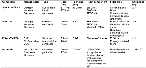
The composite restorative materials chosen for this study represented different chemical systems (Table 1). They included a conventional matrix (pure methacrylate) type as hybrid and nano composites (Spectrum TPH3); inorganic matrix (inorganic polycondensate) such as Ormocers (Admira®); ring opening epoxide (cationic polymerisation) such as Siloranes (Filteck TM P90) and Smart dentine replacement (SDR TM) which is based on the chemistry of the universal composite but differs in the modulator which is incorporated in the Urethane-based dimethacrylates (a modified resin system).
2.4Placement of Restorations
Table 1. Composites employed in the present investigation
Composite Manufacturer Type Filer
( %) Filer size(µm) Resin component Filler type Shrinkage(%) SpectrumTPH3 Dentsply
Konstanz Germany
Sub-micron hybrid composite
57.1 vol
77.5 wt 10-20nm Bis-GMABis-EMA TEGDMA
Silicon dioxide
Boro-ilicate/aluminum bariumandsilicam icro-hybrid
2.1
SDR TM Dentsply
Konstanz Germany
Flowable
composite 45 vol68 wt 4.2 EBPADMATEGDMA Modified UDMA
Barium aluminuo-fluoro-borosilicate glass
Strontium- aluminuo-Fluoro-silicate glass
3.5
FilteckTM P90 3 M
St. Paul, Minn USA
Siloranes (nanohybride composite)
55 vol.
76 wt 0.1-2 SiloxanesOxiranes Zr/SilicaPrepoly- marized fillers
< 1
Admira® Voco GmbH
Cuxhavan Germany
Ormocers
packable 56 vol77 wt 0.04-0.7 UDMA TMA-alkoxysilanes "Multifunctional-urethane and thioether(meth) acylatealkoxysilan es"
Ba-al-Borosilicate
2.5 Micro Leakage Testing
Each restored tooth was attached from one of its roots to a length of nickel-chrome wire 1 mm in diameter (Dentrum; Ispringen, Germany) passing through a hole made at the apical third of the root. The other end of the wire was then fastened to the cover of a 30-mL plastic vial (Arab Food and Medical Appliances Co, Ltd, Amman, Jordon). To ensure that the tooth hung freely and firmly in the center of the plastic vial the free end of the wire was tightly attached to the vial’s cover through a hole made in the center of the cover, and tightly fastened end of the wire was further fixed in place by sticky wax. Each restored tooth was coated with and acid-resistant protective nail varnish (Del Laboratories, Inc Uniondale, NY) except for a window including the restoration and a clear area of one millimeter width around it. The windows were prepared by placing strips of an adhesive tape, each measuring 4mm in width in a occlusal-cervical direction and 6mm in length in the mesio-distal direction. After the varnish had dried completely, the adhesive tape was removed with a fine pair of tweezers, leaving the windows (4mm x 6 mm) uncovered by the varnish. The specimens were immersed in 1% methylene blue at room temperature for 4 hours. On removal from the dye solution, the specimens were gently washed with tap water. Care was taken not to exert any pressure by the running tap water on the window part of the stained specimen.
2.6 Dye Penetration Measurements
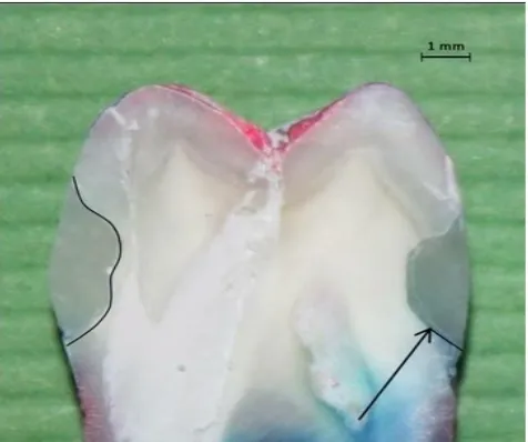
The samples were cut in the bucco-lingual direction through the center of the restoration with diamond disc (Summa Disk no. 692M; Shofu Inc, Kyoto, Japan). Each specimen was photographed using Canon camera (EOS REBEL T3i 600D, CANON INC, Shimomaruko3-chome, Ohta-ku, Tokyo, Japan). The photographs were then imported to the AutoCAD version 2009 for measurement of the outline of the buccal and lingual cavities and dye penetration at both occlusal and gingival walls / restoration (Autodesk, Inc. San Rafael, California, USA). The AutoCAD is a software application for computer-aided design (CAD) and drafting. The software supports both 2D and 3D formats. CAD often involves more than just shapes. As in the manual drafting of technical and engineering drawings, the output of CAD often must convey also symbolic information such as materials, processes, dimensions, and tolerances, according to application-specific conventions. It provides for extremely accurate to scale drawings. The drawing measurements were carried out at a magnification X20. The outline of the cavities was measured first and then the linear dye penetration at the margins of occlusal and gingival walls/restoration interfaces (Fig. 2). The percentage of dye penetration for each restoration was then calculated. Both sections of each restoration were measured and the mean microleakage was recorded as the percent for that restorations.
2.7 Statistical Analysis
Fig. 2. An AutoCAD image displaying a longitudinal section of a premolar tooth with buccal and lingual Class V restorations. The outline of both cavities
is clearly visible and the depth of dye penetration, indicated by the arrow, is shown at the gingival wall of the lingual Class V restoration
3. RESULTS
Table 2. Length fraction of intact bond interface between restorative materials and walls of Class V cavities prepared on the buccal and lingual surfaces of teeth
Cavity
walls SpectrumTPH3 SDRTMMean ±(SD)FilteckTM fValue ‘P’value
P90 Admira®
Buccal
Surface Occlusalwall 100±(0.0) 98.8±(5.2) 94.79±(6.3) 92.84±(7.6) 6.85 P=.05 Gingival
wall 70.89±(16.2) 79.05±(15.8) 77.04±(12.8) 80.69±(10.2) 1.78 P=.05
Lingual
Surface Occlusalwall 100±(0.0) 98.4±(6.9) 94.2±(6.9) 92.8±(6.9) 1.513 p>.05 Gingival
wall 72.7±(15.6) 82.3±(15.9) 78.4±(11.9) 84.5±(10.6) 2.63 p>.05
Table 3. Differences of group means among the investigated materials showing comparison of differences between means of any two materials and “T” value of 5.07
computed by “Tukey” post hoc statistics
Materials Comparison means M2 M3 M4
SpectrumTPH3
SDRTM
FiltekTMP90 Admira®
M1=100.00 M2=98.81 M3=94.79 M4=92.84
M1 – M2=1.19 M1 – M3=5.21*
M2 – M3=4.02 M1 – M4=7.16*M2 – M4=5.97* M3 – M4=1.95
(*) denotes a significant difference (P=.05) between the two means
The findings of the present investigation indicated that the SpectrumTPH3 composite display intact interface compared to FilteckTM P90 and Admira®. In addition, the SDRTM composite showed longer fraction of sound interface than Admira® (P=.04). Nevertheless, there was no significant difference in length fraction of intact interface at the occlusal wall of cavities prepared on the lingual surfaces (P>.05).
Table 4. Comparison among the studied restorative materials in term of their length fraction of the intact bond interface between the material and occlusal and gingival walls of Class V cavities prepared on buccal and lingual surfaces of teeth
Tooth Surface/Cavity wall SpectrumTPH3 SDRTM FiltekTMP90 Admira®
Length
Fraction “P” Value LengthFraction “P” Value LengthFraction “P” Value LengthFraction “P” Value Buccal
Surface Occlusal wallGingival wall 100.0*70.895 P=1.1x10
-12 98.810*
79.045 P=1.5x10
-9 94.79*
77.037 P=8.2x10
-6 92.84*
80.69 P=9.9x10
-5
Lingual
Surface Occlusal wallGingival wall 100.0*72.735 P=1.7x10⁻⁷ 98.405*82.25 P=.0002 94.137*78.353 P=2.4x10
-5 92.816*
84.526 P=.004 Occlusal
Wall Buccal surfaceLingual surface 100.0100.0 P=.165 98.8198.41 P=.421 94.7994.14 P=.392 92.8492.82 P=.497 Gingival
All cavities prepared on the buccal and lingual surfaces and restored by Spectrum TPH3 and 95% of these restored by the SDR TM composite showed complete sealing at the occlusal walls while only 50% at the buccal surface and 55% at the lingual surface of Filteck TM P90 composite did so. Similarly 45% of the cavities at the buccal and 40% at the lingual surfaces restored by Admira® composite showed complete sealing at the occlusal walls. In addition, two cavities at the buccal surface and one cavity on the lingual surface restored by Spectrum TPH3 and three cavities on both buccal and lingual surfaces restored SDR TM composites showed complete seal at the gingival walls. There was no significant difference in the length fraction of sound interface among cavities restored by the four materials regarding the gingival walls (p>0.05).
4. DISCUSSION
The selection of the restorative materials employed in this investigation was based on several factors, first, they were all indicated for Class V, secondly, the filler contents of all materials investigated was relatively similar, and thirdly, the SDR TM, Filteck TM P90 and Admira® were considered of a low polymerisation shrinkage ranging from <1% to 3.5% compared with the SpectrumTPH3 (2.1%).
It is of a particular importance to mention that Class V preparations were used on the account that they have a high C-factor, i.e. they are preparations with high ratio of bonded “flow-inactive” to free “flow-active” surfaces [12]. A butt joint enamel margin was selected because it complies with traditional enamel margin designs advocated for most preparations for posterior composite restorations [29].
Using bulk placement technique rather than incremental method following same standards used in reported studies [30]. The thermocycling regiment, using 500 cycles between 5º and 55ºC with a dwell time 30 second, was applied since it yields significant microleakage [31] Methylene blue dye was chosen for dye penetration measurement because it is simple, inexpensive and does not require the use of complex laboratory equipment. Moreover, the particle size of this dye is less than the internal diameter of the dentinal tubules (1-4 µm), thus, it is suitable for showing the permeability of dentin [32].
The use of AutoCAD software has increased the accuracy and precision of measurement of cavity outline and the length fraction of dye penetration at the restoration/tooth interface. This method provided a far more precise measurement of the actual depth of dye penetration measured on magnified apparent images up to 20 times (X20) compared with the readings obtained by conventional scoring method made on images of a stereomicroscope.
The findings of the present study showed that all four investigated restorative materials formed a superior seal with the enamel margin at the occlusal wall of the Class V cavity preparation. The results pointed out that SpectrumTPH3 had a more favorable seal than other low shrinkage composite and SDRTM was better than FilteckTMP-90 and Admira®. On the other hand, all the restorative materials exhibited interfacial microleakage at the gingival/dentine margins except few cavities restored by SpectrumTPH3 and SDR.TM These results were in agreement with previously reported findings [29,31,33].
also be ascribed to a very week interfacial bond that formed initially but failed to overcome the internal stress that resulted by the polymerisation shrinkage of the composite [11].
It is a noteworthy that despite the fact that the investigated materials were claimed of being a low polymerisation shrinkage type composites, the magnitude of the stresses associated with their shrinkage was too high to be compensated for by the possible interfacial bond between these composites and the tooth structure. This has incurred what might be described as a “de-bonded” interface as manifested by the microleakage of the contrast dye.
SDRTM displayed an intact interface with tooth structure which was far more superior compared with those shown by FilteckTM P-90 and/or Admira® at occlusal / enamel margins. This was for a probable reason that SDR TM is a low-stress composite resin which offers up to a 50% reduction in shrinkage stress compared to that of the conventional resins and it has improved flow properties compared to that of traditional posterior composites. The reduced stress in this urethane methacrylate resin could be related to its relatively slow polymerisation rate [13] which allowed for more-thorough curing of the material and therefore reduced its polymerisation stress.
FilteckTM P-90 revealed a comparable quality of bond interface with tooth structure compared with SpectrumTPH3 at occlusal/enamel margins. The probable reason for this may be explained by the fact that Siloranes system uses “ring opening” polymerisation instead of free-radical polymerisation of dimethacrylate monomers. In this “self or dark’’ polymerisation associated with the cationic mechanism, the reactive species do not become extinguished as quickly as the free radical contained within conventional methacrylate-based resin [34] and therefore, a significantly low polymerisation shrinkage occurred irrespective of the location of the margins [33].
Similarly Ormocers materials (Admira®) contain inorganic-organic co-polymers in addition to the inorganic silanated filler particles. It is synthesized through a solution and gelation process (sol-gel process) from multifunctional urethane and thioether(meth)acrylate alkoxysilanes. The Ormocers matrix is a polymer even prior to light curing. It consists of ceramic Polysiloxane, which has low shrinkage properties than the organic dimethacrylate monomer matrix seen in composites. To the Polysiloxane chains in Ormocers, polymerisable side chains were added to react during curing and form the setting matrix. These inorganic molecules are longer than Bis-GMA, which could explain the material's lower volumetric shrinkage. This could suggest that the change in compositions of composites regarding their resin systems may improve other properties of these composites, such as hardness properties rather than their bond to enamel and dentine, as found in the present study.
The results of this investigation indicated that the type of resin chemistry and orientation of the preparation (buccal/lingual) had no significant effect on the quality of bond interface between the restorative materials and the walls of Class V cavities. The four materials showed a similar performance in Class V preparation with respect to microleakage, although the composites used varied considerably in composition. This observation is in agreement with Meier et al. [29].
to reduce the polymerisation shrinkage stress and may be responsible for diminishing the formation of interfacial gap. Moreover, the differences among the materials are substantial, which include the structural and compositional differences such as size of filler, chemistry of the resin components, the unpolymerised resin content. In addition, the materials also differ in the viscosity and handling characteristics. All these factors may directly or indirectly influence the adaptation to the preparation walls of these materials before and after polymerisation. These structural differences among the materials may affect the degree of hygroscopic expansion of their resin component, in addition to influencing the bond strength and durability.
The findings of this study confirmed those previously reported which demonstrated strong bonding to enamel than to dentine or cementum [29,31]. The gingival/dentine margins showed inferior quality of bond interface, indicated by a significantly higher microleakage than the occlusal/enamel margins. The probable reason for this was that the bond strength to enamel is usually higher than to dentine. Dentine is less favorable bonding substrate due to its heterogeneous structure [35]. Enamel on the other hand, is a highly mineralised tissue composed of more than 90% of hydroxyapatite, whereas, dentine contains a substantial proportion of water and organic material primarily type I collagen. Dentine also contains a dense network of tubules that connect the pulp with the dentine-enamel junction [36]. The tubules branch, particularly near the dentine-enamel and cement-dential junctions. Generally branching of tubules are smaller and more numerous in root dentine than in crown dentine. Acid etching of the heterogeneous dentine structure results in different surface chemistries and morphologies. The orientation of dentinal tubules can affect the formation of the hybrid layer. In areas with perpendicular tubules orientation, the hybrid layer is significantly thicker than in areas with parallel tubules orientation [36]. Therefore, the dentine’s surface on the gingival floor of Class V preparation maybe a surface on which good hybrid layer formation is difficult [33]. This could well explain the markedly inferior quality of bond interface displayed between the dentine gingival cavity wall and all of the employed composite restorative materials compared to the quality of their interface at the enamel dominated occlusal cavity wall.
5. CONCLUSION
Based on the findings of the present investigation, the following rational conclusions were arrived at:
•Conclusive evidence showed that microleakage at the enamel margin was less than that at the gingival margins. This suggests that utmost care should be practiced in the selection of the restorative materials and materials such as the low polymerisation shrinkage type may not be suitable for sub-gingival preparations.
•Factors that include variation of material type, the monomer system used, and/or the orientations of Class V cavity preparation, whether on the buccal or the lingual tooth surface, bear no significant effect on the pattern of microleakage at the tooth/restoration interface.
CONSENT
We the authors of this submission hereby declare that ‘written informed consent was obtained from the patient whose extracted teeth were used in the present investigation.
ETHICAL APPROVAL
We the authors of this submission hereby declare that all experiments have been examined and approved by the appropriate ethics committee and have therefore been performed in accordance with the ethical standards laid down in the 1964 Declaration of Helsinki.”
COMPETING INTERESTS
Authors have declared that no competing interests exist.
REFERENCES
1. Ferracane JL. Resin composite-state of the art. Dent Mater.2011;27:29-38.
2. Ferracane JL. Placing dental composites a stressful experience.Oper Dent. 2008;33:247-257.
3. Sadowsky SJ. An overview of treatment considerations for esthetic restorationsa review of the literature. J Prosthet Dent. 2006;96:433-42.
4. Anseth KS, Newman SM, Bowman CN. Polymeric dental composites properties and reaction behavior of multimethacrylate dental restorations. Adv PolymSci 1995;122:177-217.
5. Ferracane JL. Developing a more complete understanding of stresses produced in dental composites during polymerisation. Dent Mater. 2005;21:36-42.
6. Weinmann W, Thalacker C, Guggenberger R. Siloranes in dental composites. Dent Mater. 2005;21:68-74.
7. Bernardo M, Luis H, Martin MD, Leroux BG, Rue T, Leitão J, DeRouen TA. Survival and reasons for failure of amalgam versus composite posterior restorations placed in a randomized clinical trial. J Am Dent Assoc. 2007;6:775-783.
8. Heintze SD. Systematic reviews. I. The correlation between laboratory tests on marginal quality and bond strength. II. The correlation between marginal quality and clinical outcome. J Adhes Dent. 2007;9Suppl1:77-106.
9. Kleverlaan CJ, Feilzer AJ. Polymerisation shrinkage and contraction stress of dental resin composites. Dent Mater. 2005;21:1150-57.
10. Bagis YH, Baltacioglu IH, Kahyaogullari S. Comparing microleakage and the layering methods of silorane-based resin composite in wide Class II MOD cavities. Oper Dent. 2009;34:578-85.
11. Davidson CL, De Gee AJ, Feilzer A. The competition between the composite-dentin bond strength and the polymerisation contraction stress. J Dent Res. 1984;63:1396-99.
12. Feilzer AJ, De Gee AJ, Davidson CL. Setting stress in composite resin in relation to configuration of the restoration. J Dent Res. 1987;66:1636-39.
15. Friedl KH, Hiller KA, Schmalz G. Placement and replacement of composite restorations in Germany. Oper Dent. 1995;20:34-8.
16. Applequist EA, Meiers JC. Effect of bulk insertion, prepolymerized resin composite balls, and beta-quartz inserts on microleakage of Class V resin composite restorations. Quentessence Int. 1996;27:253-58.
17. Lindberg A, Dijken JW, Lindberg M. Nine year evaluation of a polyacid-modified resin composite/resin composite open sandwich technique in class II cavities. J Dent. 2007;35:124-29.
18. Tjan AH, Bergh BH, Lidner C. Effect of various incremental techniques on marginal adaptation of class II composite resin restorations. J Prosthet Dent. 1992;67:62-6. 19. Duarte S, Dinelli W, Carmona da silva MH. Influence of resin composite insertion
technique in preparations with a high C-factor. Quintessence Int. 2007;38:829-35. 20. Duarte S, Saad JR. Marginal adaptation of class 2 adhesive restorations.
Quintessence Int. 2008;39:413-19.
21. Lutz F, Philips RW A.Classification and evaluation of composite resin systems. J Prosthet Dent. 1983;50:480-88.
22. Zimmerli B, Strub M, Jeger F, Stadler O, Lussi A. Composite materials composition, properties and clinical applications. A Literature Review. SchweizMonatsschrZahnmed. 2010;120:972-79.
23. Bottenberg P, Jacquet W, Alaerts, Keulemans F. A prospective randomized clinical trial of one Bis-GMA-based and two ormocer-based composite restorative systems in class II cavities five-year results. J Dent. 2009;37:198–203.
24. Duarte SJR, Botta AC, Phark JH, Sadan A. A selected mechanical and physical properties and clinical application of a new low-shrinkage composite restoration. Quintessence Int. 2009;40:631-38.
25. Ilie N, Hickel R. Investigations on a methacrylate-based flowable composite based on the SDR™ technology. Dent Mater. 2011;27:348-55.
26. Papadogiannis D, Kakaboura A, Palaghias G, EliadesG.Setting characteristics and cavity adaptation of low-shrinking resin composites. Dent Mater. 2009;25:1509-16. 27. Hakimeh S, Vaidyanathan J, Houpt ML, Vaidyanathan TK, Von Hagen S.
Microleakage of compomer Class V restorations effect of load cycling, thermal cycling, and cavity shape differences. J Prosthet Dent. 2000;83:194-203.
28. Manhart J, Schmidt M, Chen HY, Kunzelmann KH, Hickel R.Marginal quality of tooth-colored restorations in class II cavities after artificial aging. Oper Dent. 2001;26:357-66.
29. Meiers JC, Kazemi R, Meiers CD. Microleakage of packable composite resins. Oper Dent. 2001;26:121-26.
30. Sensi LG, Marson FC, Baratieri LN, Monteiro Junior S. Effect of placement techniques on the marginal adaptation of Class V composite restorations. J Contemp Dent Pract. 2005;15:17-25.
31. Wahab FK, Shaini FJ, Morgano SM. The effect of thermocycling on microleakage of several commercially available composite Class V restorations in vitro. J Prosthet Dent. 2003;90:168-74.
32. Yavuz I, Aydin AH. New method for measurement of surface areas of microleakage at the primary teeth by biomolecule characteristics of methylene blue. Biotechnol&Biotechnol Eq. 2005;19:181-87.
34. Orefice RL, Discacciati JAC, Neves AD, Mansur HS, Jansen WC. In situ evaluation of the polymerisation kinetics and corresponding evolution of the mechanical properties of dental composites. Polymer Testing. 2003;22:77-81.
35. Bala O, Uçtasli MB, Unlü I. The leakage of Class II cavities restored with packable resin-based composites. J Contemp Dent Pract. 2003;4:1-11.
36. Sturdevant JR. Clinical significance of dental anatomy, histology, physiology and occlusion. In Roberson TM, Heymann HO, Swift EJ (eds). Art and science of operative dentistry. St. Louis Elsevier Inc. 2006;15-64.
© 2014 Wahab et al.; This is an Open Access article distributed under the terms of the Creative Commons Attribution License (http://creativecommons.org/licenses/by/3.0), which permits unrestricted use, distribution, and reproduction in any medium, provided the original work is properly cited.
Peer-review history: