Clinical Ophthalmology
Dove
press
O r i g i n a l r e s e a r C h
open access to scientific and medical research
Open access Full Text article
Worsening anatomic outcomes following
aflibercept for neovascular age-related macular
degeneration in eyes previously well controlled
with ranibizumab
eric nudleman1
Jeremy D Wolfe2,3
Maria a Woodward4
Yoshihiro Yonekawa2,3
george a Williams2,3
Tarek s hassan2,3 1Department of Ophthalmology,
shiley eye Center, University of California, san Diego, la Jolla, Ca,
2Beaumont eye institute, Oakland
University William Beaumont school of Medicine, 3associated retinal
Consultants, royal Oak, 4Kellogg
eye Center, University of Michigan Medical School, Ann Arbor, MI, USA
Purpose: Antivascular endothelial growth factor injection is the mainstay of treating neovascular age-related macular degeneration (AMD). Previous studies have shown that switching treatment from ranibizumab to aflibercept led to an improvement in eyes with recalcitrant activity. Herein, we identify a unique subset of patients whose eyes with neovascular AMD were previously well controlled with ranibizumab injections were then worsened after being switched to aflibercept. Methods: This is a retrospective interventional case series. Eyes with neovascular AMD, previously well controlled with monthly injections of ranibizumab, which then developed worsening of subretinal fluid after being switched to aflibercept were included.
Results: A total of 17 eyes were included. All eyes developed increased subretinal fluid when switched from ranibizumab to aflibercept. Fourteen patients were switched back to ranibizumab after a single injection of aflibercept and had subsequent rapid resolution of subretinal fluid. Three patients continued with monthly aflibercept injections for two subsequent months and demonstrated the persistence of the increased subretinal fluid until they were switched back to treatment with ranibizumab at which time the fluid resolved. No eye had persistent decline in visual acuity.
Conclusion: Switching from intravitreal ranibizumab to aflibercept in eyes with well-controlled neovascular AMD may result in worsening in a subset of patients and resolves when therapy is switched back to ranibizumab.
Keywords: anti-VEGF; intravitreal injection; macula; optical coherence tomography; retina
Introduction
Age-related macular degeneration (AMD) is the leading cause of legal blindness in
the US.1–3 Neovascular AMD may cause vision loss secondary to subretinal fluid,
hemorrhage, intraretinal edema, or scarring.3 It is well established that vision loss from
neovascular AMD is dramatically reduced in eyes treated with intravitreal injections
of antibodies that target vascular endothelial growth factor (VEGF).4–6
Several groups have reported on patients treated with intravitreal ranibizumab or bevacizumab who have a good initial response with resolution of subretinal or intraretinal fluid, but then later become resistant to continued treatment with these agents
and develop recurrent exudation with vision loss.7 The mechanism of this acquired
recalcitrance to treatment is not known, but tachyphylaxis has been suggested.7–9 It has
been reported that switching from one anti-VEGF medication to another may lead to a favorable result with improvement in vision and resolution of fluid.10
Correspondence: Tarek s hassan associated retinal Consultants, 3535 West 13 Mile road, suite 344, royal Oak, Mi 48073, Usa email tsahassan@yahoo.com
Journal name: Clinical Ophthalmology Article Designation: Original Research Year: 2016
Volume: 10
Running head verso: Nudleman et al
Running head recto: Worsening with aflibercept conversion in neovascular AMD DOI: http://dx.doi.org/10.2147/OPTH.S109894
Clinical Ophthalmology downloaded from https://www.dovepress.com/ by 118.70.13.36 on 21-Aug-2020
For personal use only.
Number of times this article has been viewed
This article was published in the following Dove Press journal: Clinical Ophthalmology
Dovepress
nudleman et al
Recent reports have shown that switching treatment from bevacizumab or ranibizumab to aflibercept led to an improved response with decreased exudation in patients refractory to their prior treatment with other anti-VEGF drugs.11–15
Afliber-cept is a protein that is constructed by fusion of the second binding domain of the receptor VEGFR1 and the third binding domain of the receptor VEGFR2 to the crystalline portion of IgG1. It has been reported that aflibercept has a higher affinity
for VEGF than both bevacizumab and ranibizumab,16 and this
may explain its improved effect when injected in patients who have developed apparent resistance to these other agents.
Combined with a higher affinity for VEGF, aflibercept also has a longer intravitreal half-life than ranibizumab.16,17
These factors have led investigators to examine the ability to extend treatment intervals beyond 1 month in eyes treated with aflibercept. Noninferiority has been demonstrated in eyes treated with three initial monthly injections of 2 mg aflibercept, followed by injections every 8 weeks, when
compared with monthly treatments of ranibizumab.18
In this study, we present a series of patients whose eyes with neovascular AMD were well controlled for a minimum of 12 months with intravitreal ranibizumab therapy and were switched to intravitreal aflibercept in an effort to extend treat-ment intervals. This subset of eyes showed immediate clinical worsening with recurrence of subretinal fluid after a single treatment with aflibercept given within 4–5 weeks of the preceding ranibizumab injection. This is a unique subgroup of patients whose characteristics have not been previously reported. All patients were switched back to ranibizumab, and there was subsequent resolution of subretinal fluid and a return to baseline vision. Of note, this study does not aim to determine the incidence of this phenomenon nor to compare with patients who did well with conversion – it is a case series to demonstrate that worsening with aflibercept is a possibility in some unique patients.
Methods
This interventional retrospective case series included an analy-sis of 17 eyes from 17 patients from one tertiary vitreoretinal care center between June 2011 and May 2013. All eyes were well controlled (defined as having no identifiable subretinal or intraretinal fluid on optical coherence tomography) after being treated with monthly intravitreal injections of ranibizumab for a minimum of 12 months prior to switching to intravitreal afliber-cept. Exclusion criteria included poor control with ranibizumab, best corrected visual acuity (VA) of #20/200 in the treated eye, history of diabetic macular edema or retinal vein occlusion, and prior treatment other than intravitreal anti-VEGF therapy.
Institutional review board approval for the data collection and study was granted by Western Institutional Review Board. All patients underwent a thorough informed consent process for each intravitreal injection. The study was conducted in a HIPAA (Health Insurance Portability and Accountability Act) compliant fashion, and research adhered to the tenets of the Declaration of Helsinki. The data collected included sex, age, VA, dates of treatment, phakic status, and central macular thickness (CMT). All measurements of CMT were made on Heidelberg optical coherence tomography. Statistical analysis was performed using paired Student’s t-tests and Pearson cor-relations using STATA software Version 13 (StataCorp LP, College Station, TX, USA).
Results
A total of 17 eyes from 17 patients met the inclusion criteria. The mean age of the patient was 80 years (range, 58–89 years), and the series included ten men and seven women. Three eyes were phakic, and 14 eyes were pseudophakic. All study eyes received at least 12 prior intravitreal injections of ranibizumab with an average of 17 injections of ranibizumab (range, 12–29) having been given prior to switching to aflibercept. In the 12 months prior to switching to aflibercept, no eyes included in the study had evidence of subretinal hemorrhage or subretinal fluid but were being managed with monthly maintenance injections. No eyes in the study had prior treatment with any modality other than anti-VEGF therapy.
The switch to aflibercept occurred in all patients within 4–5 weeks after the preceding treatment with ranibizumab (Figure 1). All patients were then seen 4–5 weeks later for follow-up subsequent to switching medication. There was no evidence of inflammation following the change in any treated eyes. The mean CMT on the day of switching to aflibercept
was 270.1±34.8 μm (range, 223–318 μm). At 1-month
follow-up, all patients had increased intraretinal/subretinal fluid, with an increase in mean CMT to 414.5±75.8 μm (range, 291–549 μm, P,0.001).
Because of the development of increased subretinal fluid after switching to aflibercept, 14 eyes in this study were switched back to ranibizumab at the 1-month follow-up visit. In these eyes, 1 month following the return to ranibi-zumab therapy, there was a decrease in the mean CMT to 327.1±98.9 μm (range, 223–509 μm). At 3 months following the return to ranibizumab therapy, the mean per-centage change in CMT compared with the month prior to switching to aflibercept was 1.8%±5.34% (range, −12.9% to 4.7%). There was no statistically significant difference in the CMT compared with that seen in the month prior to
Clinical Ophthalmology downloaded from https://www.dovepress.com/ by 118.70.13.36 on 21-Aug-2020
Dovepress Worsening with aflibercept conversion in neovascular AMD
switching (P=0.29), and no measurable intraretinal/subretinal fluid or hemorrhage was seen in any eye at the visit 3 months after the return to ranibizumab therapy.
Symptoms of vision loss were experienced in six of 17 patients (35%) following the switch to aflibercept, all of which resolved following the return to ranibizumab therapy. The mean logMAR VA on the day of switching to aflibercept was 0.411±0.17 (range, 0.699–0.097). The mean logMAR VA at the 1-month postaflibercept injection follow-up visit was 0.52±0.14 (range, 0.699–0.301, P,0.01). Vision had returned to baseline by 3 months following the
return to ranibizumab, with a mean logMAR VA 0.405±0.17
(range, 0.70–0.097, P=0.34). A significant negative correla-tion was found between VA at the time of treatment with aflibercept and the change in VA between the time of afliber-cept injection and 1 month later (r=−0.66, P=0.004).
Despite the recurrence of subretinal fluid following the switch to aflibercept, three eyes received two more consecu-tive monthly treatments of aflibercept. The average increase in mean CMT in these eyes 1 month after the change to
aflibercept was 260±72.2 μm (range, 206–342 μm). During the subsequent 2 months of aflibercept therapy, the mean
CMT improved minimally (range, −8.2 μm to 3.1%). One
month following the eventual return to treatment with ranibizumab, the mean CMT in these eyes decreased by 250±27.87 μm (range, 206–342 μm, P=0.004). Following the return to treatment with intravitreal ranibizumab, all patients had a return to baseline CMT by 3 months, with a mean change of −14±18.5 μm (range, −33 μm to 4 μm, P=0.32) compared with the CMT prior to the switch.
Discussion
In this retrospective, observational case series, we report on a small group of patients who developed a worsening of exudation and some loss of vision following a change in intra-vitreal therapy from ranibizumab to aflibercept. This change rapidly resolved after the return to treatment with intravitreal ranibizumab. Visual acuities were only mildly affected, with less than half of the patients noting a symptomatic change. However, there was a negative correlation between VA prior
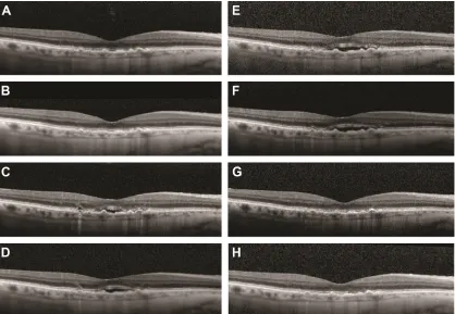
Figure 1 Worsening neovascular age-related macular degeneration after switching from ranibizumab to aflibercept.
Notes: A 78-year-old man with neovascular age-related macular degeneration was well controlled with monthly ranibizumab intravitreal injections, with complete resolution of subretinal fluid. (A, 1 month prior to conversion to aflibercept and B, the day of conversion to aflibercept). The patient was switched to aflibercept in an effort to extend the intervals, but the subretinal fluid recurred. (C, 1 month after conversion to aflibercept). Three more injections of monthly aflibercept were attempted without improvement (D–F). When he was switched back to ranibizumab, the subretinal fluid resolved (G, 1 month after reverting back to ranibizumab) and continued to do well on ranibizumab (H, 2 months after reverting back to monthly ranibizumab). Visual acuity was stable at 20/30–20/40 throughout the course, and he continues to be well controlled on monthly ranibizumab.
Clinical Ophthalmology downloaded from https://www.dovepress.com/ by 118.70.13.36 on 21-Aug-2020
Dovepress
nudleman et al
to the switch and change in vision 1 month following the switch. Those with better VA likely had more vision to lose, although no definite conclusion can be drawn from such a small number of patients. No patient in this series suffered a subretinal hemorrhage after the switch, and all patients returned to baseline VA within 3 months of returning to ranibizumab therapy.
Several recent publications have reported on improve-ment in subretinal fluid in patients switched from other anti-VEGF therapies to aflibercept. Our series demonstrates that there is a subset of patients who may have an opposite response to such a change, particularly from ranibizumab to aflibercept. The mechanism by which some patients respond more favorably than others to such a switch in medications is unknown. It is possible that there are genetic variants in VEGF-A that may confer differences in response to treatment. Abedi et al19 investigated this theory
and identified a single tagged-SNP, rs3025000, which conferred a statistically significant difference in response to treatment with either bevacizumab or ranibizumab. The authors determined that a T allele (versus C) conferred an
odds ratio of ∼3 in favor of being a positive responder to
treatment. It is possible that a genetic difference in our cohort of patients accounts for their poor response to treat-ment with aflibercept.
Conclusion
This study identifies for the first time a small subset of patients who had an immediate clinical worsening of exudative AMD after switching from intravitreal ranibizumab to intravitreal aflibercept therapy. We agree with the previous reports that most patients do well with conversion to aflibercept, but we must be aware of this unique subset of patients who worsen. Fortunately, we were able to demonstrate that this worsening was reliably reversible and there was no persistent morbidity after a return to ranibizumab therapy.
Disclosure
Jeremy D Wolfe is a consultant for Allergan, Alimera, and Genentech. George A Williams is a consultant for Allergan. Tarek S Hassan is a consultant for Allergan, Arctic Dx, Genentech, Novartis, Regeneron, and Roche. Eric Nudleman is a consultant for Allergan. Yoshihiro Yonekawa is a consul-tant for Allergan. Maria A Woodward reports no conflicts of interest in this work.
References
1. Friedman DS, O’Colmain BJ, Muñoz B, et al; Eye Diseases Prevalence Research Group. Prevalence of age-related macular degeneration in the United States. Arch Ophthalmol. 2004;122(4):564–572.
2. Buitendijk GH, Rochtchina E, Myers C, et al. Prediction of age-related macular degeneration in the general population: the three continent AMD consortium. Ophthalmology. 2013;120(12):2644–2655. 3. Velez-Montoya R, Oliver SCN, Olson JL, Fine SL, Quiroz-Mercado H,
Mandava N. Current knowledge and trends in age-related macular degen-eration: genetics, epidemiology, and prevention. Retina. 2014;34(3): 423–441.
4. Miller JW, Le Couter J, Strauss EC, Ferrara N. Vascular endothelial growth factor a in intraocular vascular disease. Ophthalmology. 2013;120(1): 106–114.
5. Rosenfeld PJ, Brown DM, Heier JS, et al; MARINA Study Group. Ranibizumab for neovascular age-related macular degeneration. N Engl
J Med. 2006;355(14):1419–1431.
6. Brown DM, Kaiser PK, Michels M, et al; ANCHOR Study Group. Ranibizumab versus verteporfin for neovascular age-related macular degeneration. N Engl J Med. 2006;355(14):1432–1444.
7. Forooghian F, Cukras C, Meyerle CB, Chew EY, Wong WT. Tachyphy-laxis after intravitreal bevacizumab for exudative age-related macular degeneration. Retina. 2009;29(6):723–731.
8. Schaal S, Kaplan HJ, Tezel TH. Is there tachyphylaxis to intravitreal anti-vascular endothelial growth factor pharmacotherapy in age-related macular degeneration? Ophthalmology. 2008;115(12):2199–2205. 9. Binder S. Loss of reactivity in intravitreal anti-VEGF therapy:
tachy-phylaxis or tolerance? Br J Ophthalmol. 2012;96(1):1–2.
10. Gasperini JL, Fawzi AA, Khondkaryan A, et al. Bevacizumab and ranibizumab tachyphylaxis in the treatment of choroidal neovasculari-sation. Br J Ophthalmol. 2012;96(1):14–20.
11. Bakall B, Folk JC, Boldt HC, et al. Aflibercept therapy for exudative age-related macular degeneration resistant to bevacizumab and ranibi-zumab. Am J Ophthalmol. 2013;156(1):15–22.
12. Yonekawa Y, Andreoli C, Miller JB, et al. Conversion to aflibercept for chronic refractory or recurrent neovascular age-related macular degeneration. Am J Ophthalmol. 2013;156(1):29–35.
13. Ho VY, Yeh S, Olsen TW, et al. Short-term outcomes of aflibercept for neovascular age-related macular degeneration in eyes previously treated with other vascular endothelial growth factor inhibitors. Am J
Ophthalmol. 2013;156(1):23–28.e2.
14. Cho H, Shah CP, Weber M, Heier JS. Aflibercept for exudative AMD with persistent fluid on ranibizumab and/or bevacizumab. Br J
Ophthalmol. 2013;97(8):1032–1035.
15. Kumar N, Marsiglia M, Mrejen S, et al. Visual and anatomical outcomes of intravitreal aflibercept in eyes with persistent subfoveal fluid despite previous treatments with ranibizumab in patients with neovascular age-related macular degeneration. Retina. 2013;33(8):1605–1612. 16. Stewart MW, Rosenfeld PJ. Predicted biological activity of intravitreal
VEGF Trap. Br J Ophthalmol. 2008;92(5):667–668.
17. Stewart MW. Aflibercept (VEGF trap-eye) for the treatment of exudative age-related macular degeneration. Expert Rev Clin Pharmacol. 2013; 6(2):103–113.
18. Heier JS, Brown DM, Chong V, et al; VIEW 1 and VIEW 2 Study Groups. Intravitreal aflibercept (VEGF trap-eye) in wet age-related macular degeneration. Ophthalmology. 2012;119(12):2537–2548. 19. Abedi F, Wickremasinghe S, Richardson AJ, et al. Variants in the VEGFA
gene and treatment outcome after anti-VEGF treatment for neovascu-lar age-related macuneovascu-lar degeneration. Ophthalmology. 2013;120(1): 115–121.
Clinical Ophthalmology downloaded from https://www.dovepress.com/ by 118.70.13.36 on 21-Aug-2020
Clinical Ophthalmology
Publish your work in this journal
Submit your manuscript here: http://www.dovepress.com/clinical-ophthalmology-journal
Clinical Ophthalmology is an international, peer-reviewed journal covering all subspecialties within ophthalmology. Key topics include: Optometry; Visual science; Pharmacology and drug therapy in eye diseases; Basic Sciences; Primary and Secondary eye care; Patient Safety and Quality of Care Improvements. This journal is indexed on
PubMed Central and CAS, and is the official journal of The Society of Clinical Ophthalmology (SCO). The manuscript management system is completely online and includes a very quick and fair peer-review system, which is all easy to use. Visit http://www.dovepress.com/ testimonials.php to read real quotes from published authors.
Dovepress
Dove
press
Worsening with aflibercept conversion in neovascular AMD
Clinical Ophthalmology downloaded from https://www.dovepress.com/ by 118.70.13.36 on 21-Aug-2020