Abstract
Primary bronchial mucoepidermoid carcinoma in the lung is relatively rare. It rarely presents with the highly malignant biological characteristic of bone marrow metastasis. We describe a case of this disease with bone marrow metastasis. A 56-year-old man with the primary manifestation of bone pain and bloodstained sputum had two abnormal shadows on the left inferior lobar bronchus and peripheral tissue of the lower lobe of the left lung, respectively. Computed tomography-guided percutaneous puncture biopsy and bone imaging confirmed the diagnosis of high-grade bronchial mucoepidermoid carcinoma with bone metastasis. However, the patient soon presented with progressive hemoglobin and platelet decline and severe multi-organ hemorrhage. Subsequently, we performed bone marrow aspiration and biopsy, which revealed malignant cells and necrosis. The patient deteriorated rapidly from the disease, and died on the 16th day of admission. We hope that this case report will increase awareness of the possibility of primary high-grade bronchial mucoepidermoid carcinoma metastasizing to the bone marrow, which might be a poor prognostic factor.
Keywords:Mucoepidermoid carcinoma, Lung, Bone marrow metastasis
Background
Primary bronchial mucoepidermoid carcinoma, a low-malignant potential tumor of bronchial gland origin, is relatively rare and comprises approximately 0.1% of all malignant lung tumors [1]. Compared with most other lung cancers, it occurs in relatively young people [2-4]. Although mucoepidermoid carcinomas are malignant tumors, they are usually indolent, with chronic progres-sion. Surgical treatment yields a favorable prognosis; the 5-year survival rate is 95% and adjuvant treatment is considered unnecessary. Mucoepidermoid carcinomas rarely present highly malignant biological characteristics, especially bone marrow metastasis. We report a case of unusually aggressive bronchial mucoepidermoid car-cinoma with bone marrow metastasis with the aim of raising awareness of the malignant biological behavior of this tumor.
Case presentation
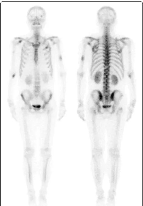
A 56-year-old man presented to our hospital complain-ing of osphyalgia, dorsalgia, and melosalgia for 2 months, and bloodstained sputum for 2 weeks. He had been a drinker and smoker for more than 30 years, but de-nied personal or family history of cancer. Physical examination on admission disclosed vertebral tender-ness. Routine blood examination revealed slightly de-creased hemoglobin (HGB, 10.5 g/dL) and platelets (PLT, 87,000/mm3). On chest computed tomography (CT), we observed two lobulated masses measuring 30 to 40 mm in diameter in the left inferior lobar bronchus (Figure 1A) and peripheral tissue of the lower lobe of the left lung (Figure 1B), respectively. We also observed left hilar lymph nodes enlargement. Magnetic resonance imaging confirmed multiple sites of bone destruction of the lumbar spine, and bone scans revealed systemic multiple abnormal hypermetabolic lesions (Figure 2). Based on the clinical and auxiliary examination find-ings, the presumptive diagnosis was lung cancer with multiple bone metastases.
On his third day in hospital, the patient underwent CT-guided percutaneous puncture biopsy of the lung and received palliative radiotherapy for the areas with * Correspondence:drlhdong@163.com
†Equal contributors
1
Department of Radiotherapy, Norman Bethune First Hospital, Jilin University, 71 Xinmin Street, Changchun 130021, China
Full list of author information is available at the end of the article
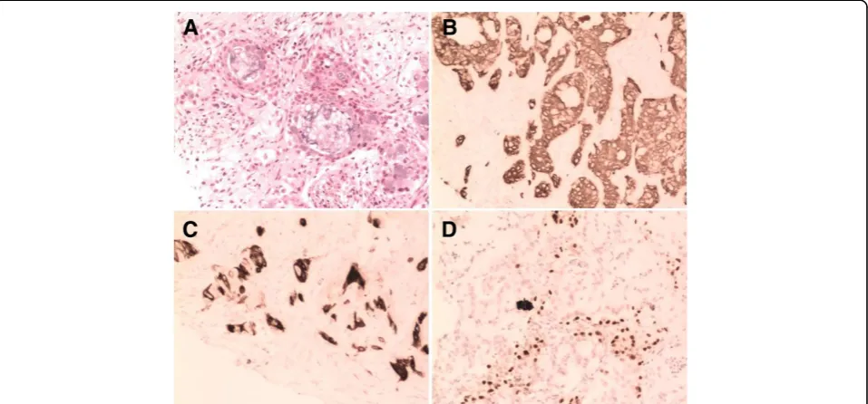
severe bone destruction. The pathological findings of the lung biopsy were poorly differentiated mucoepidermoid carcinoma (Figure 3A). Ki-67 expression was about 70%. Immunohistochemical examination revealed tumor cells were positive for cytokeratin (CK) 7, CK5/6, and thyroid
transcription factor-1 (TTF-1) (Figure 3B-D), which allowed us to determine that his condition was primary lung cancer. On the sixth day of admission, he presented with mild hemoptysis; routine blood examination re-vealed clearly decreased HGB and PLT, which were 6.2 g/dL and 56,000/mm3, respectively. As we suspected bone marrow infiltration by the cancer cells, we stopped radiotherapy and suggested that he undergo bone mar-row aspiration and biopsy. However, he continued to de-teriorate. Eight days later, we repeated the laboratory tests; Table 1 lists the results. We did not find skin pe-techiae or ecchymosis, or other evidence of bleeding. To carry out further examinations and prevent the risk of severe bleeding, we started supportive treatment com-prising drug hemostasis and transfusion of platelet, fresh frozen plasma, and red blood cell suspension. However, the patient presented with upper gastrointestinal bleed-ing without any obvious cause the followbleed-ing day, which we suspected was stress ulceration bleeding. Thus, we administered gastrointestinal decompression and gastric acid inhibition.
Eleven days later, his PLT count rose to 63,000/mm3. Bone marrow aspiration and biopsy revealed malignant cells and necrosis (Figure 4A-D). We informed the pa-tient of the diagnosis of bone marrow metastasis with bronchial mucoepidermoid carcinoma. Unfortunately, on the day he was scheduled to receive systemic chemother-apy, the patient presented with severe respiratory tract hemorrhage. Following rapid deterioration from the dis-ease, he died on the 16th day of admission.
Discussion
Based on its histopathological features, we diagnosed the present lung tumor as primary high-grade bronchial mucoepidermoid carcinoma, which is relatively rare in the lung. Histologically, it is believed that mucoepider-moid carcinomas are derived from the serous and mucus glands of the trachea and bronchi. They are classified as low or high grade according to histological appearance, Figure 1Enhanced CT scan revealing two lobulated masses measuring 30 to 40 mm in diameter in the left inferior lobar bronchus (A) and peripheral tissue of the lower lobe of the left lung (B), respectively.
Figure 2Technetium-99 m methylene diphosphonate bone scintigraphy showing metastatic involvement of the thoracic vertebra, humeri, and ribs.
cellular atypia, mitotic activity, local invasion, and necrosis. It is believed that the biological behavior is associated with differentiation [5]. The prognosis of low-grade mucoepider-moid carcinomas is much better. Only very few cases of high-grade tumors with the malignant features of rapid de-terioration and early distant metastases have extremely poor prognosis [6].
Bronchial mucoepidermoid carcinoma always occurs in the central bronchi. It appears on CT as an isolated, well-defined oval or lobulated mass with smooth margins aris-ing within the bronchus [7]. It may be associated with ob-structive pneumonia and atelectasis, or in a few cases, with cavitation and calcification. On CT imaging, the ma-jority of these tumors exhibits moderate to marked
enhancement. However, the present tumor was mainly located in the left inferior lobar bronchus and associated with an oval, spiculated mass in the peripheral lung tissue, a multicenter origin considered very rare.
Poorly differentiated bronchial mucoepidermoid carcin-oma may present with the highly malignant biological characteristics of regional lymph node metastases. How-ever, distant metastases, especially bone marrow metasta-sis, are extremely rare, and have not been reported in the available literature. Bone marrow metastasis occurs when cancer cells from a non-hematological tumor infiltrate the bone marrow via hematogenous spread or direct exten-sion from contiguous tumor deposits [8]. Malignant cells in bone marrow smears and biopsy are a key diagnostic feature. Patients typically present with anemia, bone pain, fatigue, and progressive deterioration. Changes in the per-ipheral blood are always obvious decreased HGB and PLT, but normal or increased leukocytes [8].
Metastatic carcinoma in bone marrow in the exhaus-tion phase is a rare pathophysiological form of bone marrow metastasis. Featuring rapid onset, progressive anemia, and thrombocytopenia accompanied by severe hemorrhage, infection, and even disseminated intravas-cular coagulation (DIC), it is considered a lethal compli-cation of malignant tumor [8-10]. Due to the poor performance status, systemic chemotherapy is often con-sidered a relative contraindication for such patients. Thus, the duration of survival maintained by only supportive treatment is usually very limited [10].
The diagnosis of bone marrow metastasis in our pa-tient was based on bone marrow smear and biopsy. The Table 1 Laboratory test results
Parameter At admission 6 days after admission
8 days after admission
WBC 11,700/μL 9,900/μL 9,900/μL
RBC 3,470,000/mm3 2,020,000/mm3 1,340,000/mm3
HGB 10.5 g/dL 6.2 g/dL 4.2 g/dL
PLT 87,000/mm3 56,000/mm3 30,000/mm3
PT ND 13.2 s 19.7 s
APTT ND 24.8 s 52.5 s
Fibrinogen ND 1.0 g/L 0.56 g/L
D-dimer ND ND 0.457 mg/L
Coombs (DAT) ND ND Negative
APTT, activated partial thromboplastin time;DAT, direct antibody test;HGB, hemoglobin;ND, not done;PLT, platelet count;PT, prothrombin time;RBC, red blood cell;WBC, white blood cell.
abnormal laboratory test parameters (prolonged pro-thrombin time and activated partial thromboplastin time, decreased fibrinogen, elevated D-dimer level) supported the diagnosis of DIC. All clinical features and auxiliary examinations were consistent with bone marrow failure induced by metastatic carcinoma. Regrettably, we were un-able to perform the bone marrow examination in time and administer salvage chemotherapy to this patient in view of his personal wishes and rapid deterioration from the disease.
Conclusions
This case demonstrates the ability of primary high-grade bronchial mucoepidermoid carcinoma to metastasize to the bone marrow, which, as has been the case in many other tumors, might be a poor prognostic factor in mucoepidermoid carcinoma. Thus, we suggest consider-ing the probability of bone marrow metastasis if there is progressive PLT and HGB decline without any obvious reason. Bone marrow aspiration and biopsy should be performed to confirm diagnosis as soon as possible. Re-gardless, timely salvage chemotherapy may prolong sur-vival and improve the prognosis.
Consent
We obtained written informed consent from the next of kin of the patient for publication of this case report and any accompanying images. A copy of the written consent is available for review by the Editor-in-Chief of this journal.
Competing interests
The authors declare that they have no competing interests.
Authors’contributions
ZYP, GZY, and LHD contributed equally to this work; participated in the care of the patient, data collection, and literature search; and drafted the manuscript. LMQ, TTY, and ZHD reviewed the CT images and photographed the bone marrow pathology. All authors participated in the conception and design of the study. ZYP and GZY wrote the first draft of the manuscript. All authors read and approved the final manuscript.
Authors’information
Zhenyu Pan and Guozi Yang are co-first authors.
Acknowledgments
We thank Dr. Hongguang Zhao for her expert technical assistance about bone scintigraphy.
Author details
1
Department of Radiotherapy, Norman Bethune First Hospital, Jilin University, 71 Xinmin Street, Changchun 130021, China.2Department of Pathology,
Norman Bethune First Hospital, Jilin University, 71 Xinmin Street, Changchun 130021, China.3Department of Radiology, Norman Bethune First Hospital,
Jilin University, 71 Xinmin Street, Changchun 130021, China.4Cancer Center, Norman Bethune First Hospital, Jilin University, 71 Xinmin Street, Changchun 130021, China.
Received: 3 September 2013 Accepted: 9 May 2014 Published: 21 May 2014
References
1. Leonardi HK, Jung-Legg Y, Legg MA, Neptune WB:Tracheobronchial mucoepidermoid carcinoma.J Thorac Cardiovasc Surg1978,76:431–438. 2. Yousem S, Nicholson A:Mucoepidermoid carcinoma.InWorld Health
Organization classification of tumours.Pathology and genetics of tumours of the lung, pleura, thymus and heart.Edited by Travis W, Brambilla E, Muller-Hermlink H, Harris C. Lyon: IARC; 2004:63–64.
3. Liu X, Adams AL:Mucoepidermoid carcinoma of the bronchus: a review.
Arch Pathol Lab Med2007,131:1400–1404.
Figure 4Bone marrow smear (A, B: Wright-Giemsa stain, ×1,000) and biopsy (C, D: HE stain, ×400) showing metastatic tumor cells and necrosis.
nonhematological tumors by bone marrow biopsy: a retrospective analysis of 10,112 samples.J Cancer Res Clin Oncol2009,135:687–693. 9. Ozkan M, Er O, Karahan IO, Deniz K, Coşkun R, Küçük C, Yurci A, AltinbaşM:
Rectal carcinoid tumor with bone marrow and osteoblastic bone metastasis: a case report.Turk J Gastroenterol2007,18:111–114. 10. Krishnan C, George TI, Arber DA:Bone marrow metastases: a survey of
nonhematologic metastases with immunohistochemical study of metastatic carcinomas.Appl Immunohistochem Mol Morphol2007,15:1–7.
doi:10.1186/1477-7819-12-158
Cite this article as:Panet al.:Bone marrow metastasis in primary bronchial mucoepidermoid carcinoma: a case report.World Journal of Surgical Oncology201412:158.
Submit your next manuscript to BioMed Central and take full advantage of:
• Convenient online submission
• Thorough peer review
• No space constraints or color figure charges
• Immediate publication on acceptance
• Inclusion in PubMed, CAS, Scopus and Google Scholar
• Research which is freely available for redistribution