R E S E A R C H
Open Access
Identification of two internal signal peptide
sequences: critical for classical swine fever virus
non-structural protein 2 to trans-localize to the
endoplasmic reticulum
Kang-kang Guo
1†, Qing-hai Tang
1,2†, Yan-ming Zhang
1*, Kai Kang
1and Lei He
1Abstract
Background:The membrane topology and molecular mechanisms for endoplasmic reticulum (ER) localization of
classical swine fever virus (CSFV) non-structural 2 (NS2) protien is unclear. We attempted to elucidate the
subcellular localization, and the molecular mechanisms responsible for the localization of this protein in our study. The NS2 gene was amplified by reverse transcription polymerase chain reaction, with the transmembrane region and hydrophilicity of the NS2 protein was predicted by bioinformatics analysis. Twelve cDNAs of the NS2 gene were amplified by the PCR deletion method and cloned into a eukaryotic expression vector, which was transfected into a swine umbilical vein endothelial cell line (SUVEC). Subcellular localization of the NS2 protein was
characterized by confocal microscopy, and western blots were carried out to analyze protein expression. Results:Our results showed that the -NH2terminal of the CSFV NS2 protein was highly hydrophobic and the protein localized in the ER. At least four transmembrane regions and two internal signal peptide sequences (amino acids103-138 and 220-262) were identified and thought to be critical for its trans-localization to the ER.
Conclusions:This is the first study to identify the internal signal peptide sequences of the CSFV NS2 protein and its subcellular localization, providing the foundation for further exploration of this protein’s function of this protein and its role in CSFV pathogenesis.
Background
Classical swine fever (CSF) is a highly contagious and often fatal disease of pigs and is classified by the World Organization for Animal Health (OIE) as a notifiable (previously List A) disease. The causative agent of CSF is classical swine fever virus (CSFV), a member of the Pestivirusgenus within theFlaviviridaefamily of viruses, which also contains the genera Flavivirusand Hepaci-virus(hepatitis C viruses, HCV)[1]. CSFV harbors a 12.3 kb positive-sense, single-stranded RNA genome that consists of a large open reading frame that encodes a polyprotein which is processed into 12 mature proteins,
namely, Npro, C, Erns, E1, E2, p7, NS2, NS3, NS4A, NS4B, NS5A and NS5B [2-4].
In recent years, the nonstructural NS2 protein has been thought to be functional only as an NS2/NS3 auto-protease, which is essential for high productivity of CSFVin vivo. It was speculated that the N-terminal half of NS2 is highly hydrophobic, and that p7 protein may contain a signal sequence to direct the downstream NS2 protein to the membrane [3,5,6]. Our previous study demonstrated that CSFV NS2 was a hydrophobic pro-tein and localized in the endoplasmic reticulum (ER) membrane, independently of CSFV p7 peptides. How-ever, the membrane topology and molecular mechanism of ER localization of this protein remains unclear. The biofunction of a protein is always associated with it’s subcellular localization. For instance, HCV NS2 protein, which shares great similarities with CSFV NS2 protein, localizes in the ER membrane and lead to ER stress
* Correspondence: [email protected]
†Contributed equally
1
College of Veterinary Medicine, Northwest A & F University, Yangling, Shaanxi 712100, P.R.China
Full list of author information is available at the end of the article
[7,8]. Interestingly, our results indicated that CSFV NS2 protein contains two internal signal peptide sequences, which are critical for trans-localization to the ER, and this protein probably possesses at least four transmem-brane regions. The findings are crucial for elucidating the function of CSFV NS2 protein, and also have poten-tially important implications for understanding the molecular mechanisms of pathogenesis for this econom-ically important agricultural disease.
Materials and methods
Vectors and cell culture
The pEGFP-C1 eukaryotic expression vector was pur-chased from Clontech (USA) and competent E. coli DH5a cells, which were used for cloning, were pur-chased from Tiangen Biotech (China). The pEGFP-NS2 plasmid contained the full-length NS2 gene from the CSFV virulent train Shimen. The established swine umbilical vein endothelial cell line (SUVEC) was cul-tured as previously described [9].
Antibodies and reagents
Mouse anti-GFP monoclonal antibody (mAb) and horse-radish peroxidase-conjugated goat anti-mouse antibodies were purchased from Millipore (USA). The nuclear staining dye Hoechst 33342 and ER-Tracker™ Red probe were obtained from Invitrogen (USA)
Plasmid construction and transfection
To investigate the internal signal sequences in the CSFV NS2 protein, the primers shown in Table 1 were designed according to the CSFV NS2 gene nucleotide sequences. All of the upstream primers contained a Sal-Isite, and a BamHIsite was incorporated into all of the downstream primers. Using these primers, 12
amino-terminal truncated polymerase chain reaction (PCR) products were obtained and the relative position of each amplified fragment is shown in Figure 1. PCR was car-ried out according to the following procedures (pEGFP-NS2 was used as a template): an initial denaturation step at 95°C for 5 min, followed by 35 cycles of 95°C for 30 sec, 60°C for 30 sec, 72°C for 1.5 min, and a final extension step at 72°C for 10 min. The 12 PCR products obtained, designated as NS2/1-457, NS2/13-457, NS2/ 33-457, NS2/103-457, NS2/138-457, NS2/263-457, NS2/ 337-457, NS2/1-336, NS2/1-170, NS2/1-76, NS2/202-262 and NS2/138-201 were cloned into the SalI/BamHI-sites of the expression vector pEGFP-C1 and the recom-binant plasmids were identified and verified by enzyme digestion and sequencing. SUVEC were seeded into 15 mm2 confocal dish (Costar, USA) 24 h before being transfected. When they reached 60-70% confluence, the cells were transfected with the 12 recombinant plasmids and the pEGFP-C1 control vector using the Lipofecta-mine 2000 transfection reagent, according to the manu-facturer’s instructions (Invitrogen, USA)
Western blot analysis
Protein expression was analyzed by western blot as reported previously. Briefly, whole cell extracts were prepared by washing the cells with PBS, harvesting them by scraping and then resuspending the cells in 1 mL of PBS. Following centrifugation, the cells were resus-pended in cell lysis buffer (50 mM Tris-HCl, 5 mM EDTA, 150 mM NaCl, 0.1% NP-40, 0.5% deoxycholic acid, 1 mM sodium orthovanadate, 100 μg/mL PMSF and protease inhibitors) and centrifuged at 15,000 × g for 30 min at 4°C. Cell extracts were resolved by 12% sodium dodecyl sulfate-polyacrylamide gel electrophor-esis (SDS-PAGE) and transferred to a PVDF membrane
Table 1 Primers for construction of 12 subcloning of cDNA.
Primers and sites(amino acid site of NS2) Primer sequences (5"-3”)
Anti457 CATGGATCCTCTAAGCACCCAGCCAAGG
A restriction site sequence was inserted in 5’of each primer. The underlined sequences were restriction sites.
Guoet al.Virology Journal2011,8:236 http://www.virologyj.com/content/8/1/236
(Millipore, USA). The membrane was blocked overnight with 5% skimmed milk in TNT buffer (20 mM Tris-HCl [pH 7.5], 150 mM NaCl, and 0.05% Tween 20) and then incubated with mouse anti-GFP mAb for 2 h. Detection of primary antibodies was performed with horseradish peroxidase-conjugated goat anti-mouse antibody, as appropriate. The protein bands were visualized by enhanced chemiluminescence methods according to the manufacturer’s instructions (Millipore).
Fluorescence staining and confocal microscopy
To examine the expression and subcellular localization of CSFV NS2 protein, 48 h after transfection, cells were washed with Hank’s balanced salt solution (HBSS) and incubated with Hoechst33342 at 37°C for 15 min. The cells were washed twice with HBSS and incubated with ER-Tracker™Red probe (Invitrogen, USA) at 37°C for 30 min. Cells were again washed with HBSS and then visualized by laser confocal scanning microscopy (Model LSM510 META, Zeiss, Germany).
Bioinformatics analysis
The amino acid sequences of the CSFV NS2 protein was analyzed by the DNAstar software and the online analy-sis tool available at http://us.expasy.org/tools.
Results
Construction of recombinant expressing plasmid
Using specifically designed primers, 12 amino-terminal truncated fragments of the NS2 genes were amplified by PCR and the sizes of these amplified products were veri-fied by electrophoresis (Figure 2). These PCR products were cloned into the expression vector pEGFP-C1, and
the 12 recombinant plasmids were identified and veri-fied by SalI/BamHI enzyme digestion (Figure 3) and sequence analysis.
Expression and subcellular localization of CSFV NS2 protein
Western blot analysis showed that all the target protein were correctly expressed and displayed the expected molecular weight (Figure 4). The subcellular localization of NS2 was investigated by confocal fluorescence micro-scopy. The subcloned proteins NS2/1-457, NS2/13-457, NS2/33-457, NS2/103-457, NS2/1-336, NS2/1-170 and NS2/202-262 localized in the ER, whereas, proteins NS2/138-457, NS2/263-457, NS2/1-76 and NS2/337-457 were distributed in the cytoplasm, gathering around the ER. Protein NS2/138-201 was observed in the nucleus and cytoplasm, with no obvious gathering around the ER. In the GFP positive control cells, GFP was observed in the nucleus and cytoplasm (SUVEC-GFP), and no green fluorescence was detected in the negative control SUVECs (Figure 5A and 5B).
NS2 contains two internal signal sequences and multiple domains in the ER
Hydrophilicity analysis of the CSFV NS2 amino acid sequences using the DNAstar software showed that the -NH2 terminal was highly hydrophobic; accordingly, five probable transmembrane regions were predicted at the -NH2 terminal of the NS2 protein using the online tool available at http://us.expasy.org/tools (Figure 6A and 6B). Taken together with the data regarding the subcel-lular localization of 12 the subcloned NS2 fragments, the possible locations of the transmembrane domains
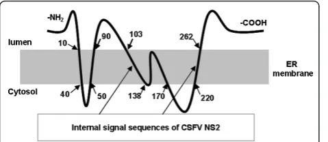
were predicted. Thus, four transmembrane domains were predicted to reside within amino acids 1-40, 50-90, 103-170 and 220-262, with two internal signal sequences likely residing within amino acids 103-138 and 220-262. The predicted model of CSFV NS2 membrane topology is show in Figure 7.
Discussion
CSF caused by virulent strains of CSFV is a hemorrhagic disease of pigs, characterized by disseminated intravas-cular coagulation, thrombocytopenia and immunosup-pression. Recently, many researchers have focused on the development of novel vaccine and diagnostic
Figure 2PCR amplification of 12 subcoloning cDNA of CSFV NS2 gene. M, DL 2000 marker; M3, Plus-DL2000 marker; 1~12, PCR products of 12 subcloning cDNA.
Figure 3Identificantion of the recombinant vector by enzyme digestion. M1, DL15000 marker; M2, DL 2000 marker; M3, Plus DL 2000 marker; 1~12, Enzyme digestion of 12 recombinant vectors by SalIand BamHI.
Guoet al.Virology Journal2011,8:236 http://www.virologyj.com/content/8/1/236
Figure 4Detection of the protein expressed by 12 subcloning cDNA of CSFV NS2 gene. M, Protein Marker; N, negative control; 1~12, Proteins encoded by 12 sub-cloning cDNA of CSFV NS2 gene
methods, however, the molecular pathogenesis of CSFV is still not well understood. Regarding the function of virus-encoded proteins, Npro, NS3 and NS5B proteins have been studied extensively. However, NS2 protein was thought to function only as an NS2/NS3 auto-pro-tease essential for the high productivity of CSFV in vivo [1,3,5,6]. To date, no reports have focused on the sub-cellular localization of NS2 protein.
Previously, researchers only speculated that CSFV NS2 protein was associated with the membrane, and it’s translocation depended on p7 peptide; however, there was no experimental data that demonstrated the mole-cular mechanism behind it’s subcellular localization. Our previous work demonstrated that, CSFV NS2 pro-tein localized in the ER of host cells. The propro-tein tag green fluorescence protein (GFP) was expressed as a fusion with CSFV NS2 protein and ER localization of the fusion protein showed no cell type specificity, and whether GFP was fused to the -COOH terminal or the -NH2 terminal did not effect translocalization of CSFV NS2 protein [7]. To reveal the molecular mechanism of ER localization of NS2 protein, the amino acid sequences of NS2 was analyzed using bioinformatics tools. The results indicated that the -NH2 terminal is
highly hydrophobic, containing at least four transmem-brane regions. Twelve subcloned cDNAs of the NS2 gene were expressed as GFP fusion proteins and confo-cal microscopy observation suggested that the proteins lacking the -NH2 terminal (NS2/337-457) distributed in cytoplasm, implying that there was no internal signal sequence in the amino acids 337-457. Proteins NS2/ 201-262 and NS2/1-170 localized to the ER, and together with the bioinformatics data, this suggested there were internal signal sequences residing within amino acids 220-262 and 103-138. A model of CSFV NS2 membrane topology was predicted, as shown in Figure 7; however further experiments are needed to investigate the natural subcellular localization of NS2 protein in CSFV infected cells. In contrast, the deletion of the NS2 gene by PCR perhaps influences translocali-zation of the target protein. Truncation by PCR deletion may have disrupted the internal signal sequences of the NS2 protein. Therefore, membrane topology was pre-dicted from confocal microscopy data and through bioinformatics analysis of NS2 amino acid sequences.
The biofunction of a protein is always associated with it’s subcellular localization. Previous studies showed that HCV NS2 protein localized in the ER independently of
Figure 6Prediction of hydrophilicity region and transmembrane region of CSFV NS2 protein (A and B). I, II, III, and IV indicate the four putative transmembrane domains.
Guoet al.Virology Journal2011,8:236 http://www.virologyj.com/content/8/1/236
p7 protein [8,10], induced ER stress of host cells and consequently played an important role not only in the regulation of the host cells physiological functions but also in the pathogenesis of HCV[11]. Interestingly, CSFV NS2 shares a high level of similarity with the HCV NS2 protein regarding subcellular localization and auto-protease activity[5,12-14]. Both were able to induce ER stress and inhibit the proliferation of the host cells [7,15], and were essential for the production of infec-tious viral particles [6,16-18]. Therefore, we speculate that CSFV NS2 protein also plays an important role in the pathogenesis of CSF. The findings of this study pro-vided a foundation for further work to reveal the bio-function of NS2 protein.
Acknowledgements
This research was supported by the China National Science Funds (30972186). We would also like thank Dr. Ming-zhu Zhai, Jing Wang, Qian Zhang and Bo-wei Zhang for their technical assistance.
Author details
1College of Veterinary Medicine, Northwest A & F University, Yangling,
Shaanxi 712100, P.R.China.2State Key Laboratory of Veterinary Biotechnology,
Harbin Veterinary Research Institute, Chinese Academy of Agricultural Sciences, Harbin, Heilongjiang 150001, P.R. China.
Authors’contributions
KKG and QHT planned and participated in all of the experiments and wrote the manuscript. YMZ designed the project. KK participated in the confocal microscopy; LH contributed to cell culturing, plasmid construction and cell transfection. All authors have read and approved the final manuscript.
Competing interests
The authors declare that they have no competing interests.
Received: 3 January 2011 Accepted: 18 May 2011 Published: 18 May 2011
References
1. T Lackner, A Muller, A Pankraz, P Becher, HJ Thiel, AE Gorbalenya, N Tautz, Temporal modulation of an autoprotease is crucial for replication and pathogenicity of an RNA virus. J Virol.78, 10765–10775 (2004). doi:10.1128/ JVI.78.19.10765-10775.2004
2. T Harada, N Tautz, HJ Thiel, E2-p7 region of the bovine viral diarrhea virus polyprotein: processing and functional studies. J Virol.74, 9498–9506 (2000). doi:10.1128/JVI.74.20.9498-9506.2000
3. EV Agapov, CL Murray, I Frolov, L Qu, TM Myers, CM Rice, Uncleaved NS2-3 is required for production of infectious bovine viral diarrhea virus. J Virol. 78, 2414–2425 (2004). doi:10.1128/JVI.78.5.2414-2425.2004
4. K Elbers, N Tautz, P Becher, D Stoll, T Rumenapf, HJ Thiel, Processing in the pestivirus E2-NS2 region: identification of proteins p7 and E2p7. J Virol.70, 4131–4135 (1996)
5. T Lackner, HJ Thiel, N Tautz, Dissection of a viral autoprotease elucidates a function of a cellular chaperone in proteolysis. Proc Natl Acad Sci USA.103, 1510–1515 (2006). doi:10.1073/pnas.0508247103
6. HR Moulin, T Seuberlich, O Bauhofer, LC Bennett, JD Tratschin, MA Hofmann, N Ruggli, Nonstructural proteins NS2-3 and NS4A of classical swine fever virus: essential features for infectious particle formation. Virology.365, 376–389 (2007). doi:10.1016/j.virol.2007.03.056
7. QH Tang, YM Zhang, L Fan, G Tong, L He, C Dai, Classic swine fever virus NS2 protein leads to the induction of cell cycle arrest at S-phase and endoplasmic reticulum stress. Virology Journal.7, 4 (2010). doi:10.1186/ 1743-422X-7-4
8. AK Yamaga, JH Ou, Membrane topology of the hepatitis C virus NS2 protein. J Biol Chem.277, 33228–33234 (2002). doi:10.1074/jbc.M202304200 9. HX Hong, YM Zhang, H Xu, ZY Su, P Sun, Immortalization of swine
umbilical vein endothelial cells with human telomerase reverse transcriptase. Mol Cells.24, 358–363 (2007)
10. IC Lorenz, J Marcotrigiano, TG Dentzer, CM Rice, Structure of the catalytic domain of the hepatitis C virus NS2-3 protease. Nature.442, 831–835 (2006). doi:10.1038/nature04975
11. D Moradpour, F Penin, CM Rice, Replication of hepatitis C virus. Nat Rev Microbiol.5, 453–463 (2007). doi:10.1038/nrmicro1645
12. V Schregel, S Jacobi, F Penin, N Tautz, Hepatitis C virus NS2 is a protease stimulated by cofactor domains in NS3. Proc Natl Acad Sci USA.106, 5342–5347 (2009). doi:10.1073/pnas.0810950106
13. L Pieroni, E Santolini, C Fipaldini, L Pacini, G Migliaccio, N LaMonica, In vitro study of the NS2-3 protease of hepatitis C virus. Journal of Virology.71, 6373–6380 (1997)
14. S Welbourn, A Pause, The hepatitis C virus NS2/3 protease. Curr Issues Mol Biol.9, 63–69 (2007)
15. XJ Yang, J Liu, L Ye, QJ Liao, JG Wu, JR Gao, YL She, ZH Wu, LB Ye, HCV NS2 protein inhibits cell proliferation and induces cell cycle arrest in the S-phase in mammalian cells through down-regulation of cyclin A expression. Virus Res.121, 134–143 (2006). doi:10.1016/j.virusres.2006.02.004 16. CT Jones, CL Murray, DK Eastman, J Tassello, CM Rice, Hepatitis C virus p7
and NS2 proteins are essential for production of infectious virus. J Virol.81, 8374–8383 (2007). doi:10.1128/JVI.00690-07
17. CL Murray, CT Jones, CM Rice, Architects of assembly: roles of Flaviviridae non-structural proteins in virion morphogenesis. Nat Rev Microbiol.6, 699–708 (2008). doi:10.1038/nrmicro1928
18. M Yi, Y Ma, J Yates, SM Lemon, Trans-complementation of an NS2 defect in a late step in hepatitis C virus (HCV) particle assembly and maturation. PLoS Pathog.5, e1000403 (2009). doi:10.1371/journal.ppat.1000403
doi:10.1186/1743-422X-8-236
Cite this article as:Guoet al.:Identification of two internal signal
peptide sequences: critical for classical swine fever virus non-structural protein 2 to trans-localize to the endoplasmic reticulum.Virology Journal 20118:236.
Submit your next manuscript to BioMed Central and take full advantage of:
• Convenient online submission
• Thorough peer review
• No space constraints or color figure charges
• Immediate publication on acceptance
• Inclusion in PubMed, CAS, Scopus and Google Scholar
• Research which is freely available for redistribution
Submit your manuscript at www.biomedcentral.com/submit