1556-6811/11/$12.00 doi:10.1128/CVI.00532-10
Copyright © 2011, American Society for Microbiology. All Rights Reserved.
The Vi Conjugate Typhoid Vaccine Is Safe, Elicits Protective Levels of
IgG Anti-Vi, and Is Compatible with Routine Infant Vaccines
䌤
†
Vu Dinh Thiem,
3Feng-Ying C. Lin,
1* Do Gia Canh,
3Nguyen Hong Son,
3Dang Duc Anh,
3Nguyen Duc Mao,
2Chiayung Chu,
1Steven W. Hunt,
1John B. Robbins,
1Rachel Schneerson,
1and Shousun C. Szu
1Eunice Kennedy Shriver National Institute of Child Health and Human Development, National Institutes of Health, Bethesda,
Maryland 20892,1and Phu Tho Provincial Health Services2and National Institute of Hygiene and
Epidemiology,3Ministry of Health, Hanoi, Vietnam
Received 8 December 2010/Returned for modification 16 February 2011/Accepted 2 March 2011
Typhoid fever remains a serious problem in developing countries. Current vaccines are licensed for indi-viduals who are 5 years old or older. A conjugate of the capsular polysaccharide (CP) ofSalmonella enterica
serovar Typhi (Vi) bound to recombinant exoprotein A ofPseudomonas aeruginosa (Vi-rEPA) enhanced Vi immunogenicity and protected 2- to 5-year-olds in Vietnam. In this study, Vi-rEPA was evaluated for use in infants. A total of 301 full-term Vietnamese infants received Expanded Program on Immunization (EPI) vaccines alone or with Vi-rEPA orHaemophilus influenzaetype b-tetanus toxoid conjugate (Hib-TT) at 2, 4, and 6 months and Vi-rEPA or Hib-TT alone at 12 months. Infants were visited 6, 24, and 48 h after each injection to monitor adverse reactions. Maternal, cord, and infant sera were assayed for IgG anti-Vi and for IgG antibodies to Hib CP and the diphtheria, tetanus, and pertussis toxins at 7, 12, and 13 months. No vaccine-related serious adverse reactions occurred. In the Vi-rEPA group, the IgG anti-Vi geometric mean (GM) increased from the cord level of 0.66 to 17.4 enzyme-linked immunosorbent assay units (EU) at 7 months, declined to 4.76 EU at 12 months, and increased to 50.1 EU 1 month after the 4th dose (95% of infants had levels of>3.5 EU, the estimated protective level). Controls had no increase of the IgG anti-Vi GM. Infants with cord anti-Vi levels of <3.5 EU responded with significantly higher IgG anti-Vi levels than those with levels of
>3.5 EU. Anti-diphtheria, -tetanus, and -pertussis toxin levels were similar in all groups. Vi-rEPA was safe, induced protective anti-Vi levels, and was compatible with EPI vaccines, and it can be used in infants. High cord IgG anti-Vi levels partially suppressed infant responses to Vi-rEPA.
Typhoid fever remains a common, serious, and difficult-to-treat disease throughout the world, including Vietnam (6, 20). In the Mekong Delta region, the incidence of typhoid in 2- to 4-year-olds is similar to that in school-age children (20). Sim-ilar findings have been reported in other Asian countries (4, 24, 30). Typhoid is still a difficult diagnosis to make. Affected infants are often unrecognized because of atypical presenta-tions, and it is often difficult to obtain adequate amounts of blood for culture, the most reliable available diagnostic test, which still identifies only 50% of cases diagnosed by bone marrow culture (the most sensitive assay) (9, 11). Lastly, it has not been possible to mobilize personnel and vaccines to im-munize the population during outbreaks of typhoid (22, 34). These data indicate that effective vaccination for typhoid should be administered as part of the routine immunization of infants.
The three licensed typhoid vaccines (parenteral inactivated whole-cell vaccine, oral attenuatedSalmonella entericaserovar Typhi Ty21a, and parenteral Vi polysaccharide) confer approx-imately 70% protection to older children and adults and do not protect young children (1, 13, 18). We planned to develop a typhoid vaccine to administer to infants as part of their routine
immunization. The immunologic properties of Vi polysaccha-ride (Vi) were improved by binding it to a recombinant
Pseu-domonas aeruginosaexoprotein A (rEPA) (33). Vi-rEPA was
89% effective at preventing blood culture-confirmed typhoid fever in 2- to 5-year-olds and induced high levels of serum IgG anti-Vi (16, 17, 21). A minimal protective level of 3.5 enzyme-linked immunosorbent assay units (ELISA units [EU]) was inferred from the level of anti-Vi 46 months after immuniza-tion (17).
We report the safety, immunogenicity, and compatibility of Vi-rEPA administered to infants concurrently with their rou-tine vaccines. The effects of maternal IgG anti-Vi levels on the infants’ antibody responses to Vi-rEPA were also measured.
MATERIALS AND METHODS
The study protocol (OH-99-CH-N050) was approved for investigation by the institutional review boards of the Eunice Kennedy Shriver National Institute of Child Health and Human Development (NICHD), National Institutes of Health (NIH), the Ministry of Health, Vietnam, and the Center for Biologics Evaluation and Research of the U.S. Food and Drug Administration.
Study design.This study was conducted in Thanh Thuy District, Phu Tho Province, Vietnam, a rural area about 85 kilometers southwest of Hanoi with
⬃78,000 residents in 15 communes and approximately 1,200 births annually. Each commune had a health center, and the district hospital provided outpatient and inpatient services. Prenatal and delivery services were provided at the com-mune health centers and the district hospital. About 60% of infants were deliv-ered at the commune health centers, and 37% were delivdeliv-ered at the district hospital.
Clinical protocol.Informed consent was obtained at prenatal visits during the third trimester. Maternal blood was obtained during labor, and cord blood was * Corresponding author. Mailing address: NICHD/NIH, 31 Center
Drive, Bldg. 31, Rm. 2A25, Bethesda, MD 20892-2423. Phone: (301) 496-0295. Fax: (301) 402-9108. E-mail: [email protected].
† ClinicalTrials.gov study NCT00342628. 䌤Published ahead of print on 16 March 2011.
730
on August 17, 2020 by guest
http://cvi.asm.org/
obtained at delivery. Only full-term newborns with birth weights ofⱖ2,500 g were enrolled. Those without maternal and cord blood or newborns born to mothers with serious medical problems were excluded. The vaccines were ad-ministered and blood samples collected on the 20th day of each month at the commune health centers.
Enrolled newborns were randomized to receive vaccines of the Expanded Program on Immunization (EPI) alone, with Vi-rEPA, or withHaemophilus influenzaetype b-tetanus toxoid conjugate (Hib-TT) (not yet used routinely in Vietnam) at 2, 4, and 6 months of age. A fourth dose of Vi-rEPA or Hib-TT was administered at 12 months. EPI vaccines administered with Vi-rEPA or Hib-TT included diphtheria-tetanus-pertussis (DTP) vaccine, oral polio vaccine (OPV), and hepatitis B vaccine at 2 and 4 months and DTP vaccine at 6 months of age. The Hib-TT group was included to provide a comparison of safety and immu-nogenicity data for an unrelated conjugate. Vi-rEPA and Hib-TT were admin-istered at different sites from EPI vaccines. Infant blood samples were collected at 7, 12, and 13 months via venipuncture. Local reactions were recorded for each injection site. Before vaccination, the infants were examined by the health staff and their axillary temperatures measured. Those without signs of infection and who had a normal temperature were vaccinated. Vaccinees were observed at the clinic for 30 min after injection. They were visited by the commune health staff 6, 24, and 48 h after each vaccination for measurement of temperature and inspection of the injection sites.
Vaccines.Vi-rEPA was synthesized and produced as described previously (15, 16). Vi-rEPA, containing a 25-g dose of Vi (Sanofi-Pasteur lot 130) (38) and rEPA in 0.2 N NaCl, 10 mM phosphate, pH 7.2, and 0.01% thimerosal, was dispensed in single-dose vials and stored at 4 to 7°C at the Pharmaceutical Development Section, Clinical Center, NIH. Hib-TT (ActHib; Sanofi-Pasteur, France) was obtained in single-dose vials containing 10g of Hib capsular polysaccharide (CP) conjugated to 24g of tetanus toxoid. DTP, OPV, and hepatitis B vaccines were obtained from the Ministry of Health, Vietnam.
Serum antibody assays. IgG anti-Vi levels in all sera were measured by ELISA, and antibody levels were expressed in EU (15, 23). IgG anti-Hib CP, anti-diphtheria toxoid (anti-DT), anti-tetanus toxoid (anti-TT), and anti-pertus-sis toxin (anti-PT) in sera of 30 randomly chosen infants/group were measured by ELISA. TT was obtained from the Lanzhou Institute of Biological Products, China, DT was obtained from the Chengdu Institute of Biological Products, China, and PT was obtained from the Wuhan Institute of Biological Prod-ucts, China. Anti-DT, -PT, -TT, and -Hib CP levels were compared among groups. For statistical analyses, antibody levels below the level of detection were assigned half of the detectable values, as follows: for Vi, 0.001 EU/ml; for TT, 0.001 IU/ml; for DT, 0.05 EU/ml; for PT, 0.077 U/ml; and for Hib, 0.01g/ml. Our estimated minimal protective level for IgG anti-Vi was 3.5 EU/ml (17).
Statistical analyses.The chi-square test or Fisher’s exact test was used to compare proportions. Antibody data were expressed as geometric means (GM) and compared by a two-sidedttest (12). The effect of the cord IgG anti-Vi level on the infant antibody response to the 4th injection was assessed by multivariate linear regression, taking into account the IgG anti-Vi level at 12 months. Ma-ternal and infant characteristics for those with cord anti-Vi levels of⬍3.5 EU at a significance level (P) of⬍0.1 in bivariate analyses were considered potential confounders in multivariate regression models. The threshold for statistical sig-nificance was aPvalue of⬍0.05.
RESULTS
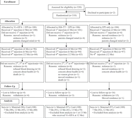
Study participants.A total of 318 healthy full-term infants (161 males and 157 females) born between 26 July 2006 and 8 March 2007 were enrolled in the study and randomized into groups (Fig. 1). Maternal and cord blood samples were col-lected from 307 subjects, cord blood only was colcol-lected from 10, and maternal blood only was collected from 1. Seventeen infants were withdrawn from the study before the first injec-tion: 10 parents changed their minds, 5 were ill on the day of vaccination, and 2 moved out of the area. As a result, 301 infants comprised the study group, with 100 in the Vi-rEPA group, 101 in the Hib-TT group, and 100 in the EPI group (Fig. 1). Another 60 were withdrawn or lost to follow-up, including 7 after the first injection, 11 after the second, 35 after the third, and 7 after the fourth: 26 parents withdrew because of con-cerns about the child’s well-being (43.3%), 16 withdrew due to
illness on the vaccination day (26.6%), 10 refused blood sam-pling (16.6%), 3 moved out of the area (5.0%), 3 gave no reasons (5.0%), and 2 withdrew due to the child’s death (3.3%).
Infants’ gestational ages ranged from 37 to 44 weeks, and birth weights were ⱖ2,500 g (median, 3,000 g). All had a normal neonatal course. Mothers’ ages ranged from 17 to 38 years (median, 24 years). The median maternal ages, gesta-tional ages, birth weights, and maternal ages were similar among the three groups.
Vaccination. Three hundred one infants received the first injection, 294 the second, 283 the third, and 167 the fourth of either Vi-rEPA or Hib-TT. The numbers of infants who re-ceived the first, second, and third injections were similar among the three groups, and the numbers that received the fourth injection were similar between the Vi-rEPA and Hib-TT groups (Fig. 1). The mean age at each injection for each of the three groups was 76 to 77 days for the first injection (range, 61 to 92 days), 137 days for the second (range, 120 to 154 days), 197 to 198 days for the third (range, 181 to 212 days), and 381 to 382 days for the fourth (range, 365 to 420 days).
Vaccine safety. There were no vaccine-related serious ad-verse events (Table 1). Only the highest temperature and max-imal local reaction for each individual were used for analyses. No differences were found among the three groups. The most common minor adverse reaction was mild fever (temperature of 38.0 to 38.4°C) on the day of first injection, which subsided the next day, in 14 to 18% of vaccinees of each vaccine group.
(i) Fever.After the first injection, 63 (20.9%) infants had a temperature of ⱖ38.0°C (maximum, 39.8°C): 22 in the Vi-rEPA group, 24 in the Hib-TT group, and 17 in the EPI group. This fever lasted for⬍24 h in 59 vaccinees and for 24 to 48 h in 4 vaccinees. One infant in the Vi-rEPA group had a tem-perature of 39.8°C 6 h after injection which had returned to normal at 24 h. After the second injection, 20 infants had a temperature of ⱖ38.0°C (maximum, 39.2°C): 7 each in the Vi-rEPA and Hib-TT groups and 6 in the EPI group. This fever lasted for⬍24 h in 18 vaccinees and for 24 to 48 h in 2 vaccinees. After the third injection, 6 infants had a tempera-ture between 38.0°C and 39.5°C: 4 in the Vi-rEPA group, 2 in the Hib-TT group, and none in the EPI group. All fevers lasted for⬍24 h. After the fourth injection, 2 infants in the Vi-rEPA group had a temperature of 38.0°C, and none of the infants in the Hib-TT group had a fever. No fever was associated with convulsions or intercurrent infections.
(ii) Local reactions.Induration ofⱖ2.5 cm was observed at 14 injection sites (10 after the first injection and 4 after the second), with a maximal size of 4 cm. All but one occurred at the DTP site, and none lasted for⬎48 h. Erythema ofⱖ5 cm was observed at 10 injection sites (8 at the DTP site and 1 each at the Vi-rEPA and Hib-TT sites); 8 incidences were 5 to 7 cm, and 1 each at the DTP and Hib-TT sites was 15 cm. All subsided within 2 days.
(iii) Systemic reactions. Inconsolable crying was reported for 17 infants after the first injection, lasting from 15 min to 6 h: seven cases lasted for⬍1 h, six for 1 to 2 h, one for 2.5 h, two for 5 h, and one for 6 h. Two deaths, not related to vaccination, were reported: a 5-month-old who received 1 in-jection of Vi-rEPA at 2.5 months of age died of septicemia
on August 17, 2020 by guest
http://cvi.asm.org/
following an operation for “nonfunctioning” kidneys, and a 4-month-old died of pneumonia 19 days after the second in-jection of Hib-TT.
Serum IgG anti-Vi.The IgG anti-Vi GM for maternal and cord sera were similar among the 3 groups (Table 2). Among mothers who wereⱖ30 years of age, 20% had anti-Vi levels of
ⱖ3.5 EU (estimated protective level), compared to 6% of those who were⬍30 years old (P⫽0.001).
At 7 months, the IgG anti-Vi GM for the Vi-rEPA group increased from the cord level of 0.66 to 17.42 EU (P⬍0.001), with 90% of infants having levels ofⱖ3.5 EU. At 12 months, it dropped to 4.76 EU (P⬍0.001), with 62% of infants having levels ofⱖ3.5 EU. At 13 months, 1 month after the fourth injection, the GM rose to 50.07 EU (P⬍0.001), with 95% of infants having levels ofⱖ3.5 EU. The serum IgG anti-Vi GM for the Hib-TT and EPI groups, in contrast, declined from cord levels of 0.55 and 0.52 EU to 0.16 and 0.05 EU, respectively, at 7 months and remained at about these levels through 13 months of age. Two infants in the Hib-TT group and another two in the EPI group had elevated anti-Vi levels at 7 months. Anti-Vi levels of these 4 infants declined at 12 months; in 2 of the infants, anti-Vi levels wereⱖ3.5 EU. These two had no
blood samples at 13 months. We were unable to verify whether these four infants received Vi-rEPA vaccine inadvertently or their serum samples were mislabeled.
Effect of cord IgG anti-Vi level on infant antibody response.
The estimated protective level (3.5 EU) was used to define high and low cord anti-Vi levels. Of the 90 infants in the Vi-rEPA group whose serum samples were collected at 7 months (1 month after the third injection), 81 were in the low group (IgG anti-Vi GM, 0.57 EU) and 9 were in the high group (IgG anti-Vi GM, 6.83 EU) (Table 3). For infants in the low group, the IgG anti-Vi GM increased from the cord level of 0.57 EU to 20.3 EU at 7 months, declined to 5.32 EU at 12 months, and rose to 60.5 EU at 13 months. For infants in the high group, however, an increase of IgG anti-Vi GM, from 1.87 to 9.13 EU, was not observed until 13 months. Nine infants in the EPI group and 6 in the Hib-TT group had high cord anti-Vi levels; their IgG anti-Vi GM declined from 11.23 EU at birth to 0.11 EU at 7 months.
IgG anti-Vi GM after injections were significantly higher at all times for infants of the low cord group than for those of the high cord group. The percentage of infants that achieved a level ofⱖ3.5 EU was higher for infants of the low cord group
FIG. 1. Flow chart of vaccinations.
on August 17, 2020 by guest
http://cvi.asm.org/
than for those of the high cord group: 93.8% versus 55.6% (P⫽0.001) at 7 months, 65.8% versus 33.3% (P⫽0.006) at 12 months, and 97.2% versus 75.0% (P ⫽0.048) at 13 months. Multiple regression analysis was used to assess whether the cord and 12-month levels independently affected the response at 13 months. The results showed that cord and 12-month levels were independently associated with the response at 13 months (P⬍0.001) and that high cord anti-Vi levels continued to partially suppress the response at 13 months.
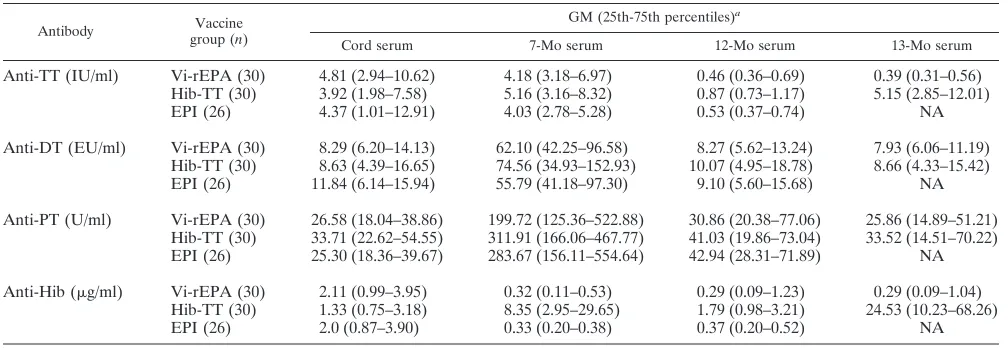
Antibody responses to tetanus and diphtheria toxoids, per-tussis toxin, and Hib CP. There were no differences in IgG GM for antibodies to DT, PT, and Hib CP between the Vi-rEPA and EPI groups at all time points (Table 4). Recipients of Hib-TT had similar levels of DT and PT antibodies to those of the Vi-rEPA and EPI groups. At 7 months, all groups had similar levels of anti-TT. The levels declined at 12 months for all groups; infants of the Hib-TT group, however, had a higher anti-TT GM than those for the Vi-rEPA and EPI groups (0.87
TABLE 1. Adverse reactions after vaccinationa
Reaction
No. of infants with reaction
First injection Second injection Third injection Fourth
injectionb
Vi-rEPA (n⫽100)
Hib-TT (n⫽101)
EPI (n⫽100)
Vi-rEPA (n⫽96)
Hib-TT (n⫽99)
EPI (n⫽99)
Vi-rEPA (n⫽92)
Hib-TT (n⫽94)
EPI (n⫽97)
Vi-rEPA (n⫽84)
Fever
38.0-38.4°C 16 18 14 5 6 6 3 2 0 2
38.5-39.5°C 5 6 3 2 1 0 1 0 0 0
⬎39.5°C 1 0 0 0 0 0 0 0 0 0
Induration ofⱖ2.5 cm
DTP site 2 2 5 1 1 2 0 0 0
Vi-rEPA or Hib-TT site 1 0 0 0 0 0 0
Erythema ofⱖ5 cm
DTP site 3 1 4 0 0 0 0 0 0
Vi-rEPA or Hib-TT site 2 0 0 0 0 0 0
Inconsolable crying
⬍4 h 5 7 2 0 0 0 0 0 0 0
ⱖ4 h 2 1 0 0 0 0 0 0 0 0
a
Axillary temperatures and induration and erythema at the injection site were recorded at 6, 24, and 48 h for each participant. No vaccine-related serious events were observed. EPI, Expanded Program on Immunization. EPI vaccines included DTP, OPV, and hepatitis B vaccines at 2 and 4 months and DTP vaccine at 6 months. Vi-rEPA or Hib-TT was given with the EPI vaccines at 2, 4, and 6 months and alone at 12 months.
b
No adverse reactions were observed in the Hib-TT group (n⫽83) after the fourth injection.
TABLE 2. Serum IgG anti-Vi levels of vaccinees
Vaccine group and parametera
Value
Maternal serum
Cord serum
7-Mo serum
12-Mo serum
13-Mo serum
Vi-rEPA
n 96 100 90 85 80
GM 0.58 0.66 17.42 4.76 50.07
25th-75th percentile 0.24–1.17 0.30–1.46 7.33–47.25 2.20–12.71 22.33–133.61
% of samples withⱖ3.5 EUb 10.4 9.0 90.0 62.4 95.0
Hib-TT
n 97 100 91 81 79
GM 0.46 0.55 0.16 0.17 0.19
25th-75th percentile 0.23–0.85 0.26–1.02 0.10–0.25 0.11–0.27 0.11–0.27
% of samples withⱖ3.5 EUb 5.2 6.0 2.2 1.2 0
EPI
n 97 100 92 81
GM 0.51 0.52 0.05 0.04
25th-75th percentile 0.20–1.25 0.20–0.98 0.02–0.09 0.02–0.08 NAc
% of samples withⱖ3.5 EUb 10.3 9.0 2.2 1.2
a
EPI, Expanded Program on Immunization. The EPI vaccines included DTP, OPV, and hepatitis B vaccines at 2 and 4 months and DTP vaccine at 6 months. Vi-rEPA or Hib-TT was given with the EPI vaccines at 2, 4, and 6 months and alone at 12 months.
b
Estimated protective level (17).
c
NA, not available.
on August 17, 2020 by guest
http://cvi.asm.org/
versus 0.46 [P ⬍ 0.002] and 0.87 versus 0.53 [P ⫽ 0.001], respectively). At 13 months (1 month after the 4th injection), recipients of Hib-TT had higher levels of anti-TT than recip-ients of Vi-rEPA (5.15 versus 0.39 [P⬍0.001]), likely due to the additional dose of Hib-TT. The 3 groups had similar cord anti-Hib IgG GM. There was a significant rise of anti-Hib 1 month after 3 injections of Hib-TT (8.35g/ml) that declined to 1.79g/ml at 12 months and rose to 24.6g/ml after the 4th injection. Similar to anti-Vi IgG responses, anti-Hib IgG re-sponses were inversely correlated with the cord level (R2 ⫽
0.92) (not shown).
DISCUSSION
In a randomized, vaccine-controlled study of infants in Viet-nam, Vi-rEPA was safe, elicited protective levels of IgG anti-Vi, and was compatible with EPI vaccines. These data show that Vi-rEPA can be added to the routine immunization of infants in countries where typhoid fever is prevalent. Immunization for
ty-phoid fever added to the EPI vaccines would have avoided the quandary that befell Tajikistan public health officials in 1996 when drinking water became contaminated with fecal material, result-ing in 8,901 typhoid cases and 95 deaths (22). Durresult-ing the peak 5 months of this outbreak, typhoid vaccine administration could not be implemented. Similar outbreaks in Vietnam and Nepal have been reported recently (5, 19). The availability of a safe, effective vaccine that elicits long-lasting immunity would be a welcome addition to the routine immunization of infants. The addition of lipopolysaccharide (LPS)-based conjugates to Vi conjugates, such
as aSalmonella entericaserovar Paratyphi A-based conjugate in
Southeast Asia, anS. entericaserovar Typhimurium-based conju-gate in Africa, and aShigella-based conjugate at both sites, could result in substantially improved health at all ages (14, 26, 36).
The suppressive effect of high levels of maternally derived IgG antibody upon infant antibody responses has been demonstrated for bacterial (diphtheria and tetanus toxoids and pertussis toxin), polysaccharide, and viral vaccines (2, 3, 7, 8, 10, 25, 26, 27, 28, 29, 31, 35). High levels of maternally derived Vi antibodies exerted
TABLE 3. Vaccinee responses to Vi-rEPA according to cord IgG anti-Vi levels
Cord level group and parametera Value
Cord blood 7-Mo serum 12-Mo serum 13-Mo serum
Cord levels ofⱖ3.5 EU (high)
n 9 9 9 8
GM 6.83 4.40 1.87 9.13
25th-75th percentiles 4.78–8.50 1.38–15.22 0.67–4.79 4.69–46.74
No. (%) of samples withⱖ3.5 EU 9 (100.0) 5 (55.6) 3 (33.3) 6 (75.0)
Cord levels of⬍3.5 EU (low)
n 81 81 76 72
GM 0.57 20.29 5.32 60.49
25th-75th percentiles 0.29–1.10 7.66–51.67 2.27–14.10 32.40–142.99
No. (%) of samples withⱖ3.5 EU 0 76 (93.8) 50 (65.8) 70 (97.2)
a
The GM at each time point were compared within the groups and between the high and low groups. Those withPvalues of⬍0.05 were as follows: high cord group, 1.87 versus 9.13 (P⫽0.04); low cord group, 0.57 versus 20.29, 20.29 versus 5.32, and 5.32 versus 60.49 (P⬍0.001); and high versus low cord group, 4.40 versus 20.29 (P⫽0.009) at 7 months, 1.87 versus 5.32 (P⫽0.02) at 12 months, 9.13 versus 60.49 (P⫽0.005) at 13 months, 55.6% versus 93.8% (P⫽0.001) at 7 months, 33.3% versus 65.8% (P⫽0.006) at 12 months, and 75% versus 97.2% (P⫽0.048) at 13 months.
TABLE 4. Serum IgG antibodies to TT, DT, PT, and Hib CP of vaccinees
Antibody Vaccine
group (n)
GM (25th-75th percentiles)a
Cord serum 7-Mo serum 12-Mo serum 13-Mo serum
Anti-TT (IU/ml) Vi-rEPA (30) 4.81 (2.94–10.62) 4.18 (3.18–6.97) 0.46 (0.36–0.69) 0.39 (0.31–0.56) Hib-TT (30) 3.92 (1.98–7.58) 5.16 (3.16–8.32) 0.87 (0.73–1.17) 5.15 (2.85–12.01)
EPI (26) 4.37 (1.01–12.91) 4.03 (2.78–5.28) 0.53 (0.37–0.74) NA
Anti-DT (EU/ml) Vi-rEPA (30) 8.29 (6.20–14.13) 62.10 (42.25–96.58) 8.27 (5.62–13.24) 7.93 (6.06–11.19) Hib-TT (30) 8.63 (4.39–16.65) 74.56 (34.93–152.93) 10.07 (4.95–18.78) 8.66 (4.33–15.42) EPI (26) 11.84 (6.14–15.94) 55.79 (41.18–97.30) 9.10 (5.60–15.68) NA
Anti-PT (U/ml) Vi-rEPA (30) 26.58 (18.04–38.86) 199.72 (125.36–522.88) 30.86 (20.38–77.06) 25.86 (14.89–51.21) Hib-TT (30) 33.71 (22.62–54.55) 311.91 (166.06–467.77) 41.03 (19.86–73.04) 33.52 (14.51–70.22) EPI (26) 25.30 (18.36–39.67) 283.67 (156.11–554.64) 42.94 (28.31–71.89) NA
Anti-Hib (g/ml) Vi-rEPA (30) 2.11 (0.99–3.95) 0.32 (0.11–0.53) 0.29 (0.09–1.23) 0.29 (0.09–1.04) Hib-TT (30) 1.33 (0.75–3.18) 8.35 (2.95–29.65) 1.79 (0.98–3.21) 24.53 (10.23–68.26)
EPI (26) 2.0 (0.87–3.90) 0.33 (0.20–0.38) 0.37 (0.20–0.52) NA
a
The GM at each time point were compared between groups: Vi-rEPA versus Hib-TT groups, Vi-rEPA versus EPI group, and Hib-TT versus EPI group. Those with Pvalues of⬍0.05 were as follows: for anti-TT GM at 12 months, 0.46 versus 0.87 (P⬍0.001) and 0.53 versus 0.87 (P⫽0.01); for anti-TT GM at 13 months, 0.39 versus 5.15 (P⬍0.001); and for anti-Hib GM at 7, 12, and 13 months, Hib-TT versus EPI group and Hib-TT versus Vi-rEPA group (allPvalues were⬍0.001). NA, not available.
on August 17, 2020 by guest
http://cvi.asm.org/
this suppressive effect on infants’ vaccine-induced anti-Vi levels at 7, 12, and 13 months of age. This suppressive effect was also related to the age-related IgG anti-Vi levels of the mothers. Fur-ther studies of this effect may be needed to guide the immuniza-tion schedule in areas where typhoid is endemic, where high levels of anti-Vi occur in women of child-bearing age (15). Another factor that might affect the immunogenicity of Vi conjugates was demonstrated recently, as extreme preterm birth (⬍28 to 30 weeks of gestation) was associated with lower serum antibody responses to several vaccines, including Vi, Hib, tetanus, and polio vaccines (7). We suggest that some “vaccine failures” could be due to either prematurity or high levels of maternally derived antibodies.
As shown for other polysaccharide vaccines, immunization with Vi polysaccharide atⱖ2 years of age induced herd immunity in Kolkata, India (32). Vi conjugate is a better immunogen than Vi at all ages and should be used widely in areas of high typhoid endemicity. In addition to prevention of acute typhoid fever, vaccination against typhoid fever should prevent the chronic car-rier state and extraintestinal complications such as hepatobiliary cancers (37).
ACKNOWLEDGMENTS
We are indebted to the parents, health staff, and local leaders of Thanh Thuy District and Phu Tho Province for their support of the program and to the following contributors to the study: George Grimes, Pharmaceutical Development Section, Clinical Center, NIH; Patricia Moyer and James F. Troendle, Division of Epidemiology, Statistics and Prevention Research, NICHD; and Arthur Karpas, NICHD.
This study was supported by funds from the Division of Intramural Research, NICHD.
REFERENCES
1.Acharya, I. L., et al.1986. Prevention of typhoid fever in Nepal with the Vi capsular polysaccharide of Salmonella typhi. A preliminary report. N. Engl. J. Med.317:1101–1104.
2.Bell, B. P., et al.2007. Immunogenicity of an inactivated hepatitis A vaccine in infants and young children. Pediatr. Infect. Dis. J.26:116–122. 3.Bjo¨rkholm, B., M. Granstro¨m, J. Taranger, M. Wahl, and L. Hagberg.1995.
Influence of high titers of maternal antibody on the serologic response of infants to diphtheria vaccination at three, five, and twelve months of age. Pediatr. Infect. Dis. J.4:846–850.
4.Brooks, W. A., et al.2005. Bacteremic typhoid fever in children in an urban slum, Bangladesh. Emerg. Infect. Dis.11:326–329.
5.Connerton, P., et al.2000. Epidemic typhoid in Vietnam: molecular typing of multiple-antibiotic-resistant Salmonella enterica serotype Typhi from four outbreaks. J. Clin. Microbiol.38:895–897.
6.Crump, J. A., and E. D. Mintz.2010. Global trends in typhoid and paraty-phoid fever. Clin. Infect. Dis.50:241–246.
7.D’Angio C. T., W. M. Maniscalco, and M. E. Pichichero.1995. Immunologic response of extremely premature infants to tetanus, Haemophilus influen-zae, and polio immunizations. Pediatrics96:18–22.
8.Fiore, A. E., et al.2003. Hepatitis A vaccination of infants: effect of maternal antibody status on antibody persistence and response to a booster dose. Pediatr. Infect. Dis. J.22:354–359.
9.Gilman, R. H., M. Terminel, M. M. Levine, P. Hernandez-Mendoza, and R. B. Hornick.1975. Relative efficacy of blood, urine, rectal swab, bone-marrow, and rose-spot cultures for recovery of Salmonella typhi in typhoid fever. Lanceti:1211–1213.
10.Glezen, W. P.2003. Effect of maternal antibodies on the infant immune response. Vaccine21:3389–3392.
11.Hornick, R. B., et al.1970. Typhoid fever: pathogenesis and immunologic control. N. Engl. J. Med.283:688–691.
12.Horne, A. D.1995. The statistical analysis of immunogenicity data in vaccine trials. A review of methodologies and issues. Ann. N. Y. Acad. Sci.754:329–346. 13.Klugman, K. P., et al.1987. Protective activity of Vi capsular polysaccharide
vaccine against typhoid fever. Lancetii:1165–1169.
14.Konadu, E., J. Shiloach, D. A. Bryla, J. B. Robbins, and S. C. Szu.1996. Synthesis, characterization and immunological properties in mice of conju-gates composed of detoxified lipopolysaccharide of Salmonella paratyphi A bound to tetanus toxoid, with emphasis on the role of O-acetyls. Infect. Immun.64:2709–2715.
15.Kossaczka, Z., et al.1999. Safety and immunogenicity of Vi conjugate vac-cines for typhoid fever in adults, teenagers, and 2- to 4-year-olds in Vietnam. Infect. Immun.67:5806–5810.
16.Kossaczka, Z., et al.1997. Synthesis and immunological properties of Vi and di-O-acetyl pectin protein conjugates with adipic acid dihydrazide as the linker. Infect. Immun.65:2088–2093.
17.Lanh, M. N., et al.1999. Persistent efficacy of Vi conjugate vaccine against typhoid fever in young children. N. Engl. J. Med.349:1390–1391. 18.Levine, M. M., et al.1999. Duration of efficacy of Ty21a, attenuated
Salmo-nella typhi live oral vaccine. Vaccine17(Suppl. 2):S22–S27.
19.Lewis, M. D., et al.2005. Typhoid fever: a massive, single-point source, multidrug-resistant outbreak in Nepal. Clin. Infect. Dis.40:554–561. 20.Lin, F. Y., et al.2000. The epidemiology of typhoid fever in the Dong Thap
Province, Mekong Delta region of Vietnam. Am. J. Trop. Med. Hyg.62:
644–648.
21.Lin, F. Y., et al.2001. Efficacy of a Salmonella typhi Vi conjugate vaccine (Vi-rEPA) in 2 to 5 year-old children. N. Engl. J. Med.344:1263–1269. 22.Mermin, J. H., et al.1999. A massive epidemic of multidrug-resistant typhoid
fever in Tajikistan associated with consumption of municipal water. J. Infect. Dis.179:1416–1422.
23.Moore, S. E., et al.2004. Birth weight predicts response to vaccination in adults born in an urban slum in Lahore, Pakistan. Am. J. Clin. Nutr.80:453–459. 24.Ochiai, R. L., et al.2008. A study of typhoid fever in five Asian countries:
disease burden and implications for controls. Bull. World Health Organ.
86:260–268.
25.Osborn, J. J., J. Dancis, and J. F. Julia.1952. Studies of the immunology of the newborn infant. II. Interference with active immunization by passive transplacental circulating antibody. Pediatrics10:328–334.
26.Passwell, J. H., et al.2010. Age-related efficacy of Shigella O-specific poly-saccharide conjugates in 1-4-year-old Israeli children. Vaccine28:2231–2235. 27.Premenko-Lanier, M., et al.2006. Maternal antibody inhibits both cellular and humoral immunity in response to measles vaccination at birth. Virology
350:429–432.
28.Santosham, M., et al.2001. Safety and antibody persistence following Hae-mophilus influenzae type b conjugate or pneumococcal polysaccharide vac-cines given before pregnancy in women of childbearing age and their infants. Pediatr. Infect. Dis. J.20:931–940.
29.Sarvas, H., S. Kurikka, I. J. T. Seppa¨la¨, P. H. Ma¨kela¨, and O. Ma¨kela¨.1992. Maternal antibodies partly inhibit an active antibody response to routine tetanus toxoid immunization in infants. J. Infect. Dis.165:977–979. 30.Sinha, A., et al.1999. Typhoid fever in children ages less than 5 years. Lancet
354:734–737.
31.Steinhoff, M. C., K. Zaman, E. Roy, R. Raqib, and E. Arifeen.2009. Effect of maternal pneumococcal antibody on infant antibody response to Streptococ-cus pneumoniae conjugate vaccine (PCV7), abstr. S17. Abstr. Annu. Conf. Vaccine Res., 27 to 29 April 2009.
32.Sur, D., et al.2009. A cluster-randomized effectiveness trial of Vi typhoid vaccine in India. N. Engl. J. Med.361:335–344.
33.Szu, S. C., A. L. Stone, J. D. Robbins, R. Schneerson, and J. B. Robbins.
1987. Vi capsular polysaccharide-protein conjugates for prevention of ty-phoid fever. Preparation, characterization, and immunogenicity in labora-tory animals. J. Exp. Med.166:1510–1524.
34.Taylor, D. N., M. M. Levine, L. Kuppens, and B. Ivanoff.1999. Why are typhoid vaccines not recommended for epidemic typhoid fever? J. Infect. Dis.180:2089–2090.
35.Van Savage, J., M. D. Decker, K. M. Edwards, S. H. Sell, and D. T. Karzon.
1990. Natural history of pertussis antibody in the infant and effect on vaccine response. J. Infect. Dis.161:487–492.
36.Watson, D. C., J. B. Robbins, and S. C. Szu.1992. Protection of mice against Salmonella typhimurium with an O-specific polysaccharide-protein conju-gate vaccine. Infect. Immun.60:4679–4686.
37.Welton, J. C., J. S. Marr, and S. M. Friedman.1979. Association between hepatobiliary cancer and typhoid carrier state. Lanceti:791–794. 38.World Health Organization.1994. Requirements for Vi polysaccharide
ty-phoid vaccine. Requirements for biological substances report no. 43. World Health Organ. Tech. Rep. Ser.840:l14.