Western University Western University
Scholarship@Western
Scholarship@Western
Electronic Thesis and Dissertation Repository
12-11-2017 10:00 AM
Identification and characterization of farR and farE as a regulator
Identification and characterization of farR and farE as a regulator
and effector of fatty acid resistance in Staphylococcus aureus
and effector of fatty acid resistance in Staphylococcus aureus
Heba Alnaseri
The University of Western Ontario Supervisor
McGavin, Martin J.
The University of Western Ontario
Graduate Program in Microbiology and Immunology
A thesis submitted in partial fulfillment of the requirements for the degree in Doctor of Philosophy
© Heba Alnaseri 2017
Follow this and additional works at: https://ir.lib.uwo.ca/etd Part of the Bacteriology Commons
Recommended Citation Recommended Citation
Abstract(
Although Staphylococcus aureus is exposed to antimicrobial fatty acids on the skin, in nasal secretions and in abscesses, specific mechanisms for regulating gene expression and intrinsic resistance in response to these fatty acids have not been reported. Through in vitro selection for increased resistance of S. aureus to linoleic acid, I identified fatty acid resistant clone FAR7, where a single nucleotide polymorphism caused a His121Tyr
substitution in an uncharacterized member of the TetR family of transcriptional regulators, which is divergently transcribed from a gene encoding a member of the resistance-nodulation-division superfamily of multi-drug efflux pumps. I named these genes farE and farR, for regulator and effector of fatty acid resistance, respectively. S. aureusΔfarER exhibited loss of inducible resistance to linoleic acid, and although FarR is a TetR family regulator which typically repress expression of a divergent gene, I found that FarR is needed to induce farE. Compared to wild type S. aureus, FAR7 exhibited increased expression of farR and farE under non-inducing conditions, and a significantly higher induced level of farE. Electrophoretic mobility shift assays revealed a FarR binding site in the farER intergenic segment, that overlaps with the +1 transcription start site of farR as determined by 5"-RACE. The variant FarR7 produced by S. aureus FAR7
failed to bind to this operator site, and nucleotide substitutions within the operator abolished binding of native FarR. Conversely, FarR and FarR7 bound equally well to a second operator site upstream of the predicted farE promoter. Therefore, like other TetR regulators, FarR represses its own expression, and a His121Tyr substitution in FarR causes
a loss of auto-repression and increases expression of both farR and farE. My data reports the first description of a specific mechanism of inducible resistance to antimicrobial fatty acids in a Gram-positive pathogen and defines a new paradigm for regulation of a divergently transcribed gene by a TetR family regulator.
Keywords
Antimicrobial fatty acids, efflux pumps, TetR family regulator, inducible resistance,
Co0Authorship(Statement((
All studies presented in this thesis were completed by Heba Alnaseri in the laboratory of Dr. Martin McGavin with assistance from co-authors as listed below. Dr. Martin McGavin contributed to the conceptualization, design, data analysis and manuscript preparation of all experiments.
Chapter 2: Inducible expression of a resistance-nodulation-division-type efflux pump in Staphylococcus aureus provides resistance to linoleic and arachidonic acids.
This chapter has been previously published:
Alnaseri H, Arsic B, Schneider JE, Kaiser JC, Scinocca ZC, Heinrichs DE, McGavin MJ. 2015. Inducible expression of a resistance-nodulation-division-type efflux pump in
Staphylococcus aureus provides resistance to linoleic and arachidonic acids. J Bacteriol 197:JB.02607-14.
Benjamin Arsic selected the fatty acid resistant clones that were sent for comparative genome sequencing. James Schneider and Zachariah Scinocca assisted in the farE
reporter gene assays. Julie Kaiser assisted with the set-up of [14C]linoleic acid uptake assays.
Chapter 3: Identification of FarR binding site responsible for auto-repression and efflux-mediated resistance to antimicrobial fatty acids in Staphylococcus aureus.
This chapter is being prepared for submission to a peer-reviewed journal.
Katherine A. Ferguson assisted with farR reporter gene assays and western blotting. James Schneider constructed the USA300ΔfakA mutant and helped with farE reporter
Epigraph(
“You may encounter many defeats, but you must not be defeated. In fact, it may be necessary to encounter the defeats, so you can know who you are, what you can rise from, how you can still come out of it.”
-Maya Angelou
(
(
(
Dedication(
Acknowledgments((
To my supervisor Dr. Martin McGavin: I owe my deepest gratitude to you for your mentorship, patience and encouragement throughout the course of my doctoral studies. Thank you for always challenging me and pushing me to think critically and boost my intellectual curiosity. I am truly fortunate to have be given the opportunity to learn from you, grow and become an independent researcher. None of this would have ever possible without your generosity, and for everything you have done for me Dr. McGavin, I will be forever in your debt.
To Dr. David Heinrichs and Dr. John McCormick: I would like to sincerely thank you for the time you devoted towards helping me. Your guidance, accessibility and kind support have been essential for my graduate studies. I am truly grateful for your engaging conversations during my committee meetings. You helped foster my confidence in myself as a scientist in more ways than you can ever imagine. To the Heinrichs and McCormick lab members: I would like to thank you for always being willing to listen to me, share your resources and for always making me feel welcomed. It was a true pleasure to have shared great conversations and plenty of laughter with all of you.
To the department of Microbiology and Immunology faculty, staff and students: I would like to thank you for the kindness you have shown me over the years. I could not have asked for a more supportive environment to grow personally and academically. To Elizabeth Coley and Fernanda Russell: I would like to especially thank you for your endless support and encouragement during the past five years. I sincerely will miss you both very much.
Table(of(Contents(
Title Page ... i!
Certificate of Examination ... ii!
Abstract ... iii!
Co-Authorship Statement ... iv!
Epigraph ... v!
Dedication ... vi!
Acknowledgments ... vii!
Table of Contents ... ix!
List of Tables ... xiii!
List of Figures ... xiv!
List of Appendices ... xvi!
List of Abbreviations ... xvii!
Chapter 1 ... 1!
1! Introduction ... 1!
1.1! Staphylococcus aureus Overview ... 2!
1.2! Emergence of Methicillin-Resistant Community-acquired Staphylococcus aureus ... 2!
1.3! Staphylococcus aureus colonization ... 3!
1.4! Staphylococcus aureus pathogenesis andclinical manifestation ... 4!
1.5! Genetic regulation of Staphylococcus aureus virulence ... 10!
1.6! Antimicrobial Resistance mechanisms of Staphylococcus aureus ... 14!
1.7! Staphylococcus aureus envelope architecture ... 15!
1.7.1! Cell envelope composition ... 15!
1.8! Fatty acid machinery of Staphylococcus aureus ... 22!
1.8.1! Phospholipid composition ... 22!
1.8.2! Phospholipid synthesis ... 22!
1.8.3! Lipids as antimicrobials ... 28!
1.9! Bacterial efflux pumps ... 34!
1.10!TetR family of transcriptional regulators and their biological functions ... 35!
1.11!Rationale and hypothesis ... 37!
1.12!Literature cited ... 40!
Chapter 2 ... 65!
2! Inducible expression of a Resistance-Nodulation-Division-Type efflux pump in Staphylococcus aureus provides resistance to Linoleic and Arachidonic Acids ... 65!
2.1! Introduction ... 66!
2.2! Materials and methods ... 68!
2.2.1! Bacterial strains and growth conditions ... 68!
2.2.2! Selection and comparative genome sequencing of fatty acid resistant (FAR) clones ... 71!
2.2.3! Strain and plasmid construction ... 72!
2.2.4! Assay of growth and bactericidal activity ... 75!
2.2.5! Assay for uptake of 14C-linoleic acid ... 75!
2.2.6! farE::lux reporter gene assays ... 76!
2.2.7! Data analyses ... 76!
2.3! Results ... 77!
2.3.4! farE contributes to persistence and growth of S. aureus in the presence
of linoleic acid ... 82!
2.3.5! The FAR7 clone exhibits increased expression of farE ... 87!
2.3.6! An H121Y substitution in FarR is sufficient for increased resistance to linoleic acid ... 87!
2.3.7! Role of farE in resistance to other uFFA ... 90!
2.3.8! Inactivation of farE promotes increased uptake of 14C-linoleic acid ... 96!
2.4! Discussion ... 99!
2.5! References ... 104!
Chapter 3 ... 112!
3! Identification of FarR binding sites responsible for auto-repression and efflux-mediated resistance to antimicrobial fatty acids in Staphylococcus aureus ... 112!
3.1! Introduction ... 113!
3.2! Materials and methods ... 114!
3.2.1! Bacterial strains and growth conditions ... 114!
3.2.2! Construction of S. aureus USA300ΔfarER and USA300ΔfakA ... 115!
3.2.3! Construction of complementation and reporter gene constructs ... 116!
3.2.4! RNA isolation and 5"-rapid amplification of complementary DNA ends ... 121!
3.2.5! Construction and purification of recombinant FarR ... 121!
3.2.6! FarR-DNA interaction studies ... 122!
3.2.7! Antibody production and western blotting ... 123!
3.2.8! Murine infection model ... 123!
3.2.9! Computer analyses ... 124!
3.3! Results ... 124!
3.3.3! Identification of farR promoter and transcription start site ... 128!
3.3.4! Expression of farR is subject to auto-regulation ... 128!
3.3.5! Identification of FarR operator sites ... 132!
3.3.6! FarR binds OPAct and OPRep ... 137!
3.3.7! OPRep contains the site of farR auto-repression and a His121Tyr substitution in FarR causes relief of auto-repression ... 139!
3.3.8! Specificity determinants of FarR binding ... 144!
3.3.9! farER and fatty acid detoxification ... 150!
3.3.10!farER influence virulence in a subcutaneous abscess infection model ... 153!
3.4! Discussion ... 155!
3.5! References ... 161!
Chapter 4 ... 165!
4! General Discussion and Conclusions ... 165!
4.1! Summary ... 166!
4.2! Limitations and future studies ... 168!
4.3! References ... 176!
Appendices ... 178!
List(of(Tables((
Table 1.1 Summary of S. aureus virulence determinants. ... 6!
Table 1.2 Major regulators of virulence determinants in S. aureus. ... 12!
Table 1.3 Summary of proposed mechanisms of uFFA toxicity. ... 30!
Table 1.4 Summary of uFFA resistance mechanisms proposed to date. ... 33!
Table 2.1 Strains and plasmids used in Chapter 2. ... 69!
Table 2.2 Primers used for construction of plasmids in Chapter 2. ... 74!
Table 3.1 Strains and plasmids used in Chapter 3. ... 117!
List(of(Figures((
Figure 1.1 Regulation of lipid homeostasis in E. coli ... 17
Figure 1.2 Regulation of lipid homeostasis in B. subtilis ... 21
Figure 1.3 Overview of fatty acid and phospholipid biosynthesis in S. aureus ... 26
Figure 1.4 Overview of exogenous fatty acid utilization by S. aureus ... 27
Figure 2.1 Linoleic acid induces expression of farE ... 80
Figure 2.2 Sensitivity of USA300 and USA300farR::ΦNΕ to the bactericidal activity of 100 µM linoleic acid. ... 81
Figure 2.3 Mutation of farE::ΦNΕ enhances sensitivity of S. aureus to toxicity of linoleic acid. ... 85
Figure 2.4 Sensitivity of USA300 and USA300farE::ΦNΕ cells to the bactericidal activity of 100 µM linoleic acid. ... 86
Figure 2.5 The FAR7 SNP causes enhanced induction of farE expression. ... 88
Figure 2.6 FAR7 is more resistant than USA300 to linoleic acid. ... 89
Figure 2.7 The variant farR7 allele, but not wild type farR, enables the ability of USA300farR::ΦNΕ to grow at inhibitory concentrations of linoleic acid. ... 92
Figure 2.8 Influence of different antimicrobial fatty acids on induction of farE, or viability of S. aureus USA300 and USA300farE::ΦNΕ. ... 94
Figure 2.9 Effect of farE::ΦNΕ and Δtet38 mutations on growth of S. aureus in the presence of 25 µM or 40 µM palmitoleic acid (PA). ... 95
Figure 2.10 Growth (A) and uptake of 14C-linoleic acid (B) following exposure of S. aureus USA300 and USA300farE::ΦΝΕ to an increase in concentration of linoleic acid. ... 98
Figure 3.1 farER contributes to resistance of S. aureus USA300 to linoleic acid. ... 126
Figure 3.5 Competition EMSA with OP2 reveals that an operator site for binding of FarR is located within OP13 ... 138
Figure 3.6 FarR and the variant FarR7 differ in binding to OP2 and OP13, both of
which span core promoter elements of PfarR. ... 143
Figure 3.7 OP4, OP5 and OP13 cross-compete with each other and nucleotide
substitutions obliterate this competition. ... 146
Figure 3.8 Multiple sequence alignment of the intergenic segment between farE and
farR among S. aureus USA300 and 12 other staphylococcal species that contain
divergent farER. ... 147
Figure 3.9 Nucleotide substitution in the -10 element of PfarR causes a relief of auto- repression. ... 149
Figure 3.10 farE::lux exhibits elevated constitutive levels of expression in the
absence of fatty acid kinase, fakA. ... 151
Figure 3.11 USA300ΔfakA exhibits enhanced resistance to linoleic acid, in a farE-
dependent manner. ... 152
Figure 3.12 USA300ΔfakA-farER exhibits reduced virulence in a murine skin abscess model of infection. ... 154
Figure 3.13 FarR binding to OPRep is partially affected by linoleoyl- CoA and
arachidonoyl-CoA. ... 160
Figure 4.1 Schematic model of the potential mechanism of PfarE regulation by FarR,
depending on exogenous fatty acid ligand. ... 172
Figure 4.2 Secondary structure prediction of farE and farR RNA using RNAfold
List(of(Appendices((
Appendix 1. ASM Journals Statement of Author’s Rights ... 178
Appendix 2. Adaptation of S. aureus USA300 during growth in TSB + 50 µM linoleic acid. ... 179
Appendix 3. Differential effect of pGYlux and pGYfarE::lux on growth of S. aureus
USA300 in TSB + 20 µM linoleic acid. ... 180
Appendix 4. Exponential phase cells are highly susceptible to the bactericidal activity of 100 µM linoleic acid (LA). ... 181
Appendix 5. Cadmium inducible expression of farR protects USA300farR::ΦΝΕ
from the bactericidal activity of 100 µM linoleic acid. ... 182
Appendix 6. FAR7, but not USA300, is able to grow in TSB containing 50 µM
palmitoleic acid (PA). ... 183
Appendix 7. fakA-farER influence virulence in a murine skin abscess model of
infection. ... 184
Appendix 8. Purification of 6×His-tagged FarR from wildtype USA300 and the
variant FAR7 using nickel affinity chromatography. ... 185
(
List(of(Abbreviations(
Δ Delta for deletion
ΔfakA fakA deletion
ΔfarER farER deletion
Δtet38 tet38 deletion
ΔfakA-ΔfarER fakA and farER double deletion
α toxin Alpha toxin
β-defensin Beta defensin
β hemolysin Beta hemolysin
β -lactamase Beta lactamase
δ toxin Delta toxin
γ toxin Gamma toxin
µCi microcurie
µg microgram
µL microlitre
µM micromolar
µm micrometre
°C Degrees Celsius
xg times gravity
5"-RACE 5"- rapid amplification of complementary DNA ends
AAP Abridged anchor primer
ABC ATP-binding cassette
ACME Arginine catabolic mobile element
Acr Acriflavine resistance
agr accessory gene regulator
AmtR Regulator of ammonium uptake
ATP Adenosine triphosphate
AUAP Abridged universal amplification primer
bp basepair
BSA Bovine serum albumin
C Carbon
CA-MRSA Community-acquired methicillin-resistant Staphylococcus aureus
Cd Cadmium
cfu Colony forming units
Cm Chloramphenicol
cme Campylobacter multidrug efflux gene
DDT Dithiothreitol
Dha Dihydroxyacetone
DMSO Dimethyl sulfoxide
EDTA Ethylenediaminetetraacetic acid
EMSA Electrophoretic mobility shift assay
Erm Erythromycin
EthR Ethionamide repressor
Fad Fatty acid degradation
Fak Fatty acid kinase
FAME Fatty acid modifying enzyme
FapR Fatty acid and phospholipid biosynthesis regulator
FAR Fatty acid resistance
FarE Effector of fatty acid resistance FarR Regulator of fatty acid resistance
FarR7 FarR protein from the variant FAR7 clone
FAS Fatty acid synthesis
G3P Glycerol-3-phosphate
Geh Glycerol ester hydrolase
GlcNAc N-acetylglucosamine
GSP Gene specific primer
HA-MRSA Hospital-associated methicillin-resistant Staphylococcus aureus
H Histidine
HapR Regulator of HA/protease gene in Vibrio cholerae
His Histidine
Ica Intercellular adhesion
IgG Immunoglobulin G
IPTG Isopropyl 1-thio-β-D- galactopyranoside
IR Imperfect repeat
IRDye Infrared dye
Isd Iron-regulated surface determinant
KCl Potassium chloride
Kb kilobase
LA Linoleic acid
Lac Los Angeles County clone
LB Luria Bertani
Luk Leukocidins
LysR Lysine regulator
M Molar
ManNAc N-acetylmannosamine
Mar Multiple antibiotic resistance
MATE Multidrug and toxic compound extrusion
MFS Major facilitator superfamily
Mg2+ Magnesium ion
MgCl2 Magnesium chloride
mL millilitre
MLST Multi-locus sequence type
mM Millimolar
Mtr Multiple transferable resistance
MurNAc N-acetylmuramic acid
NaCl Sodium chloride
ns Non-significant
OD Optical density
OP Operator
OPAct Activation operator site
OPRep Repression operator site
P Promoter
PfarE farE promoter
PfarR farR promoter
PA Palmitoleic acid
PAGE Polyacrylamide gel electrophoresis
PAL Pseudo-palindrome
PBP2a penicillin-binding protein 2a
PBS Phosphate-buffered saline
PCR Polymerase chain reaction
PHYRE Protein homology/analogy recognition engine
Pls Phosphate acyltransferase
pmol picomole
poly[d(I-C)] Poly-deoxyinosinic-deoxycytidylic acid
psi Pound-force per square inch
PtdIns Phosphphotidylinositol
PVDF Polyvinylidene difluoride
PVL Panton-valentine leukocidin
Qac Quaternary ammonium antiseptic compounds
RBS Ribosomal binding site
RLU Relative light units
RND Resistance nodulation cell division
rot Repressor of toxin
RPM Rotation per minute
RutR Regulator of pyrimidine utilization operon
sar Staphylococcal accessory regulator
sae S. aureus exoprotein
SCCmec Staphylococcal cassette chromosome
SczA Streptococcus zinc-sensing regulator
SdiA Regulator that suppresses cell division inhibitors
SDS Sodium dodecyl sulfate
SMR Small multidrug resistance
SNP Single nucleotide polymorphism
SoxS Superoxide response regulon transcriptional activator
SspA Staphylococcus serine protease
T7SS Type VII secretion system
TBE Tris-borate-EDTA
Tet Tetracycline
TSA Tryptic soy agar
TSB Tryptic soy broth
TSS Transcription start site
Tyr Tyrosine
uFFA Unsaturated free fatty acid
UTR Untranslated region
V Volt
Y Tyrosine
(
(
(
(
((
(
Chapter(1((
1(
Introduction(
1.1(
Staphylococcus,aureus,
Overview(
Staphyloccous aureus is a Gram-positive bacterium from the Firmicutes phylum that is considered as a pathobiont. It colonizes ~30% of the human population asymptomatically, and thus is a part of the resident microbiota, but also a pathogen proficient in causing diverse cutaneous and systemic infections of ranging severity (1). In his attempt to examine the underlying cause of blood-poisoning in 1881, Alexander Ogston observed micrococci with a spherical outline that stained a uniformly deep violet and grouped into clusters. He examined a series of chronic and acute abscesses and found them to contain micrococci mingled with pus. When these micrococci were injected into subcutaneous tissue of test animals, they were capable of proliferating, forming inflammatory knots, invading the tissue, and disseminating into the blood stream. In 1882, these micrococci were named Staphylococcus (2–4). Since this first description as a causative agent of human infection, S. aureus has been and continues to be a leading cause of human infectious morbidity and mortality. Over the past few decades, community-acquired methicillin-resistant S. aureus (CA-MRSA) strains have become a significant disease burden on healthcare systems worldwide (5). These strains display hyper-virulence and effective host-to-host transmission at a relatively minor fitness cost, posing a serious threat to public health systems (6). In fact, MRSA accounts for 78% of the skin and soft tissue infections presented to emergency departments in eleven cities in the United States; 98% of these infections being caused by the community-acquired MRSA (CA-MRSA) clone USA300, which is the strain of choice in our studies (7).
1.2( Emergence(of(Methicillin0Resistant(Community0
acquired(
Staphylococcus,aureus,,
found to be associated with increased length of hospitalization (13, 14). In a survey of staphylococcal infections across the United States, Canada, Latin America, Europe, and the Western Pacific region, MRSA was found to be the most prevalent cause of bacteremia, pneumonia, and skin and soft tissue infections (15). For decades, MRSA infections were mostly limited to healthcare settings such as hospitals and nursing homes; however, in the late 1990s community-acquired isolates were first reported. The prevalent clinical presentations were mainly fatal necrotizing pneumonia, septicemia, and pulmonary abscesses (16, 17). There were five CA-MRSA strains responsible for the majority of staphylococcal disease worldwide; the Midwest Clone, the Southwest Pacific Oceania Clone, the European Clone, the Pacific Clone, and the USA300 clone (18). USA300 was named after the unique profile of its pulse-field gel electrophoresis (19). This clone which has been responsible for severe disease outbreaks in the past (17, 20), is currently the predominant strain of S. aureus in North America, and accounts for 98% of infections presented to emergency departments (7, 21).
1.3(
Staphylococcus,aureus,
colonization(
S. aureus colonizes the skin and mucosal surfaces such as the nose and throat, as well as the axillae, vagina, and perineum; however, the preferred site of colonization seems to be the anterior nares (22–25). S. aureus binding to mucin is an important factor in the organism’s ability to colonize the nasopharyngeal mucosal surfaces (26). Approximately 30% of the population are transient carriers, and 20% are persistent carriers of S. aureus
in the anterior nares (27). Moreover, there is a greater possibility of recovering S. aureus
from other body sites among these carriers compared to non-carriers (1). In fact, self-infection in carriers of S. aureus has been reported as early as in the late 1950s. Williams
et al. in 1959 noticed that infectious staphylococcal strains were normally the same strains that colonized the nares in the same individual (28). Additionally, Von Eiff et al.
in 2001 reported that 82% of their sampled bacteremia patients displayed diseases caused by staphylococcal strains of endogenous origin (29). Strains obtained from the nares of these patients were identical to those in the blood up to 14 months later (28, 29).
even mites (30–32). The microbiome that makes up the skin is more variable over time when compared to that of the gut and mouth (33). The skin microbiome can vary with topography, thickness, and frequency of cutaneous appendages such as sweat, sebaceous glands, and hair follicles (34). Sebum, for instance, is a lipid-rich antibacterial coating that lubricates and protects the skin. It is secreted from the sebaceous glands and differentiated keratinocytes in the stratum corneum of the skin. These sebaceous glands are associated with the hair follicles, and support anoxic conditions allowing for selection of facultative anaerobic microorganisms. Studies have shown that Propionibacterium acnes, for instance, can grow in sebaceous glands and secrete lipases that break down sebum lipids (35). Sebum triglycerides undergo enzymatic hydrolysis by P. acnes
resulting in the release of free fatty acids on the skin (36, 37). Additionally, hormone production, life style choices such as occupation and antibiotic use, and the genetic profile of the host also contribute to the variability of the skin microbiome (38–40). Staphylococcal species are considered one of the most stable inhabitants of the skin (41). They have developed mechanisms that enable them to inhabit moist skin microenvironments, most likely by utilizing the urea component of sweat as a nitrogen source (42). Breaching cutaneous immunity on the skin provides an opportunity for pathobionts, like S. aureus, to establish infections. The introduction of foreign objects, such as indwelling medical devices, also provide a niche for staphylococcal colonization. Moreover, members of the resident skin microbiota might contribute to cutaneous immunity, as was reported by Iwase et al. in which a subset of Staphylococcus epidermidis strains evolved to produce an endopeptidase protein that cooperatively works with the host's β-defensin 2 to impede S. aureus colonization and biofilm formation (43).
1.4(
Staphylococcus,aureus
(pathogenesis(and
,
clinical(
manifestation(
enzymes; adhesion and immune evasion factors; and toxins. Secreted enzymes that are associated with S. aureus adhesion and invasion include: glycerol ester hydrolases that are involved in degradation of triacylglycerols; phosphphatidylinositol-specific lipase (PtdIns-phospholipase C); and enolases that mediate binding to laminin (44–49). Adhesion factors include a plethora of microbial surface proteins known collectively as microbial surface components recognizing adhesive matrix molecules (MSCRAMMs) (50). The acronym was first used in 1994 to describe microbial adhesins, such as fibronectin-binding proteins A and B, collagen-binding adhesin, and S. aureus surface protein G/S, that span the staphylococcal wall, and have exposed domains which recognize host proteins such as fibronectin, fibrinogen, laminin, collagen, and heparin associated polysaccharides with a high degree of specificity (50–59). Subsequently, S. aureus is able to evade host immune defenses with factors such as staphylococcal protein A which binds immunoglobulins to block phagocytosis, and toxins such as δ toxin which
Table 1.1 Summary of S. aureus virulence determinants.
Category Determinant Function Reference
Secreted enzymes
SspA (serine protease)
Cleaves fibronectin-binding protein as well
as cleaves the heavy chains of all human immunoglobulin classes. (71–74) PtdIns-phospholipase C Phosphatidylinositol-specific lipase activity
that releases glycan- PtdIns-anchored cell
surface proteins. (45, 48)
Enolase
Mediates the binding
to laminin. (45, 49)
glycerol ester hydrolase Involved in degradation of triacylglycerols, and impede human
granulocyte function. (44, 45)
Adhesion factors
Fibronectin-binding proteins A and B
Bind fibronectin,
fibrinogen and elastin. (51–54)
S. aureus surface protein G and S
Binds to the
extracellular matrix. (58, 59)
Collagen-binding adhesin
Binds collagen I and
IV. (55–57)
Adhesion and immune evasion factors activates prothrombin. Extracellular adherence protein
Binds to extracellular matrix. Also displays immunomodulatory
properties. (77–81)
Extracellular matrix protein-binding
protein
Binds to the
extracellular matrix. (82)
Bone sialoprotein-binding protein
Binds bone sialoprotein as well as
fibrinogen. (83–85)
Iron-regulated surface determinants A, B, C,
and H
IsdA binds to fibrinogen, fibronectin
and transferrin. IsdB binds hemoglobin and
hemin. IsdC binds hemin. IsdH binds
haptoglobin-hemoglobin complex. (86–92)
Immune evasion factors
Staphylokinase
Involved in plasminogen activation and defensin
inactivation. Displays anti-opsonic and
fibrinolytic properties. (93–96)
Protein A
Binds immunoglobulins, tumor-necrosis
factor-alpha receptor 1 and
von Willebrand factor. (61–63)
Staphylococcal complement inhibitor
Acts on C3 convertases to inhibit
Chemotaxis inhibitory protein
Binds to C5a and
blocks chemotaxis (98, 99)
Extracellular fibrinogen-binding
protein
Binds fibrinogen as well as complement C3. Recruits plasmin
to degrade C3 and C3b. Inactivates
complement. (100–103)
Capsular
polysaccharides Impede phagocytosis (104–106)
Multiple peptide resistance factor F
Modifies membrane phosphatidylglycerol
with L-lysine.
Mediates resistance to cationic antimicrobial peptides and defensins as well as evasion of
neutrophil killing. (107, 108)
Antimicrobial peptide sensing system
Two component histidine kinase and
response regulator system that is involved
in antimicrobial
peptides resistance. (109, 110)
α toxin
Pore-forming cytotoxin. Stimulates
inflammatory
cytokines. (66–70)
β hemolysin Cytotoxic action. (111, 112)
production.
γ toxin Hemolytic activity. (113, 114)
Enterotoxins
Superantigen with immunomodulatory properties via potent T
cell activation. (115, 116)
Toxic shock syndrome toxin 1
Superantigen with immunomodulatory properties via potent T
cell activation. (115, 117–119)
Leukocidins A, B, D, E and M.
LukA/B have cytolytic activity towards macrophages and
neutrophils. Luk D/E/M have
leukocidal activity. (120–123)
Panton-Valentine leukocidin
Pore-forming toxin with cytolytic activity towards macrophages
S. aureus can cause mild skin and soft tissue infections as well as life-threatening infections such as infective endocarditis, osteomyelitis, and sepsis. In the early 1990s, these infections were primarily nosocomial, and spread rapidly in healthcare settings. In one report, a single MRSA strain spread in two hospitals in British Colombia and Manitoba within 6 weeks from the strain’s introduction from the Punjab (128). First reports of CA-MRSA infections were documented in the United States in 1997, and since then CA-MRSA strains have proven to be hyper-virulent especially those belonging to the USA300 lineage (129). USA300 is particularly hyper-virulent for multiple reasons: first, it carries a smaller staphylococcal cassette chromosome (SCCmec) than that possessed by hospital-acquired strains, and thus has a relatively lower fitness cost (130); second, it is notorious for causing destructive infections such as necrotizing pneumonia due, in part, to the production of Panton-Valentine leucocidin (PVL) that causes leukocyte destruction and tissue necrosis (16, 131); third, it has acquired the arginine catabolic mobile element (ACME) from the skin commensal S. epidermidis which encodes determinants of resistance and virulence that enhance pathogenesis (132).
1.5( Genetic(regulation(of(
Staphylococcus,aureus,
virulence(
In addition to the cell wall associated factors, enzymes, and toxins mentioned above, S. aureus virulence is regulated extensively at the transcriptional level. This regulation is rather complex and involves a combination of multiple sensory and regulatory systems. The principal regulatory system in S. aureus is the accessory gene regulator, agr. The agr
Table 1.2 Major regulators of virulence determinants in S. aureus. The regulatory
system Genes regulated
The type of
regulation Reference
Protein A
-
(136)Accessory gene regulator (agr)
Fibronectin-binding
protein
-
(137)PVL
+
(138)SspA (V8 serine
protease)
+
(139)Leukotoxin LukE/D
+
(138)α-hemolysin
+
(140)β-hemolysin
+
(141)δ-hemolysin
+
(141)agr
+
(142)Protein A
-
(143)Staphylococcal accessory regulator
(sarA)
Fibronectin-binding
protein
+
(137)PVL
+
(138)SspA
-
(139)δ-hemolysin
+
(143)Toxic shock
syndrome toxin 1
-
(145)agr
-
(146)sarA
-
(146)Protein A
+
(147)S. aureus exoprotein (sae)
Fibronectin-binding
protein
+
(147)SspA
+
(148)α-hemolysin
+
(149)β-hemolysin
+
(149)Coagulase
+
(149)Toxic shock
syndrome toxin 1
+
(145)Repressor of toxin (rot)
Protein A
+
(150)1.6( Antimicrobial(Resistance(mechanisms(of(
Staphylococcus,aureus,,
There are two mechanisms by which S. aureus acquires resistance to antimicrobials: horizontal transfer of an existing resistance gene, and through intrinsic mutations. The first evidence of resistance in S. aureus was to penicillin, which was mediated by β
-lactamase encoded by the gene blaZ. Upon antibiotic exposure, a transmembrane signal transducer, BlaR1, undergoes proteolytic cleavage to inactivate a transcriptional repressor BlaI, and this de-repression permits expression of blaZ, encoding a β-lactamase. This
enzyme hydrolyzes the β-lactam ring of the antibiotics rendering them ineffective (152,
153). The horizontal transfer of the mec gene accounted for the emergence of methicillin resistance among S. aureus strains. The mecA encodes the membrane-bound enzyme, penicillin-binding protein 2a (PBP2a). Typical PBPs catalyze the transpeptidation reaction required for peptidoglycan cross-linkage, and PBP2a exhibits low affinity to all agents of the β-lactam class of antibiotics. The expression of mecA is regulated in a
1.7(
Staphylococcus,aureus,
envelope(architecture((
1.7.1(
Cell(envelope(composition(
Gram-positive bacteria are surrounded by a cell wall, composed of layers of peptidoglycan with attached accessory proteins and teichoic acids, as well as a cytoplasmic membrane. Teichoic acids can be either in the form of wall teichoic acids or lipoteichoic acids. Wall teichoic acids are anionic polymers of teichoic acids that are covalently anchored to the peptidoglycan layer of the cell envelope, whereas the lipoteichoic acids are those embedded into the cytoplasmic membrane via a lipid moiety. Wall teichoic acids are comprised of repetitive units of ribitol phosphate or glycerol phosphate, and are linked to the N-acetylmuramic acid (MurNAc) of the peptidoglycan through a disaccharide of N-acetylglucosamine (GlcNAc)-1-P and N-acetylmannosamine (ManNAc) which is followed by glycerol phosphate units (165, 166). In contrast, lipoteichoic acids are comprised of repetitive units of glycerol phosphate with a D-alanyl ester or an α- GlcNAc on the second hydroxyl group of the glycerol, and are linked by a
diglucosyl diacylglycerol anchor to the outer leaflet of cytoplasmic membrane (167). S. aureus utilizes the presence of wall polymers to defend itself from environmental stressors. Studies have shown that lipoteichoic acids surface-anchoring properties play an integral role in invading the microvascular endothelial cells of the brain. S. aureus
mutants deficient in lipoteichoic acid membrane anchoring were found to be impaired in their capability to penetrate the blood-brain barrier thus reducing the chance of staphylococcal central nervous system disease (168). These wall polymers have also been linked to resistance to cationic antimicrobial peptides and cationic antibiotics (169, 170).
Gram-positive bacteria are also surrounded by a single cytoplasmic membrane. The barrier function of the membrane is mediated by its phospholipid bilayer that surrounds and protects the cytoplasm. There are three major polar phospholipids in S. aureus
antimicrobial peptides (107). Furthermore, membrane phospholipids composition must be altered to accommodate environmental stressors such as fluctuations in temperature and pH, and exogenous antimicrobials.
1.7.2(
Membrane(homeostasis(
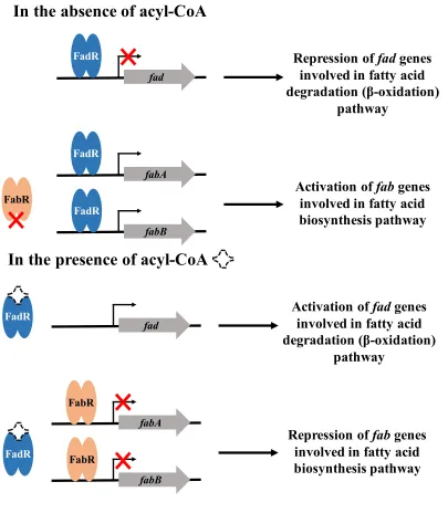
Fatty acids play important roles in microorganisms, both as an integral component of cellular membranes, as well as a source of energy. While the biosynthetic pathways for these fatty acids are rather complex and energetically expensive, a stable membrane composition must be maintained. The Gram-negative Escherichia coli utilizes the combination of transcriptional activators and repressors, belonging to the TetR and GntR families of transcriptional regulators, to control genes involved in fatty acid metabolism in order to maintain membrane homeostasis (172, 173). This homeostasis especially must be maintained upon exposure to exogenous fatty acids that could interfere with membrane fluidity. Host-derived fatty acids are either utilized as a precursor in the membrane phospholipid biosynthetic pathway or degraded via the β-oxidation pathway to
be utilized as an energy source for the bacteria. Fatty acid biosynthesis is regulated via the TetR family regulator, FabR, and fatty acid degradation is regulated via the GntR family regulator, FadR (172, 174). FadR is a repressor of the β-oxidation pathway (fad
fad
fabA
fabB
In the absence of acyl-CoA
FadR
FadR
FadR
FabR
Repression of fadgenes involved in fatty acid degradation (β-oxidation)
pathway
Activation of fabgenes involved in fatty acid biosynthesis pathway
fad
fabA
fabB
In the presence of acyl-CoA
FabR
Activation of fadgenes involved in fatty acid degradation (β-oxidation)
pathway
Repression of fabgenes involved in fatty acid biosynthesis pathway
FabR
FadR FadR
Figure 1.1 Regulation of lipid homeostasis in E. coli. Exogenous fatty acids are converted to acyl-CoAs after entry into the cell. In the absence of acyl-CoA, the transcriptional regulator FadR acts as a repressor of the fad regulon to repress fatty acid degradation, and as an activator of fabA and fabB to activate fatty acids biosynthesis. The genes fabA and fabB are also under the control of the transcriptional repressor FabR. In the presence of acyl-CoA, FadR dissociates from its operator DNA, resulting in the expression of fad genes to activate fatty acid degradation, as well as repression of the fab
Moreover, optimal membrane fluidity is governed by the fatty acid composition of phospholipids; both saturated and unsaturated. Saturated fatty acids are produced to increase membrane rigidity, whereas unsaturated fatty acids are produced to increase membrane fluidity, as is the case when the bacteria are exposed to higher and lower growth temperatures, respectively (178). For instance, Pseudomonas aeruginosa employs two desaturase systems, in addition to the Fab pathway, to regulate fatty acid biosynthesis to maintain membrane homeostasis. The first system is DesA which modifies existing membrane phospholipids to accommodate fluctuations in fluidity. The second desaturase system is DesB which alters saturated fatty acids of exogenous source, such as those lipids in the pulmonary surfactant encountered by P. aeruginosa, to produce unsaturated fatty acids to maintain optimal membrane fluidity. DesB is regulated at the transcriptional level by DesT which is a regulator that senses environmental fatty acids, and differentiates between saturated and unsaturated acyl-CoA substrates. In the presence of unsaturated acyl-CoA, DesT binds to an operator DNA located in the promoter region of
desCB, to repress transcription of these genes which are necessary for production of unsaturated fatty acids. Conversely, the presence of saturated acyl-CoA causes the release of DesT from the operator DNA and the subsequent expression of desB, which in turn introduces double bonds into the acyl-CoA products of exogenous fatty acids and restores optimal membrane fluidity (179–181).
DNA. In the presence of exogenous fatty acids, the promoters repressed by FadR are de-repressed and fatty acids are degraded to be utilized as a carbon source (184)
Furthermore, B. subtilis employs transcriptional regulation to sense the status of the intracellular pool of fatty acids to maintain lipid homeostasis and optimal membrane fluidity. The repressor FapR regulates the expression of genes involved in fatty acid and phospholipid synthesis in response to the cellular levels of malonyl-CoA. Elevated levels of malonyl-CoA are indicative of diminished fatty acid and phospholipid synthesis which in turn results in the de-repression of FapR-mediated genes that are involved in lipid biosynthetic machinery (185, 186). FapR is the first global transcriptional regulator that is highly conserved among Gram-positive bacteria (185, 187). FapR was first characterized and purified from B. subtilis, and was subsequently found to be conserved in other organisms such as S. aureus and Listeria monocytogenes. Similar to FapR in B. subtilis,
fad
In the absence of acyl-CoA
FadR
Repression of genes involved
in fatty acid degradation
fad
De-repression of genes
involved in fatty acid
degradation
In the presence of acyl-CoA
FadR
fap
Low cellular pool of malonyl-CoA
FapR
Repression of
fap
genes
involved in fatty acid and
phospholipid biosynthesis
pathway
fap
Relief of FapR mediated
repression of genes involved in
fatty acid and phospholipid
Elevated cellular pool of malonyl-CoA
FapR
A.
1.8( Fatty(acid(machinery(of(
Staphylococcus,aureus,,
1.8.1(
Phospholipid(composition((
There are three major phospholipids in S. aureus membranes: phosphatidylglycerol; lysyl- phosphatidylglycerol; and cardiolipin (171). These phospholipids play a crucial role in preserving membrane biophysical properties, and particularly, the fatty acid composition of phospholipids is the key determinant of membrane fluidity (189) . Unlike
B. subtilis that possesses a gene encoding a membrane phospholipid desaturase for the synthesis of unsaturated fatty acids, S. aureus does not possess such desaturase and thus is incapable of producing these fatty acids (182). Instead, a combination of branched- and straight-chain fatty acids are present in staphylococcal membranes. Branched-chain fatty acids are the predominant fatty acids in S. aureus phospholipids, comprising 55-65% of the total fatty acids (189, 190). Branched-chain fatty acids are synthesized from branched-chain amino acids; these include leucine- and valine-derived (iso) fatty acids, and isoleucine-derived (anteiso) fatty acids, with the latter in particular promoting increased membrane fluidity (189, 191).
1.8.2(
Phospholipid(synthesis((
The biosynthetic pathway of staphylococcal branched-chain fatty acids starts when the amino acids leucine, isoleucine and valine undergo a transamination reaction mediated by the branched-chain amino acid transaminase BAT. The resulting branched-chain α-keto
acid then undergoes a decarboxylation reaction mediated by α-keto acid dehydrogenase
(BKD), resulting in the formation of branched-chain acyl coenzyme A derivatives which are the precursor for fatty acids biosynthesis. The branched-chain acyl coenzyme A derivatives, in combination with malonyl-ACP, are then utilized by the β-ketoacyl-ACP
synthase III (FabH) to generate β-ketoacyl-ACP. This in turn undergoes a reduction
The peripheral membrane protein PlsX is responsible for transferring the acyl group from Acyl-ACP to an inorganic phosphate, forming an acylphosphate product, which can then be used by an integral plasma membrane protein, PlsY. This interaction between PlsY and the acylphosphate product, in the presence of glycerol-3-phosphate, results in the formation of Acyl-G3P, which is then acylated by the integral membrane protein PlsC, forming phosphatidic acid that represents the key intermediate in membrane phospholipid formation. Phosphatidic acid is subsequently used, in the presence of cytidine triphosphate, to synthesize cytidine diphosphate diacylglycerol through the action of phosphatidate cytidylyltransferase. Phosphatidylglycerol-phosphate is then generated upon the replacement of cytidine monophosphate with glycerol phosphate. The dephosphorylation of phosphatidylglycerol-phosphate results in the formation of phosphatidylglycerol which is the core phospholipid in S. aureus (196–201). An overview of fatty acid and phospholipid biosynthesis in S. aureus can be seen in Figure 1.3
In addition to the fatty acids produced by the fatty acid synthase machinery mentioned above, S. aureus is also capable of utilizing fatty acids of exogenous source. Upon entry into the cell by flipping across the membrane leaflet, exogenous fatty acids are processed by the fatty acid kinase machinery. This machinery includes a kinase domain protein, FakA, and a fatty acid binding protein FakB1 and FakB2 for binding saturated and unsaturated fatty acids, respectively. Exogenous fatty acids bind FakB, and after phosphorylation by FakA, are incorporated into the phospholipid directly, or indirectly after passing through an extension cycle by the FASII machinery (202). During the extension cycle, acyl chains are extended by two carbons via four enzymatic reactions. Extension is initiated by an elongation condensing enzyme, FabF, and the resulting β
-ketoacyl-ACP is then reduced by the reductase, FabG. Subsequently, β
Leucine
Valine
Isoleucine
Branched-chain
β
-keto acids
Branched-chain acyl coenzyme A derivatives
Branched-chain amino
acid transaminase
α
-keto acid
dehydrogenase
FabH
+ malonyl-ACP
β
-ketoacyl-ACP
FabG
FabZ
FabI
Saturated acyl-ACP
FabF
Long chain acyl-ACP
Acyl- PO
4PlsX
+ Glycerol 3-phosphate
PlsY
Acyl- G3P
PlsC
Phosphatidic acid
Cytidine diphosphate
diacylglycerol
Phosphatidylglycerol
phosphate
Figure 1.3 Overview of fatty acid and phospholipid biosynthesis in S. aureus.
Synthesis of staphylococcal branched-chain fatty acids starts when the amino acids leucine, isoleucine and valine undergo a transamination followed by a carboxylation reactions reaction, resulting in the formation of branched-chain acyl coenzyme A derivatives which are the precursor for fatty acids biosynthesis. The branched-chain acyl coenzyme A derivatives, in combination with malonyl-ACP, are then utilized by FabH to generate β-ketoacyl-ACP. This in turn undergoes a reduction reaction by FabG, a
Exogenous
FA
FA
FA ~ P
FakB-FA~P
FakB-FA
FakA
Acyl-PO
4Acyl-ACP
Glycerol-3-phosphate Lyso-phosphatidic acid Phosphatidic acid Membrane Phospholipids PlsX PlsY PlsC Exogenous FA
FA FA ~ P
FakB-FA~P FakB-FA FakA
Acyl-PO4 Acyl-ACP
Membrane Phospholipids PlsX
trans-2-Enoyl-ACP β-ketoacyl-ACP
β -hydroxyacyl-ACP FASII Elongation FabF/B FabG FabA/Z FabI/K Phosphatidic acid FabZ A. B.
1.8.3(
Lipids(as(antimicrobials((
The skin is the first line of defense against microbial infection. One of the host innate defense elements on the skin is antimicrobial long-chain unsaturated fatty acids. The main source of these fatty acids is the sebum secreted by the sebaceous glands and differentiating keratinocytes present in the stratum corneum of the epidermis (208). The sebum consists of squalene, wax monoesters, triglycerides, and small amounts of cholesterol and cholesterol esters (209). As the sebum’s constituents stream outwards through the hair follicle associated with the sebaceous glands, the triglycerides undergo enzymatic hydrolysis to produce unsaturated free fatty acids (uFFA) on the skin surface. The principal uFFA derived from sebaceous triglycerides is sapienic acid, which is a 16-carbon fatty acid with one degree of unsaturation that has potent bactericidal property against MRSA (208). Linoleic acid was identified as the major uFFA in human nasal secretion and is also present in S. aureus abscesses, a hallmark of S. aureus skin and soft tissue infections (210–212). Humans deficient in the production of uFFAs are more susceptible to S. aureus skin infections (213).
Table 1.3 Summary of proposed mechanisms of uFFA toxicity.
Target Mechanism Reference
Cytoplasmic
membrane Uncouple the electron transport chain (223, 224)
Cytoplasmic
membrane Impede oxidative phosphorylation (220)
Cytoplasmic membrane
Surfactant action that increases membrane
permeability (219)
Cytoplasmic membrane
Increase membrane fluidity leading to cell
lysis (218, 225)
Cytoplasmic membrane
Dissolution of the proton gradient across the membrane and subsequent release of
low molecular-weight proteins (216)
Fatty acid synthetic
machinery Enzymatic inhibition (222)
Although exogenous uFFAs display antimicrobial properties, bacterial species are equipped with intrinsic mechanisms of resistance to these uFFAs. External structures such as the outer membrane and cell wall of Gram-negative and positive bacteria, respectively, mediate protection against uFFAs through several mechanisms that are briefly reviewed here. First, transcriptomics and proteomics studies show that upon exposure to exogenous uFFAs, S. aureus upregulates genes involved in carotenoid biosynthesis, as well as those in cellular energy metabolism, peptidoglycan, and call wall biosynthetic pathways (227). Cell wall components play an integral role in S. aureus
susceptibility to antimicrobial uFFAs, as wall structure, thickness and properties can impede the entry of these uFFAs. It has been proposed that the iron-regulated surface determinant, IsdA, is involved in resistance to uFFAs where this cell wall anchored protein modifies cellular hydrophobicity. The C domain of IsdA extends into the staphylococcal cell wall, altering its charge and hydrophobicity thus rendering the cells less susceptible to host antimicrobial peptides and fatty acids that rely on hydrophobic interactions to penetrate the cellular membrane and manifest their effects (227, 228). Other reports suggest that S. aureus counteracts the uFFA-mediated increase in membrane fluidity by upregulating the production of staphyloxanthin, leading to a direct correlation between membrane stability, cellular pigmentation, and uFFAs-induced cellular killing (218, 227, 229). The production of staphyloxanthin is regulated by the sigma B (σB) regulon as a general stress response to the presence of uFFAs (227).
Table 1.4 Summary of uFFA resistance mechanisms proposed to date.
Target Mechanism Reference
Cell wall physiology
Decrease cellular hydrophobicity and thus
prevent uFFA penetration (228)
Membrane stability
Increase staphyloxanthin production to counteract the uFFA-mediated increase in
membrane fluidity (218, 227, 229)
Wall teichoic acids Prevent binding of uFFAs (216, 230)
uFFA transport
Tet38-mediated efflux of uFFAs outside
the cell (234)
uFFA processing
FAME- medicated esterification of uFFAs
1.9( Bacterial(efflux(pumps(
One of the greatest innovations in modern medicine is the introduction of antibiotics in clinical practices to treat the otherwise life-threatening infectious diseases. However, bacterial pathogens were able to circumvent this progress by developing resistance mechanisms leading to infectious diseases that are once again, a major problem in the hospital and community settings. The World Health Organization continuously urges for the development of new bacterial therapeutic targets to combat the life-threatening multidrug resistance (235, 236).
1.10(TetR(family(of(transcriptional(regulators(and(their(
biological(functions((
Transcriptional regulators are the key to most microbial adaptation strategies. These regulators normally contain a DNA- and ligand- binding domain that can be used to sense environmental signals and elicit a downstream response at the transcriptional level. There are many families of transcriptional regulators that have been grouped based on structural and functional similarities, such as the helix-turn-helix motif, which is the most common DNA-binding motif among prokaryotes (245–247). Members of these families have either an activation or a repression function, with a few families that display unconventional dual roles in activation and repression. Members of the LysR, IcIR, MarR, and Crp families of transcriptional regulators have displayed dual roles (248–251). Recently, there have been few reports of members of the TetR family of transcriptional regulators that display dual functionality (252, 253).
TetR family regulators (TFRs) have been well documented for their roles in environmental adaptation and antibiotic resistance (254). There are more than 2500 TFRs with functions ranging from multidrug resistance, biosynthesis of antibiotics, and pathogenicity in both Gram-positive and Gram-negative bacteria (255). TFRs are named after the well-characterized TetR, a repressor that controls expression of a membrane-associated protein, TetA, that exports tetracycline out of the bacterial cell. tetR and tetA
are divergently transcribed, and the tetR gene product controls expression of both tetA
and tetR. When tetracycline is present and associated with Mg2+, it binds to the TetR protein leading to a conformational change that renders TetR unable to bind to the DNA. As a result, tetR and tetA, both of which are repressed byTetR, are expressed (256–260).
governs tetracycline-resistance in Gram-negative bacteria (261–264). By 1998, it was confirmed that QacA contains 14 transmembrane domains and that it belongs to the major facilitator superfamily of transporters, and that QacR is the repressor regulating the expression of this transporter (265, 266). QacR functions as a homodimer with an N-terminal DNA-binding domain recognizing an operator sequence located downstream from the divergent qacA promoter (266–268). This binding is considered unorthodox for a repressor as it may not physically block the binding RNA polymerase directly, but rather it hinders the transition of the transcription machinery. The regulator of intercellular adhesion (ica) operon of S. epidermidis is a TFR that is responsible for regulating the synthesis of polysaccharide intercellular adhesin (PIA) and, subsequently, biofilm formation (269). Two IcaR homodimers bind cooperatively to an operator site located upstream of the start codon of icaA to repress its transcription (270, 271). Upon ligand binding of streptomycin and gentamicin, IcaR dimers undergo a conformational change in the DNA binding domain, and the subsequent transcription of the divergent
icaADBC operon (272). Cramton et al in 1999 showed that the ica locus is not only present in S. aureus, but is also implicated for biofilm formation (273); however, in S. aureus, the teicoplanin-associated locus regulator (TcaR), a member of the MarR family of transcriptional regulators, is also a repressor of the ica operon (272).
Other examples of TFRs include: the AmeR of Agrobacterium tumefaciens which is a regulator of an RND- efflux system; AmrR of Pseudomonas aeruginosa which is a regulator of an efflux pump involved in aminoglycoside resistance; and BpeR of
RND family of efflux pumps. AcrR interacts with the acr promoter located in the intergenic segment between acrR and acrAB. This interaction between AcrR and the promoter region is responsive to accumulation of cellular metabolites and bile salts as well as global stress signals. In the presence of inducing ligand, AcrR is de-repressed and functions in combination with global transcriptional activators to enable efflux pump expression (282–285). Other TFRs that have roles in lipid efflux include: CmeR from
Campylobacter jejuni and MtrR from N. gonorrhea. CmeR is a repressor of an RND-efflux pump, CmeABC, that is responsive to accumulation of amphipathic bile salts (286–289). Similarly, MtrR is a repressor of an RND pump, MtrCDE, the efflux of which enables gonococcal growth on mucosal surfaces that are enriched in fatty acids and bile salts. Additionally, MtrR is a repressor of a MarR family transcriptional regulator, FarR, which in turn is a repressor of the MFS efflux pump FarAB. Therefore, MtrR also functions as a positive regulator of the FarAB efflux pump which mediates resistance to antimicrobial fatty acids (233, 279, 290–292).
1.11(Rationale(and(hypothesis((
CA-MRSA strains are particularly efficient in colonization, transmission, and causing invasive skin and soft tissue infections (6). Our laboratory strain of choice is USA300, the dominant CA-MRSA strain in the United States, accounting for 98% of MRSA infections presenting to emergency departments (293). USA300, which was isolated in September 2000, has been linked to infection outbreaks in at least 21 American states, Canada, and Europe (132). Further, this isolate is more resistant to killing by polymorphonuclear leukocytes (132). To persist on human skin, S. aureus must cope with uFFAs; a component of human innate defense mechanisms. Although genome sequencing and molecular typing have provided a better understanding of the pathogenic success of USA300, detailed knowledge of the specific molecular determinants, especially in regards to uFFA resistance, is still lacking (294, 295). Although S. aureus
fatty acid import exceeds that of incorporation, excess free fatty acids accumulate in the membrane, ultimately resulting in cell death (202, 214, 216).
To better understand the factors that contribute to S. aureus adaptation to uFFA, our laboratory evaluated the growth of S. aureus USA300 in the presence of physiological concentrations of uFFA that would normally be encountered on the skin and in the anterior nares. Arsic et al. showed that uFFA can induce the staphylococcal proteolytic cascade (296). Additionally, Arsic et al. showed that unsaturated linoleic and sapienic acids are sub-inhibitory at 25 µM and bactericidal at 100 µM, whereas a 50 µM concentration caused a 10-12 hour lag phase after which bacteria resumed growth (296). It was established that the profile of secreted proteins and virulence factors is influenced by exogenous uFFAs, suggesting a possible regulatory mechanism of virulence in response to uFFAs.
To identify genes that may confer intrinsic resistance to uFFA, S. aureus USA300 was selected for growth at elevated concentrations of linoleic acid after which single colonies were selected for DNA isolation and genome sequencing on the Ion Torrent Platform to identify single nucleotide polymorphisms (SNP) in 7 fatty acid resistant (FAR) clones. Two of these clones contained SNP causing a His121>Tyr substitution in SAUSA300_2490 gene. Using domain enhanced lookup time accelerated BLAST, the amino acid sequence of SAUSA300_2490 had 99% homology to TFRs and structure prediction using homology modeling server, Phyre2, predicted that SAUSA300_2490
shares 99.8% amino acid sequence similarity with TFRs such as FadR, a regulator of fatty acid degradation in Thermus thermophilus.
I hereafter refer to SAUSA300_2490 as farR for regulator of fatty acid resistance, and to
SAUSA300_2489 as farE, for an effector of fatty acid resistance. Therefore, I hypothesize that farR functions to regulate expression of a fatty acid efflux pump encoded by farE
1.12(Literature(cited((
1. Wertheim HFL, Melles DC, Vos MC, van Leeuwen W, van Belkum A, Verbrugh HA, Nouwen JL. 2005. The role of nasal carriage in Staphylococcus aureus
infections. Lancet Infect Dis 5:751–62.
2. Ogston A. 1881. Report upon micro-organisms in surgical diseases. Br Med J 1:369.b2-375.
3. Ogston A. 1882. Micrococcus poisoning. J Anat Physiol 16:526–67.
4. Ogston A and WW. 1984. Classics in infectious diseases. “On abscesses”. Rev Infect Dis 6:122–8.
5. David MZ, Daum RS. 2010. Community-associated methicillin-resistant
Staphylococcus aureus: epidemiology and clinical consequences of an emerging epidemic. Clin Microbiol Rev 23:616–87.
6. Gilbert M, Macdonald J, Louie M, Gregson D, Zhang K, Elsayed S, Laupland K, Nielsen D, Wheeler V, Lye T, Conly J. 2007. Prevalence of USA300 colonization or infection and associated variables during an outbreak of community-associated methicillin-resistant Staphylococcus aureus in a marginalized urban population. Can J Infect Dis Med Microbiol J Can des Mal Infect la Microbiol medicale 18:357–62.
7. Moran GJ, Krishnadasan A, Gorwitz RJ, Fosheim GE, McDougal LK, Carey RB, Talan DA, EMERGEncy ID Net Study Group. 2006. Methicillin-resistant S. aureus infections among patients in the emergency department. N Engl J Med 355:666–674.
8. Skinner D, Keefer CS. 1941. Significance of bacteremia caused by Staphylococcus aureus. Arch Intern Med 68:851.
9. Rammelkamp CH, Maxon T. 1942. Resistance of Staphylococcus aureus to the action of penicillin. Exp Biol Med 51:386–389.
10. Jessen O, Rosendal K, Bülow P, Faber V, Eriksen KR. 1969. Changing Staphylococci and staphylococcal infections. N Engl J Med 281:627–635.
11. Parker M., Hewitt JH. 1970. Methicillin resistance in Staphylococcus aureus. Lancet 295:800–804.