Scholarship@Western
Scholarship@Western
Electronic Thesis and Dissertation Repository
5-1-2017 12:00 AM
Alternative splicing and extra-telomeric role of telomerase reverse
Alternative splicing and extra-telomeric role of telomerase reverse
transcriptase isoforms in human embryonic stem cells
transcriptase isoforms in human embryonic stem cells
Joohwan Kim
The University of Western Ontario Supervisor
Dr. Dean H. Betts
The University of Western Ontario
Graduate Program in Physiology and Pharmacology
A thesis submitted in partial fulfillment of the requirements for the degree in Master of Science © Joohwan Kim 2017
Follow this and additional works at: https://ir.lib.uwo.ca/etd Part of the Cell Biology Commons
Recommended Citation Recommended Citation
Kim, Joohwan, "Alternative splicing and extra-telomeric role of telomerase reverse transcriptase isoforms in human embryonic stem cells" (2017). Electronic Thesis and Dissertation Repository. 4571.
https://ir.lib.uwo.ca/etd/4571
This Dissertation/Thesis is brought to you for free and open access by Scholarship@Western. It has been accepted for inclusion in Electronic Thesis and Dissertation Repository by an authorized administrator of
iii
Human telomerase, a ribonucleoprotein complex consisting of a catalytic subunit (hTERT) and a RNA template component (TERC), prevents telomere attrition in human embryonic stem cells (hESCs) providing these cells with prolonged replicative capacity. Interestingly, hTERT has several alternatively spliced isoforms, most of which are
catalytically inactive. One alternative splicing event involving a complete loss of exons 7 and 8 results in a catalytically inactive isoform called β-hTERT. The β-hTERT is highly
expressed in certain cancer types and hESCs. Full-length hTERT (FL-hTERT) is known to translocate from nuclei to mitochondria when cells are exposed to either chronic (oxygen tension) or acute (hydrogen peroxide) oxidative stress. In this study, I examined the
alternative splicing of endogenous hTERT transcript in hESC, intra-cellular localization of FL-hTERT and β-hTERT in hESCs, and the effect of overexpressing recombinant hTERT on hESC mitochondria. Human ESCs (HES2 line) were cultured under 2% oxygen and 3xFLAG tagged FL-hTERT and β-hTERT were induced by doxycycline prior to 100μM H2O2
treatment. My results demonstrate that overexpression of transgenic FL-hTERT resulted in a significant increase in endogenous β-hTERT transcript level. Additionally, doxycycline and H2O2 altered the splicing pattern of hTERT in favor of β-hTERT. I demonstrate that not only the full-length- but also β-hTERT can bind TERC, and that β-hTERT overexpression inhibits telomerase activity in vitro. However, I failed to observe the mitochondrial localization of recombinant FL-hTERT or β-hTERT. Lastly, my results demonstrate the pro-apoptotic role of β-hTERT and the pro-survival effect of FL-hTERT under H2O2-induced oxidative stress.
iv
I would like to start by thanking Dr. Dean H. Betts for giving me both financial and spiritual support over three and half years of my master’s degree. He was always there to give me good pat on the back in times of discouragement, and great scientific insight to help me move forward with the research. I was very fortunate to have him as my supervisor. Without him, I could not have made this far. Once again, I thank him for the unconditional support and guidance he has given me throughout my study at Western University.
I owe greatly to our lab’s post-doctoral researcher Dr. Jonathan H. Teichroeb for many things. His expertise in cell cloning and telomerase was the greatest intellectual asset I was lucky to have access to. He never hesitated to lend me a hand with new or difficult experimental tasks, and graciously let me use his model human embryonic cell lines. Thank you, Jonathan, for your great mentorship and friendship.
When I started here, I knew very little about culturing cells, not to mention how to culture human embryonic cells. But I now feel very confident with it, thanks to Courtney Brooks our stem cell technician. Professional at work, yet very friendly and attentive as a friend, Courtney is the greatest member of the lab and friend. Thank you for your precious contribution to every aspect of my research.
v
Certificate of examination ii
Abstract iii
Acknowledgements iv
Table of Contents v
List of Tables x
List of Figures xi
List of Appendices xiv
List of Abbreviations xv
Chapter 1: Introduction and Literature Review 1
1.1Human Embryonic Stem Cells 1
1.1.1 Signaling pathways in hESC 2
1.1.2 The role of oxygen tension in embryonic stem cells 4
1.1.3 Energy metabolism of pluripotent stem cells 5
1.1.4 Telomerase and telomere in human embryonic Stem cells 7
1.2Human Telomerase Catalytic Subunit hTERT 8
1.2.1 Canonical function of hTERT 8
1.2.2 Alternative splicing of hTERT 10
1.2.3 Transcriptional regulation of hTERT 14
1.2.4 Post-translational regulation of hTERT 16
1.3Non-canonical functions of Human Telomerase Catalytic Subunit hTERT 20
1.3.1 Non-canonical functions of hTERT in Wnt/β-catenin pathway 20
vi polymerase (RdRP)
1.3.4 Mitochondrial functions of hTERT 24
1.4Rationale 25
Chapter 2: Materials and Methods 28
2.1Cell culture 28
2.1.1 Mouse embryonic fibroblasts feeder cells 28
2.1.2 Human embryonic stem cell line HES2 28
2.1.3 Establishment of monoclonal transgenic HES2 lines 29
2.1.4 Cell treatment scheme 30
2.2PCR protocols 31
2.2.1 General real-time quantitative PCR protocol 31
2.2.2 Detection and quantitation of hTERT isoforms by hydrolysis-probe assisted PCR 31
2.2.3 RNA-immunoprecipitation and PCR-assisted detection of TERC 34
2.2.4 PCR amplification of mitochondrial DNA for mitochondria DNA copy number 34
2.3Telomerase activity assay 35
2.4Immunodetection of 3xFLAG-hTERT isoforms 36
2.4.1 Western blot analysis 36
2.4.2 Immunocytochemistry 37
2.4.3 Cell count analysis for intra-nuclear localization of 3xFLAG-hTERT isoforms 38 2.5Flowcytometry 40
2.5.1 Mitochondria membrane potential 40
2.5.2 Detection of apoptosis 41
vii
3.1Basic cell characterization of human embryonic stem cells 43 3.1.1 Characterization of pluripotency markers in human embryonic stem
cells 43
3.1.2 Characterization of hTERT isoforms in human embryonic stem cells43 3.2Characterization of 3xFLAG-hTERT transgenic clones 47
3.2.1 Characterization of 3xFLAG-hTERT transgene expression level in
transgenic hESC clones 47
3.2.2 Characterization of Pluripotency in HES2 B6 and HES2 FL12 hTERT
cell lines 48
3.3Characterization of recombinant hTERT in HES2 B6 and HES2 FL12 52 3.3.1 Endogenous TERC-binding of 3xFLAG-β-hTERT and
3xFLAG-FL-hTERT subunits 52
3.3.2 Endogenous RMRP-binding of 3xFLAG-β-hTERT and
3xFLAG-FL-hTERT subunits 52
3.3.3 Telomerase activity in HES2 B6 and HES2 FL12 hESCs 52 3.3.4 Re-visiting DOX-induction of 3xFLAG-hTERT transcription in HES2
clones 53
3.43xFLAG-hTERT localization under H2O2-induced oxidative stress 58 3.4.1 Sub-cellular Western Blot Analysis of 3xFLAG-FL-hTERT and
3xFLAG-β-hTERT expressing hESC lines 58 3.4.2 Immunocytochemistry and Image analysis of 3xFLAG-FL-hTERT and 3xFLAG-β-hTERT localization in hESCs 58 3.5The effect of overexpressed 3xFLAG-hTERT on Mitochondrial Function 64
3.5.1 The effect of DOX-induced 3xFLAG-hTERT on mitochondria
viii
copy number 64
3.5.3 The effect of DOX-induced 3xFLAG-hTERT on mitochondria
biogenesis and function 65
3.6The effect of doxycycline-induced 3xFLAG-hTERT on cell survival in HES2 lines under H2O2-induced oxidative stress 71
Chapter 4: Discussion 76
4.1Pluripotency and alternative splicing of endogenous hTERT in hESC 77 4.2Transgenic HES2 line B6 and FL12 remain pluripotent following doxycycline and
H2O2 treatment 82
4.33xFLAG-hTERT in HES2 B6 and HES2 FL12 bind TERC and RMRP 86 4.4The effect of doxycycline on survival and cell death of HES2 lines under H2O2
-induced oxidative stress 88
4.5The intra-cellular localization of 3xFLAG-β-hTERT and 3xFLAG-FL-hTERT in hESCs treated with H2O2-induced oxidative stress 91 4.6Investigation of the effect of 3xFLAG-β-hTERT and 3xFLAG-FL-hTERT on
mitochondria function in hESCs 94
4.7The effect of 3xFLAG-β-hTERT and 3xFLAG-FL-hTERT on cell survival of HES2 lines under H2O2-induced oxidative stress 97 4.8Concluding remark and proposed future studies 100
Chapter 5: Reference 102
ix
hours and 12- hours post doxycycline induction 128 A4: Fig. 3.2 H2O2-induced oxidative stress and its effect on the cellular localization of
3xFLAG-hTERT 129
A5: Fig. 3.3: The effect oxygen tension on the intra-cellular localization of
3xFLAG-FL-hTERT in hESC 130
x
Table Description Page
A1: 2.1 List of PCR primers 124
xi
Figure Description Page
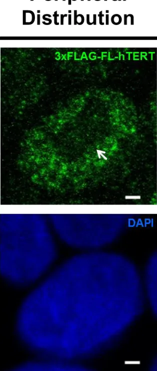
1.1 mRNA and protein structure of the major hTERT isoforms 13 1.2 Post-translational modification and function of hTERT 19 2.1 Transgene structure and TaqMan qPCR probe-/primer-binding sites 33 2.2 Representative confocal microscopy images of ‘uniform’ and
‘peripheral’ distribution of 3xFLAG-FL-hTERT within nuclei
39
3.1 HES2 is a hESC cell line that expresses various markers of pluripotency and different proportions of endogenous telomerase reverse transcriptase (TERT) isoform transcripts
45
3.2 Endogenous β-hTERT transcript is ubiquitously present at high levels in human embryonic cells grown under low oxygen tension conditions, with and without H2O2 and/or doxycycline (DOX) treatments
46
3.3 RT-qPCR and Western blot analysis of doxycycline-induced expression of 3xFLAG-hTERT isoforms in HES2 clones over 3-day time course
49
3.4 H2O2 and DOX treatments do not affect pluripotency marker expression in the 3xFLAG-β-hTERT transgenic HES2 B6 line
50
3.5 H2O2 and DOX treatments do not affect pluripotency of the 3xFLAG-FL-hTERT transgenic HES2 FL12 lines
51
3.6 Doxycycline-induced transgenic telomerase proteins (3xFLAG-β-hTERT and 3xFLAG-FL-hTERT) bind to the telomerase RNA component
54
3.7 Enrichment of RNA component of mitochondrial RNA processing endoribonuclease (RMRP by doxycycline-induced transgenic telomerase proteins (3xFLAG-β-hTERT and 3xFLAG-FL-hTERT)
xii
Figure Description Page
3.8 Altered telomerase activity levels in doxycycline (DOX)-induced transgenic telomerase proteins (3xFLAG-hTERT) in hESCs
56
3.9 DOX-induction of 3xFLAG-hTERT isoforms results in a simultaneous increase in the amount of other hTERT transcript variants containing β-deletion
57
3.10 H2O2-induced Oxidative stress and its effect on the cellular localization of 3xFLAG-β-hTERT
60
3.11 H2O2-induced Oxidative stress and its effect on the cellular localization of 3xFLAG-FL-hTERT
61
3.12 H2O2-induced effect on the intra-cellular localization of 3xFLAG-β-hTERT and 3xFLAG-FL-3xFLAG-β-hTERT in hESCs
62
3.13 Microscopic analysis of intra-nuclear localization of 3xFLAG-β-hTERT and 3xFLAG-FL-hTERT under H2O2-induced oxidative stress
63
3.14 Mitochondrial membrane potential (MMP) gating strategy and controls for flow cytometry experiment with JC-1 dye measurements
66
3.15 3xFLAG-β-hTERT and 3xFLAG-Full-Length-hTERT do not affect mitochondrial membrane potential (MMP) in hESCs
67
3.16 Doxycycline affects mitochondria copy number in hESCs 68 3.17 The effect of 3xFLAG-β-hTERT and 3xFLAG-Full-Length-hTERT
overexpression on mitochondria biogenesis regulator gene expression
xiii
3.18 DOX-induced 3xFLAG-β-hTERT and 3xFLAG-Full-Length-hTERT overexpression does not affect UCP2 and SOD2 transcript abundance levels
70
3.19 Representative plots showing stain controls and gating strategies for flowcytometry experiment with 7-AAD and Annexin V for measuring cell death upon H2O2 treatment
72
3.20 Effect of DOX-induced 3xFLAG-β-hTERT and 3xFLAG-Full-Length-hTERT on cell viability in HES2 under H2O2-induced cell stress
73
3.21 Effect of DOX-induced 3xFLAG-β-hTERT and 3xFLAG-Full-Length-hTERT on apoptosis in HES2 under H2O2-induced cell stress
74
3.22 Effect of DOX-induced 3xFLAG-β-hTERT and 3xFLAG-Full-Length-hTERT on cell death in HES2 under H2O2-induced cell stress
75
A3 3.1 The amount of 3xFLAG-hTERT in the respective HES2 clones after 6-hours and 12- 6-hours post doxycycline induction
128
A4 3.2 H2O2-induced oxidative stress and its effect on the cellular localization of 3xFLAG-hTERT
129
A5 3.3 The effect oxygen tension on the intra-cellular localization of 3xFLAG-β-hTERT and 3xFLAG-FL-3xFLAG-β-hTERT in hESCs
xiv
Appendix Description Page
A1 List of PCR primers 124
A2 List of antibodies and dyes 127
A3 The amount of 3xFLAG-hTERT in the respective HES2 clones after 6-hours and 12- 6-hours post doxycycline induction
128
A4 H2O2-induced oxidative stress and its effect on the cellular localization of 3xFLAG-hTERT
129
A5 The effect oxygen tension on the intra-cellular localization of 3xFLAG-β-hTERT and 3xFLAG-FL-hTERT in hESCs
xv ALT Alternative lengthening of telomere
APC Adenomatosis polyposis coli
BIO 6-bromoindirubin-3-oxime
BRG1 Brahma-related gene 1
BSA Bovine serum albumin
Abl Abelson murine leukemia viral oncogene homolog 1
CHIP C terminus of HSC70-Interacting Protein
CRM1 Chromosomal maintenance 1
Ct Cycle threshold
DHS DNAse I hypersensitive site
DMEM Dulbecco's modified eagle medium
DMSO Dimethylsulfoxide
DNA Deoxyribonucleic acid
DSB Double-stranded break
ESC Embryonic stem cell
ESE Exonic splicing enhancer
ESS Exonic splicing silencer
ETC Electron transport chain
FBS Fetal bovine serum
FL Full length
GSK3β Glycogen synthase kinase 3 beta
xvi HIF2A Hypoxia-inducible factor 2 alpha
HK2 Hexokinase 2
hnRNP Heterogeneous ribonucleoprotein
hPSC Human pluripotent stem cell
HSP90 Heat shock protein 90
iPSC Induced pluripotent stem cell
ISE Intronic splicing enhancer
ISS Intronic splicing siliencer
LRP Lipoprotein receptor-related protein
MEF Mouse embryonic fibroblast
MFN1/2 Mitofusin 1/2
MKRN1 Makorin ring finger protein 1
MMP Mitochondria membrane potential
mtDNA Mitochondrial DNA
mTHF L-methyl-tetrahydrofolate
mTOR Mechanistic target of rapamycin
NLS Nuclear localization signal
NMD Nonsense mediated decay
NP-40 Nonidet-P40
NS Nucleostemin
ORF Open reading frame
OXPHOS Oxidative phosphorylation
xvii PDH Pyruvate dehydrogenase
PFK Phophofructokinase
PI3K Phosphatidylinositol-4,5-bisphosphate 3-kinase
PKC Protein kinase C
Plk1 Polo-like kinase 1
PP2A Protein phosphatase 2A
PSC Pluripotent stem cell
PTB Polypyrimidine tract binding protein
PTDC Pyrrolidinethiocarbamate
RdRP RNA-dependant RNA Polymerase
RISC RNA-induced silencing complex
RMRP RNA component of the mitochondrial RNA-processing endoribonuclease
RNA Ribonucleic acid
RNP Ribonucleoprotein
ROS Reactive oxygen species
RT Reverse transcriptase
S1P Sphingosine-1-phosphate
siRNA Small-interfering RNAs
snRNP Small nucleolar ribonucleoprotein
SSEA Stage-specific embryonic antigen
TBE Tris/Borate/EDTA
TCA Tricarboxylic acid
xviii TEN Telomerase essential N-terminal
TERC Telomerase RNA component
TERT Telomerase reverse transcriptase
THF Tetrahydrofolate
TNF Tumor necrosis factor
Chapter 1: Introduction and Literature Review
1.1 Human Embryonic Stem Cells
1.1.1 Signaling pathways in hESCs
Typical PSC media for feeder-free growth conditions includes factors such as insulin-like growth factor (IGF) a strong stimulant for the canonical phosphatidylinositol-3 kinase (PI3K) signaling pathway [17,18]. PI3K/AKT signaling is crucial for maintenance of hESC pluripotency [19, 20]. PI3K/AKT is a signaling pathway best known for its regulatory role in cell cycle regulation. Its function is especially important in actively proliferating cell types such as cancer cells and hESCs. A microarray experiment by Armstrong et al. revealed a highly active PI3K/AKT signal-transduction in undifferentiated ESCs [18]. Inhibition of PI3K/AKT in hESCs resulted in a significant loss of core pluripotency marker (OCT4, NANOG, SOX2) expression, confirming the requirement of PI3K signaling in the
maintenance of hESC pluripotency. When activated, AKT1 initiates a signaling cascade by activating mTOR and by inhibiting GSK3[19]. Inhibition of PI3K/AKT signaling by small molecules such as LY294002 or by removal of PI3K activators leads to an eventual loss of pluripotency in hPSCs [19, 20, 23]. The mechanisms of how PI3K controls the pluripotent state in hPSCs are unclear. It is speculated that PI3K/AKT regulates protein synthesis through mTOR, thereby regulating the protein synthesis of core factors of the pluripotency network [21]. Inhibition of mTOR by rapamycin results in similar effects to that of AKT inhibition, triggering spontaneous hPSC differentiation [19, 24]. GSK3 inhibition by AKT signaling may also contribute to PSC maintenance through Wnt/β-catenin-mediated expression of c-myc, a key regulator of PSC maintenance [25, 26]. This mechanism could also apply in hPSCs, but has not been evaluated fully.
the self-renewal of hESCs (H1 and H9 cell line) under feeder-free conditions [27-29]. In presence of BIO, Wnt-signaling sustained the expression of Oct-3/4, Rex1, and Nanog. The effect was reversed when BIO was withdrawn from the culture, resulting in spontaneous multi-differentiation of hESCs. However, the consequences of blocking GSK3 are not limited to Wnt/β-catenin pathway, and can affect many other pathways through mTOR
stabilization/cross-talk between signaling pathways. For instance, inhibition of GSK3 via BIO results in increased phosphorylated Smad2/3 in hESCs, a downstream Nodal signaling pathway characteristic of the undifferentiated state of hESCs [26]. In support of this view, contradicting results emerged afterwards, demonstrating that Wnt/β-catenin activation by Wnt3A/BIO does not support the self-renewal of hPSCs in long term cultures [30-32]. Moreover, their functional reporter assay showed that β-catenin-mediated transcription activity was nearly absent in undifferentiated hESCs (H1 and H9 cell lines). Further studies with a chimeric (estrogen-receptor tagged) β-catenin model showed that
4-hydroxytamoxifen-induced activation of Wnt/β-catenin signaling resulted in the
1.1.2 The role of oxygen tension in embryonic stem cells.
The natural niche of early embryonic cells is enriched with essential growth factors and the proper microenvironment for the maintenance of self-renewal and pluripotency, and this often includes low oxygen tensions [36, 37]. Early embryonic cells grow under low oxygen availability starting from fertilization and continuing through implantation and fetal development. During implantation, a hypoxic environment is created due to the absence of oxygen provided from maternal circulation [37]. The uterine surface typically has oxygen concentrations of 2% in early pregnancy. The oxygen tension rises to approximately 8% once the maternal vasculature is established [38]. Oxygen acts as the final electron carrier in the ETC of OXPHOS-dependent energy metabolism in mammalian cells [39]. During early embryo development, cells adapt to the low oxygen environment by switching between OXPHOS-dependent and glycolysis-dependent energy production[40].
hESCs derived from the early embryo grow relatively well under ambient oxygen tension (~21%) without apparent negative consequences. However, closer examination revealed the effect of oxygen tension on altered hESC morphology [42-44], pluripotency and differentiation marker expression [44, 45], cloning efficiency [43], chromosomal stability[44] and differentiation capacity [42, 43]. In the majority of the studies, low oxygen tension (2– 5%) improved human ES cell pluripotency and maintenance [44– 47]. In addition, low oxygen condition (5%) was shown to improve the efficiency of iPS generation from human dermal fibroblast cells [48], further supporting the notion that stemness of hESC is
In murine ESCs, hypoxia-inducible-factor1 alpha (HIF1A) plays a central role in regulating the cellular response to oxygen availability, mediating the expression of many oxygen-related genes [50]. While HIF1A controls a broader spectrum of oxygen-dependent genes, HIF2A is a direct upstream regulator of POU5F1 (OCT4) in mouse ES cells [51]. In hESCs, HIF1A is only transiently expressed for approximately 48 hours following hypoxic exposure [52], suggesting a long-term hypoxic regulation by a factor other than HIF1A. Indeed, HIF2A was described to be responsible for the regulation of energy metabolism and self-renewal in hESCs grown under 5% oxygen tensions [53].
1.1.3 Energy Metabolism of Pluripotent Stem Cells
Prior to vasculature formation, early-stage embryonic cells grow in a hypoxic environment (1-5%) [38, 53]. PSCs such as hESCs and mouse epiblast-derived stem cells (mEpiSC) exhibit low-level cellular respiration and down-regulation of electron transport chain (ETC) activity, while enzymes involved in glycolytic pathways are up-regulated [11]. This reliance on glycolytic metabolism is partly explained by the hypoxic
mitochondria and increased metabolism via OXPHOS [60]. Glycolytic energy metabolism of hESCs serves to protect the cells from DNA damage by minimizing reactive oxygen species (ROS) generated through OXPHOS [55]. Although oxygen consumption is decoupled from ATP generation in ESCs, some level of oxygen consumption appears to be important,
(SAM) production by modulating tetrahydrofolate’s (THF) conversion to L-methyl-tetrahydrofolate (mTHF). Threonine deprivation in mESCs leads to the reduction in the trimethylation of histone 3 lysine 4, increasing the propensity to differentiate [63].
The importance of mitochondria is also seen in reprogramming of somatic cells to induced pluripotent stem cells (iPSCs). During conversion of somatic cells to a pluripotent state, depleting mitofusin proteins (MFN1/2) promotes the reprogramming efficiency by increased mitochondria fission [64]. Similarly, the reprogramming efficiency of somatic cells into iPSCs is enhanced three- to four-fold when glycolysis is promoted, and is reduced when glycolysis is inhibited [48]. Several pieces of evidence suggest that mitochondrial maturation and subsequent rise in intra-cellular level of reactive oxygen species (ROS) are required signaling elements in regulating both stem cell function and differentiation potential [54, 63– 65]. Conversely, use of potent antioxidant vitamin C improves somatic cell reprograming efficiency by reducing intra-cellular ROS and by modifying the epigenetic landscape [68]. The significance of mitochondrial function and nuclear-mitochondrial cross-talk in stem cell biology remains yet poorly understood. The functional significance of
mitochondria-dependent metabolites in the self-renewal, pluripotency, and induction of PSCs calls for a closer investigation.
1.1.4 Telomerase and Telomere in Human Embryonic Stem Cells
alternative lengthening of telomere (ALT) pathway are utilized to maintain telomere lengths. In human cells, ALT is utilized to a lesser extent and telomerase activity is the primary means for maintaining telomere homeostasis. A ribonucleoprotein called telomerase is highly upregulated in hESCs for this reason, and the stable telomerase activity and long telomere lengths are characteristic of hESCs. Telomere lengthening via telomerase is activated as early as the blastocyst stage of embryo development in mice and high telomerase expression becomes absent in most adult somatic cells with exception of the germ line [69] and activated lymphocytes [65-69]. Of note, several stem cell types, their proliferative capacity
notwithstanding, exhibit low to moderate telomerase activity and experience telomeric shortening. To date, most telomerase/telomere studies have been conducted with mouse embryonic stem cells (mESCs). In mESCs, the loss of telomerase activity results in telomere shortening, genomic instability, and reduced growth rates. Moreover, PSCs with long
telomere lengths exhibit higher chimera success rate in tetraploid complementation assays than those with short telomeres [72]. These results suggests that telomere maintenance by telomerase is crucial for the proper functioning of pluripotent stem cells.
1.2 Human Telomerase Catalytic Subunit hTERT
1.2.1 Canonical Function of hTERT
The telomerase ribonucleoprotein (RNP) complex was first discovered in
components or the failure to dimerize result in loss of telomerase activity [76]. hTERC is a non-coding RNA that acts as a template for telomere sequence elongation. Using hTERC, hTERT adds multiple hexameric DNA repeats (TTAGGG) to the telomere ends. This
canonical function of telomerase is strictly regulated at multiple levels. Transcriptionally, the pre-messenger RNA of hTERT gets spliced to generate various alternative transcripts (23 different alternatively spliced transcripts to date) with disrupted catalytic domain, ultimately resulting in a decreased pool of mRNA for catalytically active hTERT [75, 76]. Several studies have observed that the splicing pattern changes in a cell type-, and developmental stage-dependent manner, suggesting that alternative splicing is likely a tightly controlled regulatory mechanism for telomerase activity [77– 80]. However, the means or the effect of alternative splicing of hTERT on telomerase activity remains largely unknown (see section 1.2.2). Telomerase activity is also regulated post-translationally through phosphorylation and ubiquitination (see section 1.2.4). hTERT has a relatively short half-life of approximately 24 hours due to its fast-turnover rate through ubiquitination [83]. Interestingly, the RNA
component hTERC is rarely the limiting factor for telomerase activity [73, 82]. Unlike hTERT, hTERC is ubiquitously expressed in most cell types, which appears to be
independent of TERT levels [75]. However, in a pathological setting such as in the premature aging syndrome dyskeratosis congenita where TERC could be mutated or lacking, telomerase activity is inhibited[83, 84].
1.2.2 Alternative splicing of hTERT
sequences [87].The canonical mode of RNA splicing entails joining of exonic sequences via the removal of the noncoding intron sequences. Splicing events occur on the nascent pre-mRNA, simultaneously with transcription [86, 87]. The event is orchestrated by a large and dynamic ribonucleoprotein called the spliceosome. A spliceosome is composed of snRNP core components (U1, U2, U4, U5, and U6) and other accessory proteins (~300 proteins) [90]. An exon may be constitutively included in the final transcript (constitutive splicing) or be conditionally included (alternative splicing), leading to alternative transcripts. Various local cis-regulatory sequences such as exonic splicing enhancers (ESEs), exonic splicing silencers (ESSs), intronic splicing enhancers (ISEs), and intronic splicing silencers (ISSs) regulate alternative splicing with the help of trans-acting elements such as snRNPs
(activators), hnRNPs (repressors), and polypyrimidine tract binding protein (PTB)[89-91]. A clear understanding of splicing mechanisms is hampered by the vast number of splicing factors (>500), and by involvement of other cellular processes that affect spliceosome assembly. The selection of cis-elements is influenced by the molecular kinetics of transcription, 5′-end capping, and 3′-end polyadenylation [92, 93]. The complexity of
alternative splicing is compounded by the competition of various transcripts for the splicing machinery [95].
referred to as FL-hTERT) constitutively spliced transcript (transcript with sixteen
uninterrupted exons) results in a TERT protein capable of reverse transcriptase activity [97, 98].
The α-deletion hTERT (hereby noted as α-hTERT) results from a partial in-frame deletion (36bp) of exon 6, leading to the partial loss of RT-motif A. This catalytically inactive form of hTERT is a dominant negative inhibitor of telomerase activity when
overexpressed in telomerase positive immortal cells [98, 107]. Considering the relatively low transcript abundance of the α-hTERT [108, 110], the level of telomerase activity inhibition by α-hTERT may be insufficient for it to have significant impact on telomere maintenance of indefinitely dividing cells. Following the discovery of the α-hTERT, Hisatomi et al.
discovered another catalytically inactive variant (γ-hTERT) with two missing RT-motifs [110]. Like the α-hTERT, the γ-hTERT was suggested as a negative regulator of telomerase activity. However, γ-hTERT transcript levels, relative to those of α-hTERT and β-hTERT, are extremely low or nearly undetectable in all tested cell types [111-112]. Deletions at α and β sites can occur simultaneously on a single transcript generating truncated and catalytically inactive α-β-hTERT [112]. The function, splicing mechanisms, and protein stability of this isoform yet remain to be elucidated.
1.2.3 Transcriptional Regulation of hTERT
An intricate regulation of hTERT expression is orchestrated by several transcription factors and DNA/histone modifying complexes at the promoter region. HSP90 (whose involvement is important for telomerase DNA binding and association with hTERC [113], [123, 124] was found to interact with the hTERT promoter in immortalized and cancer cells, regulating hTERT transcription [116]. Inhibition of HSP90 causes hTERT transcription and telomerase activity to decrease, which occurs concurrently with the diminished interaction of HSP90 with the hTERT promoter [116]. It is postulated that HSP90 may associate with GC-box-binding transcription factors such as Sp1 [116]. GC-boxes are important regulatory regions of the hTERT promoter that lacks a TATA-box [126, 127]. The hTERT promoter contains a minimum of five GC-boxes, and mutations in these regions severely impair the transcriptional activity at the site [116]. Notably, Sp1’s interaction with the hTERT promoter is not disrupted by inhibition of HSP90, but there has been a report on their cooperative coordination of other GC-box-containing gene expression [119], suggesting an auxiliary function of HSP90 in Sp1-driven hTERT transcription. The significance of Sp1 in hTERT regulation is a controversial one. As demonstrated by Cheng et al., both Sp1 and its related transcription factor Sp3 are required at the hTERT promoter for its transcription [120]. This agrees with previous reports that Sp1-family proteins generally activate hTERT transcription. Conversely, the repressive role of Sp1/3 has also been reported in normal human somatic cells [121]. Notably, the epigenetic state of the hTERT promoter appears to have little effect on Sp1 binding. Conversely, binding of Sp1/3 does not affect H3K9 (histone 3 lysine 9) acetylation or trimethylation status. It is postulated that hTERT transcriptional
epigenetic state of the target site [121]. Nonetheless, the heterochromatin-like state of the hTERT gene must become more open landscape for transcription factors’ access to the GC-boxes and the eventual promoter activity in telomerase positive cells [120].
E-box transcription factors, Myc family proteins, and USF1/2 are believed to be the key de-repressors of the hTERT promoter via the recruitment of histone-modifying
complexes [120]. The E-box transcription factors were found to be clustered around the hTERT promoter, possibly to promote an open conformation of the site. However, such occupation by E-box transcription factors at the hTERT promoter is seen even in telomerase-negative cells. This implies an involvement of additional factors to initiate the remodeling of heterochromatins [120]. In the same study, it was demonstrated that the rate of hTERT transcription was directly proportional to the establishment of a nucleosome-free region (also known as a DNAse I hypersensitive site (DHS; euchromatic) in the hTERT promoter of telomerase positive cells [120]. Chromatin remodeling events are not unique for GC-boxes as they can also take place at estrogen response elements (EREs) in the hTERT promoter of estrogen-receptor-α-positive cells [122]. In addition, retinoic acid hormone receptor family proteins can repress hTERT transcription by reducing H3K9 acetylation (change to
1.2.4 Post-translational activation/repression of hTERT
hTERT is primarily regulated at the transcriptional level [113, 114], but regulation by post-translational mechanisms, such as phosphorylation and ubiquitination, has been reported and thought to be important [113, 115]. New discoveries regarding the post-translational regulation of hTERT are continuously emerging.
Two known mechanisms of post-translational regulation of hTERT are by
phosphorylation and ubiquitination (Figure 1.2). The best characterized degradation process of hTERT occurs through ubiquitination by Makorin Ring Finger Protein 1 (MKRN1,
Activation of hTERT is primarily regulated by a series of phosphorylation events caused by various kinases and adaptor proteins. Phosphorylation of hTERT by protein kinase C (PKC) isoenzymes alpha, beta, delta, epsilon, and zeta phosphorylate hTERT, thereby enhancing telomerase activity [144, 145]. The mechanism of this remains unclear. hTERT contains a consensus bipartite nuclear localization signal (NLS) consisting of two clusters of basic amino acids (222RRR224 and 236KRPRR240)[137]. Additionally, putative
Akt-phosphorylation sites are located at 220GARRRGGSAS229 and 817AVRIRGKSYV826, and the phosphorylation of serine 227 (but not serine 824) is required for nuclear translocation of hTERT [138]. Chung et al. [137] predicted that the observed post-translational modification by Akt may result in a conformational change of hTERT, generating new interaction sites for binding partners like p65 of nuclear factor kappa-light-chain-enhancer of activated B cells (NF-κB) family proteins [139]. Similarly, Polo-like kinase 1 (Plk1) is believed to augment telomerase activity by promoting the nuclear retention of hTERT (thereby preventing the proteosomal degradation of hTERT in the cytoplasm) [140]. In support of this,
overexpression of Plk1 resulted in increased hTERT protein levels without changing the steady-state transcript level of hTERT [140]. However, Plk1 does not phosphorylate hTERT, nor does it affect its interaction with hTERT [140]. Conversely, extra-nuclear localization of hTERT is regulated by the phosphorylation of Src tyrosine kinase at tyrosine 707 [141]. Src-mediated tyrosine 707 phosphorylation enables binding of Ran GTPase to hTERT, and the subsequent nuclear export via the chromosomal maintenance 1 (CRM1) export receptor [142]. 14-3-3 protein can bind hTERT and prevent its cytoplasmic localization by inhibiting CRM1-hTERT interaction [143]. Contrarily, dephosphorylation of hTERT by the
localization, Src (mitochondrial)-mediated phosphorylation at Y707 was also shown to be responsible for the depletion of mitochondrial hTERT following H2O2-treatment [141]. In the same study, H2O2 treatment led to the reduced activity of mitochondria-resident Akt,
1.3 Non-canonical functions of Human Telomerase Catalytic Subunit hTERT
1.3.1 Non-Canonical Functions of hTERT in Wnt/β-catenin pathway
hTERT participates directly with Wnt/β-catenin to regulate downstream gene
expression [145]. Wnt-signaling governs cell proliferation, and its involvement is critical for proper axis formation during embryo development. Three known modes of Wnt-signaling pathways are Wnt/calcium pathway, planar cell polarity pathway, and the canonical Wnt/β-catenin pathway [145]. The canonical signaling cascades starts when extra-cellular signaling Wnt family proteins bind to the N-terminal extra-cellular cysteine-rich domain of Fizzled (Fz) family receptor and lipoprotein receptor-related protein (LRP)-5/6 [145]. The binding of Wnt to Fz and LRP-5/6 leads to the dephosphorylation of a negative regulator of
Wnt-signaling called Axin. This subsequently causes the disruption of protein degradation machinery (consisting of adenomatosis polyposis coli (APC), protein phosphatase 2A (PP2A), glycogen synthase kinase 3 β (GSK3β), which otherwise marks β-catenin for degradation by ubiquitination. The stabilized β-catenin eventually accumulates in nuclei to initiate the cellular response with the help of other transcription factors such as T-cell factor/lymphoid enhancing factor (TCF/LEF) and BRG1 [146]. It was demonstrated by Park
feed-forward regulation between Wnt-signaling and hTERT expression. BRG1, on the other hand, suppresses hTERT expression by forming a ternary complex with HDAC2, leading to
deacetylation of H3K9ac and H4ac at the transcription start site of hTERT in human cancer cells [152].
1.3.2 Non-Canonical Functions of hTERT in NF-kB pathway
Several groups have shown that TERT regulates NF-κB-dependent gene transcription in the absence of any telomere-mediated effects [156, 166-168]. The NF-κB signaling
pathway is a master regulator of cell stress, inflammatory response, and developmental signaling [169- 171]. NF-κB protein family proteins include p50, p52, RelA (p65), RelB, c-Rel. The canonical NF-κB signaling involves dimerization of p65/p50 subunits (stabilized through the degradation of I-kappa-B-alpha (IκBα)) to regulate the expression of target genes. NF-κB is strongly activated in cancer cells, which led more researchers to investigate the potential regulatory cross-talk between hTERT and NF-κB. The studies have revealed that NF-κB positively regulates TERT by directly binding to its promoter [157]. In agreement, it was demonstrated that hTERT’s nuclear translocation and telomerase activity/expression were enhanced by the binding of p65 in MM.1S cells [173, 174].
Additionally, Ghosh et al. recently discovered that hTERT directly regulates the expression of the NF-κB-dependent genes by binding to p65, and localizing to a subset of NF-κB sites, on promoters of IL-6, TNF-α and IL-8 [166, 167]. In the same study, results indicated that TERT is not merely a co-factor, but a requirement for optimal p65 binding to a few NF-κB target genes when NF-κB signaling was induced by TNF-α stimulation [153]. Ghosh et al.
Not much is known about NF-κB signaling with respect to the pluripotency and self-renewal of PSC. However, the gene expression/proteome profiling of undifferentiated and differentiated ESCs show that components of the NF-κB signaling pathway are enriched in undifferentiated hESC, and downregulated during differentiation. Immunohistochemistry of RelA showed that it is only present in the nucleus of undifferentiated hESC, indicating active NF-κB signaling. To test the potential importance of NF-κB signaling in the maintenance of pluripotency, a specific NF-κB inhibitor sodium pyrrolidinethiocarbamate (PTDC) was added to the cells. PTDC prevents RelA (p65) entry into the nucleus. Inhibition of NF-κB signaling by PTDC caused massive cell differentiation within ESC colonies, as well as significant cell death, demonstrating that NF-κB, or at least the nuclear translocation of hTERT mediated by NF-κB molecule p65, is essential to maintain ESC pluripotency [18]. The downregulation of NF-κB signaling was also observed in spontaneously differentiating human ESCs. In another study with three independent human iPSC lines, the augmented level of NF-κB signaling was confirmed. Moreover, the knockdown of NF-κB resulted in decreased Nanog and Oct-3/4 expression levels, suggesting an important role of NF-κB in the maintenance of undifferentiated human PSCs [161]. In contrast, NF-κB signaling is
suppressed in undifferentiated mouse ESCs and activated in differentiating mouse ESCs [176, 177]. Torres et al. reported that a pluripotency transcription factor Nanog represses NF-κB signaling to promote pluripotency. Taken together, the studies point to the importance of NF-κB signaling and its cross-talk with hTERT in modulation of pluripotency in human PSCs.
1.3.3 Non-Canonical function of hTERT as an RNA-dependant RNA Polymerase (RdRP)
In more recent years, several reports show that TERT may regulate gene expression at the post-transcriptional level in an extra-telomeric fashion [157, 178, 179]. Maida et al. showed that hTERT can interact with non-TERC RNA species called RNA component of the mitochondrial RNA-processing endoribonuclease (RMRP) to form a ribonucleoprotein complex that has RNA-dependent RNA polymerase activity. The resulting reverse
transcriptase TERT-RMRP complex generates double stranded RNA (dsRNA) using RMRP as a template RNA strand. The dsRNAs are subsequently processed by Dicer1 into 22-nucleotide small-interfering RNAs (siRNAs) that selectively cleave and silence themselves. RMRP is a 267 nucleotide-long non-coding RNA that is highly expressed in many human and murine tissues. Notably, RMRP expression is activated by Wnt/β-catenin and YAP proteins in human carcinoma cells [166], suggesting TERT-RMRP complex may be a part of much broader signaling pathway. The canonical role of RMRP is implicated in mitochondrial DNA replication where it initiates the replication process by cleaving another mitochondrial RNA (inhibits progression of mitochondrial DNA replication) bound at the priming site [167]. Mutations in the RMRP gene are associated with cartilage hair hypoplasia (CHH) [182, 183]. Homozygous deletion of RMRP in mouse results in embryonic lethality,
indicating RMRP is essential for early mouse embryonic development [165]. Sharma et al.
(a team led by a pioneering scientist to show mitochondrial effects of hTERT) suggested the mitochondrial hTERT binds different mitochondrial RNAs and carries out its tentative mitochondrial functions (see section 1.2.4) in TERC-independent manner [170]. The study raised possibilities that there may be more RNA binding partners other than RMRP to regulate gene expression in the mitochondria.
with BRG1 and nucleostemin (NS) to form a TBN (TERT, BRG1, and Nucleostemin) complex with RdRP activity. The complex produces dsRNAs homologous to centromeric alpha-satellite (alphoid) repeat elements. The dsRNA is then processed into siRNAs that target the centromeric alphoid regions to promote the recruitment of heterochromatin assembly complexes to the site. In a different study by the same group, it was shown that siRNA generated via RMRP-hTERT RdRP and RNA-induced silencing complex (RISC) negatively regulated PHYHIPL transcript level whose cellular functions are yet unclear [171]. Even though there has been no report to indicate the functional significance of hTERT as TERC-independent RdRP in hESCs, the mounting evidence of it in other cell types
suggest hTERT may carry out important nuclear and mitochondrial roles in hESCs independently of TERC.
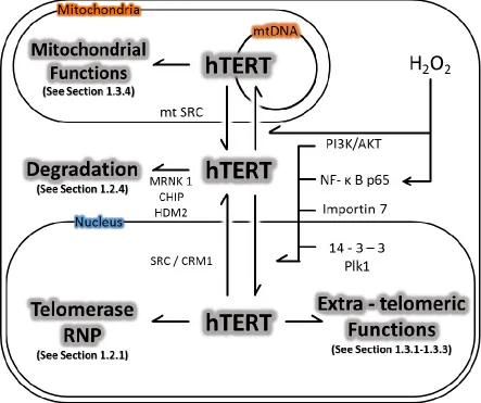
1.3.4 Mitochondrial Functions of hTERT
responsible for repressing OXPHOS in PSCs), reduced mitochondrial superoxide, mitochondrial DNA (mtDNA) content, and mitochondria mass [174].
It was suggested that ectopically expressed hTERT lead to increased H2O2-mediated mtDNA damage through increased availability of metal ions [175]. However, other studies demonstrate that FL-hTERT protects mtDNA from H2O2-induced oxidative stress[142], [174]. In addition, FL-hTERT protects mtDNA from other genotoxins such as ethidium bromide and UV-light. In general, hTERT appears to augment mitochondrial function and maturation. A growing body of research suggests that TERT has important effects on mitochondrial function in a telomere-independent manner [187, 190-193]. However, the mechanism by which TERT protects mitochondrial function is not completely understood, and remains to be elucidated
1.4 Rationale
Mitochondria in undifferentiated hESCs are localized to the peri-nuclear space, and remain low in copy number and are punctuate in morphology [60]. In contrast, hESC differentiation is accompanied by the formation of a reticular network of maturing
mitochondria with a simultaneous increase in OXPHOS and ROS production [66]. Taken together, mitochondrial function and biogenesis is key to understanding the regulation of pluripotency in undifferentiated hESCs and in early differentiation processes [178].
196-198]. Thus, hTERT has various functions in the nucleus and mitochondria. Interestingly, a number of these hTERT functions require catalytically activity of hTERT [181] and is often inhibited by catalytically inactive forms [98, 108, 187]. Many past studies [175–177, 187, 188] and our lab’s previous work by Radan et al. [184]demonstrated that high oxygen tension and oxidative stress on cells induce the extra-nuclear/mitochondrial localization of hTERT. Radan et al. additionally showed that pharmacological inhibition of telomerase activity, but not TERC, resulted in spontaneous differentiation of hESC. Similarly, disrupting β-splicing by morpholino oligonucleotides in hESC resulted in loss of pluripotency,
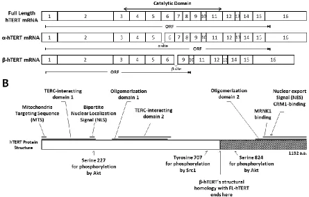
indicating the importance of hTERT and its non-catalytic variants in stem cell function [184]. Yang et al. [185] had also shown the importance of hTERT in hESC maintenance through knockdown studies. β-hTERT, like its full-length counterpart, has an intact MTS [172], RNA-interacting domains [199, 200], and oligomerization sequence[188], and retains some phosphorylation sites for nuclear import [201, 152] and export [142] (Figure 1.1). Listerman and colleagues recently demonstrated that β-hTERT is capable of mitochondrial localization in HeLa cells [102]. However, the cause and the pattern of β-hTERT localization is currently unknown. This mitochondrial localization may allow β-hTERT to alter the function of FL-hTERT in any subcellular compartment by sequestering key functional components such as specific RNA components or by disrupting the formation of a functional homodimer of catalytically active hTERT subunits.
reverse-transcriptase incompetent hTERT mutant abrogated this effect [173]. Considering β-hTERT is a reverse-transcriptase incompetent form, there may exist an interaction between FL-hTERT and β-FL-hTERT in mitochondria to fine-tune mitochondria function, possibly through hetero-dimerization. Collectively, targeting of β-TERT to mitochondria and its potential to modulate mitochondrial function and growth may imply biological significance in the regulation of pluripotency, self-renewal and differentiation potential of PSCs.
Hypothesis: Extra-telomeric hTERT β-isoform localizes to mitochondria under H2O2 -induced oxidative stress, modulating mitochondria function in hESCs.
Objectives:
1. To characterize FL-hTERT and β-hTERT expression and their role in modulating telomerase activity in hESCs.
2. To characterize the mitochondrial localization pattern of FL-hTERT and β-hTERT in hESCs under H2O2-induced oxidative stress conditions.
Chapter 2: Material and Methods
2.1 Cell Culture
2.1.1 Mouse Embryonic Fibroblasts Feeder Cells
Mouse embryonic fibroblasts (MEF) secrete several soluble growth factors to enhance the self-renewal and pluripotency of hESCs. For this reason, hESCs are often co-cultured with irradiated MEF-feeder cells. Four drug (neomycin, hygromycin, puromycin, and 6-thioguanine)-resistant MEF(4DR-MEF) cells were purchased for the future use in drug-selection of cloned transgenic hESC populations. 4DR-MEF was cultured inDMEM medium (ThermoFisher) containing 10% fetal bovine serum, 1X Non-Essential Amino Acid (NEAA; ThermoFisher) under 5% CO2 at 37oC. A bulk batch of MEFs was γ-irradiated to halt the cell proliferation and was cryo-preserved in small aliquots. γ-irradiated 4DR MEFs (approximately 60,000 cells and 106 for each well of 6-well plate and 10cm dish,
respectively) were thawed and allowed to settle onto the culture dishes or plates for one day prior to being used as feeder-layer for hESC culture.
2.1.2 Human Embryonic Stem Cell Line HES2
The human ESC line HES2 was originally established in a fertility clinic in Singapore [190] and was purchased from ATCC for utilization in this study. For maintenance purposes, HES2 cells were cultured on γ-irradiated 4DR MEFs layer for a single passage (3-4 days to reach 70-80% confluency). hESC growth medium (83% KnockOutTM DMEM/F-12
(ThermoFisher)) was refreshed daily. The stably growing HES2 cells were subsequently expanded onto a feeder-free basement membrane GeltrexTM(Life technologies) in 2% O2 (37oC, in 5% CO2) for experimental use. Fresh Essential 8TM Medium (ThermoFisher) was supplied daily for feeder free hESC culture. Cells were treated with 100μM H2O2 for up to 4 hours.
2.1.3 Establishment of monoclonal transgenic HES2 lines.
The lack of a reliable hTERT-specific antibody has been a challenge in studying FL-hTERT, β-FL-hTERT, and other TERT isoforms. To address this problem, my lab established clonally selected transgenic hESC cell lines capable of doxycycline-inducible overexpression of 3xFLAG-tagged FL-hTERT and 3xFLAG-tagged β-hTERT (hereby called HES2 ‘FL’ and HES2 ‘B’ for 3xFLAG-FL-hTERT and 3xFLAG-β-hTERT lines, respectively), enabling the use of commercially available anti-FLAG antibody to trace/isolate overexpressed FL-hTERT and β-hTERT transgenic proteins in hESCs (Fig. 2.1 A). Briefly, endogenous FL-hTERT and β-hTERT transcripts were reverse-transcribed and PCR-amplified into library DNA sequence using hTERT specific primers (appendix A1: Table 2.1). The library hTERT sequences were extended to include Gateway LR-Clonase (ThermoFisher) adaptor sequences attL1 (5’GTACAAAAAAGCAGGCT), attL2 (TGGGTCGAAAGAACATG3’), 5’Kozak sequence (GCCGCCACC), 3xFLAG sequence
Nagy Lab, Mt Sinai Hospital). The correct orientation and sequence of ligated inserts was validated by DNA sequencing (Robarts Genomic facility). The entry vector plasmid and two other PB plasmid constructs containing PiggyBac rtTA gene (transactivator of gene of interest (GOI) expression; genome-integrated), and PiggyBac transposase gene (facilitates integration of GOI and rtTA into the genome; short-lived without genomic integration) were introduced to HES2 genome via electroporation (170V, 1050 µF). Successfully transformed HES2 cells were selected by blasticidin (10μg/ml; 1 day) and G418 (1mg/ml; 7 days). The surviving cell clusters were single-celled and clonally picked to establish the final transgenic HES2 clones.
2.1.4 Cell treatment scheme
Preliminary experiments were carried out to determine the optimal treatment time and dose of doxycycline for the induction of hTERT transgene expression in HES2 clones
2.2 PCR protocols
2.2.1 General Real-Time quantitative PCR protocol
A phenol-chloroform extraction protocol was performed for RNA isolations as outlined in the product instructions (TRIzol reagent; ThermoFisher). Isolated RNA was reverse transcribed to cDNA using SuperScript III (ThermoFisher). cDNA concentrations were estimated with a NanoDrop (ThermoFisher) spectrophotometer and equal amount of template was ensured for each set of PCR reactions. RT-qPCR reactions consisted of SensiFASTTM (Bioline) PCR mix, gene-specific primer sets, and cDNA template. DNA polymerase was heat-activated at 95oC for 2 minutes prior to PCR cycles. Temperature cycling conditions were 95 oC for 10 seconds (denaturation), pre-determined temperature (primer specific; appendix A1: Table 2.1), 72oC for 30 seconds (elongation). HPRT1 was used as reference gene in all RT-qPCR experiments. Relative transcript quantity was determined based on ΔCt(gene of interest)ΔCt(HPRT1) values.
2.2.2 Detection and quantitation of hTERT isoforms by Hydrolysis-probe assisted PCR
TaqMan® Hydrolysis probe-assisted RT-qPCR was used to detect hTERT isoform transcripts. Exon-exon junctions unique for each of hTERT isoforms (α+β+hTERT, α-hTERT, β-α-hTERT, α-β-hTERT) were used as PCR priming sites (Fig. 2.1 B). α+β+hTERT are transcript species with α and β site intact, and includes FL-hTERT transcript. Importantly, the TaqMan primers are designed to detect any transcripts with the target junctions, meaning
FAM-conjugated DNA probes specific for the hTERT gene were employed to further increase the specificity and sensitivity of the RT-qPCR (Fig. 2.1 B). 1X TaqMan Universal PCR Master Mix (Applied Biosystems), 200nM forward and reverse primers, and 250nM hTERT specific FAM-probe were included per PCR reaction (20 μl). For reference, HPRT1 transcript
2.2.3 RNA-immunoprecipitation and PCR-assisted detection of TERC
1 x 10⁷ HES2 transgenic clonal cells were trypsinized and lysed in cold
non-denaturing buffer (20mM Tris pH8.0, 137mM NaCl, 10% Glycerol, 1% IGEPAL (Sigma), 2mM EDTA, 1X Protease Inhibitor Cocktail Set I (Calbiochem). The lysate was kept on ice for 30 minutes with constant agitation to help complete lysis. Cell debris was isolated and removed by centrifugation at 800g for 10 minutes. The cleared lysate was co-incubated with Anti-FLAG M2 Magnetic beads (30μl packed volume; Sigma), which was precleared with salmon sperm DNA (10 μg/ml) to prevent non-specific binding of nucleotides to the beads. The incubation was done at 4oC on a rotator overnight. Beads were washed 3 times with the lysis buffer and 2 times with TBE buffer (89mM Tris, 0.5mM EDTA, 89mM boric acid). TRIzol reagent (ThermoFisher) was added to the washed beads to extract bound RNA from 3xFLAG-hTERT proteins. TERC transcript abundance was quantified and compared in doxycycline-negative and doxycycline-positive sample groups.
2.2.4 PCR amplification of mitochondrial DNA for mitochondria DNA copy number
grade water. PCR Cycling conditions were 95oC for 2mins (DNA polymerase activation; 1cycle), 95oC for 10 seconds (denaturation), 62oC for 10 seconds (annealing), and 72 oC for 20 seconds (synthesis). A single copy nuclear gene, beta-2-microglobulin (B2M) was used as a reference to normalize the data to the number of genomes per sample.
2.3 Telomerase Activity Assay
To confirm the telomerase activity of 3xFLAG-tagged transgenic hTERT, a telomeric repeat amplification protocol (TRAP) assay was performed as previously described [191]. HES2 and 3xFLAG-hTERT-expressing lines were treated with doxycycline (1μg/ml) for 2 days. Cells were harvested by trypsinization and lysed in CHAPS lysis buffer (150mM KCl, 50mM HEPES pH7.4, 0.1% CHAPS). RNaseOUTTM (Recombinant RNase Inhibitor;
TSNT (AATCCGTCGAGCAGAGTTAAAAGGCCGAGAAGCGAT) and NT
(ATCGCTTCTCGGCCTTTT) were included. TSNT is semi-competitively amplified by the annealing of TS primer and its dedicated return primer NT during PCR. The primary purpose of this is to rule out potential false-negative outcomes resulting from hindered DNA
polymerase during PCR. Secondly, consistent amplification of TSNT amplicon (36bp) is indicative of equal PCR reaction efficiency. The finished PCR reaction mix was
electrophoresed through a vertical native 12% polyacrylamide gel at 400V for 45 minutes in 1X TBE buffer (89mM Tris, 0.5mM EDTA, 89mM boric acid) to resolve the product bands. The PAGE gel was stained with X1 SYBR Green I (ThermoFisher) nucleic acid dye in TBE for 30 minutes at room temperature. Bands were visualized using a Gel DocTM EZ (BioRad) and UV. The band intensity values of telomeric repeat products were first normalized to TSNT internal control band intensity.
2.4 Immunodetection of 3xFLAG-hTERT isoforms
2.4.1 Western Blot Analysis
HES2 B6 and F12 clones (n≥3/group) were grown under feeder-free condition under 2%O2 and H2O2 treatments. 3xFLAG-hTERT overexpression was induced by direct addition of doxycycline (1µg/ml) to the culture medium 48-hours prior to cell harvest. Cells were lysed and fractionated using a mitochondria isolation kit per its instructions (Mitochondria Isolation Kit for Cultured Cells; ThermoFisher). Protease Inhibitor Cocktail Set I
channel (VDAC; mitochondria marker), Prohibitin (mitochondria marker), and histone H3 (nuclear marker) (see appendix A2: Table 2.2). VDAC and histone H3 were used for
normalization. 3xFLAG- hTERT in nuclear and mitochondrial fractions was traced using an anti-FLAG antibody (Sigma) and quantified by densitometry analysis using Image LabTM (BioRad). All membranes were developed using a VersaDoc Imaging System (BioRad)
2.4.2 Immunocytochemistry
For immunofluorescence microscopy, HES2 B6 and F12 clones were grown in feeder-free conditions on ChamberSlidesTM (Nunc), and were treated with H2O2 as previously indicated (see section 2.1.4). Cells were fixed and permeabilized in 4% formaldehyde (15 minutes) and in 0.25% TritonTM X-100 for (10 minutes), respectively. Cells were washed with phosphate buffered saline three times in between steps. Cells were blocked with 10% bovine serum albumin solution for 45 minutes at 37 oC. Cells were
2.4.3 Cell count analysis for intra-nuclear localization of 3xFLAG-hTERT isoforms
2.5 Flow Cytometry
2.5.1 Mitochondria Membrane Potential
The cyanine dye JC-1 (5,5′,6,6′-tetrachloro-1,1′,3,3′-tetraethylbenzimidazolocarbo-cyanine iodide; Sigma) [192, 193] is widely used for microscopic and cytometric estimation and measurement of mitochondrial membrane potential (Δψm). Aggregates of the dye (J-aggregate) form at the high concentrations reached in energized mitochondria of cells exposed to near-micromolar external concentrations of the dye. J-aggregates are spectrally distinguishable from dye monomers. JC-1 is advantageous over other types of cationic Δψm dyes in that it selectively accumulates in mitochondria. JC-1 is excited at 488nm with the highest efficiency. When excited at 488 nm, JC-1 monomers emit green fluorescence with a maximum at 530 nm (green), whereas J-aggregates emit orange-red fluorescence with a maximum at 595 nm (orange-red). A subset of HES2, B6, and FL12 clones were treated with 1µg/ml doxycycline for 2 days. All cells were treated with 100 µM H2O2 for 2 hours to induce oxidative/apoptotic stress. Thirty minutes prior to the harvest, cells were treated with JC-1 (10 µg/ml; dissolved in DMSO) for 30 minutes in the incubator. Cells were washed with PBS (Ca2+ free) once and collected by trypsinizing/centrifugation at 300g for 5 minutes. Cell pellets were re-suspended in flow-cytometry staining buffer (Ca2+ free PBS, 0.5% BSA). Spectral readings were taken immediately with BD AccuriTM C6 using a blue laser (488nm), bandpass filters FL2 (586±20nm; J-aggregate) and FL1 (530 ±15nm; JC-1 monomer).
Twenty thousand events were recorded per group at all channels. 20μM Carbonyl cyanide 4-l (trifluoromethoxy) phenylhydrazone (FCCP; chemical dissipater of Δψm; Molecular
2.5.2 Detection of apoptosis
To assess the pro-survival effect of overexpressed 3xFLAG-hTERT in HES2, cells were stained with Annexin V-FITC (ThermoFisher)/7-amino-actinomycin (7-AAD;
eBioscience), following 100 µM H2O2 treatment. Annexin V/7-AAD staining provides a way to discriminate between early apoptosis and late apoptosis. When a cell undergoes apoptotic cell death, phosphatidyl serine (PS; normally on the inner leaflet of plasma membrane) becomes exposed on the outer leaflet of the plasma membrane. Annexin V binds PS with high affinity and marks early-apoptotic cells. 7-AAD is a DNA intercalating dye that can enter dead cells with compromised membrane integrity. A subset of HES2, B6, and FL12 clones were treated with 1µg/ml doxycycline for 2 days. All cells were treated with 100 µM H2O2 for 2 hours to induce oxidative/apoptotic stress. Following cell harvest by
trypsinization/centrifugation (as outlined above), cells were re-suspended in Annexin V-staining buffer (10mM HEPES, pH 7.4, 140 mM NaCl, 2.5 mM CaCl2) to 106 cells per/ml. Annexin V-FITC (15 minutes, at room temperature) and 7-AAD (5 minutes, at room temperature) were added to a 100 µl aliquot of the resuspension. Spectral readings were taken immediately with BD AccuriTM C6 using a blue laser (488nm), bandpass filters FL3 (670 nm longpass; 7-AAD) and FL1 (530±15nm; FITC Annexin V-FITC). A total of 30000 events were recorded per group at both channels.
2.6 Statistical Analyses
All statistical analyses were performed using Prism 6 (Graph Pad Inc.), and are
Chapter 3: Results
3.1 Basic cell characterization of human embryonic stem cells
3.1.1 Characterization of pluripotency markers in human embryonic stem cells
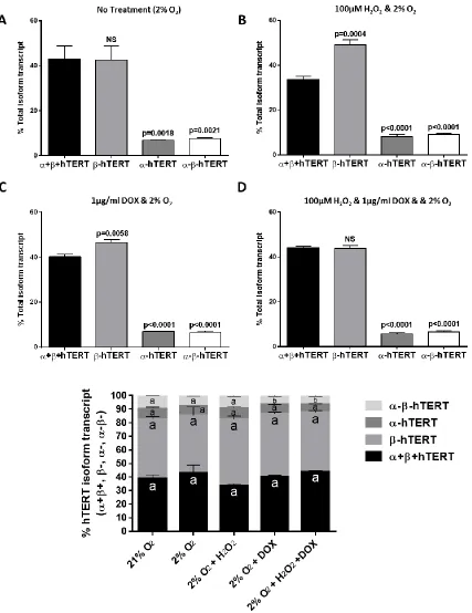
Human embryonic stem cells (HES2 cell line) grow as flat and compact round colonies indicative of the primed pluripotent stem cell state [194] (Fig. 3.1 A). Assessed by immunofluorescence microscopy, HES2 cells were positive for pluripotency markers SSEA-4 and OCT-SSEA-4, but negative for SSEA-1 (Fig. 3.1 C). Transcripts of the α+β+TERT isoform (includes FL-hTERT transcript) and the most prominent hTERT splice variants (β-, α-, α-β-) were detected in HES2 cells that had been cultured under 21% oxygen tension (Fig. 3.1 B). Both β-hTERT and α+β+hTERT transcript levels were detected at about 40% of hTERT variant pool measured by TaqMan RT-qPCR (Fig. 2.1), and were significantly higher in abundance (~5-10 fold higher) than α-hTERT and α-β-hTERT transcript levels (Fig. 3.1 B).
3.1.2 Characterization of hTERT isoforms in human embryonic stem cells
Fig. 3.2: Endogenous β-hTERT transcript is ubiquitously present at high levels in human embryonic cells grown under low oxygen tension conditions, with and without H2O2 and/or doxycycline (DOX) treatments. hESCs (HES2 cell line) were grown under low oxygen conditions
(2% O2) with A) no treatment, B) 100μM H2O2, C) 1μg/ml DOX, D) 100μM H2O2 and 1μg/ml DOX,
E) Combined representation of %hTERT isoform change under conditions specified in A-D.
3.2 Characterization of 3xFLAG-hTERT transgenic clones
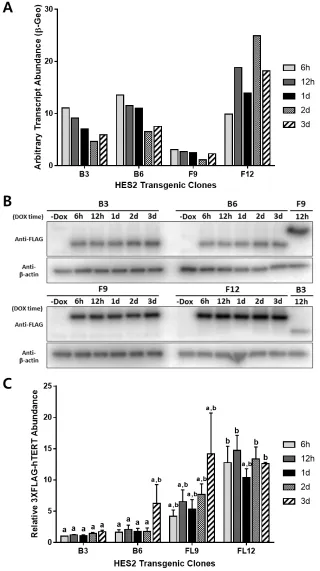
3.2.1 Characterization of 3xFLAG-hTERT transgene expression level in transgenic hESC clones
3.2.2 Characterization of Pluripotency in HES2 B6 and HES2 FL12 hTERT cell lines
HES2 B6 and FL12 were treated with DOX (2 days; 1 μg/ml) and with H2O2 (final 4 hours; 100μM) to assess the potential side effect of DOX and H2O2 treatment on pluripotency
Fig. 3.3: RT-qPCR and Western blot analysis of doxycycline-induced expression of 3xFLAG-hTERT isoforms in HES2 clones over 3-day time course. HES2 clones (HES2 B3 and B6 for DOX-inducible 3xFLAG-β-hTERT, and HES2 FL9 and FL12 for DOX-inducible 3xFLAG-FL-hTERT) were cultured under 2% O2 and were treated with varying times (h = hours, d = days) of doxycycline (DOX) prior to harvest. A)
Fig. 3.4: H2O2 and DOX treatments do not affect pluripotency marker expression in
3.3 Characterization of DOX-induced recombinant hTERT in HES2 B6 and HES2 FL12
3.3.1 Endogenous TERC-binding of 3xFLAG-β-hTERT and 3xFLAG-FL-hTERT subunits
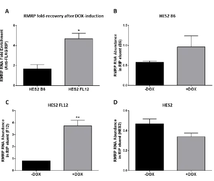
RNA-immunoprecipitation (RIP) using a FLAG antibody was performed to determine whether the 3xFLAG-hTERT transgenic proteins have the ability to bind to the telomerase RNA-component (TERC). TERC recovery from the RIP eluents of DOX-treated samples and non-DOX-treated samples were compared. Overexpression of 3xFLAG-hTERT by
doxycycline prior to RIP resulted in approximately 60-fold increase in TERC recovery for 3xFLAG-FL-hTERT, and 10-fold increase for 3xFLAG-β-hTERT (Fig. 3.6 A, B, C).
Doxycycline-treated HES2 (non-transgenic; negative control) did not result in any change in TERC recovery, and resulted in a very low TERC PCR-amplification (Fig. 3.6 D).
3.3.2 Endogenous RMRP-binding of 3xFLAG-β-hTERT and 3xFLAG-FL-hTERT subunits
3.3.3 Telomerase activity in HES2 B6 and HES2 FL12 hESCs
The effect of 3xFLAG-β-hTERT and 3xFLAG-FL-hTERT overexpression on total telomerase activity of HES2 B6 and FL12 cell lines was investigated by the telomerase repeat amplification protocol (TRAP) assay (Fig. 3.8). Negative controls (heat-inactivated lysate and no-lysate control) did not generate any product bands (Fig.3.8 A). Doxycycline in non-transgenic HES2 had no effect on the endogenous telomerase activity (Fig. 3.8 B top). DOX-induction of recombinant β-hTERT in HES2 B6 resulted in ~30% decrease in the total telomerase activity, whereas DOX-induction of recombinant FL-hTERT in HES2 FL12 had no effect (Fig. 3.8 B bottom left and right, respectively).
3.3.4 Re-visiting DOX-induction of 3xFLAG-hTERT transcription in HES2 clones
Since overexpression of 3xFLAG-FL-hTERT did not result in an the expected increase in telomerase activity in HES2 FL12 (Fig. 3.8 B), we re-investigated the 3xFLAG-hTERT transgene expressions and endogenous hTERT isoform transcript abundance in the trangenic HES2 clones. The transcript level of different hTERT isoforms in HES2 FL12 and HES2 B6 was examined after DOX-induction by hydrolysis-probe-assisted RT-qPCR. The addition of DOX in HES2 FL12 resulted in ~500-fold increase in FL-hTERT transcript, but suprisingly a ~300-fold increase in β-hTERT transcript, and ~200-fold increase in α-β-hTERT transcript was also observed compared to non-induced controls (Fig.3.9 A). Interestingly, the HES2 B6 cell line expressed only the β-deletion-containing TERT isoforms upon doxycycline
Fig. 3.6: Doxycycline-induced transgenic telomerase proteins (3xFLAG-β-hTERT and 3xFLAG-FL-hTERT) bind to the telomerase RNA component (TERC). Cells were grown under 2% O2 and were treated with 1μg/ml doxycycline (DOX) for two days prior to harvest. DOX-induced 3xFLAG-hTERT isoforms were isolated from their respective clones by immunoprecipitation using anti-FLAG-magnetic beads. Eluted proteins were subsequently denatured to release any bound RNA for downstream RT-qPCR analysis. A)
Comparison of results in HES2 transgenic lines, and individual TERC enrichment data from
Fig. 3.8: Altered telomerase activity levels in doxycycline (DOX)-induced transgenic telomerase proteins (3xFLAG-hTERT) in hESCs. Cells were grown under 2% O2 and were
treated with 1μg/ml doxycycline for two days prior to harvest. A) Representative DNA gel of
telomerase-repeat amplification assay by qPCR. Relative telomerase activity between samples is
reflected in the intensity and the range of telomerase product bands. H.I. = Heat-Inactivated B) Band
intensity of amplified telomerase product was measured by densitometry analysis and represented as relative telomerase activity. Data were normalized to internal control band intensities (n=4, error
Fig. 3.9: DOX-induction of 3xFLAG-hTERT isoforms results in a simultaneous increase in the amount of other hTERT transcript variants containing β-deletion. A)