Original Article
Transforming growth factor beta2 promotes the formation of
the mouse cochleovestibular ganglion in organ culture
JUNKO OKANO
1,2, TOSHIYA TAKIGAWA
1, KENJI SEKI
1, SHIGEHIKO SUZUKI
2, KOHEI SHIOTA
1,3and MAKOTO ISHIBASHI*
,11Department of Anatomy and Developmental Biology, 2Department of Plastic and Reconstructive Surgery and 3Congenital Anomaly Research Center, Graduate School of Medicine, Kyoto University, Kyoto, Japan
ABSTRACT The inner ear structures are derived from the otic vesicle (OV) which is formed by thickening and invagination of the otic placode of the surface ectoderm. A number of neuroblasts, which arise from epithelial cells of the otic vesicle, delaminate and differentiate into neurons of the cochleovestibular ganglion (CVG). We have found that transforming growth factor-βββββ2 (Tgfβββββ2 ) was expressed in the otic epithelium at the OV stages between embryonic days (E) 9.5 and 11.5 and that anteroventrolateral localization of its expression in the OV overlapped with that of NeuroD, which is a marker of delaminating CVG precursors. The expression of TGFβββββ type I and type II receptors in the otic epithelium and the nuclear localization of phosphorylated-Smad2 in both the otic epithe-lium and CVG suggested that TGFβββββ2 signaling plays some roles in CVG formation. In order to examine the roles of TGFβββββ2 in differentiation of the inner ear, otic vesicle explants of E10.5 mouse embryos were treated in vitro with TGFβββββ2 or the TGFβββββ type I receptor kinase inhibitor, SB431542. Addition of TGFβββββ2 peptide to the culture led to enlargement of the CVG, while the inhibitor reduced its size. These findings strongly imply that TGFβββββ2 contributes to the development of the CVG in mouse embryos.
KEY WORDS:
TGF
β
, epithelial-mesenchymal transformation, delamination, neuronal differentiation
Introduction
The mammalian ear is a delicate sensory organ for hearing and balance and is one of the most complex structures in the body. The inner ear primordium, the otic placode, first appears as a thickening of the surface ectoderm on which the neural plate neighbors. The otic placode invaginates to form the otic vesicle, which subse-quently detaches from the surface ectoderm and form the inner ear structures such as the semicircular canals, vestibule and cochlea (Rossant, 2002).
The cochleovestibular ganglion (CVG) is formed by the cells which delaminate and migrate from the anteroventral portion of the otic vesicle (Carney et al., 1983) and the neural crest-derived cells (D’Amico-Martel and Noden, 1983; Rubel and Fritzsch, 2002). It has been shown that a large area of the anteroventrolateral part of the OV gives rise to the CVG neuroblasts in mice (Fritzsch, 2003) and these cells are among the first cell types to be specified during the early OV stages (Fekete and Wu, 2002; Hemond and Morest, 1991; Hossain et al., 2000). Recently, several genes have been identified that play crucial roles in development of the CVG. For
*Address correspondence to: Dr. Makoto Ishibashi. Department of Anatomy and Developmental Biology, Graduate School of Medicine, Kyoto University, Yoshida Sakyo-ku, Kyoto 606-8501, Japan. Fax: +81-75-751-7529. e-mail: ishibash@anat1.med.kyoto-u.ac.jp
web: http://www.anat1.med.kyoto-u.ac.jp/index-j.htm
Abbreviations used in this paper: BDNF, brain derived neurotrophic factor; BMP, bone morphogenetic protein; CVG, cochleovestibular ganglion; E, embryonic day; Fgf, fibroblast growth factor; IGF, insulin-like growth factor; NGN, neurogenin; NT, neurotrophin; OV, otic vesicle; TGF, transforming growth factor.
0214-6282/2005/$25.00 © UBC Press
Printed in Spain www.intjdevbiol.com
example, the differentiation of the CVG depends on two basic helix-loop-helix (bHLH) factors, Neurogenin1 (NGN1) and NeuroD. Ma
et al. (1998) reported that the CVG was not formed in Ngn1 null mutants. In NeuroD mutant mice, migration of CVG precursors was compromised and there was considerable size reduction of the CVG (Liu et al., 2000; Kim et al., 2001). The roles of some growth factors have also been implicated in inner ear development. Double mutants of fibroblastgrowthfactor (Fgf ) 3 and Fgf10
factor-1 (IGF-1) has been suggested to be necessary for CVG differentiation (Camarero et al., 2001; Camarero et al., 2002; Camarero et al., 2003).
Transforming growth factor-β (TGFβ) is a secreted peptide which promotes cell survival during the development of the central and peripheral nervous systems (Sporn etal., 1987; McFarlane and Cooper, 1993). Three isoforms of TGFβ (TGFβ1, 2 and 3) show distinct spatial and temporal expression patterns in mouse embryos, suggesting that they play various roles during embryo-genesis (Flanders et al., 1991; Johnson et al., 1993). Blottner et al. (1996) reported that implantation of gelform soaked with TGFβ2 rescued all the injured neurons of the preganglionic sympathetic neurons in the spinal cord, though the mechanisms remain to be clarified. It has been shown that TGFβ1 is a potent survival factor for motor neurons of rat embryos (Martinou et al.,
1990) and it has also been shown that the TGFβ2/3 promote the survival of midbrain dopaminergic neurons (Poulsen et al., 1994). Recently, Farkas et al. (2003) showed that both TGFβ2 and TGFβ3 are required for induction of midbrain dopaminergic neu-rons as well. They showed that TGFβ2/3 were expressed in the notochord and floor plate and that neutralization of TGFβ2/3 abolished induction of dopaminergic neurons in rat primary cul-tures and E2 chick embryos.
Although the pattern of Tgfβ2 expression during inner ear development has been reported previously (Pelton et al., 1990; Millan et al., 1991; Schmid et al., 1991), their description was not conferred at the OV stages. In this study, we examined the
Fig. 1. Expression patterns of Tgfβββββ2, Neurogenin1(Ngn1), NeuroD and Bmp4 in the mouse developing OV. Tgfβ2 expression was detected in the anterior wall (arrowhead) of the otic vesicle at E9.5 (A) and became more intense at E10.0 (B). At E10.5, Tgfβ2 transcripts were also detected in the posterior part of the vesicle as well as in the anterior wall (C). Neurogenin1 was also expressed in the anterior region between E9.5 and 10.5 (D,E,F). Note that its expression domain partially overlapped with that of Tgfβ2. NeuroD was expressed in the same region as Neurogenin1 in the epithelium as well as in the neuronal precursors of the CVG between E9.5 and E10.5 (G,H,I). Bmp4 was expressed in the posterior wall (arrow) of the OV at E9.5 (J), E10.0 (K) and E10.5 (L) and also in the anterior wall (arrowhead) at E10.0 (K) and E10.5 (L). Dotted circles indicate the contour of the OV. The thick arrow and the asterisk in (G) show the developing CVG and the facial ganglion, respectively. Direction is indicated in (L) for all panels (A, anterior; D, dorsal). Scale bar, 100 µm.
Fig. 2. Detailed expression patterns of Tgfβββββ2 and NeuroD by section RNA in situ hybridization. Sections of the anterior (A,B,E,F) and posterior (C,G) parts of the OV were stained with Tgfβ2 (A,C,E,G) and NeuroD (B,F) probes. (D) and (H) indicate the plane of each section. At E10.5, Tgfβ2 was detected in the ventrolateral region of the anterior OV (A) (see arrow), while NeuroD was expressed in both ventrolateral and ventral regions (B)(see arrowheads). NeuroD was also detected in the delaminated precursors (B). In the posterior OV, Tgfβ2 was observed in the dorsolateral region where NeuroD was not expressed (C). At E11.5, Tgfβ2 was expressed in the ventral region ( (E), double arrow) in addition to the ventrolateral region (E, arrow) of the anterior OV. On the other hand, NeuroD expression was observed in a small number of the ventral epithelial cells of the anterior OV
expression patterns of Tgfβ2 in the developing OV of mouse embryos by RNA in situ hybridization. We also treated cultured mouse embryonic OVs with TGFβ2 or TGFβ type I receptor kinase inhibitor and analyzed their effects on the developing OV. Our data suggest that TGFβ2 may contribute to inner ear development, especially to the formation of the CVG.
Results
The expression of Tgfβββββ2 overlaps that of Neurogenin and NeuroD in the early otic vesicle
Tgfβ2 expression in the otic epithelium was examined at OV stages (E9.5-E11.5) by RNA in situ hybridization. At E8.5-9, Tgfβ2 expression was not detected in the otic placode or pit. At E9.5, when the otic vesicle formed, Tgfβ2 transcripts were detected in the anteroventrolateral region of the otic vesicle (Fig. 1A). At E10.0 and E10.5, Tgfβ2 was more intensely expressed than at E9.5 in the anteroventrolateral wall and also came to be detectable in the posterodorsolateral wall (Fig. 1 B,C). Transverse sections re-vealed that its expression was confined to the epithelium of the otic vesicle, but not in the surrounding mesenchyme (Fig. 2 A,C).
The neuronal precursors for the CVG delaminate from the anteroventral wall of the OV between E9.5 and E11.5 with its peak at E10.5 (Carney et al., 1983). We compared the expression patterns of Tgfβ2 with those of the neural bHLH genes Ngn1 and
NeuroD, which have essential roles in CVG development (Ma et al., 1998; Liu et al., 2000). Ngn1 expression was first detected in the anteroventrolateral region of the otic epithelium at E9.0 when the otic placode began to invaginate (Raft et al., 2004). At E9.5 and E10.0, Ngn1 was expressed in the anteroventrolateral wall of the OV and its expression domain partially overlapped with that of
Tgfβ2 (compare Fig. 1 A,B and D,E).
NeuroD was first faintly expressed in the same region as Ngn1
at E9.0 (Liu et al., 2000; Raft et al., 2004). Its expression became intense in the delaminating precursors and CVG cells by E9.5 (Fig. 1G). The epithelial domains of Ngn1 and NeuroD expression overlapped with each other up to E11.5 (Fig. 1 D-I). The expression domain of NeuroD in the otic vesicle also partially overlapped with that of Tgfβ2 (compare Fig. 1 A,B,C and G,H,I). To analyze their expression patterns in detail, we performed section RNA in situ
hybridization. At E10.5, both Tgfβ2 and NeuroD transcripts were found in the anteroventrolateral regions of the otic vesicle (Fig. 2 A,B), although their expression patterns were not exactly identical.
Tgfβ2 was expressed more intensely in the luminal and interme-diate layers than in the outer layers of the otic epithelium (Fig. 2A), whereas NeuroD was more intensely expressed in the otic
epithelial cells near the mesenchyme (Fig. 2B). Tgfβ2 expression was also observed in the posterodorsolateral otic epithelium (Fig. 2C) where NeuroD was not detectable (data not shown). At E11.5, when the endolymphatic duct became more overt and the cochlear anlage expanded ventrally, Tgfβ2 expression still remained on the anteroventral wall of the OV, although the expression domain slightly shifted to the outer layers in the ventral region (Fig. 2E).
NeuroD was also detected in a few cells of the same domain (Fig. 2F). Robust expression of Tgfβ2 was also detected in the posteroventrolateral wall at E11.5 (Fig. 2G).
Bone morphogenetic protein (BMP) 4 is another member of TGFβ superfamily which has been shown to be expressed in the developing OV of both chick and mice (Wu and Oh, 1996; Morsli
et al., 1998; Raft et al., 2004). To further clarify the expression domain of Tgfβ2, we compared its expression with that of Bmp4. At E9.5, Bmp4 was expressed in the posterodorsal wall of the OV (Fig. 1J). At E10 and later, it was also detectable as a streak domain in the anterior wall (Fig. 1 K,L). These two loci correspond to the anterior and posterior cristae (Wu et al., 1996). The expres-sion domains of Tgfβ2 and Bmp4 seemed to be very close to each other but not to overlap.
In summary, Tgfβ2 transcripts were localized in the antreoventrolateral wall and in the posterolateral region of the otic vesicle at E9.5-11.5. Tgfβ2 expression overlapped with that of
NeuroD in the anteroventolateral compartment.
TGFβββββ type I and type II receptors and phosphorylated Smad2 are present in the developing OV
To examine whether TGFβ type I and type II receptors are expressed in the developing OV, we used RNA in situ hybridiza-tion. Since their expression levels were too low to be detected with digoxigenin-labeled probes, we performed RT-PCR, which is much more sensitive than RNA in situ hybridization, using OVs of E10.5 (35-39 somites) embryos. The embryonic heart tissue, which expresses those receptors most strongly at this stage (Mariano et al., 1998), was used as a positive control. The OVs showed comparable expression levels of both TGFβ type I and
type IIreceptors to the embryonic hearts (Fig. 3). Fig. 3. Expression of TGFβββββ type I and II receptors in the developing OV.
One µg of RNA extracted from E10.5 otic vesicles was subjected to RT-PCR. The heart was used as a positive control. Robust expression of both receptors was observed in the otic vesicle (27 cycles). Three independent experiments were performed and the representative data are shown.
Next, to confirm that TGFβ signaling is actually active in the OV, we carried out an immunohistochemical study using an anti-phosphorylated Smad2 antibody. Phosphorylation of Ser465 and Ser467 of Smad2 is required for the Smad2/Smad4 complex formation and TGFβ signal transduction (Nakao et al., 1997; Abodollah et al., 1997; Souchelnytskyi etal., 1997; de Caestecker
et al., 1998). Nuclear localization of phosphorylated Smad2 indi-cates that TGFβ signaling is conveyed to the nucleus in the cells (Heldin et al., 1997; Massague et al., 1998; Massague and Wotton, 2000). Intense nuclear staining was observed in the anteroventolateral wall of the otic epithelium and the outer cells of the CVG (Fig. 4), indicating that TGFβ may signal in these regions. There were phosphorylated Smad2-positive cells in the neural tube (Fig. 4) as previously reported by Farkas et al. (2003).
TGFβββββ2 increases the size of the cochleovestibular ganglion in cultured otic vesicles
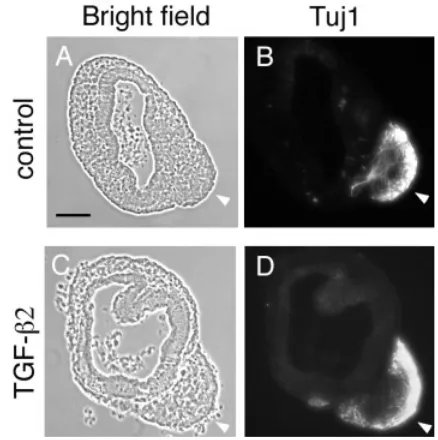
Since Tgfβ2 was expressed in the anteroventrolateral wall of the OV, we assumed that TGFβ2 might play some role in CVG develop-ment. To test this hypothesis, we cultured the otic vesicles of E10.5 mouse embryos in which CVG precursors delaminate most actively from the otic epithelium (Carney et al., 1983) and treated them with TGFβ2. Within 16 hours in culture a mass of cells migrated out of the anteroventrolateral region of the OV and formed a CVG-like structure (Fig. 5). Many of the cells forming this structure were positive for Tuj1, an early neuronal marker (Fig. 6). Our observation along with the study of Camarero et al. (2003) suggests that this structure was likely a newly-formed CVG in the culture.
To examine the effects of TGFβ2 on the differentiation of OVs, we treated cultured OVs with recombinant TGFβ2 protein. With TGFβ2, 6 of 14 OV explants (42.9%) produced the CVG after 4 hours incubation whereas no CVG was formed in the control cultures (Fig. 5; Table 1). Likewise, when the explants were incubated for 6 hours, 15 of 24 cultured vesicles (62.5%) produced the CVG with TGFβ2 whereas only 4 of 25 (16.0%) formed without TGFβ2 (Table 1). Moreover, the size of the CVG was increased with TGFβ2 (compare the top and bottom panels in Figs. 5,6) when compared to the control samples (Figs. 5,6), suggesting that TGFβ2 accelerates CVG formation.
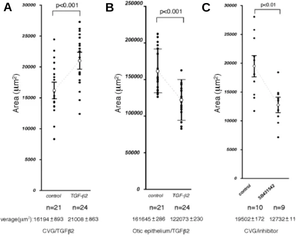
To quantitate the effects of TGFβ2 on CVG development the area of the CVG on photographs was measured using the tech-nique of Camarero et al. (2003). The area of the CVG treated with TGFβ2 was increased by 1.4-fold (P< 0.001, Fig. 7A). We also
measured the area of the remaining epithelium of the OV and found that the average area of the treated group was significantly smaller than that of the control group (P< 0.001, Fig. 7B), suggesting that TGFβ2 promotes CVG formation at the expense of the epithelial cells of the OV.
To determine whether TGFβ2 increases the number of CVG precursors or cell proliferation of neuroblasts, we performed immunostaining with anti-phosphorylated histone3, a proliferation marker. There was no clear difference between the control and TGFβ2-treated tissues (data not shown), suggesting that TGFβ2 increases the number of CVG precursors instead of promoting cell proliferation.
Next, we treated OVs with the TGFβ type I receptor kinase inhibitor SB431542 (5µM) in order to block the endogenous TGFβ signaling. The area of the CVG treated with SB431542 was Fig. 5. Organ culture of otic vesicles from E10.5 embryos. Top and bottom panels show controls and the culture treated with TGFβ2 (10 ng/ml), respec-tively. Incubation periods are indicated at the top. 0 h represents the beginning of the culture. The already formed CVG was removed from the explants before culture. After 4 hours, there was no CVG in controls, while several vesicles already produced the CVG (arrowheads) when treated with TGFβ2 protein (see Table 1). After 16 h, larger CVGs (arrow-heads) were formed with TGFβ2 than in controls. Scale bar, 100 µm.
Fig. 6. Tuj1 immunohistochemistry of OV culture.(A,B) Controls. (C,D)
significantly reduced when compared to the control vesicles (P< 0.01, Fig. 7C).
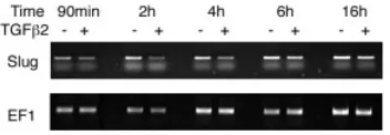
Slug expression is downregulated in the TGFβββββ2-treated OV It was previously reported that TGFβ2 upregulates the expres-sion of Slug, a member of the Snail family that is involved in delamination of endothelial cells in the embryonic heart (Romano and Runyan, 2000). We have found that Slug was expressed in the developing OV of the mouse embryo (unpublished data). Since the delamination of CVG precursors from the OV is a similar phenom-enon to epithelial-mesenchymal transformation (EMT), we exam-ined by semi-quantitative RT-PCR whether the expression of Slug
is affected with TGFβ2 in the OV. mRNA was extracted from the whole culture including both the vesicle and CVG after various incubation intervals (Fig. 8). The intensity of the PCR bands was measured and normalized by that of EF1 products. The relative intensity of the bands from TGFβ2-treated OVs to the control was 0.624 (1.5h), 0.409 (2h), 0.569 (4h), 0.631 (6h) and 0.886 (16h), indicating that Slug mRNA was substantially reduced in culture treated with TGFβ2.
Discussion
In this study we found that Tgfβ2 is expressed in the epithelium of the mouse OV in a distinct pattern. TGFβ type I and type II receptors and phosphorylated Smad2 were also detected in the
In our present study Tgfβ2 showed an intense and dynamic expression pattern in the developing OV. It was expressed in the anteroventrolateral otic epithelium in the proNS compartment at E9.5 and then also detected in the posterodorsolateral wall by E10.5. At E11.5, the latter Tgfβ2 expression domain shifted to the posteroventrolateral wall. It was noteworthy that the expression domain of Tgfβ2, at least partially, overlapped with those of nuclear phosphorylated Smad2 and NeuroD. NeuroD is a marker of delaminating CVG precursors in the mouse OV (Ma et al., 1998; Raft et al., 2004). This result suggests that TGFβ2 is likely to signal to NeuroD -positive cells in paracrine and/or autocrine fashions.
NGN1 is another neurogenic bHLH transcription factor which is expressed in the same population of the otic epithelium. Both
Ngn1 and NeuroD knockouts showed CVG defects (Ma et al.,
Interval TGFβββββ2 Total explants CVG(+) explants (%) CVG(-) explants(%)
4 hours - 14 0 (0.0) 14(100) + 14 6(42.9)* 8(57.1) 6 hours - 25 4(16.0) 21(84.0) + 24 15(62.5)* 9(37.5) *Significantly different from controls by Chi-square test (P<0.01)
TABLE 1
FORMATION OF CVG IN EXPLANTS CULTURED WITH TGFβββββ2
Fig. 7. The size of the CVG and otic epithelium in culture. The areas of the CVG (A)
and otic epithelium (B) were estimated on microphotographs by NIH Image program (http://rsb.info.nih.gov/nih-image/). The vertical axis indicates areas (µm2). Dots
repre-sent individual values. Open circles indicate the average areas with standard errors. The total numbers of the explants (n) and the average areas are indicated at the bottom of each panel. The area of the CVG with TGFβ2 was significantly larger than control (P<0.001). In contrast, the area of the epithelium with TGFβ2 was significantly smaller than control (P<0.001). (C) A TGFβ signaling inhibitor, SB431542 (5 µM), significantly decreased the size of the CVG (P<0.01).
A
B
C
otic vesicle, suggesting that TGFβ signaling is under-way during inner ear development.
Brigande et al. (2000) proposed that the spherical OV can be divided into eight compartments by the anterior-posterior, dorsal-ventral and medial-lateral boundaries and that different parts of the inner ear are derived from different compartments. Alsina et al. (2004) designated the anteroventral domain as “proneural sensory territory” (proNS) because proneural and neurogenic genes, in-cluding Neurogenin1 and NeuroD, are expressed in this domain. They observed that CVG precursors delami-nated around the boundary between proNS and its posterior region.
1998; Liu et al., 2000; Kim et al., 2001) and downregulation of
NeuroD in Ngn1 mutants implied the requirement of Ngn1 for
NeuroD expression. According to Sun et al. (2001), Smad1, a mediator of TGFβ signaling, forms a transcriptional complex with NGN1. Therefore, it may be interesting to determine whether TGFβ2 signals to NeuroD -positive cells through Ngn1.
The Ngn1 and NeuroD expression domains seemed to be restricted to the anterior compartment by TBX1. Tbx1 is one of T-box genes and is expressed in the developing OV (Raft et al., 2004). Its expression showed a posterior-anterior gradient first at E9.5, suggesting that TBX1 specifies the posterior compartment of the OV. Indeed, the Ngn1 and NeuroD expression domains expanded in Tbx1 knockouts, leading to the duplication of the CVG rudiment (Raft et al., 2004). Therefore, it is intriguing to examine whether Tbx1 expression is downregulated by TGFβ2 treatment.
It has been reported that Bmp4 is also expressed in the developing OV. Wu and Oh (1996) suggested that BMP4 plays important roles in specification of sensory primordia of the otic epithelium. The previous and present studies demonstrated that the expression domains of Bmp4 and Tgfβ2 were very close to each other but not overlapped in the anterior and posterior parts of the OV. Since addition of TGFβ2 to OV culture led to increased size of the CVG (a mass of sensory neurons), it may be possible that BMP4 and TGFβ2 cooperatively specify sensory neuron precursors in the developing OV.
We showed that exogenous TGFβ2 increases the size of the CVG and that inhibition of the endogenous signaling suppresses the CVG formation. The data suggest that TGFβ2 may have increased the number of cells which are to become delaminating CVG precursors and/or their proliferation during CVG develop-ment. Although TGFβ has been shown to stimulate the proliferation of spiral ganglion cells (Hansen et al., 2001), we observed that the size of otic vesicles treated with TGFβ2 was significantly reduced after 16 hours in culture (Fig. 6C) while the size of the CVG was increased. It is possible that more cells are specified as delaminat-ing CVG precursors with exogenous TGFβ2 at the expense of the epithelial cells, though the possibility cannot be excluded that TGFβ2 accelerates proliferation of the neuronal precursors while it decreases proliferation of non-neuronal precursors.
Recently Lawoko-Kerali et al. (2004) reported that the NeuroD-positive domain of the OV might be further divided into two
regions. The medial region was positive for GATA3, a zinc finger transcription factor and thought to produce the cochlear part of the CVG. The GATA3-negative region was thought to produce the vestibular part. Although these two parts of the ganglion are morphologically separated in vivo, we cannot distinguish the two parts in organ culture. Still, it is intriguing to determine which part of the CVG is increased in size after TGFβ2 treatment by exam-ining GATA3 expression.
In spite of defects in other parts of the inner ear (Sanford et al., 1997), the CVG phenotype in Tgfβ2 mutants has not been reported to date. The inner ear phenotypes of the mutants included the defects of the spiral limbus in the basal cochlear turn and Rosenthal’s canal. The primary auditory neurons were normally formed in the mutants, but the interdental cells overlying the spiral limbus were undifferentiated. It should be noted that such anomalies of the inner ear in Tgfβ2 mutants were restricted to nonsensory areas, which is not consistent with our in vitro data. It is likely that the phenotype of the sensory area was rescued in vivo by other members of the
Tgfβ family while that of the nonsensory area was not. This posterior domain did not express the proneural gene NeuroD. According to Fekete and Wu (2002), the nonsensory area of the cochlear duct arises from the posteroventrolateral compartment. Therefore, the inner ear defects in null mutants may be related to the posterior domain of Tgfβ2 expression.
It is not known whether Tgfβ1/3 rescue the CVG phenotype though they are expressed in or near the OV. TGFβ1 begins to be immunolabeled in the periotic mesenchymal cells surrounding the developing OV, which form the capsule of the inner ear (Frenz et al. 1992). Other factors might also compensate the Tgfβ2 func-tion, since TGFβ2 works synergistically with other factors such as BDNF and glial cell line-derived neutrophic factor (GDNF) in the nervous system (Sometani et al., 2001; Krieglstein et al., 2002; Sometani et al., 2002).
Slug, a zinc finger-type transcription factor, has been reported to play a crucial role in EMT during neural crest cell differentiation (Sefton et al., 1998; del Barrio and Nieto, 2002). Romano and Runyan (2000) suggested that Slug is an essential target of TGFβ2 signaling during EMT of endocardial cushion formation. Delamination of CVG precursors from the otic epithelium is similar to EMT in terms of loss of epithelial character. However, in our RT-PCR study, Slug mRNA was downregulated when cultured OVs were treated with TGFβ2, which is not consistent with the previous report (Romano and Runyan, 2000). One possible explanation for the discrepancy between the studies is as follows: Slug functions in the epithelial cells to cause transformation and is quickly downregulated in the transformed cells. Although addition of TGFβ2 led to increase of CVG cells and decrease of epithelial cells, the analysis of the culture explants showed that Slug mRNA was seemingly downregulated in our experiments. Another pos-sibility is that the delamination of CVG precursors is an unrelated phenomenon to EMT. Delaminated precursors do not seem to go through mesenchymal cell stages. It is rather similar to migration of neuroblasts from the ventricular zone to the mantle zone in the developing neural tube. Therefore, it is possible that Slug plays a different role from that in neural crest development and is differ-ently regulated by TGFβ2 signaling in the OV. Further study is required to elucidate how TGFβ2 regulates Slug expression and transformation of the otic epithelial cells and whether Slug is involved in transformation of the otic epithelial cells.
Materials and Methods
RNA in situ hybridaization
Embryos were collected between E9.0 and E11.5. The noon on the day of finding a vaginal plug was designated as E0.5. Whole mount and section RNA in situ hybridization was carried out as previously described (Wilkinson, 1992; Ishii et al., 1997). Embryos were fixed overnight in 4% paraformalde-hyde at 4oC, rinsed in PBS and dehydrated in 25% sucrose for section or
in methanol for whole mount. Transverse sections (20µm) were mounted on APS-coated slides (Matsunami, Japan). Murine Tgfβ2 probe (Pelton et al., 1991) was kindly provided by Dr. Moses. NeuroD (Lee et al. 1995) and Ngn1 probes (Cau et al.1997) were gifts from Dr. Kageyama. Digoxigenin-labeled sense and antisense probes were synthesized by digoxigenin RNA labeling kit (Roche Diagnostics Corporation, Indianapolis, IN, USA). Whole mount embryos were cleared in 80% glycerol.
Immnohistochemistry
Embryos at E10.5 were fixed for 1 h in 4% paraformaldehyde at 4ºC, followed by routine procedures for embedding in paraffin. Transverse sections (6µm) were mounted in a serial order on slides. Sections were treated in 10mM sodium citrate buffer at 95ºC for 20 min to unmask antigens. After incubation with the blocking reagent (Nacalai tesque, Japan) for 20 min, sections were incubated with primary antibodies overnight at 4ºC. The anti-phospho-Smad2 antibody and anti-phosphory-lated histone 3 antibody were purchased from Cell Signaling (Beverly, MA) and Upstate (Lake Placid, NY), respectively. Unimmuned rabbit IgG was used as a negative control. Immunoreactivity was visualized by ABC kit (Vector Laboratories, USA) with 0.04 % NiCl2. Cultured otic vesicles were fixed for 30 min in 4% (w/v) paraformaldehyde at room temperature. Mouse anti-Tuj1 mAb (Covance, Berkeley, CA, USA) was diluted at 1:1000 with blocking solution. Alexa 568-conjugated goat anti-mouse antibody (Mo-lecular Probes, USA) was used at 1:200. Stained samples were embedded in OCT compound and sectioned at 8µm. Microphotographs were taken on AxioPlan (Zeiss, Germany) or VB-7010 (Keyence, Japan). Adobe Photoshop was used to process the photographs.
Semi-quantitative RT-PCR
Otic vesicles with surrounding mesenchyme were dissected out from embryos at E10.5. They were treated in 1 unit/ml dispase for 10 minutes at 37ºC. After rinsing in PBS briefly, the surrounding mesenchyme was removed from otic vesicles. Hearts were also dissected out as a positive control. RNA from otic vesicles or heart tissues was isolated using TRIZOL (Invitrogen, Tokyo, Japan). The first strand complementary DNA (cDNA) was synthesized with random primers by Superscript First-Strand Synthe-sis System (Invitrogen, Tokyo, Japan). PCR was performed with HotStarTaq (QIAGEN, Tokyo, Japan). PCR conditions were: 30sec at 94oC, 30sec at
58ºC and 1 min at 72ºC for 27, 30 or 33 cycles. Products were electrophore-sed on 1.5% agarose gel. Primers were as follows: For TGFβ typeI receptor (511bp product),
Forward: 5’-GCCATAACCGCACTGTCA-3’, Reverse: 5’-ATGGGCAATAGCTGGTTTTC-3’; For TGFβ typeII receptor (437bp product),
Forward: 5’-CCCGGGGCATCGCTCATCTC-3’,
Otic vesicles were dissected out as described above. CVGs which had already developed were removed and then vesicles were transferred into four-well plates (NUNC, Roskilde, Denmark). They were incubated at 37ºC in a water-saturated atmosphere containing 5% CO2 (Leon et al., 1995). The standard culture medium consisted of M199 medium with Earle’s salts (Gibco, Tokyo, Japan) supplemented with 2mM glutamine (Nacalai tesque,
Kyoto, Japan), 1% antibiotic-antimycotic (10,000 units/ml penicillinG, 10,000
µg/ml streptomycin and 25 µg/ml amphotericinB; Gibco, Tokyo, Japan) and 0.1% BSA (Wako, Tokyo, Japan). Recombinant human TGFβ2 protein (Peprotech EC Ltd, London, UK) was added in culture at 10 ng/ml. The TGFβ inhibitor, SB431542, was purchased from Tocris Cookson Ltd (Bristol, UK) and dissolved at a concentration of 10 mM in DMSO as a 2000 x stock solution. As SB431542 was dissolved in DMSO, 0.05% DMSO was used as a negative control in each experiment.
Statistical analysis of otic vesicle and CVG areas
Areas of otic vesicles or CVGs were measured on their photomicro-graphs using Adobe Photoshop and NIH Image. Mann-Whitney U-test was used to assess significance of the data. For Table 1, 2◊2 Chi square test and Fisher’s test was used.
Acknowledgements
We thank Dr. Moses for Tgfβ2 probe, Dr. R. Kageyama for Ngn1 and NeuroD probes, Dr. Y. Bessho for technical assistance and Drs. T. Miura, S. Yamada and T. Okano for helpful discussion and comments. Also we thank Dr. Murray S. R. Smith for critical reading of the manuscript. This study was supported by The Japanese Ministry of Education, Culture, Sports, Science and Technology (Grant number 15689004, 16015264, 15066201) and Kato Memorial Bioscience Foundation.
References
ABDOLLAH, S., MACIAS-SILVA, M., TSUKAZAKI, T., HAYASHI, H., ATTISANO, L. and WRANA, J.L. (1997) TbetaRI phosphorylation of Smad2 on Ser465 and Ser467 is required for Smad2-Smad4 complex formation and signaling. J Biol Chem 272: 27678-85.
ALSINA, B., ABELLO, G., ULLOA, E., HENRIQUE, D., PUJADES, C. and GIRALDEZ, F. (2004) FGF signaling is required for determination of otic neuroblasts in the chick embryo. Dev Biol 267: 119-34.
ALVAREZ, Y., ALONSO, M.T., VENDRELL, V., ZELARAYAN, L.C., CHAMERO, P., THEIL, T., BOSL, M.R., KATO, S., MACONOCHIE, M., RIETHMACHER, D. and SCHIMMANG, T. (2003) Requirements for FGF3 and FGF10 during inner ear formation. Development 130: 6329-38.
BIANCHI, L.M., CONOVER, J.C., FRITZSCH, B., DECHIARA, T., LINDSAY, R.M. and YANCOPOULOS, G.D. (1996) Degeneration of vestibular neurons in late embryogenesis of both heterozygous and homozygous BDNF null mutant mice.
Development 122: 1965-73.
BLOTTNER, D., WOLF, N., LACHMUND, A., FLANDERS, K. C., UNSICKER, K. (1996)TGF-beta rescues target-deprived preganglionic sympathetic neurons in the spinal cord. Eur J Neurosci 8: 202-10
BRIGANDE, J.V., KIERNAN, A.E., GAO, X., ITEN, L.E. and FEKETE, D.M. (2000) Molecular genetics of pattern formation in the inner ear: do compartment bound-aries play a role? Proc Natl Acad Sci USA 97: 11700-6.
CAMARERO, G., AVENDANO, C., FERNANDEZ-MORENO, C., VILLAR, A., CONTRERAS, J., DE PABLO, F., PICHEL, J.G. and VARELA-NIETO, I. (2001) Delayed inner ear maturation and neuronal loss in postnatal Igf-1-deficient mice.
J Neurosci 21: 7630-41.
CAMARERO, G., LEON, Y., GOROSPE, I., DE PABLO, F., ALSINA, B., GIRALDEZ, F. and VARELA-NIETO, I. (2003) Insulin-like growth factor 1 is required for survival of transit-amplifying neuroblasts and differentiation of otic neurons. Dev Biol 262: 242-53.
CAMARERO, G., VILLAR, M.A., CONTRERAS, J., FERNANDEZ-MORENO, C., PICHEL, J.G., AVENDANO, C. and VARELA-NIETO, I. (2002) Cochlear abnor-malities in insulin-like growth factor-1 mouse mutants. Hear Res 170: 2-11. CAMENISCH, T.D., MOLIN, D.G., PERSON, A., RUNYAN, R.B.,
GITTENBERGER-DE GROOT, A.C., MCDONALD, J.A. and KLEWER, S.E. (2002) Temporal and distinct TGFbeta ligand requirements during mouse and avian endocardial cush-ion morphogenesis. Dev Biol 248: 170-81.
CAU, E., GRADWOHL, G., FODE, C. and GUILLEMOT, F. (1997) Mash1 activates a cascade of bHLH regulators in olfactory neuron progenitors. Development
124: 1611-21.
CHAI, Y., ITO, Y. and HAN, J. (2003) TGF-beta signaling and its functional significance in regulating the fate of cranial neural crest cells. Crit Rev Oral Biol Med 14: 78-88.
D’AMICO-MARTEL, A. (1982) Temporal patterns of neurogenesis in avian cranial sensory and autonomic ganglia. Am J Anat 163: 351-72.
DE CAESTECKER, M.P., PARKS, W.T., FRANK, C.J., CASTAGNINO, P., BOTTARO, D.P., ROBERTS, A.B. and LECHLEIDER, R.J. (1998) Smad2 transduces common signals from receptor serine-threonine and tyrosine ki-nases. Genes Dev 12: 1587-92.
DEL BARRIO, M.G. and NIETO, M.A. (2002) Overexpression of Snail family members highlights their ability to promote chick neural crest formation. Devel-opment 129: 1583-93.
ERNFORS, P., VAN DE WATER, T., LORING, J. and JAENISCH, R. (1995) Complementary roles of BDNF and NT-3 in vestibular and auditory develop-ment. Neuron 14: 1153-64.
FARINAS, I., JONES, K. R., BACKUS, C., WANG, X. Y., REICHARDT, L. F. (1994) Severe sensory and sympathetic deficits in mice lacking neurotrophin-3. Nature
369: 658-61
FARKAS, L.M., DUNKER, N., ROUSSA, E., UNSICKER, K. and KRIEGLSTEIN, K. (2003) Transforming growth factor-beta(s) are essential for the development of midbrain dopaminergic neurons in vitro and in vivo. J Neurosci 23: 5178-86. FEKETE, D.M. and WU, D.K. (2002) Revisiting cell fate specification in the inner
ear. Curr Opin Neurobiol 12: 35-42.
FLANDERS, K. C., LUDECKE, G., ENGELS, S., CISSEL, D. S., ROBERTS, A. B., KONDAIAH, P., LAFYATIS, R., SPORN, M. B., UNSICKER, K. (1991) Localiza-tion and acLocaliza-tions of transforming growth factor-beta s in the embryonic nervous system. Development 113: 183-91
FRENZ, D.A., GALINOVIC-SCHWARTZ, V., LIU, W., FLANDERS, K.C. and VAN DE WATER, T.R. (1992) Transforming growth factor beta 1 is an epithelial-derived signal peptide that influences otic capsule formation. Dev Biol 153: 324-36.
FRITZSCH, B. (2003) Development of inner ear afferent connections: forming primary neurons and connecting them to the developing sensory epithelia. Brain Res Bull 60: 423-33.
HANSEN, M.R., VIJAPURKAR, U., KOLAND, J.G. and GREEN, S.H. (2001) Reciprocal signaling between spiral ganglion neurons and Schwann cells involves neuregulin and neurotrophins. Hear Res 161: 87-98.
HELDIN, C.H., MIYAZONO, K. and TEN DIJKE, P. (1997) TGF-beta signalling from cell membrane to nucleus through SMAD proteins. Nature 390: 465-71. HEMOND, S.G. and MOREST, D.K. (1991) Ganglion formation from the otic
placode and the otic crest in the chick embryo: mitosis, migration and the basal lamina. Anat Embryol (Berl) 184: 1-13.
HOSSAIN, W.A. and MOREST, D.K. (2000) Fibroblast growth factors (1, FGF-2) promote migration and neurite growth of mouse cochlear ganglion cells in vitro: immunohistochemistry and antibody perturbation. J Neurosci Res 62: 40-55.
ISHII, Y., FUKUDA, K., SAIGA, H., MATSUSHITA, S. and YASUGI, S. (1997) Early specification of intestinal epithelium in the chicken embryo: a study on the localization and regulation of CdxA expression. Dev Growth Differ 39: 643-53. JONES, K.R., FARINAS, I., BACKUS, C. and REICHARDT, L.F. (1994) Targeted disruption of the BDNF gene perturbs brain and sensory neuron development but not motor neuron development. Cell 76: 989-99.
JOHNSON, M. D., JENNINGS, M. T., GOLD, L. I., MOSES, H. L. (1993) Transform-ing growth factor-beta in neural embryogenesis and neoplasia. HumPathol 24: 457-62
KIM, W.Y., FRITZSCH, B., SERLS, A., BAKEL, L.A., HUANG, E.J., REICHARDT, L.F., BARTH, D.S. and LEE, J.E. (2001) NeuroD-null mice are deaf due to a severe loss of the inner ear sensory neurons during development. Development
128: 417-26.
KRIEGLSTEIN, K., STRELAU, J., SCHOBER, A., SULLIVAN, A. and UNSICKER, K. (2002) TGF-beta and the regulation of neuron survival and death. J Physiol Paris 96: 25-30.
LAWOKO-KERALI, G., RIVOLTA, M.N., LAWLOR, P., CACCIABUE-RIVOLTA, D.I., LANGTON-HEWER, C., VAN DOORNINCK, J.H. and HOLLEY, M.C. (2004) GATA3 and NeuroD distinguish auditory and vestibular neurons during development of the mammalian inner ear. Mech Dev 121: 287-99.
LEE, J.E., HOLLENBERG, S.M., SNIDER, L., TURNER, D.L., LIPNICK, N. and WEINTRAUB, H. (1995) Conversion of Xenopus ectoderm into neurons by NeuroD, a basic helix-loop-helix protein. Science 268: 836-44.
LEON, Y., VAZQUEZ, E., SANZ, C., VEGA, J. A., MATO, J. M., GIRALDEZ, F., REPRESA, J., VARELA-NIETO, I. (1995) Insulin-like growth factor-I regulates cell proliferation in the developing inner ear, activating glycosyl-phosphatidylinositol hydrolysis and Fos expression. Endocrinonlogy 136: 3494-503
LIU, M., PEREIRA, F.A., PRICE, S.D., CHU, M.J., SHOPE, C., HIMES, D., EATOCK, R.A., BROWNELL, W.E., LYSAKOWSKI, A. and TSAI, M.J. (2000) Essential role of BETA2/NeuroD1 in development of the vestibular and auditory systems. Genes Dev 14: 2839-54.
MA, Q., CHEN, Z., DEL BARCO BARRANTES, I., DE LA POMPA, J.L. and ANDERSON, D.J. (1998) neurogenin1 is essential for the determination of neuronal precursors for proximal cranial sensory ganglia. Neuron 20: 469-82.
MARIANO, J. M., MONTUENGA, L. M., PRENTICE, M. A., CUTTITTA, F., JAKOWLEW, S. B. (1998) Concurrent and distinct transcription and translation of transforming growth factor-beta type I and type II receptors in rodent embryogenesis. Int J Dev Biol 42: 1125-36
MARTINOU, J.C., LE VAN THAI, A., VALETTE, A. and WEBER, M.J. (1990) Transforming growth factor beta 1 is a potent survival factor for rat embryo motoneurons in culture. Brain Res Dev Brain Res 52: 175-81.
MASSAGUE, J. (1998) TGF-beta signal transduction. Annu Rev Biochem 67: 753-91.
MASSAGUE, J. and WOTTON, D. (2000) Transcriptional control by the TGF-beta/ Smad signaling system. EMBO J 19: 1745-54.
MCFARLANE, S. and COOPER, E. (1993) Extrinsic factors influence the expres-sion of voltage-gated K currents on neonatal rat sympathetic neurons. J Neurosci 13: 2591-600.
MIETTINEN, P.J., EBNER, R., LOPEZ, A.R. and DERYNCK, R. (1994) TGF-beta induced transdifferentiation of mammary epithelial cells to mesenchymal cells: involvement of type I receptors. J Cell Biol 127: 2021-36.
MILLAN, F.A., DENHEZ, F., KONDAIAH, P. and AKHURST, R.J. (1991) Embryonic gene expression patterns of TGF beta 1, beta 2 and beta 3 suggest different developmental functions in vivo. Development 111: 131-43.
MORSLI, H., CHOO, D., RYAN, A., JOHNSON, R. and WU, D.K. (1998) Develop-ment of the mouse inner ear and origin of its sensory organs. J Neurosci 18: 3327-35.
NAKAO, A., AFRAKHTE, M., MOREN, A., NAKAYAMA, T., CHRISTIAN, J.L., HEUCHEL, R., ITOH, S., KAWABATA, M., HELDIN, N.E., HELDIN, C.H. and TEN DIJKE, P. (1997) Identification of Smad7, a TGFbeta-inducible antagonist of TGF-beta signalling. Nature 389: 631-5.
PELTON, R.W., DICKINSON, M.E., MOSES, H.L., HOGAN, B.L., MILLER, D.A. and NOMURA, S. (1990) In situ hybridization analysis of TGF beta 3 RNA expression during mouse development: comparative studies with TGF beta 1 and beta 2. Development 110: 609-20.
POULSEN, K.T., ARMANINI, M.P., KLEIN, R.D., HYNES, M.A., PHILLIPS, H.S. and ROSENTHAL, A. (1994) TGF beta 2 and TGF beta 3 are potent survival factors for midbrain dopaminergic neurons. Neuron 13: 1245-52.
RAFT S, NOWOTSCHIN S, LIAO J, MORROW BE. (2004) Suppression of neural fate and control of inner ear morphogenesis by Tbx1. Development 131: 1801-12
ROMANO, L.A. and RUNYAN, R.B. (2000) Slug is an essential target of TGFbeta2 signaling in the developing chicken heart. Dev Biol 223: 91-102.
ROMANO LA, RUNYAN RB (1999) Slug is a mediator of epithelial-mesenchymal cell transformation in the developing chicken heart. Dev Biol. 212: 243-54.
ROSSANT, J., TAM, PPL (2002) Mouse Development. Development of the Mouse Inner Ear, pp. 539-565.
SANFORD, L.P., ORMSBY, I., GITTENBERGER-DE GROOT, A.C., SARIOLA, H., FRIEDMAN, R., BOIVIN, G.P., CARDELL, E.L. and DOETSCHMAN, T. (1997) TGFbeta2 knockout mice have multiple developmental defects that are non-overlapping with other TGFbeta knockout phenotypes. Development 124: 2659-70.
SCHMID, P., COX, D., BILBE, G., MAIER, R. and MCMASTER, G.K. (1991) Differential expression of TGF beta 1, beta 2 and beta 3 genes during mouse embryogenesis. Development 111: 117-30.
SEFTON, M., SANCHEZ, S. and NIETO, M.A. (1998) Conserved and divergent roles for members of the Snail family of transcription factors in the chick and mouse embryo. Development 125: 3111-21.
SOMETANI, A., KATAOKA, H., NITTA, A., FUKUMITSU, H., NOMOTO, H. and FURUKAWA, S. (2001) Transforming growth factor-beta1 enhances expres-sion of brain-derived neurotrophic factor and its receptor, TrkB, in neurons cultured from rat cerebral cortex. J Neurosci Res 66: 369-76.
SOMETANI A, NOMOTO H, NITTA A, FURUKAWA Y, FURUKAWA S. (2002) 4-Methylcatechol stimulates phosphorylation of Trk family neurotrophin receptors and MAP kinases in cultured rat cortical neurons. J Neurosci Res. 70: 335-9. SOUCHELNYTSKYI, S., TAMAKI, K., ENGSTROM, U., WERNSTEDT, C., TEN DIJKE, P. and HELDIN, C.H. (1997) Phosphorylation of Ser465 and Ser467 in the C terminus of Smad2 mediates interaction with Smad4 and is required for transforming growth factor-beta signaling. J Biol Chem 272: 28107-15.
SPORN, M.B., ROBERTS, A.B., WAKEFIELD, L.M. and DE CROMBRUGGHE, B. (1987) Some recent advances in the chemistry and biology of transforming growth factor-beta. J Cell Biol 105: 1039-45.
SUN, D., VANDERBURG, C.R., ODIERNA, G.S. and HAY, E.D. (1998) TGFbeta3 promotes transformation of chicken palate medial edge epithelium to mesen-chyme in vitro. Development 125: 95-105.
SUN, Y., NADAL-VICENS, M., MISONO, S., LIN, M.Z., ZUBIAGA, A., HUA, X., FAN, G., GREENBERG, M.E. (2001) Neurogenin Promotes Neurogenesis and Inhibits Glial Differentiation by Independent Mechanisms. Cell 104: 365-376 WILKINSON, D.G. (1992) In situ hybridization; a practical approach, Whole-mount
in situ hybridization of vertebrate embryos., pp. 75-84.
WU, D.K. and OH, S.H. (1996) Sensory organ generation in the chick inner ear. J Neurosci 16: 6454-62