Original Article
Identical triplets and twins developed from isolated
blastomeres of 8- and 16-cell mouse embryos supported
with tetraploid blastomeres
ANDRZEJ K. TARKOWSKI*, WACLAW OZDZENSKI and RENATA CZOLOWSKA
Department of Embryology, Institute of Zoology, Faculty of Biology, Warsaw University, Warsaw, Poland
ABSTRACT We studied the developmental potential of single blastomeres from early cleavage mouse embryos. Eight- and sixteen-cell diploid mouse embryos were disaggregated and single blastomeres from eight-cell embryos or pairs of sister blastomeres from sixteen-cell embryos were aggregated with 4, 5 or 6 tetraploid blastomeres from 4-cell embryos. Each diploid donor embryo gave eight sister aggregates, which later were manipulated together as one group (set). The aggregates were cultured in vitro until the blastocyst stage, when they were transferred (in sets) to the oviducts of pseudopregnant recipients. Eighteen live foetuses or pups were obtained from the transfer (11.0% of transferred blastocysts) and out of those, eleven developed into fertile adults (one triplet, one pair of twins and four singletons). In all surviving adults, pups and living foetuses, only diploid cells were detected in their organs and tissues as shown by analysis of coat pigmentation and distribution of glucose phosphate isomerase isoforms. In order to explain the observed high rate of mortality of transferred blastocysts, in an accompanying experiment, the diploid and tetraploid blastomeres were labelled with different fluorochromes and then aggre-gated. These experiments showed the diploid cells to be present not only in the inner cell mass (ICM) but also in the trophectoderm. The low number of diploid cells and the predominance of tetraploid cells in the ICM of chimaeric blastocysts might have been responsible for high postimplantation mortality of our experimental embryos.
KEY WORDS:
mouse, 8-16-cell embryo, isolated diploid blastomere, tetraploid carrier blastomere, triplet
Introduction
Over many decades, a variety of experimental approaches, such as reduction of the volume of the zygote or blastomeres, bisection of morulae and blastocysts to produce twins, aggrega-tion of cleaving embryos to form chimaeras and separaaggrega-tion of the embryo into single blastomeres, showed that the early mamma-lian embryos, including mouse embryos, are very flexible in their developmental potential. Traditionally, in literature, the blas-tomeres that originate from 2-, 4- and 8-cell embryos are desig-nated as 1/2, 1/4, 1/8 blastomeres and the single blastomeres, which are able to develop into normal fertile adults, are termed ‘totipotent’. In animals such as the rabbit, sheep and cattle, normal adults have been obtained from 1/2, 1/4 and 1/8 blas-tomeres (Moore et al., 1968; Willadsen, 1981; Willadsen and Polge, 1981; Johnson et al., 1995). However, it seemed that in the mouse, only 1/2 blastomeres and not the 1/4 and 1/8 blastomeres, can develop into normal animals (Tarkowski, 1959a,b; Mullen et
*Address correspondence to: Dr. Andrzej K. Tarkowski. Department of Embryology, Institute of Zoology, Faculty of Biology, Warsaw University, Miecznikowa 1, 02-096 Warsaw, Poland. Fax: +48-22-5541-210 or 203. e-mail: akt@biol.uw.edu.pl
Abbreviations used in this paper: BSA, bovine serum albumin; GPI, glucose phosphate isomerase; hCG, human chorionic gonadotrophin, ICM: inner cell mass.
0214-6282/2005/$25.00
© UBC Press Printed in Spain
www.intjdevbiol.com
in various tissues of the body and in the extra-embryonic mem-branes of the chimaeric foetuses/animals. In addition, she found, that several foetuses/pups/animals derived solely from the single blastomere. This indicated that, in the mouse, single 1/4 and 1/8 blastomeres are capable to give rise to a complete animal, provided, however, that they were supported by carrier blas-tomeres. Subsequently, similar results were obtained by Tsunoda et al. (1987) and Pinyopummin et al. (1994) when they used parthenogenetic embryos as the source of carrier cells.
We have described previously, an experimental system in which the carrier blastomeres were tetraploid rather than diploid (Tarkowski et al., 2001). This approach drastically reduced the chance of ‘contamination’ of the tissues of the resulting embryo with the cells derived from carrier blastomeres and the over-whelming majority of mice thus produced were derived solely from the single, diploid blastomere. Using this system we produced identical twins from 1/4 blastomeres derived from a single embryo (Tarkowski et al., 2001). In the present study we have success-fully used this procedure to obtain identical triplet and twin mice from single 1/8 blastomeres and pairs of 1/16 blastomeres. These results show that up to the 8-cell stage, at least some blastomeres retain the potential to develop into an animal, provided that they were supported by tetraploid carrier cells.
Results
Postimplantation development and survival of 2n↔↔↔↔↔4n chimaeric blastocysts
Total number of 210 chimaeric blastocysts were transferred to 27 recipient females. Each recipient received one set of blasto-cysts which developed from sister chimaeric aggregates con-structed from a single diploid blastomere obtained from a single 8-cell embryo (or a pair of sister blastomeres from the 16-cell embryo) and 4 to 6 tetraploid carrier blastomeres (Figs. 1,2). Thus, within each set of blastocysts, the diploid blastomeres were genetically identical because they originated from one disaggre-gated cleaving embryo. The tetraploid blastomeres were usually mixed after disaggregation, so they originated from more than one embryo. Implantation occurred only in 21 females, which received altogether 163 blastocysts. Only half of the transferred blasto-cysts (86/163) implanted (Table 1). In one female all 8 blastoblasto-cysts constituting one set had implanted. In four females 7 out of 8 blastocysts had implanted and in sixteen remaining pregnant
females 1 to 6 blastocysts had implanted. Seven out of 21 pregnant females terminated pregnancy prematurely (as re-vealed by replacement of mucus with epithelial cells in the vaginal smears). Two pregnant females were sacrificed on the 15th day of pregnancy, eleven females were sacrificed on the 20th and 21st days and only one female littered naturally. The number of early resorptions in all sacrificed pregnant females was high (69.8% of all blastocysts implanted). The size of resorption sites indicated that embryos must have died before or around the middle of pregnancy. In the second half of pregnancy the mortality was much lower and affected only 9.3% of implanted embryos: six foetuses died between 12th and 16th day of pregnancy and two foetuses shortly before term.
Eleven females, which were sacrificed at term, had very small litters: four females had live twins, four females had just a single live pup and three females had only dead foetuses. The female that littered naturally had three live pups.
Foetuses and pups were obtained from chimaeric aggregates that differed in type of diploid blastomeres (1/8 or 2/16) and in the number of carrier tetraploid blastomeres (4, 5 or 6) (Table 1). Because the five experimental variants differed considerably in the number of embryos produced, transferred and implanted (Table 1), it is difficult to draw any definite conclusion about the possible superiority of any of these variants. One can only conclude that with the exception of the ‘1/8 + 5 x 1/4’ variant, in all other variants some embryos developed to full-term foetuses or pups.
Postnatal survival of diploid neonates
Among 15 full term foetuses/pups delivered naturally or by Caesarian section there were four pairs of identical twins and one set of identical triplets. In three pairs of twins only one member from each pair survived (one pup died soon after birth and two pups died within two days, with one of them displaying varus deformity of hind legs). From the single litters one pup died at the age of one month. All the remaining 11 pups including one set of triplets (Fig. 3), one pair of twins and 6 singletons survived to adulthood. Among the 11 adults there were four males (including one pair of twins) and seven females (including triplets). All adult animals were fertile and females produced at least one litter.
Distribution of diploid and tetraploid cells in chimaeric foet-uses/animals and their foetal membranes
The contribution of diploid and tetraploid components to the organs and tissues and to the yolk sac and amnion of experimen-tal specimens was established by analysis of the distribution of GPI isoforms. Because, in our hands, the sensitivity of the GPI
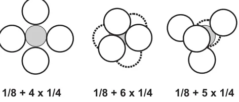
Fig. 1. Schematic representation of the spatial arrangement of a diploid 1/8 blastomere (gray) aggregated with tetraploid blastomeres (white). The aggregates were either ‘two-dimensional’ (four tetraploid blastomeres) or ’three-dimensional’ (five and six tetraploid blastomeres).
assay was at the level of about 6%, the potential contribution of the tetraploid component to any exam-ined tissue lower than 6% might have been undetec-ted. We analysed 20 foetuses/pups/adults: three live 15-day foetuses, one dead early foetus (aged about 13 days), four full term foetuses (two dead in womb and two that did not survive the Caesarian section), one 1-month old animal and eleven adults. The following 13 organs/tissues were examined in adults and full term foetuses/neonatal pups: blood, brain, eyes, heart, in-testine, liver, lungs, spleen, kidneys, adrenals, gonads, muscles and tongue. The number of examined organs/ tissues varied between specimens (4-12), but in the majority of cases ranged between 9 and 11 (in adult triplets 10 tissues were examined; in adult twins, 4 tissues were examined in one twin and 9 tissues in another twin). In 19 specimens (foetuses/pups/adults), in all examined organs/tissues, only the diploid compo-nent was detected. One foetus, which probably died around 13th day but was recovered on the 20th day, was examined in toto: it contained both GPI isoforms, but the diploid GPI variant predominated.
peared to be an equal participation of both variants and in six specimens the tetraploid variant predominated (these estimates were based on visual comparison of the intensity and size of 1A and 1B bands on cellulose acetate plates).
A Number of recipients (= number of transplanted sets)a/ number of pregnant recipients
B Transplanted blastocysts / number of blastocysts transplanted to pregnant recipients C Total number of implantations
D Number of resorptions + dead foetuses (% in pregnant recipients)
E Number of live foetuses and pups /number of adults (% in pregnant recipients)
aEach recipient received one set of blastocysts; each set consisted of blastocysts
developed from aggregates carrying sister blastomeres from a single diploid embryo.
bAmong 27 recipients 23 recipients received a set of 8 blastocysts, 3 recipients a set of 7
blastocysts and one recipient a set of 5 blastocysts.
cIncluding three foetuses recovered in two females on the 15th day of pregnancy. d One pair of twins in which both co-twins survived to adulthood.
e Including one set of identical triplets.
Distribution of diploid and tetraploid cells in chimaeric blastocysts
Our goal was to construct blastocysts in which the diploid cells would contribute solely to the ICM and the trophectoderm would contain only the tetraploid cells. We constructed aggregates that differed in the number of tetraploid blastomeres and their spatial arrangement around the single diploid blastomere (Figs. 1,2). Despite very careful transfer of the aggregates to the drops of culture medium, the original position of tetraploid blas-tomeres within the aggregate was not always pre-served. Morever, once the aggregates underwent compaction, the relative position of diploid and tetraploid blastomeres was no longer visible. In order to learn what was their distribution within morulae and blastocysts, we produced chimaeric aggregates in which diploid and tetraploid blas-tomeres were labelled with different fluorescent dextrans.
Confocal microscopy analysis showed that in four out of five morulae, the diploid 1/8 blastomere had divided once or twice (giving 2 and 4 cells, respectively) and only one morula contained single undivided diploid blastomere. In these five morulae the number of tetraploid blastomeres varied be-tween 8 and 16. Only in one morula, 2 diploid blastomeres occupied an internal position and were almost entirely surrounded by 8 carrier tetraploid blastomeres (type of aggregate: 1 diploid 1/8 blas-tomere + 4 tetraploid 1/4 blasblas-tomeres) (Fig. 4A). In other four morulae (1/8 + 4 x 1/4 and 1/8 + 6 x 1/4)
Fig. 3. Female triplets of BAMIZ origin, which developed from sister 2/16 blas-tomeres, at the age of 5 weeks.
The yolk sac was examined in 3 adults, 3 living 15-day foetuses and 4 dead foetuses. In two cases only the diploid GPI variant was detected and in the remaining eight cases both the diploid and tetraploid variants were present: in two specimens there
ap-Type of
Total 27b/21 210/163
(100.0) POSTIMPLANTATION DEVELOPMENT OF CHIMAERIC BLASTOCYSTS
diploid blastomeres were only partially surrounded by tetraploid blastomeres, or were located outside the tetraploid carrier blas-tomeres.
We also examined 9 blastocysts. Three blastocysts contained diploid cells in the ICM and in the overlying polar trophectoderm. In five blastocysts, the diploid cells formed a distinct group in the ICM and they were also present in polar and mural trophectoderm (Fig. 4B), or only in mural trophectoderm (Fig. 4 C,D). In one blastocyst the diploid cells were mostly located in the trophecto-derm.
These observations show that in the majority of our chimaeric embryos, the diploid cells were present not only in the ICM, but also in the trophectoderm. We believe that the relatively low number of diploid cells in the ICM and a substantial contribution of tetraploid cells to the ICM, were probably responsible for the
high mortality of our experimental embryos.
Discusssion
The organisation of the mammalian egg and the early embryo and the mechanisms responsible for the progressive determination and differentiation of embryonic cells, have remained a subject of continu-ous interest. The results of experimental studies, accumulated during several decades of the last century, point towards the regulative capability of the blastomeres of the early embryos. In mice, the indi-vidual blastomeres, or two or more embryos aggre-gated together during cleavage, are able to develop into normal adult. This led to the idea that in mice, in contrast to many other species where the develop-ment is programmed by the polar distribution of organelles and molecules in the egg and early em-bryo, the embryonic cells are developmentally ‘naïve’ in the early phases of embryogenesis and their fate is determined epigenetically according to their spa-tial position in the embryo (the ‘inside-outside’ hy-pothesis, Tarkowski and Wroblewska, 1967). How-ever, in the last decade, Gardner (1997, 2001) and Zernicka-Goetz (reviewed in 2002, 2004), suggested that although in extreme experimental conditions the early mammalian embryos can display great regula-tive capabilities, the normal development relies mostly on an inherent pattern of organisation of the egg and early embryo. We believe, however, that the ob-served predisposition of certain parts of the egg, or certain blastomeres, to contribute to specific parts of the embryo, does not prove that these cells are already irrevocably committed to specific fates. As long as the fate of blastomeres can be steered experimentally in different directions (for instance into the ICM versus the trophectoderm), there is no ground to view them as being irreversibly deter-mined.
The easiest and the most informative way of learning whether a blastomere has the unrestricted developmental abilities of the zygote, (i.e. it is totipo-tent), or whether its developmental potential is al-ready constrained, is to track the development of an
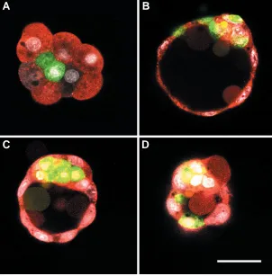
Fig. 4. Embryos which developed from aggregates of diploid blastomeres injected with with FITC-conjugated dextran (green), and tetraploid blastomeres injected with with rhodamine-conjugated dextran (red) and cultured in vitro. Fixed embryos were examined with confocal microscope. Bar 50 µm. (A) A morula composed of two diploid blastomeres (green) surrounded by eight tetraploid blastomeres (red) developed from an aggregate composed of one 1/8 blastomere and four 1/4 blastomeres. The diploid blastomeres were almost entirely surrounded by tetraploid blastomeres. The embryo was cultured for 24 h. (B) Optical section of a blastocyst developed from one diploid 1/ 8 blastomere and six 1/4 tetraploid blastomeres, after 48 h in culture. Diploid cells are present in the ICM and polar and mural trophectoderm. (C,D) Two optical sections of a blastocyst developed from one diploid 1/8 blastomere and four tetraploid 1/4 blas-tomeres, after 48 h in culture. (C) A group of diploid cells situated in the ICM is covered by tetraploid polar trophectoderm. (D) In the tangential section, two diploid cells are situated in the mural trophectoderm. Vesicular structures in the blastocysts’ cavities are artifacts.
isolated, single blastomere. Mammalian blastomere can be called truly totipotent if it is able to develop into a foetus with extra-embryonic membranes that eventually develops into a fertile adult. However, the blastomere isolated from the embryo in late cleavage may not be able to complete embryogenesis - not because of its restricted capabilities to differentiate into various embryonic and extra-embryonic tissues - but because (as it happens in the mouse) it has no time to produce, before cavita-tion, enough number of cells to allocate some of them to the ICM. In order to properly implant in the mother’s uterus and to start to grow, the mammalian embryo must allocate the large number of cells to the first extraembryonic structure – the trophectoderm. In the mouse, cavitation begins as the embryo approaches the 32-cell stage (Smith and McLaren, 1977) and its ICM consists of about 25% of the total number of cells (Rands, 1985). By the time
A
B
of cavitation, single 1/4 and 1/8 blastomeres will divide into 8 and 4 cells respectively and as a result will form the blastocyst with either very small ICM, or trophoblastic vesicles completely devoid of ICM (Tarkowski and Wroblewska, 1967). Consequently, these embryos are unable to develop normally after implantation (Rossant, 1976) and the only tactic available to assess the developmental potential of such blastomeres, is to support them with subsidiary cells. Kelly (1975, 1977) used genetically distinct diploid blastomeres as the subsidiary cells and was able to prove that 1/4 and 1/8 blastomeres can contribute to embryonic and also to extra-embryonic tissues of chimaeras. On some occasions, the single blastomeres were able to form the whole non-chimaeric animals, provided, however that subsidiary blastomeres contrib-uted solely to the extra-embryonic tissues. Although these results are considered to be the evidence of the totipotency of the individual 1/4 and 1/8 blastomeres, none of these blastomeres were self-sufficient in creating the new individuals with supporting foetal membranes. The experiments of Kelly and others who followed her experimental design (Tsunoda et al., 1987; Pinyopummin et al., 1994; Tarkowski et al., 2001; the present study), have demonstrated that the 1/4 and 1/8 blastomeres are able to form the body of the embryo/foetus/animal but only when supported with carrier cells (tetraploid blastomeres or diploid blastomeres derived from genetically distinct, normal or parthe-nogenetic embryos). Although the classic definition of ‘totipo-tency’ does not fully apply in this situation, the extraordinary ability of these isolated blastomeres to give rise to a complete fertile animal, justifies the usage of the word ‘totipotency’, though in the restricted meaning of this term.
In the present study we used tetraploid blastomeres as subsid-iary cells to support the diploid blastomere. This experimental system takes advantage of the negative selection of tetraploid cells against diploid cells in the embryonic (but not extra-embry-onic) tissues of 2n/4n mosaics (Tarkowski et al., 1977) and of 2n↔4n chimaeras (Lu and Markert, 1980; Nagy et al., 1990; Goto and Takagi, 1998; Tarkowski et al., 2001).
The present study confirms the conclusion of Kelly (1975, 1977) and Tsunoda et al. (1987) that at least some blastomeres of the 8-cell mouse embryo and sister pairs of 16-cell embryo, are able to develop into a normal individual. We were able to produce one set of triplets, five pairs of twins and a number of singletons. All eleven adults were fertile. We did not detect the contribution of the tetraploid component to the body of adults, pups and full term foetuses. The type of pigmentation (in coat and/or eyes) was always characteristic of the diploid component. Also, the GPI analysis of four to ten organs/tissues confirmed presence of the diploid GPI isoform only. However, because of the limited sensi-tivity of the assay, we cannot exclude the possibility of low, i.e. below 6%, presence of the tetraploid component in experimental animals. It was shown previously that conceptuses and animals produced from 2n↔4n chimaeric embryos might occasionally contain traces of tetraploid cells in some tissues (Nagy et al., 1990; Wang et al., 1997b; Tarkowski et al., 2001; Goto et al., 2002). In our experimental chimaeras the tetraploid cells were eliminated from the body of foetuses/pups/adults, even though the tetraploid component initially predominated. The initial ratio between 4n and 2n cells was greatly in favour of the 4n compo-nent: 4-6:1 in regard to the number of cells and 8-12:1 in regard to the cellular volume. We used four to six tetraploid blastomeres
in order to enclose the diploid blastomere inside the aggregate and thus to form blastocyst in which diploid cells would be present exclusively in ICM. However, the experiments with blastomeres marked with different fluorochromes showed that the diploid blastomere were often located not only in ICM but also in polar and mural trophectoderm. Thus, the contribution of the diploid compo-nent to the ICM was often lower than expected and in conse-quence might have hampered normal morphogenesis. There was also no evident advantage in making ‘three-dimensional’ versus ‘two-dimensional’ aggregation chimaeras (Fig. 1). The predomi-nance of tetraploid cells in the ICM, resulting from the excess of 4n blastomeres in the aggregates, might have been another factor contributing to the death of the diploid embryo. It was shown previously that tetraploid embryos of the same genotype as those used in our experiments, initially form apparently normal egg cylinders, but eventually, at the beginning of gastrulation (8th day of development) their morphogenesis deviates from the normal course and the embryos die (Tarkowski, et al., 1977).
In contrast to the embryo proper, the tetraploid cells from the ICM successfully contribute to and survive until birth in the yolk sac. Moreover, in 6 out of 10 examined cases, the tetraploid cells predominated in the yolk sac. Because we observed the same phenomenon in diploid↔triploid mouse chimaeras (Suwinska et al., 2005), this may reflect the general tendency of polyploid cells to colonise preferably the yolk sac and/or survive better in this extra-embryonic structure.
Evaluation of the developmental potential of a single blas-tomere from a given developmental stage is informative, but much more valuable is the comparison of the developmental potential of all sister blastomeres, i.e. 2 of 2, 4 of 4 etc. In the mouse, Mullen et al. (1970), Tsunoda and McLaren (1983), Togashi et al. (1987), Wang et al. (1997a) and Sotomaru et al. (1998) produced twins from isolated sister 1/2 blastomeres, but the efficiency was quite low. So far there are no reports of birth and successful postnatal development of mouse quadruplets. However, the production of quadruplets in the sheep (Willadsen, 1981) and triplets and quadruplets in cattle (respectively Willadsen and Polge, 1981 and Johnson et al., 1995) by separation of blastomeres of the 4- or 8-cell embryos shows that, at least in some mammalian species, all blastomeres at this developmental stage are truly totipotent and do not require support of carrier blastomeres.
blas-tomeres of some 4-cell mouse embryos may differ in their fate and that this non-equivalence arises from a particular pattern of cell division during the 2 → 4-cell stage. Aggregates of blas-tomeres taken from 4-cell embryos develop to term or display developmental abnormalities if they are formed from specific vegetal blastomeres. However, so far there is no evidence that the individual blastomeres in the 4-cell embryo differ at cyto-logical and/or molecular level. The first morphocyto-logical differ-ences between blastomeres are observed after the 4th cleav-age division with the appearance of polar and apolar cells (for review see Johnson and McConnell, 2004). Even then, in the 16-cell embryo the polar and apolar cells though morphologi-cally distinct, are developmentally labile, because aggregates of each cell type can form blastocysts and normal conceptuses (Ziomek et al., 1982). This suggests that at the preceding developmental stage, i.e. in the 8-cell embryo, the majority if not all of the isolated blastomeres (provided each is supported with carrier cells) should be able to develop into normal mouse. We believe that failure to produce octuplets from 8-cell mouse embryo is due to imperfect techniques rather than to a restricted developmental potential of some sister 1/8 blastomeres. How-ever, the confirmation of this belief should await further studies.
Materials and Methods
Animals were kept under 14 h light/10 h dark regime. Embryos were obtained from 1.5 - 6.5 month old females, albino or pigmented and homozygous for 1a or 1b allele of glucose phosphate isomerase (GPI).
Diploid (2n) embryos
In most experiments cycling BAMIZ females were used as donors of 2n embryos. These outbred albino mice were obtained from a cross between BALB/c and MIZ mice selected for Gpi-1a/Gpi-1a homozygosity. Females
were crossed with BAMIZ males and inspected for plugs in the morning (the day of plug = the first day of pregnancy). The plugged females were sacrificed on the 3rd day between 9.30 and 12.30 and the 8- to 16-cell embryos were recovered from oviducts in M2 medium (Fulton and Whittingham, 1978). The zona pellucida was removed by incubation in 0.5% pronase (Sigma) in Ringer solution. The blastomeres were disaggre-gated by incubation (minimum 15 minutes) in Ca2+ - and Mg2+ - free M2
medium at 37ºC followed by pipetting in the same medium. Each embryo was manipulated individually and the sister blastomeres (octet of 1/8 or 2/ 16 blastomeres) were stored in a separate M2 drop under liquid paraffin at 37ºC in an atmosphere of 5% CO2 in air, until used for aggregation with tetraploid blastomeres.
In three experiments, cycling F1(C57BL/10 x CBA/H) females mated with F1 males were used as donors of diploid 8- and 16-cell embryos (GPI and coat characteristics given below).
Tetraploid (4n) embryos
In most experiments embryos used for tetraploidization originated from F1(C57BL/10 x CBA/H or a reverse cross) and from F1(C57/BL/6 x CBA/ H) females. Females were mated with F1 males of the same genotype. These animals are agouti and have the 1B isoform of GPI. The females were induced to ovulate with pregnant mare’s serum gonadotrophin (PMSG; Folligon, Intervet) and human chorionic gonadotrophin (hCG; Chorulon, Intervet) given 47-49.5 h apart in doses of 7.5 - 10 IU each. The females were sacrificed 42-45 h after hCG injection. Recovered 2-cell embryos were electrofused according to the modified technique of Kubiak and Tarkowski (1985) in a fusion chamber filled with 0.25 M glucose supplemented with 100 µM CaCl2.2H20 and 100 µM MgSO4.7H2O, with the distance between electrodes varying between 220 and 290 µm. Embryos aged 43.5 - 49 h after hCG injection were oriented with the interblastomeric
surface perpendicular to the lines of the electric field and were subjected to two pulses of 40 Volts d.c. and 25 µs duration, given twice (the orientation of embryos was corrected after the first stimulus, if required). Fusion usually took place within the first hour after treatment. Embryos that had not fused were subjected to a second round of pulses. Fused embryos were cultured in KSOM medium (Erbach et al., 1994) (Specialty Media) under standard
conditions. Those embryos that had cleaved within one hour after fusion were eliminated. About 24 h later the 4-cell embryos (considered to be tetraploid) were released from zona pellucida and disaggregated as described above. Isolated blastomeres were pooled in drops of M2 medium until aggregation with diploid blastomeres.
In three experiments 2-cell embryos used for tetraploidization originated from hormonally-stimulated BAMIZ females mated to BAMIZ males (GPI and coat characteristics as described for diploid embryos). These were recovered 47 - 51.5 h after hCG injection and at 48-53 h they were subjected to electric pulses as described earlier. Fused embryos were cultured and disaggregated as F1 embryos.
Aggregation of diploid and tetraploid embryos
Aggregation was performed between 1.00 and 6.00 p.m. Each set of diploid blastomeres obtained from 8-cell or 16-cell embryos was processed separately. Diploid 8-cell blastomeres (1/8 blastomeres) and pairs of sister diploid 16-cell blastomeres (2/16 blastomeres) were placed with tetraploid 4-cell blastomeres (1/4 blastomeres) in a solution of phytohemagglutinin (300 µg/ml) (Sigma) in BSA-free M2 medium in agar-coated embryological watch glass for 1-3 minutes. Within each set, all sister diploid blastomeres (1/8 or 2/16) were surrounded with four, five or six tetraploid blastomeres (Figs. 1,2). After the blastomeres had stuck firmly together, the chimaeric aggregates were placed in drops of KSOM medium. After 48 h of culture the chimaeric aggregates had developed into large blastocysts which were transferred to recipient females.
Transplantation of chimaeric blastocysts
Each set of chimaeric blastocysts (usually 8, occasionally 7 or 5) was transplanted separately on the first day of pseudopregnancy to the oviduct of a recipient female. The recipients (age: 2.5 - 5 months) were MIZ, BAMIZ and F1 females mated with F1 vasectomised males. Females were anaesthetized with 0.25-0.36 ml of 6 mg/ml of water solution of Nembutal (Serva) injected intraperitoneally.
Monitoring of pregnancy and delivery of young
Starting on the 9th day after mating, vaginal smears were taken daily from each recipient female. Females showing signs of proestrus or oestrus were sacrificed and the uterus was inspected for resorptions. Most of the pregnant females were sacrificed in the afternoon on the 20th day or in the morning on the 21st day and the foetuses recovered by Caesarian section were fostered by females which had littered the same night or 24 h earlier. The experimental animals reared successfully were tested for fertility by crossing with normal males or females and after giving birth to one or more litters they were sacrificed for GPI analysis.
GPI assay
Samples of tissues/organs were frozen in small amount of redistilled water and stored at -20ºC. After thawing the samples were homogenized, centrifuged at 14000 rpm and the supernatant was collected. Electrophore-sis was carried out on TITAN III-H plates (Helena BioSciences) according to the method of Buehr and McLaren (1985). From 4 to 10 tissues/organs were examined in each experimental foetus/animal.
Labelling with fluorescent dextrans
One-cell diploid and tetraploid embryos were microinjected with 2pl of 2mg/ml solution of dextran conjugated with fluorescein or rhodamine (70000 MW; Molecular Probes) in 2 mM PIPES buffer (pH 7.4; Sigma) and 0.140 mM KCl (Sigma).
pregnancy. After removal of cumulus cells with hyaluronidase (Sigma) (500µg/ml in Ca2+- and Mg2+-free phosphate buffered saline (PBS, Biomed)
the zygotes were injected with FITC-conjugated dextran and transplanted to the oviduct of MIZ females mated to F1 vasectomized males (1st day of pseudopregnancy). 48 h after transplantation they were recovered as 8-cell embryos and disaggregated as described previously.
Embryos used for tetraplodization originated from hormonally-stimu-lated F1 females mated with F1 males. They were recovered between 42 and 45 h post hCG injection as two-cell embryos and subjected to electrofusion 2-4 h later. Fused (now 1-cell) embryos were injected with rhodamine-conjugated dextran and cultured in vitro in KSOM medium for
about 24 h. Embryos that divided into four cells were disaggregated and the single blastomeres were used for aggregation with diploid blastomeres.
Diploid and tetraploid blastomeres were aggregated as described earlier and cultured in KSOM medium for 1-2 days. They were fixed as morulae or blastocysts for 20 min in 2% paraformaldehyde in Ca2+- and
Mg2+-free PBS, stained for 10 min at 37ºC in fluorescent chromatin-specific
dye DRAQ-5 (10 µM in PBS; Biostatus Ltd) and transferred to droplets of PBS in a viewing chamber. The embryos were examined with confocal laser microscope LSM 510 Zeiss.
Acknowledgements
This work was partly financed by a grant from the State Committee for Scientific Research (Grant 3 P04C 074 22). We wish to thank Professor Marek Maleszewski and Dr. Ewa Borsuk for injecting eggs with fluorescent dextrans, Mr. Darek Maluchnik for help in examining chimaeric embryos in confocal microscope and in preparing figures, Dr. Anne McLaren for helpful discussions and Professor Michael Bedford for making comments on the manuscript. We are most thankful to Professor Malgorzata Kloc for final shaping of the manuscript.
References
BUEHR, M. and McLAREN, A. (1985). Expression of glucose-phosphate isomerase in relation to growth of the mouse oocyte in vivo and in vitro. Gamete Res.
11:271-281.
ERBACH, G.T., LAWITTS, J.A., PAPAIOANNOU, V.E. and BIGGERS, J.D. (1994). Differential growth of the mouse preimplantation embryo in chemically defined media. Biol. Reprod. 50: 1027-1033.
FULTON, B.P. and WHITTINGHAM, D.G. (1978). Activation of mammalian oocytes by intracellular injection of calcium. Nature 273: 149-151.
GARDNER, R.L. (1997). The early blastocyst is bilaterally symmetrical and its axis of symmetry is aligned with the animal-vegetal axis of the zygote in the mouse.
Development 124: 289-301.
GARDNER, R.L. (2001). Specification of embryonic axes begins before cleavage in normal mouse development. Development 128: 839-847.
GOTO, Y., MATSUI, J. and TAKAGI, N. (2002). Developmental potential of mouse tetraploid cells in diploid↔tetraploid chimeric embryos. Int. J. Dev. Biol. 46:
741-745.
GOTO, Y. and TAKAGI, N. (1998). Tetraploid embryos rescue embryonic lethality caused by an additional maternally inherited X chromosome in the mouse.
Development 125: 3353-3363.
HOPPE, P.C. and WHITTEN, W.K. (1972). Does X chromosome inactivation occur during mitosis of first cleavage. Nature 239, 520.
JOHNSON, M.H. and McCONNELL, J.M.L. (2004). Lineage allocation and cell polarity during mouse embryogenesis. Semin. Cell Dev. Biol. 15: 583-597.
JOHNSON, W.H., LOSKUTOFF, N.M., PLANTE, Y. and BETTERIDGE, K.J. (1995). Production of four identical calves by the separation of blastomeres from an in vitro derived four-cell embryo. Vet. Rec. 137: 15–16.
KELLY, S.J. (1975). Studies of the potency of the early cleavage blastomeres of the mouse. In The Early Development of Mammals (Eds. M. Balls and A.E.
Wild). Cambridge University Press, Cambridge, London, New York, Melbourne, pp. 97-105.
KELLY, S.J. (1977). Studies of the developmental potential of 4- and 8-cell stage
mouse blastomeres. J. Exp. Zool. 200: 365- 376.
KUBIAK, J.Z. and TARKOWSKI, A.K. (1985). Electrofusion of mouse blastomeres.
Exp. Cell Res. 157: 561-566.
LU, T-Y. and MARKERT, C.L. (1980). Manufacture of diploid/ tetraploid chimeric mice. Proc. Natl. Acad. Sci. USA 77: 6012-6016.
MOORE, N.W., ADAMS, C.E. and ROWSON, L.E. (1968). Developmental potential of single blastomeres of the rabbit egg. J. Reprod. Fert. 17: 527-531.
MULLEN, R.J., WHITTEN, W.K. and CARTER, S.C. (1970). Studies on chimeric mice and half-embryos. Annual Report of the Jackson Laboratory. Bar Harbour,
Maine, pp 67-68.
NAGY, A., GOCZA, E., DIAZ, E.M., PRIDEAUX, V.R., IVANYI, E., MARKKULA, M. and ROSSANT, J. (1990). Embryonic stem cells alone are able to support fetal development in the mouse. Development 110: 815-821.
PAPAIOANNOU, V.E., MKANDAWIRE, J. and BIGGERS, J.D. (1989). Develop-ment and phenotypic variability of genetically identical half mouse embryos.
Development 106: 817-827.
PINYOPUMMIN, A., TAKAHASHI, Y., HISHINUMA,M. and KANAGAWA, H. (1994). Development of single blastomeres from 4-cell stage embryos after aggregation with parthenogenones in mice. Jpn. J. Vet. Res. 42: 119-126.
PIOTROWSKA-NITSCHE, K., PEREA-GOMEZ, A., HARAGUCHI, S. AND ZERNICKA-GOETZ, M. (2005). Four-cell mouse blastomeres have different developmental properties. Development 132: 479-490.
PIOTROWSKA-NITSCHE, K. and ZERNICKA-GOETZ, M. (2005). Spatial arrange-ment of individual 4-cell stage blastomeres and the order in which they are generated correlate with blastocyst pattern in the mouse embryo. Mech. Dev.
122: 487-500.
RANDS, G.F. (1985). Cell allocation in half- and quadruple-sized preimplantation mouse embryos. J. Exp. Zool. 236: 67-70.
ROSSANT, J. (1976). Postimplantation development of blastomeres isolated from 4- and 8-cell mouse eggs. J. Embryol. Exp. Morph. 36: 283-290.
SMITH, R. and McLAREN, A. (1977). Factors affecting the time of formation of the mouse blastocoele. J. Embryol. Exp. Morph. 41: 79-92.
SOTOMARU, Y., KATO, Y. and TSUNODA, Y. (1998). Production of monozygotic twins after freezing and thawing of bisected mouse embryos. Cryobiology 37:
139-145.
SUWINSKA, A., OZDZENSKI, W., WAKSMUNDZKA, M. and TARKOWSKI, A.K. (2005). Experimentally produced diploid<->triploid mouse chimaeras develop up to adulthood. Mol. Reprod. Dev. 72: 362-376. (DOI 10.1002/mrd.20350).
TARKOWSKI. A.K. (1959a). Experiments on the development of isolated blas-tomeres of mouse eggs. Nature 184: 1286-1287.
TARKOWSKI, A.K. (1959b). Experimental studies on regulation in the development of isolated blastomeres of mouse eggs. Acta Theriol. 3: 191-267.
TARKOWSKI, A.K., OZDZENSKI, W. and CZOLOWSKA, R. (2001). Mouse single-tons and twins developed from isolated diploid blastomeres supported with tetraploid blastomeres. Int. J. Dev. Biol. 45: 591-596.
TARKOWSKI, A.K., WITKOWSKA, A. and OPAS, J. (1977). Development of cytochalasin B-induced tetraploid and diploid/tetraploid mosaic mouse em-bryos. J. Embryol. Exp. Morph. 41: 47-64.
TARKOWSKI, A.K. and WROBLEWSKA, J. (1967). Development of blastomeres of mouse eggs isolated at the 4- and 8-cell stage. J. Embryol. Exp. Morph. 18:
155-180.
TOGASHI, M., SUZUKI, H., MIYAI, T. and OKAMOTO, M.T. (1987). Production of monozygotic twins by splitting of 2-cell stage embryos in mice. Jpn. J. Anim. Reprod. 33: 51-56.
TSUNODA, Y. and McLAREN, A. (1983). Effects of various procedures on the viability of mouse embryos containing half the normal number of blastomeres.
J. Reprod. Fert. 69: 315–322.
TSUNODA, Y., YASUI, T., OKUBO, Y., NAKAMURA, K. and SUGIE, T. (1987). Development of one or two blastomeres from eight-cell mouse embryos to term in the presence of parthenogenetic eggs. Theriogenology 28: 615-623.
WANG, M., KATO, Y. and TSUNODA, Y. (1997a). Effects of several factors on the monozygotic twin production in the mouse. J. Reprod. Dev. 43: 91-95.
blasto-cyst injection. Mech. Dev. 62: 137-145.
WILLADSEN, S.M. (1981). The developmental capacity of blastomeres from 4- and 8-cell sheep embryos. J. Embryol. Exp. Morph. 65: 165-172.
WILLADSEN, S.M. and POLGE, C. (1981). Attempts to produce monozygotic quadruplets in cattle by blastomere separation. Vet. Rec. 108: 211-213.
ZERNICKA-GOETZ, M. (2002). Patterning of the embryo: the first spatial decisions in the life of a mouse. Development 129: 815-829.
ZERNICKA-GOETZ, M. (2004). First cell fate decisions and spatial patterning in the early mouse embryo. Semin. Cell Dev. Biol. 15: 563-572.
ZIOMEK, C.A., JOHNSON, M.H. and HANDYSIDE, A.H. (1982). The developmen-tal potential of mouse 16-cell blastomeres. J. Exp. Zool. 221: 345-355.