Case Report Open Access
Key points in Diabetes Insipidus´ Diagnosis
Mara Capra*
1, Ana Raisa Jatahy Santos
1, Sabrina Pinto Ferreira
1, Moacir Benazzi
1, Larissa Bianca
Paiva Cunha de Sá
1, Helen Paula Apparicio Medeiros
1, Mirella Hansen de Almeida
1,2, Alberto Krayyem
Arbex
1,3,41Endocrinology Division, IPEMED Medical School, Rio de Janeiro, Brazil
2Division of Endocrinology, Central Air Force Hospital, Rio de Janeiro, Brazil
3Diabetology Department, Malteser Franziskus Krankenhaus Flensburg, Germany
4Former Visiting Scholar, Harvard TH Chan School of Public Health, Boston, USA Received: January 17, 2018; Published: January 23, 2018
*Corresponding author: Rua dos Jasmins, 326. Jardim Laguna, Linhares – Espírito Santo, Brazil, Tel: ; Email:
ISSN: 2574-1241
Biomed J Sci & Tech Res
Introduction
Diabetes insipidus (DI) is characterized by profuse diuresis, usually greater than 3L/24h of urine, with osmolality below 300mOsm/kg [1]. It has two main forms: Central (also known asneurogenic) DI is characterized by low levels of antidiuretic hormone, also known as vasopressin (AVP)[2]; nephrogenic DI is
characterized by difficulties concentrating urine due to resistance to
AVP action in the kidneys. AVP is the main determiner of free water clearance in the body. It acts mainly in the kidneys where it alters water permeability in the cortical tubules and the medulla. Water is reabsorbed by osmosisdue to hypertonicity within the medullary interstitium, and then taken back into systemic circulation [3]. Two other forms of DI are gestational DI and primary polydipsia (dipsogenic DI). Neither of these results from a defect in the neurohypophysis or the kidneys, but both participate in differential diagnoses of diabetes. The predominant manifestations of DI are polyuria, polydipsia, and nocturia, which also indicate diabetes mellitus (DM), another important differential diagnosis.
Laboratory tests, particularly 24-hour urine collection to determine the volume of urine and concentration of serum electrolytes, urine and plasma osmolality, and glucose and AVP levels, must be carefully analyzed,as small differences are crucial to a correct diagnosis. We report a case of central diabetes insipidus (CDI), accompanied by a literary review. CDI has many possible causes. According to the literature, the main causes of CDI and their frequencies are: idiopathic-30%; malign or benign tumors of the brain or neurohypophysis-25%; cranial surgery0-20%; and cranial trauma-16% [4]. As this disease is often undiagnosed, we will stress the importance of noting discrete abnormalities in laboratory results to make a differential, early diagnosis so that appropriate therapy can be started to improve the patient’s quality of life.
Case Report
The patient, a 56-year-old Caucasian male, was referred to a tertiary endocrinology clinic by a Family Health Program physician. He had a history of mild systemic arterial hypertension and DM
Abstract
Diabetes insipidus is mainly characterized by polyuria, urinary volume over 3 L/day or 40mL/kg/day in adults, leading to subsequent polydipsia; these features are also present in most cases of diabetes mellitus. We report the case of a 56-year-old man initially misdiagnosed with and treated for diabetes mellitus; he was eventually diagnosed with central diabetes insipidus following further laboratory tests. We have also conducted a thorough review of the literature relating to the physiology, diagnosis, and treatment of diabetes insipidus. The patient, who complained of polyuria, polydipsia and weight loss, and had fasting glucose levels of 108 mg/dL, was already using metformin to treat type 2 diabetes. He then developed hypoglycemic symptoms, and pre- and postprandial capillary glycaemia between 70 and 120 mg/dL, as a result of which metformin was suspended. Nevertheless, polyuria and polydipsia persisted. Based on a plasma osmolality of 305,5 mOsm/kg, urinary density of 1005 g/mL and low arginine vasopressin levels, a diagnosis of central diabetes insipidus was made and treatment with desmopressin was initiated. Because the symptoms of centraldiabetes insipidus and uncontrolled type 2 diabetes mellitus overlap, it is important to consider clinical presentations carefully in order to make a differential diagnosis.
Keywords: Central diabetes insipidus; Osmolality; Desmopressin; Polyuria; Polydipsia
type2 (DM2) diagnosed five years previously. He spent two years
receiving treatment for DM2, and experienced frequent episodes of hypoglycemia, in addition to benign prostatic hyperplasia. The patient had experienced frequent episodes of hypoglycemia, as a result of which he had had two emergency hospital visits at which he reportedtaking metformin (2g/day in two doses), enalapril (10mg/ day), and doxazosin (2mg/day). Capillary glycemia varied between 28 and 236mg/dL. His main concerns were hypoglycemia and
intense thirst. At our initial consultation, on 25 September, 2015, he was currently using metformin 500mg/day; he weighed 87.1kg, his height was 1.72 m, and his body mass index (BMI) was 29.4kg/m2.
Laboratory tests were requested, pre- and postprandial glycemia were monitored, and medication was maintained. Two months later (May 5, 2015), the patient returned to clinic asymptomatic. Glycaemia varied between 81 and 166 mg/dL. Furthertest results are shown in (Tables 1 & 2).
Table 1: Results of Blood Tests.
Tests Reference value October 2015 March 2016 June 2016
Cortisol (µg/dL) 05-25 10.4 11
Hemoglobin (g/dL) 12.8-17.8 14.21
Hematocrit (%) 40-54 43.4
Fasting glycemia (mg/dL) 70-99 116 125 125
HbA1c (%)* Up to 5.7 6.2 6.2 6.4
Urea (mg/dL) 13-43 22 22
Creatinine (mg/dL) 0.7-1.3 0.82 0.87
Uric acid (mg/dL) Up to 6.5 6.3 7.4
Sodium (mEq/L) 136-145 141 142 143
Potassium (mEq/L) 3.5-5.3 5.0 4.9 4.7
TSH (mU/L)* 0.4-5 2.46
Free T4 (ng/dL)* 0.7-1.8 0.68
OGTT 2h (mg/dL)* Up to 155 118
Cortisol after 1mg dexamethasone (µg/dL) <1.8 <1.8
AVP (pg/ml) Up to 6.5 1.5
Plasma osmolality (mOsm/kg) 280-295 305.5
Note: *HbA1c: glycosylated hemoglobin, TSH: thyroid-stimulating hormone, T4: thyroxine, OGTT: oral glucose tolerance test, AVP: arginine vasopressin.
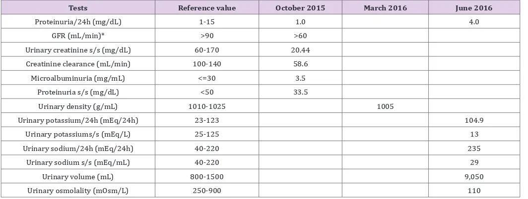
Table 2: Results of Urine Tests.
Tests Reference value October 2015 March 2016 June 2016
Proteinuria/24h (mg/dL) 1-15 1.0 4.0
GFR (mL/min)* >90 >60
Urinary creatinine s/s (mg/dL) 60-170 20.44
Creatinine clearance (mL/min) 100-140 58.6
Microalbuminuria (mg/mL) <=30 3.5
Proteinuria s/s (mg/dL) <50 33.5
Urinary density (g/mL) 1010-1025 1005
Urinary potassium/24h (mEq/24h) 23-123 104.9
Urinary potassiums/s (mEq/L) 25-125 13
Urinary sodium/24h (mEq/24h) 40-220 235
Urinary sodium s/s (mEq/mL) 40-220 29
Urinary volume (mL) 800-1500 9,050
Urinary osmolality (mOsm/L) 250-900 110
Note: *GFR: glomerular filtration rate,s/s (single sample).
On clinical examination, hepatic steatosis was seen on total abdominal ultrasonography; fundus oculi were normal; arterial pressure was 140/90mmHg; weight was 91kg; abdominal
circum-ference was 111cm; and BMI was 30.7kg/m2. Adrenal insufficiency
and increase in abdominal circumference. Medications were main-tained, and orlistat (120 mg twice a day) was prescribed, in addi-tion to a 1500kcal diet. At the patient’s next visit on November 27th,
2015, 2 months later, his weight loss was 5.5kg, BMI was 29.4kg/ m2, and his abdominal circumference had reduced to 103cm. DI was
investigated due to urine volume of 3350mL/day. He had already stopped taking metformin. Orlistat was stopped and further tests were requested. Submitted tests performed on October 08th, 2015.
On March 21th, 2016, the patient returned to the clinic
complaining of intense thirst and urinating excessively. His arterial blood pressure was 160/110mmHg and his weight 86.9 kg. He no longer reported hypoglycemia. Plasma osmolality, which was calculated using the equation 2×(Na+K)+(glycemia/18), was 300.74mmol/L (reference values 280-285mmol/L). Further test results ashown in (Tables1 & 2). On June 28th, 2016, he attended
with the results of tests carried out on June 1, 2016 to confirm the
diagnosis is shown in Table 1. The patient ingested 7250mL/dayof
fluid and urinated 7100mL/day. We hypothesized that central DI
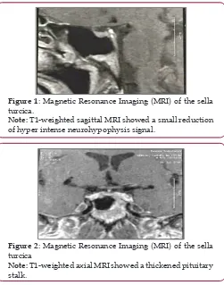
(CDI) was the most probable diagnosis, and conducted further tests onJune 28, 2016 (see (Tables 1 & 2). Magnetic resonance imaging (MRI) of the sella turcica showed a thickened pituitary
stalk andconfirmed the diagnosis of CDI (Figures 1 and 2). AVP
replacement therapy was prescribed. T1-weighted sagittal MRI showed a small reduction of hyper intense neurohypophysis signal.
Figure 1: Magnetic Resonance Imaging (MRI) of the sella turcica.
Note: T1-weighted sagittal MRI showed a small reduction of hyper intense neurohypophysis signal.
Figure 2: Magnetic Resonance Imaging (MRI) of the sella turcica
Note: T1-weighted axial MRI showed a thickened pituitary stalk.
Literature Review
Pathophysiology and Epidemiology
DI is characterized by polyuria, the excessive secretion of diluted urine, in adults of volumes greater than 3L/day. DI may result from
disorders related to AVP. Insufficient synthesis of AVP is a sign
of CDI, neurogenic diabetes, or vasopressin-responsivediabetes. Further, a decrease in renal sensitivity to AVP is a sign of NDI or non-AVP-responsive DI [5,6]. CDI is a heterogeneous condition
characterized by polyuria and polydipsia due to deficiency of
AVP. In some patients, it is caused by destruction or degeneration of neurons originating in the supraoptic and paraventricular nuclei of the hypothalamus. Known causes of such lesions include germinoma, craniopharyngioma, Langerhans cell hystiocytosis,
inflammatory, autoimmune, or vascular disorders, trauma resulting
from surgery or accident and, in rare cases, inherited genetic defects in synthesis of AVP. However, most cases are considered idiopathic [6]. Maintaining the water balance in healthy humans is due to three factors: thirst, AVP, and the kidneys [7]. Plasma osmolality is maintained within a narrow range varying between 285 and 295mOsm/kg, and this depends on the balance between water input (controlled by the physiological sensation of thirst) and renal water secretion (controlled by AVP secretion and action). Physiologically, plasma osmolality is the main thirst regulator.
When an increase in plasma osmolality is detected by osmotically sensitive neurons (osmoreceptors) located in the anterior hypothalamus, AVP is liberated. This increases resorption of water in the renal collecting tubules, stimulating thirst. The increase in ingested water reduces plasma osmolality to levels at which the control of urine production mediated by the increase in AVP maintainsosmolality within its normal range [5,6,8]. CDI may becongenital, and may have a genetic cause. However, it is acquired in 95% of cases [9,10]. Familial CDI is the most common genetic form. It has a similar incidence in both sexes and most frequently
presents as a progressive deficiency in the secretion of AVP. Acquired
causes include any surgical, traumatic, ischemic, infectious, or
infiltrative lesion or tumor in the hypothalamic-hypophyseal region
leading to destruction of AVP-producing neurons or impeding transportation of this hormone by the pituitary stalk [5,10,11]. Several tumors of the central nervous system may cause DI, but craniopharyngioma is the main etiology. Other tumors that may cause DI are germinoma, meningioma, glioma, and astrocytoma [5,6,10,12,13]. Cranial trauma resulting in pituitary shaft contusion may be a cause of DI [14]. Transitory DI is the main complication of transsphenoidal surgery [15]. However, craniotomies in which neoplasms are resected often result in permanent DI [5]. The main infectious causes of DI are meningitis, encephalitis, sellar- and super-sellartuberculomas, and infection with cytomegalovirus or neurosyphilis [5,6,10,16,17]. Other causes may be lymphocytic hypophysitis, aneurysms, thromboses, Sheehan syndrome, and multiple sclerosis [5,6,18-20].
Diagnosis
The diagnosis of DI is made on the basis of the patient’s clinical history, laboratory tests, and imagingresults [21].The main clinical symptoms of DI are polyuria and polydipsia, which manifest
regardless of thetime period.The laboratory findings that indicate DI are persistent hyposthenuria, with specific density below
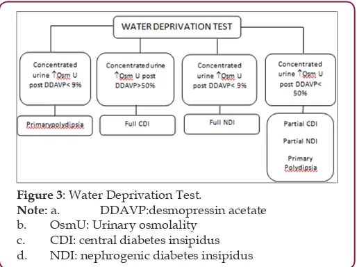
Plasma osmolality (POSM) is normal or slightly high, according to the patient’s thirst and the amount of water ingested. Determining POSM or basal serum sodium is not diagnostically useful because values in patients with CDI, NDI and primary polydipsia (PP) are normal and may overlap. Nonetheless, if these values are clearly above normal levels (POSM>295 mOsm/kg and Na>143mEq/L) in conditions where ingestion of water is unrestricted, a diagnosis of PP is excluded. Consequently, a differential diagnosis must be made between CDI and serious NDI [5,6,10,11]. For a differential diagnosis, DDAVP (10µg subcutaneously) is administered, and after one and two hours assessment, non-concentrated urine indicates NDI, and concentrated urine, CDI [21]. In cases where a cause for polyuria cannot be found, water deprivation is advised [21]. Water deprivation is carried out in two stages [22]: First, during a period of water fasting, the patient’s weight (after hourly urine elimination) and urine osmolality are checked every hour, and plasma osmolality and serum sodium every two hours [22].
Figure 3: Water Deprivation Test.
Note: a. DDAVP:desmopressin acetate
b. OsmU: Urinary osmolality c. CDI: central diabetes insipidus d. NDI: nephrogenic diabetes insipidus
Second, 40 µg of intranasal desmopressin is administered, free
fluids given orally, and plasma osmolality and urinary osmolality
checked one and two hours, respectively, after administration of desmopressin [21]. This is illustrated in (Figure 3). In CDI cases, the gold standard imaging techniqueto search for tumors or other pathologies in the hypothalamic-hypophyseal region is MRI. MRI may also reveal expanding lesions in the sella turcica region (e.g.,
tumors, abscesses, infiltrative diseases, hypophysitis, etc.) and
thickening of the pituitary stalk [23]. In up to 80% of CDI cases, the posterior pituitary bright spot is visualized on MRI. However, this is also observed in up to 20% of normal people or people who have NDI, which may be secondary to depletion of AVP caused by chronic, excessive secretion. Patients with a diagnosis of idiopathic
CDI with no evidence of tumors, infiltrative diseases, infectious
diseases or metastatic diseases on initial MRI images should have subsequent MRIs because, in some cases, tumors may appear a few years after diagnosis [24].
Differential Diagnosis
Differential diagnoses could be PP, CDI or NDI [21]. PP includes psychiatric diseases, absence of nocturia, or episodic polyuria. A urinary volume greater than 18L is highly suggestive
of PP. However, most CDI patients have moderate dehydration, a decrease in urine volume (6 to 12L), and a decrease in the
glomerular filtration rate [21]. In CDI, patients develop polyuria
after cranial trauma or neurosurgery; it is also found in patients with clinical evidence of sella turcica or metastatic tumors (lung and breast), granulomatous diseases (Langerhans cell histiocytosis, sarcoidosis, and tuberculosis), and infectious diseases (pituitary abscesses, meningitis, encephalitis, and neurosyphilis) [25]. NDI is characterized by the kidney’s inabilityto concentrate urine despite normal concentrations of AVP. It may be caused by hydroelectrolytic disorders (hypopotassemia, hypercalcemia), chronic renal disease or ingestion of DI-inducing substances (lithium, amphotericin B, methotrexate, etc.). A rare form is familial CDI, caused by mutation of the receptor gene V2 [5,6,10,11].
Treatment
Specific treatment for CDI is AVP replacement. The medicine
of choice is Desmopressin, an AVP-analog with minimal pressor effect, greater diuretic activity and extended half-life, with a 6 to 24 h duration of action [26].
Medicationformats aretablets (0.1 and 0.2mg), nasal spray (0.1 mg/mL), nasal solution (0.1 mg/mL), and injectable solution (4 µg/ mL). The best therapeutic form will depend on the physician and the particular characteristics of the patient. The number of doses for optimal control of the disease will also depend on the patient’s clinical characteristics [26-28].
Other Therapeutic Options
In cases of CDI with mild or moderate polyuria, chlorpropamide (CLORP) (125 to 500mg/day) may be trialed. Doses greater than 500mg/day do not produce any greater therapeutic effect and may potentiate the action of AVP. CLORP is contraindicated in pregnant women and children due to risk of hypoglycemia [5,10,24,29]. Thiazide diuretics such as hydrochlorothiazide (50 to 100mg/day) may alternatively be trialed if CLORP produces anun satisfactory response. Indapamide has a similar effect [5,6,10,11]. Carbamazepine (200 to 400 mg/day) is indicated in patients unresponsive to the previous options. It stimulates the secretion of AVP by hypothalamic neurons and, in some cases, increases renal sensitivity to the hormone. The use of carbamazepine is limited by side effects including skin rashes, diplopia, blurred vision, drowsiness, nausea, vomiting, and ataxia [6,10-12].
Discussion
Diabetes Insipidusrepresentsa small part of diabetes cases in our population. DM2 account for the great majority of cases of Diabetes [1]. Complaints of polydipsia and polyuria are common in patients with uncontrolled DM2. In our patient’s case, his complaints at the initial consultations suggested likely uncontrolled DM2. However, continuous monitoring of capillary glycemia and glycosylated
hemoglobin showed that the patient did not have significant
receiving treatment for DM2, and experienced frequent episodes of hypoglycemia. In such cases, it is also important to exclude adrenal
insufficiency; the basal cortisol level was greater than 10µg/L, which helped exclude adrenal insufficiency. We did not investigate
possible insulinoma because there was no classic Whipple’s triad
of hypoglycemic symptoms and no significant weight gain, both
of which are typical of insulinomas. Differential analysis depends on exclusion criteria that compare symptoms and laboratory anomalies to diseases that are similar to NDI.
Urine density analysis is a differential criterion. In DM2, this is high, above 1020 mmol/L, whereas in neurogenic DI urine is hypotonic, i.e., with a density less than 1010mmol/L [30]. In our patient’s case, the symptoms of polyuria and polydipsia, alteration in arterial pressure, being overweight and slight hyperglycemia aroused suspicion of DM2, so treatment was initiated. The patient experienced side effects due to the use of oral hyperglycemic agents and rigorous diet. When he started treatment at our medical clinic, his urinary density of 1005 g/mLindicated DI. The plasma sodium level of 143mEq/L, characteristic of hypernatremia, wasan important differential indicator ofCDI; in hypernatremia, plasma sodium levels are greater than 135 mEq/L, whereas in NDI, hyponatremia is observed [31]. Another differential parameter was plasma osmolality, calculated by the formula POSM=2×(Na+K)+(glycemia/18), which, in our patient’s case was 300.74mOsm/kg. When osmolality is greater than 282mOsm/kg, the thirst mechanism is activated, and the POSMreference value varies between 280 and 295 mOsm/kg. POSM is a measurement that indirectly assesses a patient’s degree of hydration [19]. The patient’s urinary osmolality was 110mOsm/kg, and, in central DI, the reference value is below 300mOsm/kg. PP is another possible differential diagnosis. Nonetheless, an important aspect encountered by the patient was intense nocturia, which harmed his sleep quality, a complaint that is infrequent in PP patients.
Nocturia is the first symptom to manifest in DI. However, most
patients do not complain about it until diuresis exceeds 4000mL/ day [20]. CDI is characterized by polyuria and polydipsia, which exacerbates excessive nocturia, leading to alteration of sleep patterns, fatigue and irritability [21]. The patient ingested 7250mL
of fluid and had diuresis of 7500mL. Therefore, diuresis was 87.2mL/kg, which falls within the definition of polyuria, a volume of urine greater than 45-50mL/kg in 24h. Total AVP deficiency would
lead to diuresis greater than 18,000mL/24h [22]. Slightly elevated
diuresis suggests partial AVP deficiency, which may explain why the
AVP value in this case was 1.5pg/mL. The reference value for CDI
is below 1.0pg/dL [6,11]. The next step was cranial MRIto confirm
disease etiology. The most common hypophyseal alterations in DI are thickening of the pituitary stalk and fading of the posterior pituitary bright spot visualized in a T1 weighted image on MRI. The
thickening of the pituitary stalk confirmed the diagnosis, and AVP
replacement treatment was initiated [6, 11] (Figures 1 and 2).
Today, most of CDI previously classified as idiopathic have been now verified as an autoimmune etiology. Some features of the
present case suggest an autoimmune pathogenesis. The imaging
findings on MRI, showing a thickened pituitary stalk and a reduction
of hyperintense neurohypophysis signal, favor an autoimmune CDI caused by a lymphocytic infundibulo-neurohypohysitis. To search for the presence of antihypothalamus and antipituitary antibodies in patient’s serum could have contributed to clarify these aspects, unfortunately the patient did not perform these exams.
Conclusion
This case report highlights the differential diagnoses and possible causes of DI. In the case, we report small abnormalities in laboratory tests, which initially were not noticed by
nonspecialists,making it difficult to reach anearly diagnosis.
Moreover, the frequency of radiological abnormalities in patients
with CDI is ill defined. After clinical investigation, CDI, also known as
neurogenic DI, was diagnosed based on abnormalitiesin laboratory tests and morphologic characteristics of the neurohypophysis on
MRI. Today, CDI cases previously classified as idiopathic have been now verified to have an autoimmune pathogenesis. CDI is a disease
that causes many problems to those who suffer from it, including a reduction in their quality of life. For this reason, early diagnosis based on differentiation fromPP, careful note of small abnormalities in laboratory tests, and particularly careful patient historyare key points to commencement of early treatment in order torestore these patients’ well-being and quality of life.
References
1. Cefalu WT (2016) The Journal Of Clinical and Applied Research and Education, Diabetes Care; Standards of Medical Care In Diabetes. American Diabetes Association 39(1).
2. Earley LE, Orloff J (1962) The mechanism of antidiuresis associated with the administration of hydrochlorothiazide to patients with vasopressin-resistant diabetes insipidus. J Clin Invest 41(11):1988-97.
3. Rochdi MD, Vargas GA, Carpentier E, Oligny-Longpré G, Chen S, et al. (2010) Functional characterization of vasopressin type 2 receptor substitutions (R137H/C/L) leading to nephrogenic diabetes insipidus and nephrogenic syndrome of inappropriate antidiuresis: implications for treatments. Mol Pharmacol 77(5): 836-845.
4. Hadjizacharia P, Beale EO, Inaba K, Chan LS, Demetriades D (2008) Acute diabetes insipidus in severe head injury: a prospective study. J Am Coll Surg 207(4): 477-484.
5. Vilar L, Leal E, Oliveira L, Lyra R Alterações da vasopressina (2001) In Coronho V, Petrolano A, Santana EM, , Pimenta LG (Eds). Tratado de endocrinologia e cirurgia endócrina. Belo Horizonte: Guanabara-Koogan, pp.316-333.
6. Reeves WB, Bichet DG, Andreolli TE (2003) The posterior pituitary and water metabolsim. In: Wilson JD, Foster DR, Kronenberg HM, Larsen PR (Eds.) Williams Textbook of Endrinology. 9th
(Edn) Philadelphia, PA W B Saunders, pp.341-429.
7. Los EL, Deen PM, Robben JH (2010) Potential of Nonpeptide (Ant) agonists to Rescue Vasopressin V2 Receptor Mutants for the Treatment of X-linked Nephrogenic Diabetes Insipidus. J Neuroendocrinol 22(5): 393-399.
8. Wong LL, Verbalis JG (2002) Systemic disease associated with disorders of water homeostasis. Endocrinol Metab Clin 31(1): 121-140.
9. Kwon TH, Nielsen J, Møller HB, Fenton RA, Nielsen S, et al. (2009) Aquaporins in the kidney. Handb Exp Pharmacol 190: 95-132.
11. Charmandari E, Brook CG (1999) 20 years of experience in idiopathic central diabetes insipidus. Lancet 353(9171): 2212-2213.
12. Naves LA, Vilar L, Costa AC, Domingues L, Casulari LA (2003) Distúrbios na secreção e ação do hormônio antidiurético. Arq Bras Endocrinol Metab 47(4): 467-481.
13. Rittig S, Jensen AR, Jensen KT, Pedersen EB (1998) Effect of food intake on the pharmacokineticsand antidiuretic activity of oral desmopressin (DDAVP) in hydrated normal subjects. Clin Endocrinol (Oxf) 48(2): 235-241.
14. Richardson DE, Robinson AG (1985) Drugs Five Years Later: Desmopressin. Ann Intern Med 103: 228-239.
15. Boulgourdjian EM, Martinez AS, Ropelato MG, Heinrich JJ, Bergada C (1997) Oral desmopressine treatment of central diabetes insipidus in children. Acta Pediatr 86(11):1261-1212.
16. Singer I, Oster JR, Fishman LM (1997) The management of diabetes insipidus in adults. Arch Intern Med 157(12): 1293-3016.
17. Figueiredo DM, Rabelo FL (2009) Diabetes insipidus: principais aspectos e análise comparativa com diabetes mellitus. Semina: Ciências Biológicas e da Saúde 30(2): 155-162.
18. Macedo CS, Simões AC, Riyuzo MC, Bastos HD (2006) Diagnóstico e resposta terapêutica em dois pacientes com Diabetes insipidus nefrogênico. Revista Paulista de Pediatria 24(1): 78-84.
19. Guyton AC, Hall JE (2011) Tratado de fisiologia médica.12th (edn) Rio de
Janeiro: Guanabara Koogan. 28: 213-222.
20. Devin JK (2012) Hypopituitarism and central diabetes insipidus: perioperative diagnosis and management. Neurosurg Clin N Am 23(4): 679-689.
21. Ghirardello S, Garrè ML, Rossi A, Maghnie M (2007) The diagnosis of children with central diabetes insipidus. J Pediatr Endocrinol Metab 20(3): 359-376.
22. Xian-ling WA, Ying-qian WA, Yi-ming MU (2012) Caution on diagnosis of idiopathetic central diabetes insipidus 10: 1683-1685.
23. Rocha JL, Friedman E, Boson WL, De Marco L (2000) Diabetes insipidus nefrogênico: conceitos atuais de fisiopatologia e aspectos clínicos. Arquivos Brasileiros de Endocrinologia & Metabologia 44(4): 290-299. 24. Robertson GL (1995) Diabetes insipidus. Endocrinol Metab Clin North
Am 24(3): 549-572.
25. Pivonello R, Colao A, Di Somma C, Facciolli G, Klain M, et al. (1998) Impairment of Bone Status in Patients with Central Diabetes Insipidus. J Clin Endocrinol Metab 83(7): 2275-2280.
26. Pivonello R, Faggiano A, Arrichiello P, Di Sarno A, Di Somma C, et al. (2001) Central diabetes insipidus and heart: effect of acute arginine vasopressin deficiency and replacement treatment with desmopressin on cardiac performance. Clinic Endocrinol (Oxf) 54(1): 97-106. 27. Baylis PH, Cheetham T (1998) Diabetes insipidus. J R Coll Physicians
Lond 79(1): 84-89.
28. Robinson AG, Verbalis JG (1997) Diabetes insipidus. Curr Ther Endocrinol Metab 6:1-7.
29. Eisenbarth GS, Verge CF (1998) Immunoendocrinopathy syndromes. In Wilson JD (Eds.) Williams Textbook of Endocrinology. 9th (edn)
Philadelphia, PA: Saunders pp.1651-1662.
30. Scherbaum WA, Bottazzo GF (1983) Autoantibodies to vasopressin cells in idiopathic diabetes insipidus: evidence for an autoimmune variant. Lancet 1(8330): 897-901.
31. Scherbaum WA, Bottazzo GF, Czernichow P, Wass JAH, Doniach D (1985) In Role of autoimmunity in central diabetes insipidus. Czernichow P, Robinson AG, (Eds.) Diabetes insipidus in man. International Symposium, Paris, January 1984. Front Horm Res Basel Karger 13: 232-239.
Submission Link: http://biomedres.us/submit-manuscript.php
Assets of Publishing with us • Global archiving of articles
• Immediate, unrestricted online access • Rigorous Peer Review Process • Authors Retain Copyrights • Unique DOI for all articles
http://biomedres.us/ This work is licensed under Creative