Metalloproteinases In Medicine
Dove
press
R E V I E W
open access to scientific and medical research
Open Access Full Text Article
Matrix metalloproteinases in bone development
and pathology: current knowledge and potential
clinical utility
Hai Po Helena Liang* Joshua Xu*
Meilang Xue
Christopher J Jackson Sutton Arthritis Research Laboratory, Institute of Bone and Joint Research, Kolling Institute, Sydney Medical School Northern, University of Sydney, St Leonards, NSW, Australia
*These authors contributed equally to this work
Abstract: Matrix metalloproteinases (MMPs) are degrading enzymes that have a pivotal function in extracellular matrix remodeling. More than half of the MMP members are expressed by bone and cartilage cells under physiological or pathological conditions such as rheumatoid arthritis, osteoarthritis, and osteoporosis. Through studies on the various bone diseases and on genetically modified mouse models in which one or more of the MMPs or their associated proteins and down-stream signaling molecules have been targeted, it is becoming increasingly evident that MMPs and other players in their cellular pathway play a pivotal role in bone development and remodeling. This review details the latest findings related to MMPs and bone development and pathology. Keywords: bone diseases, mouse models, gelatinases, collagenases, vascular endothelial growth factor, activated protein C, bone morphogenetic proteins, transforming growth factor
Introduction
There are two vital processes during bone development that are responsible for bone formation, intramembranous ossification and endochondral ossification. Intramem-branous ossification is a process that forms most of the craniofacial skeleton through
the direct differentiation of mesenchymal cells into bone.1 In contrast, endochondral
ossification is a process of bone development in which cartilage is used as a template
for bone morphogenesis.1 Endochondral ossification is responsible for the
develop-ment of most of the bones including the long bones. In this process, mesenchymal cells undergo condensation and then differentiate into chondrocytes, which proliferate
and undergo hypertrophy before dying.1 This leaves cavities for the invasion of blood
vessels and bone-forming/bone-remodeling cells such as osteoclasts, osteoblasts,
and bone marrow.2 Following this, ossification gradually occurs along the cartilage
template to replace it with bone.2 Successful bone growth requires a mechanically
stable cartilage model that can be degraded during ossification to allow for mineral
deposition and ultimately bone formation.2
The recruitment, survival, and function of osteoclasts and osteoblasts are essential for the prevention of metabolic bone diseases such as osteoporosis. In addition, these cells play an important role in promoting bone regeneration in pathological conditions such as rheumatoid arthritis (RA) and osteoarthritis (OA). RA and OA are characterized by synovitis, a destruction of the cartilage and the surrounding extracellular matrix
(ECM).3 Remodeling of ECM in bones is therefore important for mediating bone
development and repair.4,5 ECM remodeling in other areas including skin and blood
Correspondence: Christopher J Jackson Sutton Arthritis Research Laboratory, Level 10, Kolling Institute, Royal North Shore Hospital, St Leonards, NSW 2065, Australia
Tel +61 2 9926 4814 Fax +61 2 9926 6269
Email [email protected]
Journal name: Metalloproteinases In Medicine Article Designation: REVIEW
Year: 2016 Volume: 3
Running head verso: Liang et al
Running head recto: MMPs in bone development and pathology DOI: http://dx.doi.org/10.2147/MNM.S92187
Metalloproteinases In Medicine downloaded from https://www.dovepress.com/ by 118.70.13.36 on 24-Aug-2020
For personal use only.
This article was published in the following Dove Press journal: Metalloproteinases In Medicine
12 December 2016
Dovepress
Liang et al
vessels has also been indicated to mediate many different physiological and pathological processes, including wound
healing and angiogenesis.6–9
Matrix metalloproteinases (MMPs) are a family of zinc-dependent ECM-degrading enzymes, which has been
estab-lished to play a crucial role in ECM remodeling,10 and are the
key enzymes responsible for cleaving structural components of the ECM such as collagen and gelatin, thereby enabling
the ECM to degrade and regenerate.10 More than half of the
MMP members are expressed in their active form by bone and cartilage cells under physiological and pathological conditions, and these MMPs are not only thought to have a pivotal involvement in bone and cartilage matrix degradation, but also important for osteoclast, osteoblast and osteocyte viability and functions, as well as chondrocyte proliferation
and differentiation.8 This review provides an update on the
role of MMPs in bone development and remodeling.
Overview of the classification,
structure, expression, and regulation
of MMPs
The 23 members of MMPs in humans (MMPs 1 through to 28, with MMP-4, MMP-5, MMP-6 and MMP-22 being removed from the classification due to duplication, and MMP-18 not found in humans) can be categorized into several different subtypes based on their structural and substrate affinity: collagenases (MMP-1, MMP-8 and MMP-13), gelatinases (MMP-2 and MMP-9), stromelysins (MMP-3, MMP-10 and MMP-11), matrilysins (MMP-7 and MMP26, membrane-type metalloproteinase (MMP-14, MMP-15, MMP-16, MMP-17 and MMP-24), and others (MMP-12, MMP-19, MMP-20, MMP-21, MMP-23, MMP-25, MMP-27 and MMP-28). Each member of these MMPs is encoded by a different gene, except for MMP-23, which is encoded by two identical genes
(MMP-23A and MMP-23B ) on chromosome 1.11 All MMPs
share three common structures: the predomain (PRE) for protein secretion, the prodomain (PRO) responsible for the regulation of protein function, and the zinc-dependent
cata-lytic endopeptidase motif,12 which are necessary for substrate
processing. MMPs are expressed as inactive proenzymes that can be activated by the proteolytic cleavage of the propeptide to reveal its active site. The cleavage of the propeptide and acti-vation of all MMPs, with the exception MMP-23, is thought to be mediated by a cysteine amino acid in the molecule’s prodomain, via extracellular proteinases and other MMPs, in a
process called “cysteine switching”.13 The regulation of MMP
expression and activity occurs at multiple levels, including
gene transcription,14 translation and secretion of the inactive
proenzyme,15 and proenzyme activation and inactivation via
signaling from cytokines, growth factors, integrins, and ECM
proteins.13,16 Examples of classic MMP activators include the
activator protein-1, nuclear factor kappa B, tumor necrosis
factor-alpha, transforming growth factor beta (TGFb), as well
as certain interleukins. The main inhibitors of MMPs are the
tissue inhibitors of MMPs (TIMPs) secreted by the ECM.17
Roles of MMPs and TIMPs during
bone development, remodeling, and
repair
Currently, the most abundantly expressed and functionally important MMPs in bone and cartilage cells during normal skeletal development are identified to be MMP-2 (also known as gelatinase A), MMP-9 (gelatinase B), MMP-13 (collage-nase 3), MMP-14 (membrane-type 1 MMP), and MMP-16 (membrane-type 3), based on in vivo studies using MMP gene knockout mice and preclinical experimental arthritis models, as well as investigations of human genetic diseases
involving MMP gene mutations.8 The roles of each of these
MMPs and TIMPs are discussed as follows.
MMP-2
MMP-2, also known as gelatinase A, is a 72 kDA gelatinase that can cleave type I, IV, V, VII, and XI collagens along with aggrecans, gelatins, fibronectin, laminin, large tenascin-C,
and elastin.18 Inactivating mutations of the human gene
expressing MMP-2 were first identified in patients with inherited multicentric osteolysis and arthritis or the “vanish-ing bone” syndrome. This autosomal recessive condition was first reported in large Saudi Arabian families in which
interfa-milial marriage had occurred.19,20 The patients suffered from
severe arthropathy, osteoporosis, and subcutaneous nodules with distinctive craniofacial defects that included
exophthal-mos, brachycephaly, and flattened nasal bridges.21 Children
with multicentric osteolysis and arthritis exhibit normal birth weight but stunted growth rates in terms of both height and
weight.21 By examining the population suffering from these
distinctive bone phenotypes, the disease gene was identified
at 16q12-21, a region encoding the expression of MMP-2.22
MMP-2 knockout mice have been used to elucidate the function of MMP-2 on bone. MMP-2 knockout mice, when compared to wild-type mice, were initially shown to have almost completely normal early development, with only a slight delay in bone development and were smaller in size
at birth.23 However, further studies found that the long bones
of MMP-2 knockout mice had osteopenia with the tibiae of
many of these mice spontaneously becoming fractured.24 It is
Metalloproteinases In Medicine downloaded from https://www.dovepress.com/ by 118.70.13.36 on 24-Aug-2020
Dovepress MMPs in bone development and pathology
postulated that the reduced bone integrity in MMP-2 knock-out mice is caused by disruptions to the osteocytic canalicular
networks in bone.24 The canalicular spaces are locations in
which mineral ion exchanges occur during the resorption and
mineralization of the bone matrix.25 Thus, disruption to these
canalicular networks could possibly inhibit the mineraliza-tion and hardening of bone. An increase in MMP-2 protein expression has also been detected in fracture callus, and bone mineralization may be one aspect by which MMP-2 mediates
bone remodeling during fracture repair.26 Nyman et al
sup-port this postulation as they found that the ratio of mineral to collagen is decreased in the bone of MMP-2 knockout
mice.27 Along with the reduced bone mineralization density,
the diaphysis cortex in the tibia of MMP-2 knockout mice
is 23% thinner than the wild type.27 MMP-2 deficiency has
also been shown to reduce tibia and femur length in adult
mice.28 Histological analysis also revealed less dense and
very disordered trabeculae.21 Since the osteocytic canalicular
networks of MMP-2 knockout mice are repaired following transplantation of wild-type periosteum, MMP-2 may
medi-ate its effects on bone via a local mechanism.24 Thus, MMP-2
appears to play a role in maintaining bone mineral density and strength, possibly via its local action on the ECM to produce canalicular networks.
The craniofacial defects observed in MMP-2 knockout mice are also comparable to those seen in humans with multicentric osteolysis and arthritis as a result of the inac-tivating mutation to the MMP-2 gene. The knockout mice have shorter, broader snouts along with hypertelorism and
smaller jaws.21 It has been postulated that MMP-2 acts via
type I collagen, as its inhibition of type I collagen cleav-age caused similar craniofacial defects in mice as MMP-2
knockout.29 There is also abnormal bone development of
the calvaria, which is the only bone reported to undergo sclerosis with increased bone deposition and osteocyte loss which is likely due to the increased osteoblastic activity in
the calvarie.24 No changes in blastocyst activity have been
detected in the long bones of MMP-2 knockout mice.24 These
findings are interesting as ex vivo studies have shown poor proliferation of osteoblasts with MMP-2 knockout calvarial
bone marrow stromal cells.21 In agreement, targeted
siRNA-mediated MMP-2 knockout decreases osteoblast proliferation
ex vivo.21 The different findings in vivo and ex vivo can
pos-sibly be explained by the fact that MMP-2 knockout in mice can directly induce the expression of osteopontin and bone
sialoprotein.30 While osteopontin has been shown to
medi-ate an increase in osteoclast activity, bone sialoprotein can
enhance osteoblast differentiation and activity.31,32 Thus, the
varying expression of these two proteins in different bones can account for either increased bone reabsorption or bone growth. Ultimately, there is evidence that MMP-2 affects bone development via its effect on osteoclast and osteoblast activity and proliferation; however, the mechanisms are still not fully elucidated.
MMP-9
MMP-9, also known as gelatinase B, is a 92 kDa collagenase that has high specific degradative activity for denatured collagens in the ECM. It can cleave elastin and native type IV, V, and XI collagens, but not non-native type I collagen,
proteoglycan, or laminins.18,33 MMP-9 is also able to cleave
non-ECM molecules such as substance P, amyloid beta pep-tide, and myelin basic protein. The expression of MMP-9 varies through the stages of development. In early develop-ment, MMP-9 is expressed in trophoblasts and osteoclasts, suggesting that it plays a role in implantation and bone
resorption.34,35 With maturity, MMP-9 is primarily expressed
in inflammatory cells during diseases such as RA and cancer.5
There are numerous postulated methods by which MMP-9 can influence bone development and strength. Long bones in MMP-9 knockout mice are 10% shorter than those of the
wild-type mice.33 These knockout mice also demonstrate an
accumulation of lengthened hypertrophic chondrocytes at the
growth plate, despite normal proliferation of these cells.33,36 It
has been postulated that MMP-9 is required for the cleavage of galectin-3 to prevent accumulation of late hypertrophic
chondrocytes.37 These findings suggest that MMP-9 plays a
role in influencing the structural properties of whole bones through its activity at the growth plate during endochondral bone formation. This is supported by evidence that MMP-9 null mice have delayed skeletal growth plate vascularization and ossification of hypertrophic cartilage which is confined to
just the growth plate cartilage.33 Bone marrow transplantation
of MMP-9 null mice corrects all bone growth defects, sug-gesting that the critical cells expressing MMP-9 are of bone
marrow origin.33 Interestingly, the MMP-9 null mice do not
have any issues with implantation or development into fertile adults, suggesting that alternate physiological processes can compensate for some roles of MMP-9.
MMP-9 can also influence the bending strength and toughness of bone. MMP-9 knockout mice have improved connectivity density of the tibia trabeculae at the metaphysis
with no overall volume changes.27 Yield strength, defined as
the force required before plastic deformation, was slightly higher in the MMP-9 knockout mice when compared with the wild types. Interestingly, however, the bones of MMP-9
Metalloproteinases In Medicine downloaded from https://www.dovepress.com/ by 118.70.13.36 on 24-Aug-2020
Dovepress
Liang et al
knockout mice were more brittle.27 Although the mechanisms
are not fully known, it is postulated that MMP-9 may affect ECM proteins such as type I collagen to alter ECM
organiza-tion and thereby influence bone strength and brittleness.27,38
Recently, MMP-9 has been reported to also be necessary for the regulation of gene pathways that are required for osteoclastogenesis, through its proteolytic interaction with
the histone H3 N-terminal tail (H3NT).39 Further studies must
be conducted to better understand the mechanisms underlying these bone pathologies in MMP-9 null mice.
MMP-13
Cartilage consists of chondrocytes and a large amount of ECM, which is made up of proteoglycans, aggrecans, and collagens, mainly type II. Type II collagen is extremely resistant to degradation by most proteinases because of its triple-helical structure. Only the classical collagenases including MMP-1, MMP-8, and MMP-13, and to a much less extent, MMP-14, are able to degrade type II collagen fibrils and denature them into gelatin, which can then be subsequently digested into small peptides by the
gelatin-ases.40 Due to its preferential digestion of type II collagen
over other collagen types, MMP-13, which is derived from chondrocytes, synovial cells, and osteoblasts, is considered to be the most important collagenase for the degradation of cartilage.3,41
The proenzyme form of MMP-13 is ~60 kDa in size
and can be activated by MMP-2, MMP-3, MMP-14, and
plasmin.42 The expression and type II collagen
degrada-tion activity of the 48 kDa active MMP-13 can also be
upregulated by ECM proteins such as type X collagen43
and periostin.44 There is now mounting evidence to suggest
that increased MMP-13 activity is associated with articular cartilage degeneration and joint pathology typical of OA in animal models, and that MMP-13 expression is critical
for OA disease progression.43–45 Therefore, the development
of pharmacologic inhibitors of MMP-13 in recent years seems to be an effective strategy to modify OA disease
outcome.46,47 In addition, a MMP-13 activity probe seems
to be useful for in vivo imaging in OA disease diagnosis
and differentiating disease severity.48,49 MMP-13-deficient
mice also show significantly decreased disease severity in the antibody-induced arthritis model, thus implicating the important role of MMP-13 as a regulator of inflammation and revealing it as a potential therapeutic target for inflam-matory arthritis.50
MMP-13 knockout mice exhibit a normal lifespan and are fertile, with no gross phenotypic abnormalities. However,
these mice have demonstrated marked defects in their growth plate cartilage, with a significant increase in the hypertrophic chondrocyte zones but normal chondrocyte proliferation, and a delay in endochondral ossification, particularly at the secondary ossification center. The mice also show increased trabecular bone mass but irregular bone spicules. This sub-sequently results in impairment of bone matrix organization and cartilage ECM remodeling in these animals, which leads to a reduction in both toughness and fracture resistance of their long bones, but ultimately the mice show no notable
abnormalities in their long bone development.51–53
MMP-13 is a downstream target of the transcription factor Cbfa1/Runx2 in hypertrophic chondrocytes, as Cbfa1 knockout mice fail to express MMP-13 during fetal
develop-ment.54,55 C-maf deficiency, on the other hand, markedly
sup-presses collagen degradation by MMP-13, thereby resulting in abnormal terminal differentiation of chondrocytes which
leads to prolongation of the chondrocyte hypertrophic state.56
Hence, MMP-13 has been implicated in the initiation of bone resorption.
MMP-9 and MMP-13 double knockout mice have exacerbated phenotypes compared to MMP-9 or MMP-13 single-knockout mice, including disorganized architecture of the hypertrophic chondrocyte zone and increased number of terminally differentiated hypertrophic chondrocytes. In contrast to MMP-13 single-knockout mice but similar to MMP-9 single-knockout mice, the double knockouts show a reduction of trabecular bone formation at the growth plate. Interestingly, there is no abnormality in aggrecan degrada-tion in the MMP-9 and MMP-13 double knockouts, despite the absence of the aggrecan cleavage product in these mice, suggesting that other mechanisms exist for the removal of
aggrecan during cartilage to bone transitions.33 This is
con-sistent with a subsequent study on an aggrecan knock-in mouse strain that is resistant to cleavage by all MMPs in the
aggrecan proteinase-sensitive interglobular domain.57
In humans, missense mutations in MMP-13 cause a genetic bone disorder called spondyloepimetaphyseal dysplasia characterized by defective growth and abnormal modeling of the spine and long bones in childhood, which spontaneously resolves by adolescence. These transient disease phenotypes appear to be caused by the late exit of chondrocytes from the growth plate, despite normal differ-entiation, consistent with the findings from knockout mice
models.58 Further studies on the role of MMP-13 in bone
remodeling in experimental arthritis mice models will likely provide better insights into the pathological process of such human bone disorders.
Metalloproteinases In Medicine downloaded from https://www.dovepress.com/ by 118.70.13.36 on 24-Aug-2020
Dovepress MMPs in bone development and pathology
MMP-14
MMP-14 is a membrane-bound protein that is expressed
highly by periskeletal and skeletal tissue.59,60 MMP-14
knock-out mice have a very high mortality, with 33% of these dying due to wasting before they are weaned, and at 50–90 days
nearly all these mutant mice die due to wasting.12 MMP-14
knockout mice also display abnormal cranial bone formation, short snouts, hypertelorism, and dome-shaped skulls along with growth impairment with respect to both weight and body
size.12 MMP-2 and MMP-14 double knockout mice also die
immediately after birth,61 and there may be some functional
overlap between these two MMPs, as the cranial defects in 14 deficiency are very similar to those seen in
MMP-2-deficient mice.12,24 It has been postulated that the MMP-14
knockout mouse inhibits fibroblast growth factor signaling, which is believed to be essential in intramembranous
ossifica-tion of cranial bones.62 In addition, MMP-14 null mice show
decreased MMP-2 activity, as expected since MMP-14 is the
major activator of MMP-2.63,64
The bone mass of MMP-14 knockout mice is greatly
reduced, with joints displaying articular cartilage loss.12
This is due to the greater bone resorption and reduced
bone formation in MMP-14 knockout mice.65,66 MMP-14
knockout mice elicit these effects via intrinsic deficits to the osteogenic cells as opposed to other systemic causes. By culture-expanding populations of marrow-derived osteogenic cells from MMP-14 mice, it was found that these cells in particular display impaired osteogenic capacity and
collagen-degrading activity.12 This may be mediated by the
reduced ability for MMP-14 knockout animals to release
CD44 from the cell membrane,67 as CD44 is important in
osteocyte cell–cell signaling with its knockout producing
shortened long bones and reduced osteoclast activity.67,68
MMP-14 may also interact with transduction signaling cascades, with a novel function of MMP-14 being its
mech-nosensory role in osteocytes.69,70
While the calcification of metaphyseal growth plates at birth is normal in MMP-14-deficient mice, there are
signifi-cant delays in secondary epiphyseal ossification.12,65 In
addi-tion, vascularization of the epiphyseal growth cartilage does not occur, which is a finding comparable to that of MMP-9
knockout mice.33 Reconstituted MMP-14 activity has been
shown to act on type II collagen-expressing cells to
amelio-rate skeletal dysplasia and rescue chondrocyte proliferation.71
Overall, it is believed that MMP-14 deficiency compromises apoptosis of chondrocytes and cartilage breakdown,
prevent-ing normal endochondral ossification.72 Thus, MMP-14 plays
an important role in bone development and growth.
MMP-16
MMP-16 present in osteoblasts and osteocytes41 acts by
breaking down ECM proteins such as high-density fibrillar type I collagen, thus promoting bone growth and
develop-ment.73 The mechanism of ECM degradation via MMP-16
is necessary for the proper functioning of mesenchymal
cells expressed on skeletal tissue.73,74 In fact, mice lacking
MMP-16 demonstrate stunted growth due to the decreased
viability of these mesenchymal cells.73 MMP-16 has been
examined along with MMP-14, as both enzymes have similar
molecular structures and tissue expression patterns.73,75 In the
double knockout of MMP-14 and MMP-16, the mortality was even higher than that of MMP-14 alone as described earlier, with the majority dying within the first day of birth due to developmental abnormalities. In contrast, the single knockout
of MMP-16 did not result in any premature deaths.73 This
suggests that MMP-16 is not essential for post-embryonic bone development in mice, unlike MMP-14. Furthermore, the double knockout of MMP-14 and MMP-16 led to even more pronounced craniofacial deformities and cortical bone
shortening than that observed in the single deficiencies.73 The
loss of both the MMPs also elicited greater nuclei apoptosis in the bone-lining cells along with reduced chondrocyte proliferation and cartilage remodeling, when compared to single knockout of the MMP-14 gene. These observations not only are tied to the loss of bone collagenolytic activity, but also imply that MMP-16 can partly compensate for the
action of MMP-14.73
TIMPs
TIMP-1 and TIMP-2 are the most studied natural inhibitors of the MMPs and the most highly expressed during bone development. TIMP-1 is expressed in chondrocytes of all zones in the growth plate, as well as in osteoblasts,
osteo-cytes, and osteoclasts.41,76 Mice overexpressing TIMP-1 in
osteoblasts were found by Geoffroy et al77 to have increased
trabecular bone volume and decreased bone turnover, hence indicative of an important regulatory role in bone forma-tion and remodeling that is likely to be dependent on its MMP inhibitory activity. TIMP-2 has also been found to be
expressed by hypertrophic chondrocytes and osteoblasts,41
and both TIMP-1 and TIMP-2 are able to directly stimulate the bone-resorbing activity of osteoclasts in vitro at physi-ological concentrations, and this is likely to be independent of
their inhibition of MMPs.78 Other TIMP members including
TIMP-3 and TIMP-4 are also expressed in bone and joint tissues, and an increased expression of these have been
associated with OA.79,80 TIMP-3 knockout mice were found
Metalloproteinases In Medicine downloaded from https://www.dovepress.com/ by 118.70.13.36 on 24-Aug-2020
Dovepress
Liang et al
to have mild cartilage degradation similar to that observed in osteoarthritic patients, as a result of increased type II
col-lagen degradation.81 On the other hand, overexpression of
TIMP-3 in hematopoietic cells was shown to result in fatal
osteosclerosis in mice.82 Most recently, the gene
expres-sions of TIMP-1, TIMP-2, and TIMP-3 were all found to be increased in the process of osteocytic differentiation during
bone matrix mineralization.83
Interactions of MMPs with other
proteins in bone development,
remodeling, and repair
Other proteins that interact with the aforementioned MMPs in bone development and disease are briefly described as follows and are summarized in Table 1.
TGF
b
The actions of MMPs on bone turnover and remodeling can be exerted via its interaction with various other proteins. Both MMP-2 and MMP-9 can regulate the bioavailability and
bioactivity of TGFb, a molecule that affects the mechanical
properties and composition of bone matrix.27,84,85 When TGFb
signaling is increased in mice, the modulus and hardness
of their bones decrease.85 In addition, MMP-14 expressed
by osteoblasts has also been reported to have the ability to
activate TGFb, thereby maintaining the survival of
osteo-blasts upon completion of bone matrix synthesis during bone
remodeling and driving them to differentiate into osteocytes.86
Conversely, TGFb is able to significantly upregulate
MMP-13 expression in osteoblasts, thereby inducing changes in osteoblast morphology that in turn promotes osteoclastic
bone resorption.87
Bone morphogenetic proteins
Bone morphogenetic proteins (BMPs) are multifunctional
secreted growth factors that also belong to the TGFb
super-family.88 Conditional and tissue-specific knockout mouse
models provide strong evidence on the pivotal roles of BMPs, as well as their receptors and effectors, in bone
for-mation and resorption.88–90 Inhibition of the BMP receptor
and its downstream signaling by a pharmacological ligand significantly reduces the expression and activity of both MMP-2 and MMP-9, thereby obstructing tissue regeneration and remodeling in teleost fish Poecilia latipinna following
amputation.91 Similarly in mice with BMP type IA receptor
(BMPRIA) deficiency specifically in osteoblasts, MMP-9 expressed by osteoclasts was significantly reduced, impair-ing bone resorption and finally resultimpair-ing in increased bone
mass during early development.90 Additionally, Choi et al92
reported that downregulation of MMP-9 activity promotes
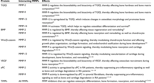
Table 1 Proteins that interact with MMPs and TIMPs in bone development and disease
Protein Interacting MMPs Effects
TGFb MMP-2 MMP-2 regulates the bioavailability and bioactivity of TGFb, thereby affecting bone hardness and bone matrix composition27,84,85
MMP-9 MMP-9 regulates the bioavailability and bioactivity of TGFb, thereby affecting bone hardness and bone matrix composition27,84,85
MMP-13 MMP-13 is upregulated by TGFb, which induces changes in osteoblast morphology and promotes bone resorption87
MMP-14 MMP-14 activates TGFb, which helps to regulate osteoblast differentiation and survival86
BMP MMP-2 MMP-2 is regulated by BMP, thereby affecting tissue regeneration and remodeling91
MMP-9 MMP-9 is regulated by BMP, thereby affecting bone resorption and remodeling, as well as chondrocyte commitment90–92
Wnt/b-catenin MMP-2 MMP-2 is regulated by Wnt/b-catenin signaling, thereby modulating chondrocyte function and affecting growth plate organization, cartilage formation, and endochondral ossification during bone development105
MMP-9 MMP-9 is upregulated by Wnt/b-catenin signaling, thereby modulating bone resorption and cartilage degradation90,100,105
MMP-13 MMP-13 is regulated by Wnt/b-catenin signaling, thereby modulating vascularization of cartilage during development, bone formation, and remodeling102–105
VEGF MMP-9 MMP-9 regulates the bioavailability and bioactivity of VEGF, thereby affecting osteoclast recruitment during bone resorption108–110
aPC MMP-2 MMP-2 activity is upregulated by aPC in RA patients, thereby suppressing pro-inflammatory signaling as well as bone and cartilage degradation in RA patients.116–118
MMP-9 MMP-9 activity is downregulated by aPC in synovial fibroblasts, thereby suppressing pro-inflammatory signaling as well as bone and cartilage degradation in RA patients117,119
TIMPs All MMPs MMP inhibitors that can regulate osteocytic differentiation, bone formation, resorption, and remodeling41,76–83 Abbreviations: aPC, activated protein C; BMP, bone morphogenetic protein; MMP, matrix metalloproteinase; RA, rheumatoid arthritis; TGFb; transforming growth factor beta; TIMP, tissue inhibitor of MMPs; VEGF, vascular endothelial growth factor.
Metalloproteinases In Medicine downloaded from https://www.dovepress.com/ by 118.70.13.36 on 24-Aug-2020
Dovepress MMPs in bone development and pathology
BMP-2-induced chondrocyte commitment of the mouse C3H10T1/2 stem cell line via modulation of glycogen
syn-thase kinase-3b signaling.
Wnt/beta-catenin
Wnt proteins are a large glycoprotein family that play important roles in various development and cell renewal
processes,93,94 and there is increasing evidence that suggests
that the canonical Wnt/b-catenin signaling pathway may be
involved in cartilage destruction in RA and OA.94–96 Various
research groups have reported that the expressions of several Wnt proteins, as well as the expressions of Wnt inhibitors known as the frizzled-related proteins, were altered in the synovium and cartilage of RA and OA patients compared to
normal controls.97–99 BMP-induced Wnt/b-catenin
signal-ing was demonstrated to regulate the expressions of TGFb,
MMP-2, MMP-9, MMP-13, and TIMP-1 in various bone cells including osteoblasts, osteoclasts, and articular
chon-drocytes,90,100–105 which can ultimately regulate bone and
cartilage formation, development, and remodeling, and hence have important implications in bone disease progression.
Vascular endothelial growth factor
Vascular endothelial growth factor (VEGF), an important angiogenic factor that acts mainly on endothelial cells, but
also stimulates osteoblasts and OA chondrocytes,106,107 has
been implicated to regulate endochondral ossification and
bone formation.108 The bioavailability and activity of VEGF is
believed to be at least partially regulated by MMP-9.108,109 In
addition, MMP-9 has been suggested to specifically regulate the ECM-bound VEGF, exerting direct chemotactic activity on osteoclasts, thereby affecting their recruitment during
bone resorption.110
Activated protein C
Activated protein C (aPC) is best known for its anticoagu-lant activity. However, it also has potent ability to promote
growth and dampen inflammation.111–114 aPC increases
osteoblast viability and signaling, promotes recombinant human recombinant BMP-2-induced ectopic bone formation, and enhances angiogenesis in vivo in a protease-activated
receptor-1-dependent manner,115 suggesting that aPC may
have the potential to be used in conjunction with rhBMP-2
as a therapeutic for bone repair.115 APC was first reported to
play a role in regulating MMP activity by Nguyen et al116
who showed that aPC directly activates MMP-2 in human umbilical vein endothelial cells. In RA, aPC co-localizes with and selectively upregulates and activates MMP-2, while at the same time inhibiting MMP-9 in synovial fibroblasts and
monocytes, resulting in suppressing of the pro-inflammatory
signaling in RA patients.117,118 This is explained by the finding
that endogenous MMP-9 but not MMP-2 promotes rheuma-toid synovial fibroblast survival, inflammation, and cartilage
degradation resulting in potential bone loss.119
Conclusion
In summary, bone is a highly dynamic and continuously remodeling tissue during development, homeostasis, and tissue repair. Half of the MMP members have been detected in bone tissue during development and in pathological condi-tions involving bone degradation. Knockout mouse models have confirmed the crucial functions of MMPs during bone development and regeneration. MMPs and proteins that modulate or are modulated by MMPs are potential targets for improving bone tissue repair. Better understanding of the regulatory mechanisms of MMPs and further elucidation of their cellular pathways in ECM remodeling is necessary for future therapeutic development.
Disclosure
The authors report no conflicts of interest in this work.
References
1. Ortega N, Behonick DJ, Werb Z. Matrix remodeling during endochon-dral ossification. Trends Cell Biol. 2004;14(2):86–93.
2. Mackie EJ, Ahmed YA, Tatarczuch L, Chen KS, Mirams M. Endochon-dral ossification: how cartilage is converted into bone in the developing skeleton. Int J Biochem Cell Biol. 2008;40(1):46–62.
3. Takaishi H, Kimura T, Dalal S, Okada Y, D’Armiento J. Joint diseases and matrix metalloproteinases: a role for MMP-13. Curr Pharm
Bio-technol. 2008;9(1):47–54.
4. Behonick DJ, Xing Z, Lieu S, et al. Role of matrix metalloproteinase 13 in both endochondral and intramembranous ossification during skeletal regeneration. PloS One. 2007;2(11):e1150.
5. Vu TH, Werb Z. Matrix metalloproteinases: effectors of development and normal physiology. Genes Dev. 2000;14(17):2123–2133. 6. Hua H, Li M, Luo T, Yin Y, Jiang Y. Matrix metalloproteinases in
tumorigenesis: an evolving paradigm. Cell Mol Life Sci. 2011;68(23): 3853–3868.
7. Deryugina EI, Quigley JP. Tumor angiogenesis: MMP-mediated induction of intravasation- and metastasis-sustaining neovasculature.
Matrix Biol. 2015;44–46:94–112.
8. Paiva KB, Granjeiro JM. Bone tissue remodeling and development: focus on matrix metalloproteinase functions. Arch Biochem Biophys. 2014;561:74–87.
9. Xue M, Le NT, Jackson CJ. Targeting matrix metalloproteases to improve cutaneous wound healing. Expert Opin Ther Targets. 2006; 10(1):143–155.
10. Le NT, Xue M, Castelnoble LA, Jackson CJ. The dual personalities of matrix metalloproteinases in inflammation. Front Biosci. 2007; 12:1475–1487.
11. Nagase H, Visse R, Murphy G. Structure and function of matrix metal-loproteinases and TIMPs. Cardiovasc Res. 2006;69(3):562–573. 12. Holmbeck K, Bianco P, Caterina J, et al. MT1-MMP-deficient mice
develop dwarfism, osteopenia, arthritis, and connective tissue disease due to inadequate collagen turnover. Cell. 1999;99(1):81–92. 13. Ra HJ, Parks WC. Control of matrix metalloproteinase catalytic activity.
Metalloproteinases In Medicine downloaded from https://www.dovepress.com/ by 118.70.13.36 on 24-Aug-2020
Dovepress
Liang et al
14. Yan C, Boyd DD. Regulation of matrix metalloproteinase gene expres-sion. J Cell Physiol. 2007;211(1):19–26.
15. Clark IM, Swingler TE, Sampieri CL, Edwards DR. The regulation of matrix metalloproteinases and their inhibitors. Int J Biochem Cell
Biol. 2008;40(6–7):1362–1378.
16. Stamenkovic I. Extracellular matrix remodelling: the role of matrix metalloproteinases. J Pathol. 2003;200(4):448–464.
17. Hadler-Olsen E, Fadnes B, Sylte I, Uhlin-Hansen L, Winberg JO. Regulation of matrix metalloproteinase activity in health and disease.
FEBS J. 2011;278(1):28–45.
18. Shimokawa Ki K, Katayama M, Matsuda Y, et al. Matrix metallopro-teinase (MMP)-2 and MMP-9 activities in human seminal plasma.
Mol Hum Reprod. 2002;8(1):32–36.
19. Al Aqeel A, Al Sewairi W, Edress B, Gorlin RJ, Desnick RJ, Martignetti JA. Inherited multicentric osteolysis with arthritis: a variant resembling Torg syndrome in a Saudi family. Am J Med Genet. 2000;93(1):11–18. 20. Al-Mayouf SM, Majeed M, Hugosson C, Bahabri S. New form of
idiopathic osteolysis: nodulosis, arthropathy and osteolysis (NAO) syndrome. Am J Med Genet. 2000;93(1):5–10.
21. Mosig RA, Dowling O, DiFeo A, et al. Loss of MMP-2 disrupts skeletal and craniofacial development and results in decreased bone mineralization, joint erosion and defects in osteoblast and osteoclast growth. Hum Mol Genet. 2007;16(9):1113–1123.
22. Martignetti JA, Aqeel AA, Sewairi WA, et al. Mutation of the matrix metalloproteinase 2 gene (MMP2) causes a multicentric osteolysis and arthritis syndrome. Nat Genet. 2001;28(3):261–265.
23. Itoh T, Ikeda T, Gomi H, Nakao S, Suzuki T, Itohara S. Unaltered secre-tion of beta-amyloid precursor protein in gelatinase A (matrix metallo-proteinase 2)-deficient mice. J Biol Chem. 1997;272(36):22389–22392. 24. Inoue K, Mikuni-Takagaki Y, Oikawa K, et al. A crucial role for matrix metalloproteinase 2 in osteocytic canalicular formation and bone metabolism. J Biol Chem. 2006;281(44):33814–33824.
25. Marenzana M, Shipley AM, Squitiero P, Kunkel JG, Rubinacci A. Bone as an ion exchange organ: evidence for instantaneous cell-dependent cal-cium efflux from bone not due to resorption. Bone. 2005;37(4):545–554. 26. Lieu S, Hansen E, Dedini R, et al. Impaired remodeling phase of fracture repair in the absence of matrix metalloproteinase-2. Dis Model Mech. 2011;4(2):203–211.
27. Nyman JS, Lynch CC, Perrien DS, et al. Differential effects between the loss of MMP-2 and MMP-9 on structural and tissue-level proper-ties of bone. J Bone Miner Res. 2011;26(6):1252–1260.
28. Madsen DH, Jurgensen HJ, Ingvarsen S, et al. Differential actions of the endocytic collagen receptor uPARAP/Endo180 and the collagenase MMP-2 in bone homeostasis. PloS One. 2013;8(8):e71261. 29. Egeblad M, Shen HC, Behonick DJ, et al. Type I collagen is a genetic
modifier of matrix metalloproteinase 2 in murine skeletal development.
Dev Dyn. 2007;236(6):1683–1693.
30. Mosig RA, Martignetti JA. Loss of MMP-2 in murine osteoblasts upregulates osteopontin and bone sialoprotein expression in a circuit regulating bone homeostasis. Dis Model Mech. 2013;6(2):397–403. 31. Gordon JA, Tye CE, Sampaio AV, Underhill TM, Hunter GK, Goldberg
HA. Bone sialoprotein expression enhances osteoblast differentiation and matrix mineralization in vitro. Bone. 2007;41(3):462–473. 32. Shapses SA, Cifuentes M, Spevak L, et al. Osteopontin facilitates bone
resorption, decreasing bone mineral crystallinity and content during calcium deficiency. Calcif Tissue Int. 2003;73(1):86–92.
33. Vu TH, Shipley JM, Bergers G, et al. MMP-9/gelatinase B is a key regulator of growth plate angiogenesis and apoptosis of hypertrophic chondrocytes. Cell. 1998;93(3):411–422.
34. Reponen P, Sahlberg C, Munaut C, Thesleff I, Tryggvason K. High expression of 92-kDa type IV collagenase (gelatinase) in the osteo-clast lineage during mouse development. Ann N Y Acad Sci. 1994; 732:472–475.
35. Alexander CM, Hansell EJ, Behrendtsen O, et al. Expression and function of matrix metalloproteinases and their inhibitors at the maternal-embryonic boundary during mouse embryo implantation.
Development. 1996;122(6):1723–1736.
36. Kojima T, Hasegawa T, de Freitas PH, et al. Histochemical aspects of the vascular invasion at the erosion zone of the epiphyseal cartilage in MMP-9-deficient mice. Biomed Res. 2013;34(3):119–128. 37. Ortega N, Behonick DJ, Colnot C, Cooper DN, Werb Z. Galectin-3 is
a downstream regulator of matrix metalloproteinase-9 function during endochondral bone formation. Mol Biol Cell. 2005;16(6):3028–3039. 38. Nyman JS, Reyes M, Wang X. Effect of ultrastructural changes on the
toughness of bone. Micron. 2005;36(7–8):566–582.
39. Kim K, Punj V, Kim JM, et al. MMP-9 facilitates selective proteolysis of the histone H3 tail at genes necessary for proficient osteoclastogen-esis. Genes Dev. 2016;30(2):208–219.
40. Krane SM. Petulant cellular acts: destroying the ECM rather than creating it. J Clin Invest. 2001;107(1):31–32.
41. Haeusler G, Walter I, Helmreich M, Egerbacher M. Localization of matrix metalloproteinases, (MMPs) their tissue inhibitors, and vascular endothelial growth factor (VEGF) in growth plates of children and adolescents indicates a role for MMPs in human postnatal growth and skeletal maturation. Calcif Tissue Int. 2005;76(5):326–335. 42. Knauper V, Will H, Lopez-Otin C, et al. Cellular mechanisms for
human procollagenase-3 (MMP-13) activation. Evidence that MT1-MMP (MT1-MMP-14) and gelatinase a (MT1-MMP-2) are able to generate active enzyme. J Biol Chem. 1996;271(29):17124–17131.
43. Neuhold LA, Killar L, Zhao W, et al. Postnatal expression in hyaline cartilage of constitutively active human collagenase-3 (MMP-13) induces osteoarthritis in mice. J Clin Invest. 2001;107(1):35–44. 44. Attur M, Yang Q, Shimada K, et al. Elevated expression of periostin in
human osteoarthritic cartilage and its potential role in matrix degrada-tion via matrix metalloproteinase-13. FASEB J. 2015;29(10):4107–4121. 45. Wang M, Sampson ER, Jin H, et al. MMP13 is a critical target gene dur-ing the progression of osteoarthritis. Arthritis Res Ther. 2013;15(1):R5. 46. Hu W, Zhang W, Li F, Guo F, Chen A. Bortezomib prevents the expres-sion of MMP-13 and the degradation of collagen type 2 in human chondrocytes. Biochem Biophys Res Commun. 2014;452(3):526–530. 47. Ruminski PG, Massa M, Strohbach J, et al. Discovery of N-(4-fluoro-3-methoxybenzyl)-6-(2-(((2S,5R)-5-(hydroxymethyl)-1,4-dioxan-2-yl) met hyl)-2H-tetrazol-5-yl)-2-methylpyrimidine-4-carboxamide. A highly selective and orally bioavailable matrix metalloproteinase-13 inhibitor for the potential treatment of osteoarthritis. J Med Chem. 2016; 59(1):313–327.
48. Lim NH, Meinjohanns E, Meldal M, Bou-Gharios G, Nagase H. In vivo imaging of MMP-13 activity in the murine destabilised medial menis-cus surgical model of osteoarthritis. Osteoarthritis Cartilage. 2014; 22(6):862–868.
49. Lim NH, Meinjohanns E, Bou-Gharios G, et al. In vivo imaging of matrix metalloproteinase 12 and matrix metalloproteinase 13 activities in the mouse model of collagen-induced arthritis. Arthritis Rheumatol. 2014;66(3):589–598.
50. Singh A, Rajasekaran N, Hartenstein B, et al. Collagenase-3 (MMP-13) deficiency protects C57BL/6 mice from antibody-induced arthritis.
Arthritis Res Ther. 2013;15(6):R222.
51. Inada M, Wang Y, Byrne MH, et al. Critical roles for collagenase-3 (Mmp13) in development of growth plate cartilage and in endochondral ossification. Proc Natl Acad Sci U S A. 2004;101(49):17192–17197. 52. Stickens D, Behonick DJ, Ortega N, et al. Altered endochondral bone development in matrix metalloproteinase 13-deficient mice.
Develop-ment. 2004;131(23):5883–5895.
53. Tang SY, Herber RP, Ho SP, Alliston T. Matrix metalloproteinase-13 is required for osteocytic perilacunar remodeling and maintains bone fracture resistance. J Bone Miner Res. 2012;27(9):1936–1950. 54. Jimenez MJ, Balbin M, Lopez JM, Alvarez J, Komori T, Lopez-Otin C.
Collagenase 3 is a target of Cbfa1, a transcription factor of the runt gene family involved in bone formation. Mol Cell Biol. 1999;19(6):4431–4442. 55. Jimenez MJ, Balbin M, Alvarez J, et al. A regulatory cascade involv-ing retinoic acid, Cbfa1, and matrix metalloproteinases is coupled to the development of a process of perichondrial invasion and osteogenic differentiation during bone formation. J Cell Biol. 2001; 155(7):1333–1344.
Metalloproteinases In Medicine downloaded from https://www.dovepress.com/ by 118.70.13.36 on 24-Aug-2020
Dovepress MMPs in bone development and pathology
56. MacLean HE, Kim JI, Glimcher MJ, Wang J, Kronenberg HM, Glim-cher LH. Absence of transcription factor c-maf causes abnormal termi-nal differentiation of hypertrophic chondrocytes during endochondral bone development. Dev Biol. 2003;262(1):51–63.
57. Little CB, Meeker CT, Hembry RM, et al. Matrix metalloproteinases are not essential for aggrecan turnover during normal skeletal growth and development. Mol Cell Biol. 2005;25(8):3388–3399.
58. Kennedy AM, Inada M, Krane SM, et al. MMP13 mutation causes spondyloepimetaphyseal dysplasia, Missouri type (SEMD(MO). J
Clin Invest. 2005;115(10):2832–2842.
59. Ohuchi E, Imai K, Fujii Y, Sato H, Seiki M, Okada Y. Membrane type 1 matrix metalloproteinase digests interstitial collagens and other extracellular matrix macromolecules. J Biol Chem. 1997; 272(4):2446–2451.
60. Kinoh H, Sato H, Tsunezuka Y, et al. MT-MMP, the cell surface activa-tor of proMMP-2 (pro-gelatinase A), is expressed with its substrate in mouse tissue during embryogenesis. J Cell Sci. 1996;109(Pt 5): 953–959.
61. Oh J, Takahashi R, Adachi E, et al. Mutations in two matrix metal-loproteinase genes, MMP-2 and MT1-MMP, are synthetic lethal in mice. Oncogene. 2004;23(29):5041–5048.
62. Hatch NE. FGF signaling in craniofacial biological control and pathological craniofacial development. Crit Rev Eukaryot Gene Expr. 2010;20(4):295–311.
63. Oblander SA, Zhou Z, Galvez BG, et al. Distinctive functions of membrane type 1 matrix-metalloprotease (MT1-MMP or MMP-14) in lung and submandibular gland development are independent of its role in pro-MMP-2 activation. Dev Biol. 2005;277(1):255–269. 64. Strongin AY, Collier I, Bannikov G, Marmer BL, Grant GA, Goldberg
GI. Mechanism of cell surface activation of 72-kDa type IV collage-nase. Isolation of the activated form of the membrane metalloprotease.
J Biol Chem. 1995;270(10):5331–5338.
65. Zhao W, Byrne MH, Wang Y, Krane SM. Osteocyte and osteoblast apoptosis and excessive bone deposition accompany failure of col-lagenase cleavage of collagen. J Clin Invest. 2000;106(8):941–949. 66. Holmbeck K, Bianco P, Pidoux I, et al. The metalloproteinase
MT1-MMP is required for normal development and maintenance of osteo-cyte processes in bone. J Cell Sci. 2005;118(Pt 1):147–156. 67. Kajita M, Itoh Y, Chiba T, et al. Membrane-type 1 matrix
metallo-proteinase cleaves CD44 and promotes cell migration. J Cell Biol. 2001;153(5):893–904.
68. Hughes DE, Salter DM, Simpson R. CD44 expression in human bone: a novel marker of osteocytic differentiation. J Bone Miner Res. 1994;9(1):39–44.
69. Eisenach PA, Roghi C, Fogarasi M, Murphy G, English WR. MT1-MMP regulates VEGF-A expression through a complex with VEGFR-2 and Src. J Cell Sci. 2010;123(Pt 23):4182–4193.
70. Kulkarni RN, Bakker AD, Gruber EV, et al. MT1-MMP modulates the mechanosensitivity of osteocytes. Biochem Biophys Res Commun. 2012;417(2):824–829.
71. Szabova L, Yamada SS, Wimer H, et al. MT1-MMP and type II col-lagen specify skeletal stem cells and their bone and cartilage progeny.
J Bone Miner Res. 2009;24(11):1905–1916.
72. Holmbeck K, Bianco P, Chrysovergis K, Yamada S, Birkedal-Hansen H. MT1-MMP-dependent, apoptotic remodeling of unmineralized cartilage: a critical process in skeletal growth. J Cell Biol. 2003; 163(3):661–671.
73. Shi J, Son MY, Yamada S, et al. Membrane-type MMPs enable extra-cellular matrix permissiveness and mesenchymal cell proliferation during embryogenesis. Dev Biol. 2008;313(1):196–209.
74. Loffek S, Schilling O, Franzke CW. Series “matrix metalloproteinases in lung health and disease”: biological role of matrix metalloprotein-ases: a critical balance. Eur Respir J. 2011;38(1):191–208. 75. Szabova L, Yamada SS, Birkedal-Hansen H, Holmbeck K.
Expres-sion pattern of four membrane-type matrix metalloproteinases in the normal and diseased mouse mammary gland. J Cell Physiol. 2005;205(1):123–132.
76. Bord S, Horner A, Beeton CA, Hembry RM, Compston JE. Tissue inhibitor of matrix metalloproteinase-1 (TIMP-1) distribution in normal and pathological human bone. Bone. 1999;24(3):229–235. 77. Geoffroy V, Marty-Morieux C, Le Goupil N, et al. In vivo inhibition
of osteoblastic metalloproteinases leads to increased trabecular bone mass. J Bone Miner Res. 2004;19(5):811–822.
78. Sobue T, Hakeda Y, Kobayashi Y, et al. Tissue inhibitor of metallo-proteinases 1 and 2 directly stimulate the bone-resorbing activity of isolated mature osteoclasts. J Bone Miner Res. 2001;16(12): 2205–2214.
79. Su S, Grover J, Roughley PJ, et al. Expression of the tissue inhibitor of metalloproteinases (TIMP) gene family in normal and osteoarthritic joints. Rheumatol Int. 1999;18(5–6):183–191.
80. Huang W, Li WQ, Dehnade F, Zafarullah M. Tissue inhibitor of metalloproteinases-4 (TIMP-4) gene expression is increased in human osteoarthritic femoral head cartilage. J Cell Biochem. 2002; 85(2):295–303.
81. Sahebjam S, Khokha R, Mort JS. Increased collagen and aggrecan degradation with age in the joints of Timp3(-/-) mice. Arthritis Rheum. 2007;56(3):905–909.
82. Shen Y, Winkler IG, Barbier V, Sims NA, Hendy J, Levesque JP. Tissue inhibitor of metalloproteinase-3 (TIMP-3) regulates hematopoiesis and bone formation in vivo. PloS One. 2010;5(9):e13086.
83. Prideaux M, Staines KA, Jones ER, Riley GP, Pitsillides AA, Farqu-harson C. MMP and TIMP temporal gene expression during osteocy-togenesis. Gene Expr Patterns. 2015;18(1–2):29–36.
84. Dallas SL, Miyazono K, Skerry TM, Mundy GR, Bonewald LF. Dual role for the latent transforming growth factor-beta binding protein in storage of latent TGF-beta in the extracellular matrix and as a structural matrix protein. J Cell Biol. 1995;131(2):539–549.
85. Balooch G, Balooch M, Nalla RK, et al. TGF-beta regulates the mechanical properties and composition of bone matrix. Proc Natl
Acad Sci U S A. 2005;102(52):18813–18818.
86. Karsdal MA, Larsen L, Engsig MT, et al. Matrix metalloproteinase-dependent activation of latent transforming growth factor-beta controls the conversion of osteoblasts into osteocytes by blocking osteoblast apoptosis. J Biol Chem. 2002;277(46):44061–44067.
87. Karsdal MA, Fjording MS, Foged NT, Delaisse JM, Lochter A. Trans-forming growth factor-beta-induced osteoblast elongation regulates osteoclastic bone resorption through a p38 mitogen-activated protein kinase- and matrix metalloproteinase-dependent pathway. J Biol Chem. 2001;276(42):39350–39358.
88. Biver E, Hardouin P, Caverzasio J. The “bone morphogenic proteins” pathways in bone and joint diseases: translational perspectives from physiopathology to therapeutic targets. Cytokine Growth Factor Rev. 2013;24(1):69–81.
89. Kamiya N, Mishina Y. New insights on the roles of BMP signaling in bone – A review of recent mouse genetic studies. BioFactors. 2011; 37(2):75–82.
90. Kamiya N, Ye L, Kobayashi T, et al. BMP signaling negatively regulates bone mass through sclerostin by inhibiting the canonical Wnt pathway.
Development. 2008;135(22):3801–3811.
91. Rajaram S, Murawala H, Buch P, Patel S, Balakrishnan S. Inhibition of BMP signaling reduces MMP-2 and MMP-9 expression and obstructs wound healing in regenerating fin of teleost fish Poecilia latipinna.
Fish Physiol Biochem. 2016;42(2):787–794.
92. Choi YA, Kang SS, Jin EJ. BMP-2 treatment of C3H10T1/2 mesen-chymal cells blocks MMP-9 activity during chondrocyte commitment.
Cell Biol Int. 2009;33(8):887–892.
93. Ge X, Ma X, Meng J, Zhang C, Ma K, Zhou C. Role of Wnt-5A in interleukin-1beta-induced matrix metalloproteinase expression in rab-bit temporomandibular joint condylar chondrocytes. Arthritis Rheum. 2009;60(9):2714–2722.
94. Rabelo Fde S, da Mota LM, Lima RA, et al. The Wnt signaling pathway and rheumatoid arthritis. Autoimmun Rev. 2010;9(4):207–210. 95. Luyten FP, Tylzanowski P, Lories RJ. Wnt signaling and osteoarthritis.
Bone. 2009;44(4):522–527.
Metalloproteinases In Medicine downloaded from https://www.dovepress.com/ by 118.70.13.36 on 24-Aug-2020
Dovepress
Metalloproteinases In Medicine
Publish your work in this journal
Submit your manuscript here: https://www.dovepress.com/metalloproteinases-in-medicine-journal
Metalloproteinases In Medicine is an international, peer reviewed, open access journal that aims to provide a platform for the discus-sion and dissemination of knowledge about the role that metallo-proteinases – such as matrix metallometallo-proteinases (MMP), ADAMs, ADAMTSs, and astacins, as well as their inhibitors – play in diseases.
The manuscript management system is completely online and includes a very quick and fair peer review system, which is all easy to use. Visit http://www.dovepress.com/testimonials.php to read real quotes from published authors.
Dove
press
Liang et al
96. Usami Y, Gunawardena AT, Iwamoto M, Enomoto-Iwamoto M. Wnt signaling in cartilage development and diseases: lessons from animal studies. Lab Invest. 2016;96(2):186–196.
97. Nakamura Y, Nawata M, Wakitani S. Expression profiles and functional analyses of Wnt-related genes in human joint disorders. Am J Pathol. 2005;167(1):97–105.
98. Imai K, Morikawa M, D’Armiento J, Matsumoto H, Komiya K, Okada Y. Differential expression of WNTs and FRPs in the synovium of rheumatoid arthritis and osteoarthritis. Biochem Biophys Res Commun. 2006;345(4):1615–1620.
99. Sen M, Lauterbach K, El-Gabalawy H, Firestein GS, Corr M, Carson DA. Expression and function of wingless and frizzled homologs in rheumatoid arthritis. Proc Natl Acad Sci U S A. 2000;97(6):2791–2796. 100. Hiyama A, Sakai D, Risbud MV, et al. Enhancement of interverte-bral disc cell senescence by WNT/beta-catenin signaling-induced matrix metalloproteinase expression. Arthritis Rheum. 2010;62(10): 3036–3047.
101. Ma B, van Blitterswijk CA, Karperien M. A Wnt/beta-catenin negative feedback loop inhibits interleukin-1-induced matrix metalloprotein-ase expression in human articular chondrocytes. Arthritis Rheum. 2012;64(8):2589–2600.
102. Papathanasiou I, Malizos KN, Tsezou A. Bone morphogenetic protein-2-induced Wnt/beta-catenin signaling pathway activation through enhanced low-density-lipoprotein receptor-related protein 5 catabolic activity contributes to hypertrophy in osteoarthritic chondrocytes.
Arthritis Res Ther. 2012;14(2):R82.
103. Chen M, Zhu M, Awad H, et al. Inhibition of beta-catenin signal-ing causes defects in postnatal cartilage development. J Cell Sci. 2008;121(Pt 9):1455–1465.
104. Nakashima A, Tamura M. Regulation of matrix metalloproteinase-13 and tissue inhibitor of matrix metalloproteinase-1 gene expression by WNT3A and bone morphogenetic protein-2 in osteoblastic differentia-tion. Front Biosci. 2006;11:1667–1678.
105. Tamamura Y, Otani T, Kanatani N, et al. Developmental regulation of Wnt/beta-catenin signals is required for growth plate assembly, cartilage integrity, and endochondral ossification. J Biol Chem. 2005;280(19):19185–19195.
106. Pufe T, Harde V, Petersen W, Goldring MB, Tillmann B, Mentlein R. Vascular endothelial growth factor (VEGF) induces matrix metal-loproteinase expression in immortalized chondrocytes. J Pathol. 2004;202(3):367–374.
107. Mayr-Wohlfart U, Waltenberger J, Hausser H, et al. Vascular endothelial growth factor stimulates chemotactic migration of primary human osteoblasts. Bone. 2002;30(3):472–477.
108. Ortega N, Wang K, Ferrara N, Werb Z, Vu TH. Complementary inter-play between matrix metalloproteinase-9, vascular endothelial growth factor and osteoclast function drives endochondral bone formation.
Dis Model Mech. 2010;3(3–4):224–235.
109. Bergers G, Brekken R, McMahon G, et al. Matrix metalloproteinase-9 triggers the angiogenic switch during carcinogenesis. Nat Cell Biol. 2000;2(10):737–744.
110. Engsig MT, Chen QJ, Vu TH, et al. Matrix metalloproteinase 9 and vascular endothelial growth factor are essential for osteoclast recruitment into developing long bones. J Cell Biol. 2000;151(4): 879–889.
111. Bouwens EA, Stavenuiter F, Mosnier LO. Mechanisms of anticoagu-lant and cytoprotective actions of the protein C pathway. J Thromb
Haemost. 2013;11(Suppl 1):242–253.
112. Weiler H. Regulation of inflammation by the protein C system. Crit
Care Med. 2010;38(2 Suppl):S18–S25.
113. Griffin JH, Zlokovic BV, Mosnier LO. Protein C anticoagulant and cytoprotective pathways. Int J Hematol. 2012;95(4):333–345. 114. Griffin JH, Zlokovic BV, Mosnier LO. Activated protein C: biased for
translation. Blood. 2015;125(19):2898–2907.
115. Shen K, Murphy CM, Chan B, et al. Activated protein C (APC) can increase bone anabolism via a protease-activated receptor (PAR)1/2 dependent mechanism. J orthopaed Res. 2014;32(12):1549–1556. 116. Nguyen M, Arkell J, Jackson CJ. Activated protein C directly
acti-vates human endothelial gelatinase A. J Biol Chem. 2000;275(13): 9095–9098.
117. Xue M, March L, Sambrook PN, Jackson CJ. Differential regulation of matrix metalloproteinase 2 and matrix metalloproteinase 9 by activated protein C: relevance to inflammation in rheumatoid arthritis. Arthritis
Rheum. 2007;56(9):2864–2874.
118. Buisson-Legendre N, Smith S, March L, Jackson C. Elevation of activated protein C in synovial joints in rheumatoid arthritis and its correlation with matrix metalloproteinase 2. Arthritis Rheum. 2004; 50(7):2151–2156.
119. Xue M, McKelvey K, Shen K, et al. Endogenous MMP-9 and not MMP-2 promotes rheumatoid synovial fibroblast survival, inflam-mation and cartilage degradation. Rheumatology. 2014;53(12): 2270–2279.
Metalloproteinases In Medicine downloaded from https://www.dovepress.com/ by 118.70.13.36 on 24-Aug-2020