O R I G I N A L R E S E A R C H
Irradiation increases the immunogenicity of lung
cancer cells and irradiation-based tumor cell vaccine
elicits tumor-speci
fi
c T cell responses in vivo
This article was published in the following Dove Press journal: OncoTargets and Therapy
Lumeng Luo Minghe Lv Xibing Zhuang Qi Zhang Tiankui Qiao
Department of Oncology, Jinshan Hospital, Medical Center of Fudan University, Shanghai 201500, People’s Republic of China
Background:During the past decades, great efforts have been built to develop lung cancer vaccines. Whole tumor cell lysate (TCL) are ideal sources of antigens for cancer vaccine
design, which however have limited efficacy due to insufficient immunogenicity. Recently,
radiotherapy has been closely related to immunotherapy. Numerous studies have demon-strated the regulatory effect of irradiation (IR) on tumor immune response.
Purpose:To explore the immunogenicity modulation effect of IR on lung cancer cells.
Methods:RNA-sequence and qPCR assay was used to evaluate the change of tumor antigens expression after repeated X rays radiation on A549 cells. Vaccine based on TCL of irradiated Lewis lung cancer cells (IR-LLC) was established; therapeutic effect of TCL (IR-LLC) was examined in xenografted tumor model of mice. Flow cytometry was conducted to evaluate the rate of immune cells in spleen; ELISA was used to detect the level of cytokines in plasma.
Immunohistochemistry was performed to evaluate the infiltrations of T-cell in tumor tissues;
TIMER analysis was used to explore the correlations between tumor antigen expressions and
the abundances of immune infiltrates.
Results:IR upregulated the expression of tumor antigens in A549 cells. Compared to the
control group and unirradiated tumor cell vaccine, TCL(IR-LLC) had a significantly stronger
anti-tumor effect in mice bearing with LLC xenografts. TCL(IR-LLC) significantly increased
matured DCs and total CD4+ T cells but downregulated Tregs and PD-1+ CD8+ T cells in
mice spleen; TCL(IR-LLC) vaccine upregulated the level of IFN-γand IL-4 while decreased
IL-10 in serum; increased infiltrations of CD4+ T-cells and CD8+ T-cells were observed in
the tumor issues of mice immunized with TCL(IR-LLC). Tumor antigens including FN1, MFGE8, MMP2, MYL9 may contribute to the enhanced T-cell response.
Conclusion:This study confirmed the immunogenicity modulation effect of IR in NSCLC cells, indicating IR might be an effective strategy to enhance the anti-tumor immunity of cancer cell vaccine.
Keywords:lung cancer, whole tumor cell vaccine, irradiation, tumor antigen, T cell
Introduction
Non-small-cell lung cancer (NSCLCs) accounts for approximately 80% of lung cancers, and is considered the leading cause of cancer-related mortality
worldwide.1 Surgical resection of NSCLC offers a favorable prognosis for
patients with localized lung cancer.2 However, approximately 75% of patients
with NSCLC have reached stage III/IV at the time of diagnosis.3 Although
significant progress has been made in the treatments of advanced stage lung
Correspondence: Tiankui Qiao Department of Oncology, Jinshan Hospital, Medical Center of Fudan University, No.1508 Longhang Road, Jinshan District, Shanghai 201500, People’s Republic of China
Tel +86 189 3077 8086 Email qiaotk@163.com
OncoTargets and Therapy
Dove
press
open access to scientific and medical researchOpen Access Full Text Article
OncoTargets and Therapy downloaded from https://www.dovepress.com/ by 118.70.13.36 on 25-Aug-2020
cancer, survival remains poor.4 In recent years, along
with the remarkable developments in the field of cancer
immunology, new methods of immunotherapy have been studied to augment anti-tumor immune responses to
target cancer cells with less damage to normal tissue.5
One of the major methods of immunotherapy is cancer vaccines. Commonly, whole tumor cell, DNA-, RNA-, and
peptide-based vaccines are used to evoke antigen-specific
immune response.6In lung cancer, single protein or
pep-tide-based vaccines such as MUC-1 (stimuvax), MAGE-A3, and GM.CD40L have been studied in phase III clin-ical trials, the outcome of which was discouraging.7In this regard, whole tumor cell vaccine has attracted our atten-tion since it contains a complete antigen-spectrum of tumor cells. However, due to the indigenous and poor immunogenic status of tumor cells, whole tumor cell vac-cines have shown limited efficacy in early studies.8In this context, methods to enhance the immunogenicity of cancer cells have become critically important.
In recent years, radiotherapy has been closely related to immunotherapy. Stereotactic ablative radiotherapy (SABR), characterized by high dose and low fractiona-tion regimen (more than 5 Gy dose per fracfractiona-tion), has
achieved great success in the reduction of
tumors outside the irradiated field or metastasis,
show-ing an abscopal effect.9 Previous studies showed that
tumor cells could release large amounts of tumor-associated antigens (TAAs) following SABR, contribut-ing to the increase of antigen presentation and robust host immune activation. Meanwhile, irradiation (IR) could also enhance tumor immunogenicity for its phe-notypes modulation such as the increased expression of CEA and MUC-1, that ultimately enhances killing by
cytotoxic T lymphocytes.9–11 Thus, these
radiation-induced changes caused by high-dose IR provide a solution to optimize traditional autologous tumor cell
vaccines.12,13In past studies of cancer vaccines, IR was
simply used as an inactivation method to prepare tumor cell vaccines, usually with a lethal radiation dose of up to 100 Gy, which caused tumor cells to be inactivated or unable to proliferate.14–16While, in this study, we aimed to explore the immunogenic modulation effect of IR on tumor cells and vaccines.
Therefore, the antigen expression of NSCLC cell line treated with IR was investigated in the present study. Further, the lysate of irradiated whole lung cancer cell was used as a vaccine and we observed a significantly higher anti-tumor response in the mouse model of Lewis lung cancer (LLC).
Materials and methods
Cell lines and cell culture
Human NSCLC A549 cell line and LLC cell line were obtained from the Cell Bank of the Chinese Academy of
Sciences (Shanghai, People’s Republic of China). The
A549 cells and LLC cells were cultured in RPMI 1640
(Thermo Fisher Scientific, Waltham, MA, USA), and
DMEM (Thermo Fisher Scientific), respectively. Both
were supplemented with 10% FBS (Thermo Fisher
Scientific). The cells were incubated at 37°C under an
atmosphere of 5% CO2.
Cell IR
A549 cells and LLC cells were inoculated into culture
flasks and irradiated with 8 Gy X-rays at exponential
growth phase using a Trilogy linear accelerator (Varian Medical Systems, CA, USA) at a rate of 6 Gy/minute. The cells were then passaged 24 hours after IR, and the
same procedure of 8 Gy IR was repeatedfive times. After
the last IR, the irradiated A549 cells and LLC cells, termed “IR-A549”and“
IR-LLC”respectively, were collected for further experiments.
RNA-sequence analysis
The mRNA-enriched total RNA was extracted from A549 cells and IR-A549 cells at log growth phase. Library preparations were sequenced on the BGISEQ-500 platform
(BGI, Shenzhen, People’s Republic of China), and 50 bp
single-end reads were generated. Differential gene expres-sion analysis was performed using DESeq and differen-tially expressed genes (DEGs) were selected based on fold change≥2 and adjustedP-value <0.001.11,17
qRT-PCR
Total RNA was isolated with the Axyprep Multisource Total RNA miniprep Kit (Axygen, Union City, CA, USA) and reverse transcribed into cDNA with the
PrimeScript RT Master Mix (TaKaRa, Dalian, People’s
Republic of China). Then the samples were subjected to RT-PCR using a SYBR Premix Ex Taq (Tli RNaseH Plus; TaKaRa) via the Fast 7300 RT-PCR system (Applied
Biosystems, Thermo Fisher Scientific).
Preparation of tumor cell lysates (TCL)
For preparation of TCL, both IR-LLC and LLC cells weredigested in trypsin and suspended in PBS at
a concentration of 1.0×10^7 cells per mL, subjected to
OncoTargets and Therapy downloaded from https://www.dovepress.com/ by 118.70.13.36 on 25-Aug-2020
five cycles of freezing and thawing (liquid nitrogen for 5 minutes, 37°C for 5 minutes) and then centrifuged at 2,-500 rpm for 10 minutes. Afterwards, the supernatants were
collected, termed “TCL(IR-LLC)” and “
TCL(IR-LLC)” respectively. Protein concentration of TLC was
quantified by the BCA assay (MAIBIO Shanghai,
People’s Republic of China) and thefinal concentrations
were adjusted and stabilized to 215 ng/µL.
Xenograft mouse model and vaccination
The 6–8 week-old female C57BL/6J mice were
pur-chased from the Shanghai Experimental Animal Center. All animal experiments were carried out according to the National Institutes of Health Guide for the Care and Use of Laboratory animals with all experimental proto-cols approved by the Institution of Animal Care and Use Committee of Jinshan Hospital. The mice were subcu-taneously injected with 2×10^6 LLC cells in the left
flank on day 0. One week later, mice bearing LLC
tumors were divided into three groups (n=7) and vacci-nated with 0.1 mL PBS (as control group), TCL(LLC), and TCL(IR-LLC) respectively. The vaccination was administrated on day 7, 11, and 15 and tumor growth was measured with a caliper every 3 days. On day 20, all mice were euthanized, with tumors, blood, and spleens harvested for later experiments.
Flow cytometry
Splenocytes were isolated from spleens of mice and
col-lected through Ficoll-Hypaque purification. About 5×10^6
cells were used for antibody staining using PE-conjugated anti-CD11 mAb and APC-conjugated anti-CD80 mAb or FITC-conjugated CD8 mAb and PE-conjugated anti-CD279 mAb or FITC-conjugated anti-CD4 mAb and PE-conjugated anti-Foxp3 mAb APC-PE-conjugated anti-CD25 mAb (eBioscience, San Diego, CA, USA). The cell counts for CD3+, CD4+, and CD8+ T lymphocytes were assessed
with a flow cytometer (Beckman, Miami, FL, USA).
Measurements for cytokines
Serum was isolated from the blood samples of the mice and serum cytokine levels were analyzed using ELISA. The level of IL-4 was measured using Mouse IL-4 ELISA
Kit (Proteintech, USA); and the levels of IFN-γand IL-10
were measured using Mouse IFN-γELISA Kit and Mouse
IL-10 ELISA Kit (InTech, People’s Republic of China),
respectively according to the manufacturers’instructions.
Immunohistochemistry and TUNEL assay
The tumor tissues of mice were immobilized with 4%paraformaldehyde and embedded in paraffin. The tissue
sections were preincubated with BSA at room temperature, and later incubated overnight at 4 °C with rabbit anti-mouse CD8 antibody or rabbit anti-anti-mouse CD4 antibody
(Bioss, People’s Republic of China). Afterwards, they
were stained with DAB and finally examined under
a microscope. For detection of tumor cell apoptosis, the sections were stained with TUNEL kit (KeyGen, Jiangsu,
People’s Republic of China), and then incubated with
DAPI (bluefluorescence) to stain nuclei. Finally, the
sec-tions were randomly observed under a microscope and examined by IPP 6.0 software.
Analysis of tumor immune estimation
resource (TIMER)
To obtain the immunity profile of genes, tumor-infiltrating
lymphocytes (TILs) were profiled using a code provided
by TIMER, a public resource at http://cistrome.org/ TIMER. The correlations between expressions of the
selected genes and the abundances of immune infiltrates
were provided by gene module on TIMER.18
Statistical analysis
All the experiments were performed at least three times. The results were presented as mean ± SD. The statistical analysis of compared groups was assessed using 2-tailed
unpaired Student’st-tests or one-way ANOVA in case of
more than three groups.
Results
Repeated IR upregulated the expression
of tumor antigens in A549 cells
To evaluate the immunogenic modulation effect of IR,
A549 cells were exposed to 8 Gy X-rays five times,
termed “IR-A549”. The genomic profiles of A549 and
IR-A549 cells were examined by RNA-sequence.
A total of 308 DEGs (selected based on fold change
≥2 and adjustedP-value ≤0.001) was identified between
A549 and IR-A549. Then, the expressions of
cancer-specific genes (CSGs) and the potential tumor antigens,
which may provide epitopes and targeted by the immune
system were compared between these two groups.18,19
CSGs have been identified in a previous study.18 As
shown in Figure 1A, the two groups displayed 29 dif-ferentially expressed CSGs (q<0.001), among which
OncoTargets and Therapy downloaded from https://www.dovepress.com/ by 118.70.13.36 on 25-Aug-2020
a large proportion was upregulated by IR. Next, the expression of the tumor antigens containing HLA ligands and T cell epitopes, which were provided in
Tumor T cell antigen database, was analyzed.20 A total
of eight tumor antigens (FN1, MFGE8, CEACAM5, MUC1, SOX2, MMP2, MYL9, and MUC2) were found to be differentially expressed between A549 and IR-A549 cells. Consistently, the expression of these eight genes was all upregulated by IR and validated by qRT-PCR (Figure 1B).
Therapeutic vaccination involving TCL
(IR-LLC) showed enhanced anti-tumor
effect in LLC mouse model
Next the effect of IR-based lung cancer vaccine was examined in vivo. For in vivo study, we used LLC cells instead of A549 cells as required for the mouse tumor model. Vaccine based on irradiated TCL(IR-LLC)
was established by repeated freezing and thawing. One week after the mouse LLC xenograft models were con-structed, all mice were divided into three groups, includ-ing control group, TCL(LLC) group, and TCL(IR-LLC) group, injected with PBS, the TCL of unirradiated LLC, and TCL of irradiated LLC, respectively. The injection was administrated on day 7, 11, and 15 and mice were
sacrificed on day 20. As shown in Figure 2A–C,
sig-nificant differences in tumor volume and tumor weight
were observed among the three groups, with TCL(IR-LLC) group ranking the lowest. Furthermore, the apop-tosis of tumor cells was examined through TUNEL assay of the tumor tissues. Compared to control group, TCL(LLC) and TCL(IR-LLC) groups had increased apoptosis rates, and TCL(IR-LLC) group achieved the highest apoptosis rate (Figure 2D, E). These results indicated that TCL(IR-LLC) had the strongest anti-tumor effect, which may be explained by its role in increasing apoptosis of tumor cells.
A549-1
Cancer specific genes
A
B
3 2 1 0 -1 -2 -3
Tumor T cell antigen CEACAM6FBN2SOX2GLULVSTM2LCST1GDATSP
AN7
NTS IGF2KCNQ3HPDLASCL2SDK1OR51E1TFF1SSPOTP63RASGRF2EDARSCGNGLRBNMUR2RPRMLBHLHE41SESN3NOX1XKRXTRIM74
A549-2 A549-3 IR-A549-1 IR-A549-2
8
relative mRNA
expression
*
* * *
*
** **
**
A549
IR-A549 6
4
2
0 IR-A549-3
CEACAM5 FN1
MFGE8 MMP2 MUC1 MUC2 MYL9 SOX2
Figure 1Immunogenic modulation effect of irradiation (IR) in A549 cells. (A) Heat map of cancer-specific genes’mRNA expression was plotted using results of RNA sequence. Colored bars represent differential levels of mRNAs expressed in A549 and IR-A549 cells. (B) The mRNA levels of tumor T cell antigens were analyzed by qRT-PCR. The data shown are the representative of three experiments (*P<0.05; **P<0.01).
OncoTargets and Therapy downloaded from https://www.dovepress.com/ by 118.70.13.36 on 25-Aug-2020
TCL(IR-LLC) vaccine upregulated the
immune response of dendritic cells and
T-cells in the spleen
To investigate the possible mechanisms of anti-tumor effects induced by TCL(IR-LLC) vaccine, the changes in the number and fraction of immune cells were
evaluated by flow cytometry in mononuclear cells
iso-lated from the mouse spleens. More matured dendritic
cells (Figure 3A and E) and significantly higher
frac-tion of CD4+ T cells in spleens (Figure 3B and F) were observed in mice immunized with TCL(IR-LLC) than the other two groups. Meanwhile, the immune suppression-related cells, including Treg cells and PD-1+ T-cells, were downregulated by TCL(IR-LLC) injection. In Figure 3C and G, the percentages of CD25+Foxp3+Treg cells by gating on CD4+ T-cells
in the spleens were significantly reduced in the TCL
(IR-LLC) vaccine group compared with the other
groups. In addition, despite the insignificant difference
in the amounts of CD8+ T-cells among these three groups, the ratio of PD-1-expressing CD8+ T-cells by
gating on CD8+ T-cells was lowest in the group
sti-mulated by TCL(IR-LLC) (Figure 3D and H).
Collectively, these results suggested that
TCL(IR-LLC) vaccine achieved better tumor-specific immune
response of dendritic cells and T-cells in spleens.
TCL(IR-LLC) vaccine changed the secretion
of cytokines in serum
To further determine the immune status regulated by TCL (IR-LLC) injection, the secretion of several cytokines in serum of LLC mouse model was assessed by ELISA.
IFN-γ is a key cytokine in anti-tumor immunity with its
cytostatic and immune-provoking effects.21IL-4 is a key
factor of Th2 cytokines involved in humoral immunity.22
IL-10 is considered to be an important anti-immunity
cytokine.23 As shown in Figure 4A–C, among the three
groups, the TCL(IR-LLC) group showed the highest
con-centration of IFN-γ and IL-4 (P<0.05), but the lowest
concentration of IL-10 (P<0.05). These results revealed a stronger immune stimulating effect of TCL(IR-LLC) vaccine.
5000
**
*
*
*
4
3
Control
TCL(LLC)
TCL(IR-LLC)
control
TCL(LLC)
TCL(IR-LLC)
5
4
3
2
apoptosis rates(%) 1
0
**** ****
** 2
1
0
4000
3000
T
umor size (mm
3)
T
umor weight (g)
2000
1000
A
B
Control TCL(LLC) TCL(IR-LLC)
C
D
E
0
control TCL(LLC)
TCL(IR-LLC)
control TCL(LLC)
TCL(IR-LLC)
Figure 2The vaccine inhibited tumor growth and increased tumor apoptosis in Lewis lung cancer (LLC) mouse model. (A) The tumor volumes of mice were examined at day 20, calculated with the formula: volume (mm3
)=0.5× length(mm)×width^2 (mm). (B) The weight of isolated tumors at the end point of observation. Each dot represented each mouse. (C) The pictures of tumor tissues isolated from three groups. (D) Representative images of the TUNEL assay. The greenfluorescence represented apoptotic cells, whereas the cell nuclei stained with DAPI was shown in blue. Scale bar, 20μm. (E) The tumor cell apoptosis rate of the three groups. The data shown are the representative of three experiments (*P<0.05; **P<0.01; ****P<0.0001).
Abbreviations:TCL, tumor cell lysates; IR, irradiation.
OncoTargets and Therapy downloaded from https://www.dovepress.com/ by 118.70.13.36 on 25-Aug-2020
TCL(IR-LLC) vaccine increased the
abundance of immune in
fi
ltration of
T-cells in tumor tissue
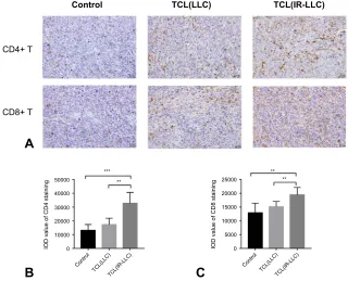
To examine whether TCL(IR-LLC) vaccine could increase TILs, immunohistochemistry was performed
to evaluate the infiltrations of CD4+ and CD8+
T-cells in tumor tissues from mice of each group. As
demonstrated in Figure 5A–C, more tumor infiltrations
of CD4+ T-cells and CD8+ T-cells were observed in mice immunized with TCL(IR-LLC) than the other two groups. In summary, these results indicated that TCL(IR-LLC) vaccine could activate more intensive
tumor-specific T-cell responses.
Tumor antigens including FN1, MFGE8,
MMP2, and MYL9 are positively
associated with immune in
fi
ltrates
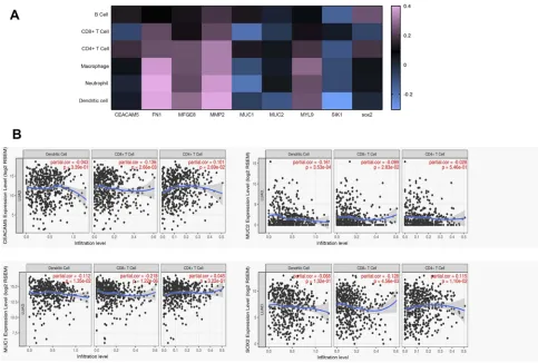
To further elucidate how IR modulates the immunogenicity of tumor cells and which antigens may contribute to induce effective immune response, we examined the tumor antigens overexpressed in irradiated A549 cells to explore their asso-ciation with immune infiltrates. The correlations between the expressions of these genes and the abundances of immune infiltrates (CD4+ T-cells, CD8+ T cells, and dendritic cells) in lung adenocarcinoma were examined through TIMER
analysis.18 Figure 6 shows positive association of FN1,
MFGE8, MMP2, and MYL9 with infiltration of CD4+
106 Q1 105 04 03 02 101
A
B
C
G
F
E
H
D
100 CD80 CD 11 CD 4 FoxP3+ CD8 CD25+ SCC PD-1 1.0M 800K 00K 400K 200K 0 1.0M 800K 600K 400K 200K 0 1.0M 800K 600K 400K 200K 0 106 105 104 103 102 101 100 106 105 104 103 102 101 100 106 105 104 103 102 101 100 106 105 104 103 102 101 100 106 105 104 103 102 101 100 106 105 104 103 102 101 100 100101 102
103
104
105
106 100
101 102 103 104 105 106
100101
102 103 104 105 106 100 101 102 103 104 105 106
100101
102 103 104 105 106 100
101 102
103
104
105
106
100101 102
103
104
105106
100
101 102 103
104
105106
100 101 102 103 104 105 106 100
101 102
103
104
105106
100
101
102 103
104
105
106
100101
102 103 104 105 106 106 105 04 03 02 101 100 106 105 04 03 02 101 100 5.56 Q2 6.76 Q4 78.1 Q3 9.59 Q1 6.36 Q2 15.5 Q4 71.3 Q3 6.85 Q1 31.0 Q2 9.02 Q4 46.9 Q3 13.1 Q1
20.5 4.08Q2
Q4 67.2 Q3 8.21 Q1 20.8 Q2 2.22 Q4 71.0 Q3 6.02 Q1
7.78 12.3Q2
Q4 72.7
Q3 7.21
Q1
5.07 5.22Q2
Q4 83.5 Q3 6.22 Q1 8.43 Q2 9.84 Q4 70.6 Q3 11.1 Q1 8.75 Q2 20 control TCL(LLC) TCL(IR-LLC) CD1
1c+ CD80+ DCs%
CD4+T lymphocyted(%) 15 10 5 0 T
reg as %(of CD4+T
cell) PD-1+ CD8+ T cells % (of CD8+ T cells) 20 *** *** 15 10 5 0 40 * * 30 20 10 0 30 * *** 20 10 0 13.5 Q4 64.7 Q3 13.0
Figure 3The vaccine-activated immune response of dendritic cells and T-cells in the spleen. The numbers of immune cells in spleens were measured byflow cytometry. (A–D) Representative images offlow cytometry. (E–H) The statistical results of proportions of dendritic cells and T-cell subsets. (AandE) The fraction of CD11c+CD80+ dendritic cells in spleens. (BandF) The fraction of CD4+ T-cells in spleens. (CandG) The ratio of Treg cells in CD4+ T-cells. (DandH) The ratio of PD-1+ T-cells in CD8+ T-cells. The data shown are the representative of three experiments (*P<0.05; **P<0.01; ***P<0.001; ****P<0.0001).
Abbreviations:TCL, tumor cell lysates; IR, irradiation; LLC, Lewis lung cancer.
OncoTargets and Therapy downloaded from https://www.dovepress.com/ by 118.70.13.36 on 25-Aug-2020
T-cells, CD8+ T cells, and dendritic cells. In contrast, the other genes including CEACAM5, MUC1, MUC2, and SOX2 showed no significant correlation or negative correla-tion with immune infiltration (Figure S1). Furthermore, the abundances of other immune infiltrates (B cells, neutrphils, macrophages) presented in Figure S1, indicated a trend roughly consistent with previously mentioned results.
These results suggested that IR increased the immunogeni-city of tumor cells possibly through the upregulation of FN1, MFGE8, MMP2, and MYL9.
Discussion
Immunotherapy plays a critical role in treatment of lung
cancer. In this field, cancer vaccine, triggering the
100
*
* *
*
** *
200
150
100
50
0
150
100
50
0 80
60
40
20
A
B
C
IL-4 pg/mL
IFN-γ
pg/mL
IL-10 pg/mL
0
control TCL(LLC)
TCL(IR-LLC)
control TCL(LLC)
TCL(IR-LLC)
control TCL(LLC)
TCL(IR-LLC)
Figure 4The vaccine changed the secretion of cytokines in serum. The levels of cytokines in serum were examined through ELISA. (A–C) The levels of IL-4, IFN-γ,and IL-10 in serum. The data shown are the representative of three experiments (*P<0.05; **P<0.01).
Abbreviations:TCL, tumor cell lysates; IR, irradiation; LLC, Lewis lung cancer.
Control
25000
20000
15000
10000
5000
0 ***
**
** **
IOD value of CD4 staining IOD value of CD8 staining
50000
40000
30000
20000
10000
0
CD4+ T
20 μm 20 μm 20 μm
20 μm 20 μm
20 μm
CD8+ T
A
B
C
TCL(LLC) TCL(IR-LLC)
Control
TCL(LLC)
TCL(IR-LLC)
Control
TCL(LLC)
TCL(IR-LLC)
Figure 5The vaccine increased the immune infiltration of T-cells in tumor tissue. (A) Representative images of immunochemistry staining for CD4+and CD8+ T-cells of tumor tissues in three groups. (B) The statistical results of the IOD value of CD4 staining in three groups. (C) The statistical results of the IOD value of CD8 staining in three groups. The data shown are the representative of three experiments (**P<0.01; ***P<0.001).
Abbreviations:IOD, integrated option density; TCL, tumor cell lysates; IR, irradiation; LLC, Lewis lung cancer.
OncoTargets and Therapy downloaded from https://www.dovepress.com/ by 118.70.13.36 on 25-Aug-2020
immune system to prevent tumor progression, has been researched for decades; however, the outcomes are
always discouraging.6One of the causes is the difficulty
in identifying the targeting tumor antigen peptide due to
tumor heterogeneity.13,24,25Previous studies also showed
the unsatisfactory effect of single protein or
peptide-based vaccines.6,26,27 In this regard, whole tumor cell
vaccine seemed a better choice because it provides a
wide spectrum of TAAs, the efficacy of which however,
is also limited.8 It may be attributed to the insufficient
immunogenicity of tumor cells.7,26 This study aimed to
find a strategy for improving the immunogenicity of
whole tumor cell vaccine. In recent years, the immune activation effect of IR has aroused much concern,
espe-cially the in situ vaccine effect of SABR in NSCLC.10
IR-induced cell death was reported to result in the exposure of numerous TAAs from damaged tumor cells and cellular debris. In addition, IR was also pro-ven, by recent studies, to alter the expressions of ICAM1, MUC1, and CEA in colon tumor cells lines,
resulting in enhanced attack by cytotoxic
T lymphocytes.28,29 Consequently, we hypothesized
that IR could be considered as a potential strategy to improve the immunogenicity of tumor cell vaccine. As
demonstrated in previous studies, radiation-related
immune response may be dose-dependent and only radiation beyond a certain threshold dose can initiate robust immune response. Hence, the dose of X-rays
used on tumor cells in this study was 8 Gy, the same
as the “ablative” dose in SABR therapy of NSCLC.9,10
CSGs provide a high possibility of encoding peptides which can be targeted by circulating lymphocytes resulting
in adaptive anti-tumoral immunity.19,20 In the present
study, most of the CSGs were upregulated after IR. Moreover, the expression of a series of tumor T cell anti-gens was proven to contain T cell epitopes and were
consistently upregulated in irradiated A549 cells.19,20 It
indicated that IR might enhance the immunogenicity of lung cancer cells, presenting the potential of IR-modulated whole tumor cell vaccine for cancer vaccine development. Unfortunately, the study of IR-treated human A549 vaccine could not be successfully conducted because constructing a xenograft A549 mouse model would require nude mice. However, the deficiency of T cell immunity in nude mice limits the study of vaccines. Thus, for in vivo experiments, we used LLC cells instead to construct a xenograft LLC mouse model
and LLC vaccine. Therapeutic efficacy of TCL(IR-LLC)
vac-cine was validated. Compared to the traditional TCL(LLC) vaccine, IR-treated LLC vaccine TCL(IR-LLC) showed enhanced inhibitory effect of tumor growth by increasing tumor cell apoptosis. As known, tumor-specific T-cell response is crucial in anti-tumor immunity.30 The activation of T-cell response is evoked by both innate immune cells and adaptive immune cells. Dendritic cells are known to be the chief antigen-presenting cells which load and cross-present tumor antigens onto MHC-I and later activate CD4+ T helper and CD8+ Figure 6Four tumor antigens were positively associated with immune infiltrates. The correlations between gene expression (FN1, MFGE8, MMP2, MYL9) and immune infiltration level (CD4+ T cells, CD8+ T cell, and dendritic cells) in lung adenocarcinoma were examined by tumor immune estimation resource analysis. (A–D) The scatterplots showing the purity-corrected partial Spearman’s correlation and statistical significance.
OncoTargets and Therapy downloaded from https://www.dovepress.com/ by 118.70.13.36 on 25-Aug-2020
cytotoxic T-cells.31,32The results of the present study demon-strated that TCL(IR-LLC) vaccine contributed to a significant increase in matured dendritic cells in mouse spleens, which may be followed by a higher antigen presenting ability. Different subpopulations of T-cells have various functions of
pro- and anti-tumor immunity.33 CD4+ T helper cells,
espe-cially Th1 cells, are essential for activation of CD8+ cytotoxic
T-cells and the priming of tumor-specific cytotoxic
T lymphocyte responses, whereas Treg cells can suppress effector T-cell-mediated anti-tumor immunity.34In this study, the TCL(IR-LLC) group obtained an increase in the proportion of total CD4+ T-cells but a decrease in the ratio of Treg cells in CD4+ T-cells. On the other hand, PD-1 is a checkpoint mole-cule on T-cells that inhibit adaptive immune responses. This study showed that TCL(IR-LLC) vaccine led to an insignificant increase in the proliferation of CD8+ T-cells and a significant decrease in the ratio of PD-1+ cells in total CD8+ cells. These results indicated that the anti-tumor immunity may be activated through the downregulation of these two types of immune
suppressor cells. Th1 cytokine IFN-γand Th2 cytokine IL-4
play an important role in anti-tumor immunity through their cytostatic and T cell-related immune-provoking effects.21,22In contrast, IL-10 is an important anti-immunity cytokine, as well as a Treg-associated cytokine.23The results of cytokines’ secre-tion in serum also showed the pro-immunity effect of the
vaccine. Moreover, the immune infiltrates of CD4+ T cells
and CD8+ T-cells in tumor tissue displayed consistent results. As the immunogenic modulation effect of IR on tumor cells had been determined through the study of A549 tumor antigen expression and the in vivo study of irradiated LLC vaccine, tumor antigens that correlated with the immune sti-mulation effect were further explored. Through TIMER ana-lysis, FN1, MFGE8, MMP2, and MYL9 containing T-cell peptide were found to be positively correlated with immune
infiltrates in lung cancer.18,20 As these genes were also
expressed in the tumor microenvironment, further study was required for validation, although partial Spearman’s correlation was tumor purity-corrected. Further experiments are also needed to study the potential mechanism. Surprisingly, pre-vious studies showed that FN1, MFGE8, MMP2, and MYL9 all promoted the malignant behavior of cancer cells and con-tributed to metastasis in different cancers.35–39Consequently, these four genes may be potential cancer immunotherapy targets for the prevention of progression, relapse, and metas-tasis. It will be more convincing if vaccines based on human irradiated autologous tumor cells are studied through human trials, and the immune activation effect of these genes is measured in vivo. This is included in our future research.
Conclusion
In summary, this study demonstrated that IR was able to
increase immunogenicity of lung cancer cells.
Furthermore, irradiated lung cancer cell vaccine showed
enhanced anti-tumor efficacy by eliciting intensive T-cell
response. It is possible for FN1, MFGE8, MMP2, and MYL9 to contribute to the enhanced T-cell response.
Further studies are required to confirm the relationship
between immune response and these upregulated genes. It may provide a new insight into immunotherapy in the
field of lung cancer.
Acknowledgment
This work was supported by the Key Subject Construction
Program of Jinshan District Health Administrative
Authority (JSZK2015A04).
Disclosure
The authors report no conflicts of interest in this work.
References
1. Siegel RL, Miller KD, Jemal A. Cancer statistics, 2019.CA Cancer J Clin. 2019;69(1):7–34. doi:10.3322/caac.21551
2. Shah R, Sabanathan S, Richardson J, Mearns AJ, Goulden C. Results of surgical treatment of stage I and II lung cancer.J Cardiovasc Surg (Torino). 1996;37(2):169–172.
3. Goldstraw P, Chansky K, Crowley J, et al. The IASLC lung cancer staging project: proposals for revision of the TNM stage groupings in the forthcoming (eighth) edition of the TNM classification for Lung cancer.
J Thorac Oncol. 2016;11(1):39–51. doi:10.1016/j.jtho.2015.09.009 4. Walters S, Maringe C, Coleman MP, et al. Lung cancer survival and
stage at diagnosis in Australia, Canada, Denmark, Norway, Sweden and the UK: a population-based study, 2004–2007.Thorax. 2013;68 (6):551–564. doi:10.1136/thoraxjnl-2012-202297
5. Seliger B. “Tumor Immunology Meets Oncology (TIMO) IX”, May 2-4, 2013, Halle/Saale, Germany. Cancer Immunol Immunother. 2014;63(3):305–311. doi:10.1007/s00262-014-1522-2 6. Oliveres H, Caglevic C, Passiglia F, Taverna S, Smits E, Rolfo C.
Vaccine and immune cell therapy in non-small cell lung cancer.
J Thorac Dis. 2018;10(Suppl 13):S1602–s1614. doi:10.21037/ jtd.2018.05.134
7. Freeman-Keller M, Goldman J, Gray J. Vaccine immunotherapy in lung cancer: clinical experience and future directions. Pharmacol Ther. 2015;153:1–9. doi:10.1016/j.pharmthera.2015.05.004 8. Le DT, Pardoll DM, Jaffee EM. Cellular vaccine approaches.Cancer
J. 2010;16(4):304–310. doi:10.1097/PPO.0b013e3181eb33d7 9. Hodge JW, Sharp HJ, Gameiro SR. Abscopal regression of antigen
disparate tumors by antigen cascade after systemic tumor vaccination in combination with local tumor radiation. Cancer Biother Radiopharm. 2012;27(1):12–22. doi:10.1089/cbr.2012.1202 10. Bernstein MB, Krishnan S, Hodge JW, Chang JY. Immunotherapy
and stereotactic ablative radiotherapy (ISABR): a curative approach?Nat Rev Clin Oncol. 2016;13(8):516–524. doi:10.1038/ nrclinonc.2016.30
11. Vanpouille-Box C, Alard A, Aryankalayil MJ, et al. DNA exonu-clease Trex1 regulates radiotherapy-induced tumour immunogenicity.
Nat Commun. 2017;8:15618. doi:10.1038/ncomms15618
OncoTargets and Therapy downloaded from https://www.dovepress.com/ by 118.70.13.36 on 25-Aug-2020
12. NajafiM, Motevaseli E, Shirazi A, et al. Mechanisms of infl amma-tory responses to radiation and normal tissues toxicity: clinical implications. Int J Radiat Biol. 2018;94(4):335–356. doi:10.1080/ 09553002.2018.1440092
13. NajafiM, Farhood B, Mortezaee K. Contribution of regulatory T cells to cancer: A review.J Cell Physiol. 2019 Jun;234(6):7983-7993. doi: 10.1002/jcp.27553.
14. Curry WT Jr., Gorrepati R, Piesche M, et al. Vaccination with irradiated autologous tumor cells mixed with irradiated GM-K562 cells stimulates antitumor immunity and T lymphocyte activation in patients with recurrent malignant glioma.Clin Cancer Res. 2016;22 (12):2885–2896. doi:10.1158/1078-0432.CCR-15-2163
15. Dillman RO, Beutel LD, Barth NM, et al. Irradiated cells from autologous tumor cell lines as patient-specific vaccine therapy in 125 patients with metastatic cancer: induction of delayed-type hypersensitivity to autolo-gous tumor is associated with improved survival. Cancer Biother Radiopharm. 2002;17(1):51–66. doi:10.1089/10849780252824073 16. Simons JW, Mikhak B, Chang JF, et al. Induction of immunity to
prostate cancer antigens: results of a clinical trial of vaccination with irradiated autologous prostate tumor cells engineered to secrete granulocyte-macrophage colony-stimulating factor using ex vivo gene transfer.Cancer Res. 1999;59(20):5160–5168.
17. Wang L, Feng Z, Wang X, Wang X, Zhang X. DEGseq: an R package for identifying differentially expressed genes from RNA-seq data.
Bioinformatics. 2010;26(1):136–138. doi:10.1093/bioinformatics/btp612 18. Li B, Severson E, Pignon JC, et al. Comprehensive analyses of tumor immunity: implications for cancer immunotherapy. Genome Biol. 2016;17(1):174. doi:10.1186/s13059-016-1028-7
19. Baldauf MC, Gerke JS, Kirschner A, et al. Tumor mutational burden is a determinant of immune-mediated survival in breast cancer.
Oncoimmunology. 2018;7(9):e1481558. doi:10.1080/2162402X.2018. 1490854
20. Olsen LR, Tongchusak S, Lin H, Reinherz EL, Brusic V, Zhang GL. TANTIGEN: a comprehensive database of tumor T cell antigens.
Cancer Immunol Immunother. 2017;66(6):731–735. doi:10.1007/ s00262-017-1978-y
21. Kursunel MA, Esendagli G. The untold story of IFN-γ in cancer biology.Cytokine Growth Factor Rev. 2016;31:73–81. doi:10.1016/ j.cytogfr.2016.07.005
22. Setrerrahmane S, Xu H. Tumor-related interleukins: old validated targets for new anti-cancer drug development.Mol Cancer. 2017;16 (1):153. doi:10.1186/s12943-017-0721-9
23. Geginat J, Larghi P, Paroni M, et al. The light and the dark sides of Interleukin-10 in immune-mediated diseases and cancer. Cytokine Growth Factor Rev. 2016;30:87–93. doi:10.1016/j.cytogfr.2016.02.003 24. Farhood B, NajafiM, Mortezaee K. CD8+ cytotoxic T lymphocytes
in cancer immunotherapy: A review.J Cell Physiol. 2019 Jun;234 (6):8509-8521. doi: 10.1002/jcp.27782.
25. NajafiM, Goradel NH, Farhood B, et al. Tumor microenvironment: interactions and therapy. J Cell Physiol. 2019;234(5):5700–5721. doi:10.1002/jcp.27425
26. Melero I, Gaudernack G, Gerritsen W, et al. Therapeutic vaccines for cancer: an overview of clinical trials.Nat Rev Clin Oncol. 2014;11 (9):509–524. doi:10.1038/nrclinonc.2014.111
27. Rosenberg SA, Yang JC, Restifo NP. Cancer immunotherapy: moving beyond current vaccines.Nat Med. 2004;10(9):909–915. doi:10.1038/ nm1100
28. Garnett CT, Palena C, Chakraborty M, Tsang KY, Schlom J, Hodge JW. Sublethal irradiation of human tumor cells modulates phenotype resulting in enhanced killing by cytotoxic T lymphocytes. Cancer Res. 2004;64(21):7985–7994. doi:10.1158/ 0008-5472.CAN-04-1525
29. Kwilas AR, Donahue RN, Bernstein MB, Hodge JW. In thefield: exploiting the untapped potential of immunogenic modulation by radiation in combination with immunotherapy for the treatment of cancer.Front Oncol. 2012;2:104. doi:10.3389/fonc.2012.00104 30. Wellenstein MD, de Visser KE. Cancer-cell-intrinsic mechanisms
shaping the tumor immune landscape. Immunity. 2018;48 (3):399–416. doi:10.1016/j.immuni.2018.03.004
31. Schiavoni G, Mattei F, Gabriele L. Type I interferons as stimulators of DC-mediated cross-priming: impact on anti-tumor response.Front Immunol. 2013;4:483. doi:10.3389/fimmu.2013.00483
32. Joffre OP, Segura E, Savina A, Amigorena S. Cross-presentation by dendritic cells. Nat Rev Immunol. 2012;12(8):557–569. doi:10.1038/ nri3254
33. Mei Y, Zhao L, Liu Y, et al. Combining DNA vaccine and AIDA-1 in attenuated salmonella activates tumor-specific CD4+and CD8+T-cell responses.Cancer Immunol Res. 2017;5(6):503–514. doi:10.1158/2326-6066.CIR-16-0240-T
34. Chraa D, Naim A, Olive D, Badou A.J Leukoc Biol. 2018. 35. Wu J, Wang Y, Xu X, et al. Transcriptional activation of FN1 and
IL11 by HMGA2 promotes the malignant behavior of colorectal cancer. Carcinogenesis. 2016;37(5):511–521. doi:10.1093/carcin/ bgw029
36. Sugano G, Bernard-Pierrot I, Lae M, et al. Milk fat globule--epidermal growth factor--factor VIII (MFGE8)/lactadherin promotes bladder tumor development. Oncogene. 2011;30(6):642–653. doi:10.1038/onc.2010.446
37. Tibaldi L, Leyman S, Nicolas A, et al. New blocking antibodies impede adhesion, migration and survival of ovarian cancer cells, highlighting MFGE8 as a potential therapeutic target of human ovar-ian carcinoma. PLoS One. 2013;8(8):e72708. doi:10.1371/journal. pone.0072708
38. Kawamata H, Nakashiro K, Uchida D, Harada K, Yoshida H, Sato M. Possible contribution of active MMP2 to lymph-node metastasis and secreted cathepsin L to bone invasion of newly established human oral-squamous-cancer cell lines. Int J Cancer. 1997;70(1):120–127.
39. Tan X, Chen M. MYLK and MYL9 expression in non-small cell lung cancer identified by bioinformatics analysis of public expression data.
Tumour Biol. 2014;35(12):12189–12200. doi:10.1007/s13277-014-2527-3
OncoTargets and Therapy downloaded from https://www.dovepress.com/ by 118.70.13.36 on 25-Aug-2020
Supplementary material
OncoTargets and Therapy
Dove
press
Publish your work in this journal
OncoTargets and Therapy is an international, peer-reviewed, open access journal focusing on the pathological basis of all cancers, potential targets for therapy and treatment protocols employed to improve the management of cancer patients. The journal also focuses on the impact of management programs and new therapeutic
agents and protocols on patient perspectives such as quality of life, adherence and satisfaction. The manuscript management system is completely online and includes a very quick and fair peer-review system, which is all easy to use. Visit http://www.dovepress.com/ testimonials.php to read real quotes from published authors.
Submit your manuscript here:https://www.dovepress.com/oncotargets-and-therapy-journal
Figure S1The correlations between gene expression and immune infiltration level were examined by tumor immune estimation resource (TIMER) analysis. (A) Heat map plotted by the correlations between gene expression and immune infiltrate level in lung adenocarcinoma through TIMER analysis. Colored bars represent differential levels of Spearman’s correlation. (B) The correlations between gene expression (CEACAM5, MUC1, MUC2, SOX2) and immune infiltration level (CD4+ T cells, CD8+ T cells, and dendritic cells) in lung adenocarcinoma.
OncoTargets and Therapy downloaded from https://www.dovepress.com/ by 118.70.13.36 on 25-Aug-2020