A NEW MAPPING METHOD EMPLOYING A MEIOTIC REC-
MUTANT OF YEAST1
SUE KLAPHOLZz AND ROCHELLE EASTON ESPOSITOZ, 3
Department of Biologyz, and Committee on Genetics: University of Chicago, Chicago, Illinois 60637
Manuscript received November 11, 1981 Revised copy accepted December 5, 1981
ABSTRACT
A rapid new mapping method has been developed for localizing a domi- nant or recessive mutation to a particular chromosome of yeast. The proce- dure utilizes the ability of strains homozygous for the spoll-1 mutation to undergo chromosome segregation without appreciable recombination during sporulation. The level of sporulation in s p o l l - l / s p o l l - I diploids is reduced and asci are often immature or abnormal in appearance; spore viability is less than 1%. The f i s t step of the mapping procedure is the construction of a haploid spoil-I strain carrying a recessive drug-resistance marker and the unmapped mutation(s). This strain is crossed to a set of three spoll-I mapping tester strains containing, among them, a recessive marker on each chromosome. The resulting spoil-I / s p o l l - l diploids are sporulated and plated on drug-containing medium. Viable meiotic products that express the drug-resistance marker due to chromosome haploidization are selectively recovered. These meiotic products are haploid f o r most, but generally not all, chromosomes. The level of disomy f o r individual chromosomes averages 19%. Each of the recessive chromosomal markers is expressed in approxi- mately a third of the drug-resistant segregants. Ninety-eight percent of these segregants show no evidence of intergenic recombination. Thus, two markers located on the same chromosome, but on different homologs, are virtually never expressed in the same drug-resistant clone. The utility of this mapping procedure. is demonstrated by confirming the chromosomal location of seven known markers, as well as by the assignment of a pre- viously unmapped mutation, spol2-1, to chromosome VIIZ. In addition, the analysis of the products of spoil-1 meiosis indicates that several markers previously assigned to either chromosome XZV or chromosome X V I Z are actually on the same chromosome.
HE most recent genetic map of Saccharomyces cereuisiae is composed of 17 linkage groups containing over 300 mapped genes (MORTIMER and SCHILD
1980,1981). There remain numerous sparsely-marked regions and three groups of linked markers, termed fragments, which have not been assigned to a par- ticular chromosome. Because of the large number of chromosomes in yeast, a rapid method for determining the chromosome on which an unmapped muta-
I T h i s work was supported by National Institute of Health grants GM 23277, GM 29182 and C.4 19265 (project 508) ( U S Public Health Service), and postdoctoral training grant CA 09273 ( U S Public Health Service) awarded to SUE KUPHOLZ
388 S. KLAPHOLZ A N D R. E. ESPOSITO
tion resides is highly desirable. Current procedures for locating a gene to a chromosome include, i n addition to standard tetrad analysis, meiotic trisomic analysis (MORTIMER and
HAWTHORNE
1973; WICKNER 1979), chromosome transfer (DUTCHER 1981), mitotic recombination (NAKAI and MORTIMER 1969) and mitotic chromosome loss or nondisjunction (KAWASAKI 1979; MORTIMER, CONTOPOULOU and SCHILD 1981). Once a gene has been mapped to a particular chromosome, meiotic tetrad analysis, random spore analysis or mitotic mapping is used to localize the gene relative to other markers and the centromere (see MORTIMER and SCHILD 1981 for review). In this paper, we describe a new and relatively rapid method for localizing an unmapped dominant or recessive mutation to a specific chromosome. This method utilizes the haploidization- without-recombination sporulation phenotype of strains homozygous for the spoll-l mutation.The recessive spoll-1 mutation, isolated by ESPOSITO and ESPOSITO (1969), causes a reduction in both sporulation and spore viability. Diploids homozygous for spoll-1 undergo premeiotic DNA synthesis (C. GIROUX, personal communi- cation; S. KLAPHOLZ, C. WADDELL and R. E. ESPOSITO, unpublished) and complete both meiotic divisions (ESPOSITO and ESPOSITO 1973, 1974; MOENS et al. 1977). The asci formed are often immature or abnormal in appearance, and the spores largely inviable.
Several lines of evidence have indicated that spoll-1 is a meiotic Rec- muta- tion. First, when genetically marked sporulating cells are transferred to the ap- propriate selective medium, no increase in the recovery of intragenic recombi- nants is observed above the mitotic level (S. KLAPHOLZ, C. WADDELL and R. E. ESPOSITO, unpublished) , Second, rare viable meiotic products can be select-
ly recovered by plating a sporulated spoll-l/spoll-1 diploid, heterozygous for a recessive drug-resistance marker, on drug-containing medium (S. KLAPHOLZ, unpublished; this study). The resulting drug-resistant colonies are haploid for most, but usually not all, chromosomes. Among these colonies, intergenic recombinants are infrequent. Thus, markers on the same chromosome show almost 100% linkage, regardless of their distance apart. Third, the meiotic Rec- phenotype conferred by spoll-1 has been confirmed by analysis of spoll-l/ spoll-1 spol3-l/spol3-l double mutant diploids (KLAPHOLZ 1980). The pres- ence of the spol3-1 mutation alone permits diploid cells to enter meiosis and execute recombination at meiotic levels. The cells then undergo a single, gener- ally equational division, resulting i n the formation of two diploid spores (KLAP-
HOLZ and ESPOSITO 1980a, b)
.
In contrast to single mutant spoll-l/spoll-1 strains, double mutant spoll-1 /spoil-1 spol3-l/spol3-1 diploids produce highly viable diploid spores. Intragenic and intergenic recombination is absent or greatly reduced in these viable meiotic products (KLAPHOLZ 1980; S. KLAPHOLZ, C. WADDELL and R. E. ESPOSITO, unpublished).R E C O M B I N A T I O N L E S S MEIOSIS 389 MATERIALS A N D M E T H O D S
Strains: The genotypes of eight s p o l l - 1 strains specifically constructed for the purpose of mapping are presented in Table 1A. The remaining strains of Saccharomyces cereuisiae em- ployed in this study are listed in Table 1B and below. The following strains were used for complementation and/or mating type testing (only the relevant genotypes are given) : 74-1A ( s p o l ) ; 1230 and 1232 (met2, p et8 ); K65-3D (ade.2, metl3, t y r l ) ; JG49-20A ( h i d ) ; K222-2B, K338-3A and K338-8A ( s p o l 2 ) ; M3 ( a d d , h i s l ) ; P65, S1336B, W85-8D and W87-4B ( m e t 4 ) ; S90-2A ( M A T a , m et 3) ; S9O-2D ( M A T a , m et3 ) ; Tester 1 (MATa, a d e l ) ; Tester 2 ( M A T a ,
a d e l ) ; Tester X and Tester Y (adel, his7, m e t 3 ) ; W60-10B ( m e t 2 ) ; W224-IC ( M A T a , his6,
TABLE 1 A . spoll mapping strains
Strain Genotype
K382-23A K382-19D
K3984D K381-9D
K399-7D K393-35C
K396-llA K396-22B
111 VIIl
v
v
VI1xv
I 1v
I 1MATa spoil ura3 canl cyh2 ade2 his7 hom3
MATa spoil ura3 canl cyh2 ade.2 his7 hom3 t y r l
111 VI11
v
VI1 VI11 XVI XI1 XI I 1xv
IVMATa spoil ura3 ade6 arg4 aro7 asp5 met14 lys2 pet17 t r p l
MATa spoil ura3 ade6 arg4 aro7 asp5 met14 lys2 pet17 t r p l
111 VI11
v
VI VI1 I X xvllxIvMATa s p o ll ura3 his2 leu1 lys l met4 pet8
M A T a spoil urn3 his2 leu1 lys l met4 pet8
111 VIIl
v
Iv
I l l XI11x
VI1MATa spoil ura3 adel his1 leu2 lys7 met3 trp5
MATa s p o l l ura3 adel his1 leu2 lys7 met3 trp5
B . Haploid strains
Strain
K231-1A
K321-2A K289-3A
K338-3A K338-8D K365-18RS K321-3C K366-7D K366-12A K366-12D K368-23RS 8392-6C K415-6A
Genotype
MATa ade2 ade5 canl his7 lys2 spoil s p o l 2 irp5
MATa adel his6 leu1 leu2 met4
M A T a d e 2 cyh2 his7 leu1 lys2 met13 s p o l l trp5 t y r l ura3
M A T a his6 met13 s p o l 2 trpl
M A T a his6 lysl met4 trpl
M A T a adel d e 2 ade6 his6 his7 leu1 lysl met4 met14 pet8 s p o l l t y r l ura3
MAT#a ade2 cyh2 his7 leu1 lys2 met13 spoil s p o l 2 trp5 t y r l ura3
M A T a ade6 arg4 aro7 asp5 met14 pet17 s p o l l ura3
M A T a ade2 am7 asp5 cyh2 his6 leu1 lys2 met13 met14 pet17 s p o i l trp5
M A T a ade2 ade6 aro7 asp5 his6 lys2 met14 pet17 spoil
MATa ade2 ade6 cdcl4 his2 his7 hom3 leu1 lysl lys2 met14 s p o l l trp5 t y r l ura3
MATa ode2 canl cyh2 his7 hom3 met13 spol spoil tyr l ura3
MATa ade2 his6 his7 leu1 pet3 ural
Gene symbols are as follows: MATa and MATa, mating type; ade, adenine; aro, aromatic amino acids; asp, aspartate; arg, arginine; can, canavanine; cdc, cell-division-cycle; c y h , cyclo- heximide; his, histidine; hom, homoserine; leu, leucine; lys, lysine; m et , methionine; p e l ,
390 S . KLAPHOLZ A N D R. E. ESPOSITO
metl4, p e t 8 ) ; W224-ID ( M A T a , hisb, metl4, pet8) and X3455-1D ( p e s 8 ) . W e obtained JG49- 20A from J. GOLIN; Testers 1 and 2 from D. PLOTKIN; Testers X and Y, and M3 from R. MALONE; W60-10B, W85-8D, W87-4B, W244-1C and W2441D from R. ROTHSTEIN; and strains 1230 and 1232 from R. WICKNER. P65, S1336B and X3455-ID were obtained from the Ber- keley Yeast Genetic Stock Center. All other strains are from our laboratory collection.
Media: The following recipes are for one liter of medium. Tetracycline (Squibb) is added at 20 pg/ml to complex and sporulation media to prevent bacterial contamination. Solid medium contains 15 gm Bacto-agar. YPDA contains 20 g m Bacto-dextrose, 20 gm Bacto- peptone, 10 gm Bacto-yeast extract and 1 ml 0.5% adenine sulfate. YPG contains 30 ml glycerol, 20 gm Bacto-peptone and 10 g m Bacto-yeast extract. Minimal contains 20 gm Bacto-dextrose, 1.7 gm yeast nitrogen base and 5 gm ammonium sulfate. Minimal plus uracil contains Minimal supplemented with 20 mg uracil. Complete contains Minimal supplemented with each of the following: 10 m g adenine sulfate; 25 mg L-arginine-HC1; 75 m g L-aspartic acid; 20 m g L-histidine-HC1; 100 mg L-leucine; 50 mg L-lysine-HC1; 20 mg L-methionine; 50 mg L-phenylalanine; 300 mg L-threonine; 50 mg L-tryptophan; 50 mg L-tyrosine and 20 mg uracil. Omission contains Complete lacking one or more amino acids or nitrogen bases. Methionine omission lacks both methionine and threonine and contains 80 mg homoserine. Cycloheximide (CYH) contains Complete with 1 ml of 0.1% cycloheximide added after auto- claving. Canavanine (CAN) contains Arginine omission with 6 ml of 1% canavanine sulfate added after autoclaving. Sporulation I1 (SPII-22) contains 20 gm potassium acetate, supple- mented with 75 ,,.g/ml of each of the following: adenine sulfate; L-arginine-HC1; L-aspartic acid; L-histidine-HC1; L-leucine; L-lysine-HCI; L-methionine; L-phenylalanine; L-threonine; L-tryptophan; L-tyrosine; and uracil; and 10 pg/ml PABA, p H 7.0. Sporulation I11 (SPIII- 22) contains 20 gm potassium acetate, 1 gm Bacto-dextrose and 2.5 gm Bacto-yeast extract, supplemented as per SPII-22.
Genetic Techniques: Standard procedures for the genetic manipulation of yeast were em- ployed (MORTIMER and HAWTHORNE 1969; SHERMAN and LAWRENCE 1974).
Mapping procedure: The general scheme for localizing an unmapped mutation to a partic- ular chromosome is as follows (See RESULTS): A strain carrying an unmapped mutation
( m u t x ) is mated to a haploid of opposite mating type containing spoll-1, can1 and q h 2 .
The diploid hybrid is sporulated, and 10-20 tetrads dissected to recover a M A T a or M A T a
s p o l l - l mutx can1 cyh2 haploid. The segregation of spoll-I in the tetrads is followed by complementation tests, described below. The haploid is then mated to each of three s p o l l - l
tester strains, containing among them one or more markers on each chromosome (Table 1A). Each test hybrid is incubated on sporulation (SPIII-22) plates for 5 days or in liquid sporu- lation (SPII-22) medium for 48 hr. Sporulated cells are streaked or plated on CAN and/or CYH media and incubated f o r about 5 days. Drug-resistant colonies of various sizes (50-100 per cross per drug selection) are picked to a YPDA master plate, incubated for 2-3 days, and replicated to the appropriate selective media to assay marker segregation.
RECOMBINATIONLESS MEIOSIS 39 1 strains were grown at 30"; spoll -IIS P Oll diploids were sporulated at 30", and s p o l l - l / sp oll -l diploids were sporulated at 34", on solid SPIII-22 medium.
Complementation testing for s p o l l - l : Haploid ascosporal colonies derived from crosses of the sp o ll - l can1 cyh2 strain to the strain carrying the unmapped mutation are mated to MATa or M A T a s p o l l - l testers. The three pairs of MATa and M A T a spoll-1 mapping testers (Table 1A) can be used for this, and in most cases, prototrophic selection on Minimal plus uracil medium can be used to obtain the diploids. The s p o l l - l / s p o l l - l diploids can be identified by several criteria: (1) reduced sporulation ability (typically 30% mature asci at 25", 20% at 30", and 10% a t 34", on SPIII-22); (2) abnormal, immature-appearing asci; (3) poor spore viability (less than 1 % ) ; and (4) reduced haploidization. The latter phenotype can be scored by replicating the diploids from SPIII-22 to CAN and CYH plates. The two to four segregants in each tetrad that are can1 and/or cyh2 will produce nearly confluent patches of growth if the diploid is SPOll/spoll-1. Few or no papillae will be found if they are homozygous s p ol l -l / spoll- 1.
Ascus dissection and micromanipulation: Asci were dissected as previously described (KLAPHOLZ and ESPOSITO 1980b). To determine the viability of intact s p o l l - l / s p o l l - 1 asci, sporulated cells of the diploid K382-19D x K393-35C (Table IA) were obtained from a 34" SPIII-22 plate, briefly treated (4 min) with 1% glusulase to aid in ascus recognition, and streaked to YPDA. Intact asci were then micromanipulated to individual positions on the plate and incubated at 30".
Photomicroscopy: Sporulated cultures of s po ll-lls poll- l (K303) and S P O l l / S P O l l (K65- 3D) were fixed in 70% ethanol for 10 min and washed with distilled water. They were then stained with 0.25 pg/ml DAPI (4', 6-diamidino-2-phenylindole), washed, and stored in 50% glycerol. Slides were prepared and photographed with fluorescence (not shown) and phase optics on a Leitz Orthoplan miscroscope.
RESULTS
Mapping rationale: Based on the haploidization-without-recombination spo- rulation phenotype of spol1-1/spol1-1 diploids, we devised a simple mapping procedure to establish the chromosomal location of an unmapped dominant or recessive mutation. The mapping scheme is outlined in Figure 1. A haploid strain carrying the spoll-I mutation, an unmapped mutation ( n u t s ) , and one or more recessive drug-resistance markers (e.g., can1 and c y h 2 ) , is con- structed and crossed to three spoll mapping tester strains (see Table IA). Each resulting M A T a I M A T a s p o l l - l / s p o l l - I m u t x / M U T x c a n l / C A N I cyh2/CYH2 diploid is also heterozygous for recessive markers on a subset of the total yeast chromosomes, contributed by the spoll-I tester parent. The diploids are sporulated and then transferred to canavanine-containing (CAN)
or cycloheximide-containing (CYH) media to select for rare viable meiotic products that are haploid for the chromosome carrying the drug-resistance allele. After several days of incubation, drug-resistant colonies are picked to master plates and analyzed for input marker segregation.
392 S. KLAPHOLZ A N D R. E. ESPOSITO
mutx S P O l l MUTx spoll
mutx spoll
CanR or CyhR meiotic segregants
Assuming mutx and AUXl are linked, ond mutx and AUX2 are unlinked:
I
Q
* Q
Absence of m u t x ouxl segregants indicates genetic linkage
R E C O M B I N A T I O N L E S S M E I O S I S
TABLE 2
Expected phenotypes of canavanine-resistant clones
393
~~ ~ ~
Phenotypes of canavanine-resistant clone+ Diploid genotype* Mut+Aux+ Mut-Aux+ Mut+Aux- Mut-Aux-
Mapping a recessiue mutation, mutx ( m u t x A U X l canl x M U T x auxl C A N I )
markers are on different chromosomes: mutx A U X I canl
M U T x auxl
___
_-_
C A N 1+
m u t x and A U X l are linked: m u t x A U X I canl M U T x auxl C A N l mutx and canl are linked: m u t x canl A U X I M U T x C A N I auxl --
____
Mapping a dominant mutrrtion, MUTx ( M U T x A U X l can1 x m u t x auxl CAN11
+
+
+
+
+
+
+
+
+
markers are on different chromosomes: M U T x A U X l canl
m u t x auxl C A N 1 M U T x and A U X l are linked: M U T x A U X l canl mutx auxl C A N 1
MUTx and canl are linked: M U T x canl A U X l m u t x C A N l auxl
+
+
+
_-_
___
-
+
-
--_____ --
+
-
+
-
-____-__
_-
* Diploids are M A T a / M A T a spoll-l/spoll-I.
t
The diploids are sporulated and plated on canavanine medium. M u t = mutant; Mutf =wild type; AUT!= auxotrophic; Aux+ = prototrophic. The
+
and-
symbols indicate that a particular phenotype is expected, and not expected, to occur, respectively.tion. Hence, the recovery of drug-resistant clones that express both the un- mapped mutation and a particular tester strain marker indicates that these loci reside on different chromosomes. When the unmapped mutation and the drug-resistance marker are present on the same chromosome, in coupling,
the mutation will be expressed in virtually all of the drug-resistant colonies. These predictions apply to both recessive and dominant unmapped mutations
(see Table 2).
394 S. K L A P H O L Z A N D R . E. ESPOSITO
By analogy with recombination-defective mutants of other organisms (see BAKER et al. 1976, for review), it seems likely that in spoll-l/spol2-l diploids, meiosis I involves the random segregation of chromosomes to each pole and meiosis I1 occurs normally. For any given chromosome this mode of segregation is expected to yield either four monosomic spores or two disomic and two null- osomic spores per ascus, in equal frequency (Figure 2). Because nullosomic spores are inviable, among the potentially viable spores 67% will be mono- somic and 33
%
disomic. Therefore, excluding other consideration, any given heterozygous recessive marker should be expressed in half of the monosomic spore clones or 33% of the total viable clones recovered from a Rec- meiosis.Ascus Viability in spoll-1 diploids: In a n organism like yeast with a large number of chromosomes, random segregation at meiosis I-generating spores nullosomic for one or more chromosomes-should result in low ascus viability. The s p o l l - l / s p o l l - l diploids employed in the experiments reported here form, on the average, 30% asci at 25", 20% asci at 30" and 10% asci at 34'. These asci are often immature or abnormal in morphology (Figure 3). The viability of asci produced by spoll-Z/spoll-Z diploids at 34" was determined by micro- manipulating 100 intact asci of diploid K382-19D
x
K393-35C onto YPDA plates. After 2 days of incubation at 30°, only one of the tetrads had formed a colony; the remaining asci showed no evidence of spore germination. This low level of ascus survival (1%) is close to that predicted for a recombination-MEIOSIS
I
SEGREGATION MEIOTIC PRODUCTSA.
kf-
-
k
+
B.
+
2+ monosomic : 2 -monosomic
2
+
disomic : 2 inviable nullosomicFIGURE %-Meiosis in a recombination-defective mutant. The
+
and - symbols representRECOMBINATIONLESS MEIOSIS
395
FIGURE 3.-phase mirrograph of cells of spoll-f/spoll-l ( A ) and SPOll/SPOff (B) d i p loids, sporulated at 25" (magnification
=
1 8 0 0 ~ ) .defective mutant undergoing random chromosome segregation at meiosis
I
(See DISCUSSION).
DISCUSSION
Crosses io analyze the segregation of previously mapped genes in spoil-1 diploids:
To
assess the utility of this mapping procedure, w e crossed a M A T a spo2l can2 cyh2 haploid strain, K3926C, carrying several previously mapped markers on a number of chromosomes, to each of thethree
M A T a spoil tester strains (Table 1). The map positions of the seven loci examined in this study are shown in Figure 4. One of these markers, a recessive sporulation- defective mutation, spoi, was mapped by tetrad analysis in this laboratory.z
>e* " 4.9 25.3
m
9
'O+
&'
Y n
Y
52.2 56.1 2.5
15.0 42.5 17.6 3 . 1 28.8
Q+\q ob*%
m
A"
19.6 45.3
396 S. K L A P H O U A N D R. E. ESPOSITO
The other map positions are from the compiled data of MORTIMER and SCHILD (1980). All of the markers are recessive, with the exception of MATa, which is condominant with MAT a .
Cross hybrids between K392-6C and the spoil-2 testers were incubated on sporulation plates at 34' for five days, and streaked to drug-containing media
(MATERIALS AND METHODS). Fifty to 100 canavanine-resistant ( CanR) and
cycloheximide-resistant (CyhR) colonies from each cross were picked to YPDA master plates after 3 to
5
days incubation at 30'. The drug-resistant colonies included a wide range of sizes and morphologies. The phenotypes of these clones were scored directly or by complementation testing of mating-capable clones.Among the 210 CanR and 240 CyhR clones initially examined, most were of a or a mating type. Nonmating clones comprised 36% of all CanR clones and 20% of all CyhR clones. In each cross, more than 98% of these nonmating clones expressed at least one recessive marker on a chromosome other than the one carrying the selected drug-resistance marker. This implies that diploid CanR and CyhR mitotic recombinants do not make a significant contribution to the population of nonmating clones (cf., DISCUSSION). The segregation of markers among nonmating and mating clones was not very different; for ex- ample, trpl was expressed in 40% of nonmating and 51% of mating clones, and aro7 was expressed i n 30% of nonmating and 39% of mating clones. Be- cause complementation testing was necessary to determine the presence of many of the markers in these crosses, the data presented below are derived only from mating-capable clones.
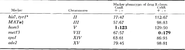
Recovery of markers on the same chromosome as the selected drug-resistance marker: As predicted (Table 2), markers in coupling with the selected drug- resistance markers were expressed in virtually all of the respective drug-re- sistant clones. The recessive hom3 allele, located on the opposite arm from
can1 on chromosome V , was expressed in 123/124 CanR clones; n e t 2 3 , linked to cyh2 on chromosome VZZ, was expressed in all of the 179 CyhR colonies
TABLE 3
Segregation of recessive mnrkers f r o m K392-6C in C a d and CyhR clones
Marker phenotype, d drug R clones
CXIR CyhR
Marker ChrDmosome
+ : -
+ : -
his7, t y r l * I I 77:47 112: 67
M A T a t I I I 5 7 ~ 6 7 98: 83
hom3 V 1 : 123 129:50
met13 V I I 67:57 0:179
s p o l X I V 63:61 86: 93
ade2
xv
79:45 98:81* his7 and t y r l cosegregated in all clones.
R E C O M B I N A T I O N L E S S M E I O S I S 397
tested (Table 3 ) . Likewise, none of the 30 CanR clones from the cross of K392-6C to tester K396-22B, and none of the CyhR clones from all three crosses, expressed the recessive tester strain marker for chromosomes V or VIZ, respectively. These results indicate that the drug-resistant clones are largely monosomic for the chromosome bearing the selected drug R locus. In contrast, only 35% of the CyhR colonies expressed hom3, and 48% of the CanR colonies expressed metl3. The other markers contributed to the crosses by K392-6C, which are not located on chromosomes V or VIZ, were recovered in similar proportions among CanR and CyhR clones (see Table 3 ) .
Recovery of recessive alleles among drug-resistant clones: Table 4 sum- marizes the segregation data for each marker contributed to the crosses by K392-6C (asterisked) or the tester strains. Excluding cases where the drug
TABLE 4
Expression of chromosomal markers in mating-capable drug-resistant clones
Phenotype of drug-resistant clones
Chronio~onie Marker
+
- Total Percent minusI
I1
I l l +
zv
VS VI VIIS VIIIS I X X XI XI1 XI11 XIVxv
XVI XVll adel his7 *t yrl *
lys2 MATa* leu2
t r p l
can1 *
hom3* his1 his2 cyh2' met13* ade6 leu1 trp5 arg4 met3 met14 asp5 lys7
spo1 *
pet8 ade2* pet17 aro7 met4 lysl 47 189 189 87 153 36 70 128 129 17 50 68 67 41 23 19 53 59 48 97 129 69 149 67 177 74 87 67 28 114 114 55 150 39 72 51 50 28 36 56 57 21 9 11 27 27 27 45 14 6 154 19 126 68 55 19 75 303 303 142 303 75 1 42 179 179 45 86 124 124 62 32 30 80 86 75 142 143 75 303 86 303 142 142 86 37 38 38 39 50 52 51 28 28 62 42 45 46 34 28 37 34 31 36 32 10 8 51 22 42 48 39 22
* Markers contributed to the cross by K392-6C.
+Chromosome 111 data do not include disomes, which are a / a nonmatsrs. Numbers in the
f
$ Chromosome VI1 data are from CanR clones only; Chromosome V and VI11 data are from column refer to (Y maters, numbers in the - column refer to a maters.
398 S . KLAPHOLZ A N D R. E. ESPOSITO
selection prevented the recovery of a particular allele, the percent of clones expressing the recessive (or, in the case of MATa, the codominant) allele of each locus ranged from 8% to 62%, with a n average value of 36%. A number of factors appear to influence how frequently a given chromosomal marker is recovered among drug-resistant clones:
1. Relative survival of disomic versus monosomic spore clones: It is known from studies of the meiotic products of triploid strains of yeast that disomy is tolerated less well for some chromosomes than for others (PARRY and Cox, 1970; CAMPBELL et al. 1981). For example, disomy for chromosome ZV, one of the longest chromosomes, is infrequently detected (PARRY and Cox 1970). W e found that trpl, marking chromosome ZV in this study, was expressed i n approximately 50% of all drug-resistant clones. If chromosome ZV disomes were completely unstable, we would expect 50% trpl monosomic and 50%
TRPl monosomic clones. A more reliable assessment of disomy for a particular chromosome can be made when both homologous chromosomes are marked (cf., item 3). Estimates of the level of disomy for a number of chromosomes are presented in a later section.
2. Relative surviual of
+
uersus - spores or spore clones: Theoretically, we expect to recover between 33% (disomes fully viable) and 50% (d’ isomes completely inviable) clones that are mutant in phenotype for a given hetero- zygous recessive marker (Figure 2 ) . However, this expectation is based on the assumption that+
and - monosomics are equally viable, and that the recovery of the alleles is not affected by the segregation of other markers (cf., items 3 and 4), Of the 27 recessive markers examined, only four were expressed in significantly fewer than a third of all drug-resistant colonies [lys7 (8%),asp5
(lo%),
pet8 (22%) and met4 (22%) (Table 4)]. At least in the case of Zys7, we have observed that spore clones carrying this mutation grow noticeably more slowly than LYS7 spore clones, even on rich YPDA medium. Thus, lys7spore clones may be at a selective disadvantage in these experiments. W e have found that choosing small colonies from CAN or CYH plates at
5
days incuba- tion enriches for these less vigorous colonies.3. The effect of another marker on the same chromosome: When two mark- ers are in coupling, one will be coincidently expressed in virtually all clones that express the other. For example, tyrl and his7 on chromosome ZZ cosegre- gated in all 303 clones tested (see also Table 3). Hence, it is apparent that if a marker displays a strong selective advantage or disadvantage, it will influence the recovery of other markers on the same homolog. A similar situation can arise for markers in repulsion, resulting in the recovery of one homolog more frequently than the other. For example, among CyhR clones from the cross K392-6C X K396-22B7 the hisl-bearing homolog of chromosome V was recov- ered twice as frequently as the one carrying can1 and hom3. Of the 45 clones tested, 62% expressed hisl, while only 31% expressed can1 and hom3 (the remaining 7
% were disomic).
RECOMBINATIONLESS MEIOSIS
TABLE 5
Phenotypes of drug-resistant clones for various tester strain chromosomal markers 399
4- :- segegation of chromosomal markers in drug R clones expressing the following alleles:* Marker on All drug R his7,
chromosome: clones tyrl MATa h o d met13 spa1 nde2
I
I1
I I I IV V VI VII VI11 IX X X I XI1 X I I I XIV
xv
XVI XVII 47:2& 87:55 36: 39 70: 72 47:38 50:36 262:41 115:27 59:27 48 : 27 97:45 127: 14698: 6 67: 19 74: 68 87: 55 67:19
20:12 21:15 27:17
47:O 43:31 45:32
13:19 36:Q 17:27
21:26 35:39 39:38 22:lO 1 9 1 7 44.d
22:13 23:17 32:20 99:15 127:27 133:41
40: 7 65:9 77:Q
22:13 27:13 35:17 18:14 23:13 27:17 25:22 56:18 53:24 40: 7 68:5 74: 3
30:2 33:3 41:3
27: 8 31:9 42: 10 23:24 36:38 37:39 26:21 42:32 47:30
27:8 31:9 42: 10
32:23 65:38 29:27 49:56 28:28 41:34 236:O 53:27 53:22 37:19 67:36 92:13 50: 6 59:16 58:47 65:# 59:16
24:14 20:12 41:24 35:24 19:18 12:20 28:37 29:30 27:ll 20:12 34:17 18:17 135:19 108:18 51:14 48:ll 33:18 23:12 28:lO 20:12 44:21 39:30
58:7 51:8
27:4 27: 5 51 :Q 27:8 35:30 59:Q
38:27 30:29
51 :O 27: 8
* Data are for mating-capable CanR and CyhR clones from crosses K392-6C with K381-9D, K396-22B and K393-35C. The three letter gene symbols refer to markers contributed by K392-6C; f and
-
refer to markers contributed by the tester strains.creased or decreased survival compared to spores bearing one or the other allele. One such example concerns the interaction between arg4 (the chromosome VZZZ marker in cross K392-6C X K381-9D) and canl. The CAN? locus codes for an arginine permease (GRENSON et al. 1966). Arginine auxotrophs with a
can2 mutation cannot grow on our synthetic media because they are unable to take up arginine. When CanR clones were selected, all 62 were Arg+, and among CyhR clones, all arg4 clones were Cans and all can1 clones were Arg+. An artifactual mapping result that can occur as a consequence of this type of interaction between genes is described below.
Mapping data for preuiously mapped markers: Among clones that expressed a particular K392-6C marker, the number that were
+
or-
in phenotype for each of the seventeen chromosomal markers contributed by one of the400 S. K L A P H O L Z A N D R . E. ESPOSITO TABLE 6
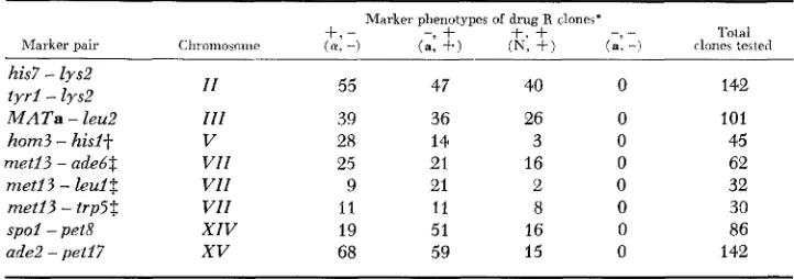
Segregation of linked markers in drug-resistant clones
Tola1
- _
Marker phenotypes of drug R clones’
+.
- - 9+
t.+
Chromosoirie ( a . -) (a, +) (E, + ) (a: -) rlones tested Marker pair
his7
-
lys2 t y r i-
lysZ M A T a-
leu2 hom3 - hisit met13 - adebt met13 - l e d $ m e t i 3 - trp5$ s p o i - pet8 ade2 - pet17I I 111 V VII VII VII XIV XV 55 39 28 25 9 I1 19 68 47 36 I4 21 21 11 51 59 40 26 3 16 2 8 16 15 142 101 45 62 32 30 86 142
* The f and - symbols refer to the phenotypes of the auxotrophic markers:
*+
= wild type and -=mutant. The symbols (Y, a and N refer to a mating type, a mating type, and non-mating, respectively. The symbols are given in the same order as the genes listed under “Marker pair.” Symbols in parentheses refer to the MATa - leu2 pair.
Jr Data are from CyhR clones only. $ Data are from CanR clones only.
( + - and -
+
monosomic clones), and those that expressed both wild-typealleles (
+
-/-
+
disomics).
The location of hom3 on the same chromosome as canl (chromosome V )
was indicated initially by the expression of hom3 in more than 99% of all CanR clones (Table 3). However, the data shown in Table 5 also suggested that hom3 was on chromosome VIZI (marked by arg4). This means that ex- pression of the Hom- phenotype appeared to require the presence of alleles on two separate chromosomes. Our inability to detect clones that are both arg4
and hom3 is due to the fact that hom3 clones are also c a d . As noted above, the canl gene in combination with arg4 prevents growth on our synthetic medium.
Unusual behavior of markers on chromosomes XIV and XVII: The behavior of the tester strain markers for chromosomes XZV (pet8) and X V Z I ( m e t 4 )
was most unexpected. In the cross of K392-6C with K393-35C (spol PET8 M E T 4
/
SPOl pet8 m e t 4 ) , pet8 and met4 exhibited 100% cosegregation: 19 clones expressed both pet8 and met4; 67 clones expressed neither (Tables 5, 6). The spol mutation, shown by tetrad analysis to be tightly linked to pet8(PD=19, NPD=O, T=O), was not expressed in any of the clones that ex- pressed pet8 and met4. Fifty-one of the 67 Pet+ Met+ clones were spol in genotype; the remaining 16 (19%) were Pet+ Met+ Spo’ and presumably disomic. These results implied that pet8, spol and met4 were located on the same chromosome, raising the intriguing possibility that chromosomes XZV
RECOMBINATIONLESS MEIOSIS 40 1 Mapping a previously unmapped mutation: The spoll mapping procedure was used to determine the chromosomal location of thc recessive spol2-1 mu- tation. Tetrad analysis had indicated that spoZ2-1 was centromere-linked (less than 55% second-division segregation), but no linkage was established be- tween spol2-l and centromere-linked markers on 15 chromosomes ( KLAPHOLZ and ESPOSITO 1980a; S. KLAPHOLZ, unpublished). The spoll mapping data compiled from 11 crosses, which places spol2-1 on chromosome VZZZ, is sum- marized in Table 7. Tetrad analysis has confirmed this result, placing spol2-1
less than 1 cM from pet3 (Table 8). The positioning of spol2-1 near pet3,
some 80 CM from the centromere, was surprising because the second-division segregation frequency for spol2-1 had indicated that it was centromere-linked. In this study both spol2-1 and pet3 exhibited 60% second-division segregation, again slightly less than the expected value of 67% for centromere-unlinked markers.
In an earlier section we described an artifactual mapping result that oc- curred for hom3, a marker in coupling with canl on chromosome V , due t o the presence of an arg mutation in one of the tester strains. The hom3 marker appeared to be on both chromosomes V and VZZZ, linked to his1 and arg4, re- spectively (Table
5).
I n contrast, this type of artifact is not observed forTABLE 7
Segregation of spol2-1 among drug R clones expressing tester strain markers'
Drug R clones expre>wg teater strain ma:ker Marher on chiomosome Total s p o l l - 1 Percent ~ p o l 2 - 1
I 12 2 17
I1 19 9 47
I11 54 26 48
IV 20 10 50
V 5 2 40
VI 12 3 25
VI1 59 25 42
r7111+ 11 0 I)
I X 27 15 56
X 10 3 33
X I 41 17 41
XI1 11 5 45
XIII 2 1 50
XIV 7 2 29
xv
37 20 54XVI 36 16 44
XVII 7 2 29
* The data are based on the analysis of a and o! CanR or CyhR clones from crosses of spoll-I spol2-2 canl haploid K231-1A with each of the fcllowing spoil-I testers: K321-2A, R365-18RS, K366-12A, K366-12D, K368-23RS, K381-9D, K393-35C and K396-22B, and from crosses of
spoll-1 spol2-I cyh2 haploid K321-3C with K366-7D, K396-llA and K398-4D (see Table 1 f o r complete genotypes).
+
Of 30 a or 01 CyhR clones from cross K321-3C x K396-11A, 14 were Arg+ spol2-I, 11 werearg4 Spo+ and 5 were Arg+ Spo+. No arg4 spol2-I clones were observed; 4 are expected if
402 S . KLAPHOLZ A N D R . E. ESPOSITO TABLE 8
Tetrad analysis of spcl2-1 versus pet3 on chromosome VI11
.ilscus type' Percent
Cross Genotype PD NPD T Total spore viability
K415-6A X K338-3,4
pet3 SPOIL' PETS spoi2-l
---
pet3 SP012
K415-6-4 X
K321-3C PET3 spol2-I
39 0 0 39 96
36 0 0 36 92
* PD = parental ditype, NPD = nonparental ditype, and T = tetratype, ascus.
markers located on chromosome VZZZ. The spol2-l gene, for example, does not appear to reside on both chromosomes V and VZZZ. This is because chromo- some VZZZ disomes are relatively stable. The s p o l l mapping cross that placed
spol2-2 on chromosome VZZZ, K321-3C (cyh2 spol2-l ARG4)
x
K398-4D(CYHZ SPO12 arg4) generated 11 arg4 Spo', 14 Arg+ spol2-1, 5 Arg' Spo' and zero arg4 spol2-I clones that were a or (Y and CyhR. W e estimate from these
data that 17% ( 5 / 3 0 ) of the clones were disomic for chromosome VZZZ. In the cross K231-1A (canl spol2-l ARG4)
x
K381-9D (CAN1 SPOl2 arg4), al- though none of the CanR clones expressed arg4, as expected, only 60% ex- pressed spol2-1. This observation clearly eliminated chromosome V as the location of spol2-I. The remaining clones that were Spo' in phenotype were most likely disomic for chromosome VZZZ (spoZ2-l ARG4/SP012 arg4). Based on our estimated disomy frequency, we would expect 72% of all Arg+ clones to be spol2-1; this is in reasonable agreement with the value observed in this cross.Disomy levels for various chromosomes: Because the level of recombination is negligible during sporulation of spoll-l/spoll-l diploids (see below)
,
we were able to closely estimate the frequency of disomy for several chromosomes carry- ing recessive markers in repulsion (+
-/-
+). I n these cases, the level of disomy is equal to the frequency of clones expressing the wild allele of each locus, i.e.,+ +
clones. The frequency of disomy for chromosome ZZZ is equal to the frequency of nonmaters ( M A T a l M A T a ) among the initial popula- tions of drug-resistant clones. The level of disomy for the seven chromosomes, determined in this and in other studies, ranged from 7% t o 28% and averaged19% (Table 9 ) .
RECOMBINATIONLESS MEIOSIS 403
TABLE 9
Disomy for indiuidual chromosomes in spoll-1 meiotic products
Percent d i m i c s among drug R clones
Chromosome CanR CyhR Average Source'
I 1
I11
V VI1
VI11
xv
X I V
26
42 26
39 33
6 27
26
34
24
14
33
10 7
-
30
26
19
14
19 7
-
-
17 11 923 14 19
17
-
28 1
27 1
1
1
2 3
7 1
24 1
1
1
2
3
4
3
17 3
12 1
3 3
3
3
18 1
3
* Source: 1 . This study; 2. S. KLAPHOLZ, C. WADDELL and R. E. ESPOSITO, in preparation;
3. S. KLAPHOLZ, unpublished; 4. S. KLAPHOLZ and R. MALONE, unpublished.
frequencies observed were equal to or slightly higher than those expected, based on the product of the individual frequencies.
The level of aneuploidy among the s p o l l - l meiotic products was also analyzed by crossing drug-resistant clones to marked haploid testers. Eight a or a CanR clones from diploids K231-1A X K366-12A and K231-1A X K366-12D were mated with haploid strains K338-8D or K289-3A7 respectively. Diploids were sporulated and 10 tetrads per cross were dissected. All of the diploids exhibited between 70% and 90% sporulation (at 30°), except for one, which produced
TABLE 10
Coincident disomy in drug-resistant colonies from cross K392-6C x K381-9D
Disomy for at least chromosomes : *
Drug R colonies I1 VI1 xv II.VII I I , X V VII,XV II.VII,XV
CanR 0.26 0.27 0.10 O.lO(0.07) 0.07(0.03) 0.03(0.03) 0.03(0.01)
CyhR 0.30 - 0.11 - 0.09( 0.03)
-
-404 S. KLAPHOLZ A N D R. E. ESPOSITO
40% asci. Spore viability was generally high, suggesting that the CanR clones were near-haploid in ploidy: 93%-98% in six of the crosses and 70% in two. (Diploids typically show greater than 90% spore viability and triploids exhibit only 20% to 30% spore viability.)
There was no evidence from these crosses for meiosis I1 nondisjunction, which would give rise to homozygous disomes. All a clones were MATa, all a clones were M A T a , and all CanR clones were c a n l , in genotype. Five of the eight clones were disomic for one (two clones) or two (three clones) of the nine chromosomes monitored. Table 11 summarizes the frequency of disomy for the different chromosomes tested. Disomy was monitored in two ways. For chromosomes carrying a heterozygous recessive marker in diploids K231- 1A
x
K366-l2A and K231-1Ax
K366-12D, the segregation of the - allelein a cross between a phenotypically
+
CanR segregant and a+
tester strain indicated disomy. For unmarked chromosomes in the s p o l l - l / s p o l l - 1 diploids, trisomic segregation patterns in crosses of the+
CanR segregants to - haploid testers indicated disomy. These results support the data presented in Tables 9 and 10, indicating that disomy for a small number of chromosomes is common among the s p o l l - 1 meiotic products.The stability of disomes for various chromosomes was tested by examining the frequency with which a disomic colony gave rise to monosomic clones when streaked to nonselective medium (YPDA). An average of 15 colonies per disomic clone were analyzed. Disome stability varied with the particular chromosome. As might be expected, the frequency with which a given disome was initially recovered was reflected in its stability. Chromosome X V disomes, for example, which were recovered at a relatively low frequency (0.12), were highly unstable: all 10 disomes segregated clones that expressed one of the two marked homologs. Chromosome ZZ disomes, on the other hand, which were recovered at a relatively high frequency (0.28) were quite stable; only one of six generated monosomic derivatives.
Intergenic recombinants are rarely detected among drug-resistant colonies:
Intergenic recombination can be measured in drug-resistant colonies by changes in the coupling of markers on the same chromosome. It is not possible, however,
TABLE 11
Disomy in canl meiotic products derived f r o m spoll-1 diploids
Chromosome tested
Ploidy of
+
clones I - lit IT.'* vir-:- IX' XII-1 X V t s l ' I + S C ' I I 'No.
+
clones: n 1 3 6 0 4 5 4 3 21 1 0 1 2 0 1 2 0
Total f clones 2 4 6 1 6 5 5 5 2
No.
+
clones: n+
1* Disomy diagnosed by crosses to haploid tester strain with - marker; spoi l- l/ spoll - l
t
Dislrmy diagnosed by crosses to haploid teste- strain with f marker; s p o l l - l / s p o f l - lRECOMBINATIONLESS MEIOSIS 405
to discern whether the recombination event occurred during meiosis or during a preceding mitosis. For two markers
in
repulsion (+
-/-
+), monosomic recombinants will be of two types:+
+
and --.
The+
+
recombinants arenot phenotypically distinguishable from nonrecombinant
+
-/
-+
disomics. To estimate the percentage of recombinant clones, twice the number of - - clones divided by the total will give a minimum estimate. T o correct for disomic recombinants, which will largely go undetected, twice the - - clones are divided by the total number of clones minus the number estimated to be disomic (i.e., minus the+ +
clones). By these criteria, no recombination was detected between markers ranging from 45 to 124 CM apart on four chromo- somes (Table 12). Based on the sample sizes examined, the maximum fre- quency of recombination between any of the marker pairs is estimated to be less than 3.2%. By random spore analysis, we would normally expect to see close to 50% meiotic recombination in each of the intervals examined.Recombination can also be measured for markers in coupling (
+
+/-
-)as the frequency of phenotypically
+
- and -+
clones (Table 12). T o correct for disomic recombinants, half of which will go undetected, one-half %he estimated number of disomic clones (based on data in Table 9) is sub- tracted from the total sample size. Only seven recombinants were detected among the 303 drug-resistant clones tested: three clones were recombinant in the met13 - cyh2 interval on chromosome VIZ (1.0% recombination), four were recombinant between h o d and can1 on chromosome V (1.3% recombi- nation) and none were recombinant in the his7-tyrl interval on chromosomeZZ
(<0.4% recombination). At least one of the chromosome VIZ and one of the chromosome V recombinant colonies were disomic for the recombinant chromosome. Three of the four hom3-ccanl recombinants were Hom+ CanR in phenotype and were derived from the same sporulated diploid; these are likely to be the clonally related products of a cell that underwent a prior mitotic recombination event (DISCUSSION).
DISCUSSION
The utility of the spoll mapping procedure: The spoll mapping procedure has a number of advantages for rapid assignment of a gene to a particular chromosome. First, it involves a minimal amount of strain construction and complementation testing. Tetrad dissection is used only in the initial construc- tion of a spoll-l haploid carrying the unmapped mutation(s). We recommend tetrad, rather than random spore, analysis, to insure that the unmapped mu- tation(s) segregates as expected, and to facilitate the identification of the
spoll-l spore clones (MATERIALS AND METHODS). The mapping strains (Table
406 S. KLAPHOLZ A N D R. E. ESPOSITO
TABLE 12
Recombinution in spoll-I diploids
Distance Marher pair C M
+-
CouFling: -
-+
ade2 - pet17 met13 - trp5$ met13 - leu18
his7 - 2ys2
can1 - his18 met13 - d e 6 8
Coupling:
-
met13 - cyh2$ tyr1 - his7 cani - horn38
++
_ _
+
MATaCoupling: ----
- MATa leu2 - MATa
45 61 85 103 108 124 15 47 1 05 30
Number of Total clones Recombinants Number Perccni.; clones tested corrected*
142 75 86 142 75 1 42 303 303 303 101 127 63 83 102 73 121 290 251 297 75 0 0 0 0
a
a
3 0 4 0* The total number of clones among which recombinants could be phenotypically detected was determined as follows. For these calculations, we assume that recombination occurs by a single crossover event between the two genetic markers, and that chromosome segregation is random at meiosis I (Figure 2 ) and equational at meiosis 11. For markers in repulsion ( f -/
- +), a single crossover followed by segregation of the two homologs to opposite poles (Figure
2A) will yield four monosomic spores, t w o of which are recombinant (+
+
and - -). and two of which are parental (+ - and - +). The+
$- recombinant cannot be distinguished phenotypically from the+
-/-+
nonrecombinant disomic. If both homologs segregate to the same pole (Figure 2B), the resulting disomic spores will be of two types (depending upon the relative orientation of the two chromosomes at meiosis 11): (1)+
-/--
and+
+/-
+
and(2)
+
-/-+
and+
-+/--.
None of these is phenotypically diagnostic of recombination;the former spore resembles the nonrecombinant parental type and the latter three spores resem- ble the nonrecombinant disomic type Thus, to correct far the undetected disomic recombinants, the estimated number of disomic clones, or the number of phenotypically
+ +
clones, 1ssubtracted from the total number of clones (see further, note
t)
For markers in coupling (+
+/-
-), following a single exchange, monosomic spores will be of four types: two recombinant (+-
and - +) and two parental (++
and - -). Both recombinant types are phenotypically distinguishable from the parentals. Disomic spore pairs will be of two types (depending upon the chromosomal orientation at meiosis 11): (1)+
+/
_ -
and.+
-/.- f and (2)+
+/-
+
and+
-/--. In the first case, both spores are nonrecombinant in phenotype. In the second case, only the latter spore is recombinant. To cor- rect f o r the undetected disomic recombinants, one half of the number of clones estimated to be disomic for the chromosome in question (based on data in Table 9) is substracted from the total number of clones (see further note, $).)These values are based on the corrected totals. For markers in repulsion, the percent recombinants is equal to twice the frequency of -
-
clones. For markers i n coupling, the percent recombinants is equal to the frequency of+
- plus -+
clones. For+
MATa/- MATa the recombinants are equal to the frequency of +a and -a clones.$: CyhR clones are monosomic for chromosome VZZ (leul, met23, irp5 and cyh2) and CanR clones are monosomic for chromosome V ( h i d , hom3 and cani) (Results; E. KLAPHOLZ, un- published). This is taken into account in estimating the number of disomics among the total.
RECOMBINATIONLESS MEIOSIS 407
strains, which contain a total of 16 centromere-linked markers, can also be used in standard crosses since s p o l l - l is recessive to wild type.
Second, both dominant and recessive mutations can be mapped by this procedure (see Table 2). We present data for several recessive and one semidominant marker. The s p o l l mapping method can also be used to map two (or more) mutations which in combination confer a particular phenotype. In this case, clones that express the mutant phenotype will not express two or more of the tester strain chromosomal markers. Meiotic trisomic analy- sis, the most commonly used method for assigning a gene to a specific chromo- some when linkage to known markers has not been established, is only useful for mapping single recessive alleles (MORTIMER and
HAWTHORNE
1973; WICKNER 1979). In addition to its utility for mapping dominant and recessive mutations, all of the spoll-1 strains are marked with the ura3-2 mutation to permit one to conveniently determine the chromosomal location of integrated recombinant DNA vectors carrying the URA3 gene.Third, the yield of informative meiotic colonies for linkage analysis is high. Each chromosomal marker is expressed in approximately a third of all drug- resistant clones (Table
4).
Since recombination is virtually absent during spo- rulation of spoll-2/spoll-I diploids, very few diagnostic clones are needed to eliminate a given chromosome as the location of an unmapped mutation (e.g.,Table 7).
Finally, the mapping strains were constructed to provide an internal control for the level of recombination. If cyh2 (chromosome VIZ) is chosen as the marker to provide a drug selection for meiotic products, the chromosome VIZ marker in each tester strain (Table 1A) provides a means to monitor recombi- nation on that chromosome. If can2 (chromosome V) is selected, hon3 can be included in the initially constructed s p o l l - l mutant haploid to permit moni- toring of recombination on chromosome V.
Several potential sources of error in interpretation of data may be encount- ered with the spoll mapping procedure. One of these is illustrated by the mapping data for hom3 (chromosome V )
.
The hom3 marker was not expressed in clones that expressed either the chromosome V or the chromosome VZZZ tester strain marker, suggesting that the Hom- phenotype was the combined effect of two independent alleles (Table 5 ) . The explanation for this result, however, is trivial. All of the hom3 clones were can2, due to the nearly com- plete linkage of markers on the same chromosome. The presence of canl andarg4, the chromosome VZZZ marker, in the same clone is incompatible with growth on our synthetic media, as described in RESULTS.
An added cautionary note is that in some genetic backgrounds it may be more difficult to obtain a high yield of meiotic products. Some spolI-l/spoll-l
408 S. K L A P H O L Z A N D R . E. ESPOSITO
recombination events on two chromosomes. In addition, we have observed that sporulation of cells at 34", as opposed to 25", results in a several-fold reduction in surviving diploid cells.
Is there recombination during sporulation of spol 1 diploids? Liquid plating experiments with s p o l l - l / s p o l I - l diploids, heteroallelic at trp5 (chromosome VZZ) and ura? ( V ) , reveal that mitotic gene conversion occurs at wild-type levels at both 25" and 34" (S. KLAPHOLZ, C. WADDELL and R. E. ESPOSITO, unpublished). No increase in the production of recombinant prototrophs at these loci, or at lys2 (ZZ), is detected when s p o l l - l / s p o l l - l diploids undergo meiosis, compared to the lo3 to lo4 -fold rise observed in wild-type cells.
I n this study, intergenic recombinants were only rarely detected among the selected meiotic products of s p o l l - l / s p o l l - l diploids sporulated at 34" (Table 12). I n this case, it was not possible to determine whether the recombination events took place during sporulation or during a preceding mitosis. Two lines of evidence suggest that at least some of the recombinants were mitotic in origin. First, three of the four can1 - horn? recombinants were derived from
a single sporulated diploid, K392-6C
x
K393-35C, and had the same recombi- nant genotype. The clonal relationship among the recombinants suggests that the recombinant chromosome arose during vegetative development of the diploid. Second, at least two of the seven recombinants were disomic f o r the recombinant chromosome. This implies that if recombination does occur during sporulation, it does not lead to proper meiosis I disjunction, as one might expect. Clones that exhibited a recombination event were nonrecombinant in all other intervals tested, indicating that they do not represent a small subpopulation of fully recombination-proficient cells.RECOMBINATIONLESS MEIOSIS 409
One of the consequences of random chromosome segregation during the first division of meiosis in an organism with a high number of chromosomes is the production of largely inviable asci. The term [
( i / ) "
+
Z(x)
(%)
"-'I
(Fig- ure 5 ) defines the frequency with which an ascus will give rise to a potentially viable colony (n=haploid number of chromosomes). For n=16, we expect approximately 2% of all asci to give rise to a viable colony. Furthermore, viable asci should contain either four [(X)"
] or two [C
(i/)i
(%>"-'
I
viable spores. In the latter case, the surviving spores should be sisters (Figure 2). In diploids, where ascus survival is extremely poor, these predictions are difficult to test. To address this problem we have been analyzing s p o l l - l tetra- ploids, where ascus survival is increased because of the higher chromosome copy number. Preliminary studies of the meiotic products of s p o l l - l tetraploids in- dicate that the majority of surviving asci contain two viable spores.
In addition to the ascus inviability expected from random meiosis I chromo- some segregation, several other factors will influence the recovery of viable
spol2 meiotic products. By selecting specifically for the expression of a reces- sive drug-resistance marker, the number of viable ascal clones is reduced by half. In addition, based on studies of triploid meiosis (PARRY and Cox 1970; CAMPBELL et al. 1981), it is clear that only a small portion of aneuploid spore clones are viable. Based on our studies thus far, we estimate that ascus viability is approximately 1
%
on nonselective medium.Some chromosomes were recovered as disomes more frequently than others. Of several chromosomes studied, disomes for ZZ and ZZZ were most frequent, approaching the theoretical expectation of 33%, disomes for VIZ, VZZZ and
XZV were intermediate in frequency (17%-24%), and those for V and X V
were least frequent (7% and 12%, respectively. It is important to note that these values are likely to depend to some extent on the particular markers used to monitor disomy, as well as other markers present in the cross, as discussed in RESULTS. The recovery of particular disomes correlated well with the stability of the disomes. This is consistent with the view that disomy for each chromo- some arises at the same frequency but that the recovery of an aneuploid clone depends upon how well a given disome is tolerated.
Mapping results: The mapping data for five of the seven previously mapped markers (i.e., t y r l , his7, M A T a , met23 and ade2) were unambiguous. Of the 101 to 142 clones tested in each case, none expressed both the marker being mapped and the respective tester strain chromosomal marker (Tables 5 and 6 ) . We had previously been unable to map the spol2-l mutation by tetrad analysis even though its second-division segregation frequency (less than 55
%
) indi- cated that it was centromere-linked (see KLAPHOLZ and ESPOSITO 1980a). Thes p o l l mapping procedure placed spol2-2 on chromosome VZZZ. Additional tetrad analysis showed that it was <I cM from pet3 and thus, approximately 80 cM from the centromere on the right arm.
The spoll mapping data that assigned the Hom- phenotype to two chromo- somes had a trivial explanation, as noted above. There appear to be no other
n
i =1
n
410 S. KLAPHOLZ A N D R. E. ESPOSITO
% . A " $ $
.
E Q . 3 E
m O U E
z 2 2 %
& a & - - ,
U " "
c +
a, Q ,E $ * G a l
$ 2
m Ez . 2 2
8
$ % *
g
p
Y
q
+
0 2
11-,
B E . $ 3 0 '
k 8 %
4
$
g';
&
4 5 %
p m j
? 8 S
VI
$$$.$
3 . 5 O E Z% - - 2
alz
p b p
2 6 . 6
2
.3 Yw g . 5
g% 4 E 3 3 h ' g 9 P
g ~ Z ~ . Z
;
2 w . 2g
g.2: g E
E m ; e g
0 4 2
g- Q E , ,
U &2 8 G 2
.2 W E " C , ,o 2 8 'P
0 a; g o G U
E - _ c l g . 2
E H % E a
- 8 . 2 4 c m
5
g g02
L
. ' @ 2
-
T 3 m . " o$ , F l o * * o *
w . 2 ~ 9
E z
al % a - %
g
,"3 3 2
M - 4 F
I
8 2 . 2s
~ Q w *
w E g h . 2
5
g * = m0 0
&.S
2
z g z g z
6
b 2 40 0
$6 4'
a z
3 m - d o Es?s
8 2. Z P Z E .
'6 A
z2
m a a l n
p P U
*
.e a l a 0