SEGMENTAL ANEUPLOIDY AS A PROBE
FOR
STRUCTURAL GENES
IN DROSOPHILA: MITOCHONDRIAL MEMBRANE ENZYMES
STEPHEN J. O’BRIENZ AND RICHARD C. GETHMANN
Laboratory of Molecular Aging, Gerontology Research Center, NICHD-NIH, Baltimore City Hospitals, Baltimore, Maryland 21224 and Department of Biological Sciences,
University of Maryland Ballimore County, Catonsuille, Maryland 21228
Manuscript received January 16, 1973
Revised copy received May 24, 1973
ABSTRACT
A method for detecting possible structural genes in D. melanogaster based on gene dosage dependency is presented. By making thirty crosses between Y-autosome translocations, and a n attached-4 cross, it is possible to produce large duplications (approximately 150 salivary gland chromosome bands in length) for every autosomal region with the exception of 83DE. The useful- ness of the technique was demonstrated by dosage dependency of three known gene-enzyme systems: a-glycerophosphate dehydrogenase-I, alcohol dehydro- genase and malate dehydrogenase. A screen for genes affecting two enzymes localized on the inner membrane of the mitochondrion, a-glycerophosphate oxidase ((uGPO) and succinic dehydrogenase (SHD), produced a dosage- sensitive region i n each case. Region 50C-52E affected aGPO activity and re- gion 28D-29F affected SDH activity. The latter region apparently includes the malic dehydrogenase-I gene. The methodology and limitations of the technique are discussed.
O V E R thirty genes in Drosophila have been described which specifically affect
the appearance or kinetics of certain macromolecules
(
O’BRIEN and
MAC-
INTYRE
1971). These genes, most of which are enzyme structural genes, have
been detected and mapped by one of the following standard procedures. These
include the educated surmise of a biochemical defect from the nature of a mor-
phological phenotype; e.g., the enzymic defects in pigment production detected
in various eye color mutants. A second approach has been the search for nutri-
tional mutants which specify genes for biosynthetic enzymes. A third and more
generally successful technique has been the detection of iso-allelic enzyme differ-
ences (allozymes) following gel electrophoresis of flies from natural populations.
The widespread polymorphism of these loci-39
%
in D.
pseudoobscura
(LEWON-
TINand
HUBBY
1966) and
54%
in D.
melanogaster
(O’BRIEN
and MACINTYRE
1969)-has provided a number of electrophoretic variants which have been use-
ful
in biochemical genetic localization studies.
Research supported by postdoctoral fellowship No. 5-F02-GM-4.9, 633-02 from the National Institute of General
’
Present address: Viral Biology Branch, National Cancer Institute, National Institutes of Health, Bethesda, Maryland Medical Sciences to S.J.O.20014.
156
s.
J.
O’BRIEN
AND R.c.
GETHMANNThe use of mutagens, such as ethylmethanesulfonate (EMS)
,
which is so use-
ful
in producing alleles for known loci, has not been so successful in the detection
of
new gene enzyme systems.
The
tremendous mutagenic efficiency of
EMS
generates lethal mutations at many loci, which makes the construction of homo-
zygous mutagenized chromosomes exceedingly difficult.
We
describe here the
use of a technique for detecting autosomal structural genes based upon gene-
dosage-dependent enzyme activity in segmental aneuploids.
The methodology makes use of the collection of Y-autosome translocations
throughout the autosomal genome of
D. melanogaster.
These were produced and
analyzed recently by the
Seattle-& Jolla Drosophila Laboratories
(LINDSLEY
et al.
1972). The cross of two translocations with adjacent autosomal breakpoints
produces four progeny types: the two parental translocations, a fly with a
deficiency of the region between the two translocation breakpoints, and a sibling
with
a
duplication for the same region. Hence, it is possible by pair-wise crosses
of selected Y-autosome translocations to generate interstitial deficiencies for at
least 85% of the autosomal genome, and duplications for all of the autosomal
regions except 83DE,
which contains an aneuploid-lethal locus.
The procedure we have employed involves the comparative enzyme analysis
of flies containing a duplication for a given region
us.
their diploid siblings. We
would expect that the duplicated region containing the structural gene for a
studied enzyme would possess enzyme levels elevated 50% over the normal
diploid (GRELL
1962). Hence, a duplication to diploid ratio of 1.5f would indi-
cate that the region in question contained the structural gene. Once a trisomic
region was implicated in enzyme elevation, it was further dissected genetically
with included translocations to more precisely localize the responsible locus.
The success of a probe f o r gene-dosage-sensitive structural genes of enzymes
depends upon three assumptions about the gene in question: (1) the gene is
nuclear, (2) the locus
is
autosomal,
(3)
gene dosage
is
the rate-limiting step of
measurable enzyme activity. Since,
a priori,
it is difficult to assess the validity of
any of these assumptions, they merely provide alternative explanations for
negative results.
A N E U P L O I D Y A N D S T R U C T U R A L G E N E S
157
MATERIALS A N D M E T H O D S
Culturing techniques: Each cross (Table 1) was made with two males and two females in vials with five replicas. The parents were transferred to a fresh vial every third day for three successive transfers. Flies were grown on a high sucrose-yeast extract medium a t 25" in the absence of chloramphenicol ( NASH and
BELL
1968).Production of the segmental aneuploids: The basis for these crosses are described in
LINDSLEY
et al. (1972).
A
cross between any two different translocation heterozygotes will produce four types of offspring: males and females which are heterozygous for one of the two parental trans- locations (and are euploid), and males and females which are aneuploid for the region between the two autosomal breakpoints of the translocations. One of these (either the males or the fe- males) will be deficient for the region in question, and the other will be the reciprocal duplica- tion.In the initial screen, it was decided to construct and examine only duplications. There are three reasons for this decision and each bears on the observation that duplications are generally healthier than deficiences:
First,
one can produce viable deficiencies for 85% of the autosomal genome with the available translocations, while the entire genome (except the 83DE region) can be duplicated with these translocations. Second, because of the small size necessary for de- ficiency surtival, it would take approximately 80 crosses t o examine the 85% which are amen- able to deficiency recovery. With the duplications, the entire autosomal genome can be sampled with 30 crosses. Finally, the use of deficiencies would depend upon the detection of a diminished enzyme activity in aneuploids relative to euploids. Since the general health of deficiencies is usually poor, and because Minutes (there are a t least 34 different autosomal Minute loci, any one of which will show the Minute phenotype when hemizygous) are known to cause reduced levels of enzyme activity (FARNSWORTH 1965), the possibility of false putatives due t o factors other than structural gene dosage would be immense. These factors would not be expected to elevate enzyme levels in duplications of the same region.It is possible, however, that duplication aneuploidy could cause a drop in enzyme activity due to the size of the duplication and its overall effect on the fly. This generalized drop could mask an increase in the activity of a locus included within the duplicated region. Therefore, duplication sizes were chosen which would minimize the occurrence of this masking effect.
The size of the duplications produced in this study was approximately 105 salivary bands. This size was chosen as one which would permit survival to adulthood of about 50% of the terminal duplication aneuploid zygotes (Table 4 of
LINDSLEY
et al. [1972]). Additionally, the survival of each duplication was estimated (Table 1 ) by theR . V.
index.R. V.
(Relative Via- bility) is the number of duplications recovered divided by the number of regular euploid progeny of the same sex. Since the frequency of progeny from alternate disjunction (which are the regu- lar euploid progeny) is approximately the same as the frequency of progeny from adjacentI
disjunction (which are the duplications and deficiencies), the ratio of the duplications to regular progeny will give an estimation of the i-requency of duplications that survive to adulthood. If all of the duplications survive, the R. V . index would be approximately 1.00. Several crosses in which the R. V.'s were less than 50% also had duplication progeny with reduced aGPO, aGPDH, and SDH activities. These regions were subsequently dissected genetically with crosses using other translocations with autosomal breakpoints within the regions.Three basic types of crosses were made: (1) those which produced intersitial aneuploids where both translocations were broken in the same autosomal arm (left or right), (2) those which produced intersitial aneuploids that spanned the centromere-thus the translocations were broken in different autosomal arms, and (3) those which produced terminal aneuploids. Each of these types of crosses will be considered separately.
The majority of crosses listed in Table 1 were Type (1). For this type of cross, there are three basic rules of thumb to considor when selecting the parents:
158
s.
J. O'BRIEN AND R.c.
GETHMANNTABLE 1
The biochemical and biological characteristics of thirty-one segmental aneuploids (duplications) of D. melanogaster
No. Phenotype of Sex Activity DP/Act. +l",lS Region Approx. size r d R.V.9 progeny duplication of Dp &PDF' aGP012 SDHIS
21A 25A 25A 27E 27E 30F 30F 35BC 35BC 38C 38C 41 40 4m3C 43c 45F 45F 47E 47E 50C 50C 52E 52E 54F 54F 57B 57B 59B 59B 60F 61A 64E 61.E 67C 67C 70C 70C 74A 74A 76F 76E 79D 74A 79D 79D 83CD 83EF 86B 86B 88C 88C 91B 91B 93F 93F 96A 96A 97F 97F 1OOF IOlA l02F
101A 102F 145 115 130 180 155 100 100 115 I10 170 150 130 135 110 115 185 165 140 135 120 95 21 5 125 175 1 60 165 1 65 150 130 150 140 1443
+2 J96
J96 H52 H52 L52 L52 RI5 RI5 BllO B177 BllO D20 RI55 RI55 L23 B107 L93
RI4 L1103 HI49 RI4
L107 HI49 P59 L107
B141 +4 B141 GI22 GI22 HI56 HI56 D228 D228 5147 A112 J1625
J1625 D228 L132 J1625 L1366 R36 G48 R36 A89 G48 B93 A89 G73 B93 RI28 G73 B I O ~ ~ 1 1 0 3
$2 P59
$7 RI28
-
+*
4
+B
k
0.41 0.43 0.58 0.81 0.49 0.63 0.29 0.50 0.35 0.45 0.71 0.89 1.01 0.68 0.56 0.19 0.73 0.33 0.64 0.00 0.64 0.06 0.15 0.72 0.69 0.43 0.54 0.83 0.73 0 27
-
-
1282 C y S m
8
287 y C y B B
P
418 HwCv 0 414 y C y B B0
364 H w C y 9
270 y C y B B
8
315 y C y B B
8
166 Hw Cy
8
82 y C y B B
P
25 C y B8
82 ScoB
P
294 H w C y 0 525 y C y B BP
143 H w C yP
1120 y C y ScoB8
240 y U b x S b B
8
357 HwUbx 0 180 y U b x B B
76 HwUbx
P
27 y U b x B BP
130 y U b x B0
460 y U b x B
8
302 HwUbx
0
426 y U b x B
8
499 HwUbx
P
14% y U b x B B 0 337 HwUbx0
81 yUbxBB 0 195 HwUbx
0
711 y U b x S b B
8
869 UbxB
8
869
{
g i B \ Q0.87 1.03 0.95 1.45 0.69 0.87 - 1.09 1.20 0.88 0.78 0.97 1.2 0.88 0.97 0.9314 0.44 1.01 0.9314 1.11 0.99 0.65 0.85 0.81 1.0 0.68 1.11 - 1.04 1.02
-
1.46 1.00 0.73 0.92 1.09 0.94 0.95 1.08 - 0.73 0.97 - 0.99 0.88 - 0.90 0.86 - 0.79 0.95 - 0.82 0.87 - 0.71 0.96- 1.OF 0.89 - 0.52 1.09 - 0.77 0.95 1.0 0.85 0.94 0.65 0.77 1.01
-
0.93 0.76-
0.62 1.00 - 0.73 0.89 - 0.94 0.99- 0.73 0.70
1.02 0.98
-
1.07 0.92_
_
--
1 Unless indicated otherwise, all female parents were of the genotype C(I)RM, y/T(Y;Z)/Cy o r C(I)RM, ys bb/T(Y;3)/In(3LR)TM6, U b F b e and the male parents were YsX.YL, I n ( l ) E N , y/T(Y;Z)/Cy or YsX.YL, y/T(Y;3)/In(3LR)TM6, Ubx6rb e, where Cy is either In(2L+2R)Cy, Cy en2 or In(2LR)SMI, als C y cns spa.
YflX-YL, In(l)EN,y/YsX.YL, In(l)EN,y; In(BLR)SMZ/Sco.
YsX.YL, I n ( l ) E N , y/T(Y;2)LiIO/Sco because Y breaks in L110, B107, and R14 are all in
YL.
See text for explanation.4 YsX.YL, In(l)EN, y; In(3LR)TM6, Ubx67b e/Sb.
5 T f Y r 2 U162 has lost B S .
of the same sex. See text f o r further explanation.
10 Ratio of activity in duplication divided by activity detected in euploid sibling of the same sex.
A N E U P L O I D Y A N D S T R U C T U R A L G E N E S
159
Y S and B S for Y L ) , in the cross wherc the
Y breaks are in opposite arms, the regular euploid
progeny will be y + W , and are the products of alternate disjunction in both parents. Since the sex chromosomes are marked with yellow, the aneuploids (products of adjacent-I disjunction in both parents), will be yBS@ and y + y + B + (phenotypically yellow double-Bar, and Hairy wing Bar+, respectively). The duplication will show one phenotype and the deficiency will show the other.In some regions, translocations with opposite Y arm-breaks were not available (e.g., region 47E-50C; 5OG52E i n Table 1 ) . Therefore, crosses between translocations with the same Y arm- breaks were made, and these crosses produce the four classes of progeny with the same visible phenotype, y+Bs. In order to recognize the aneuploid progeny, we substituted the marker SCO for C y in the male translocation parent (see the “Report of the Seattle-La Jolla Drosophila Laboratories”, supplement to Drosophila Inform. Seru. 47: 1971 and
LINDSLEY
et al. 1972). From the segregation of the Sco and C y markers from the translocation and sex chromosomes, it is POS-sible to determine the phenotype of the aneuploid duplication.
There is one potential problem which manifests itself in the crosses involving same-Y-arm breaks. Approximately 5
%
of all the offspring recovered from Y-autosome translocation crosses are the products of adjacent I1 disjunction in both parents (R. C. GETHRIIANN, unpublished). These offspring will be aneuploid for the region i n question; however, the duplication from ad- jacentI disjunction will be phenotypically indistinguishable from the deficiency from adjacent
IT disjunction. Likewise, the deficiency from adjacentI disjunction is phenotypically the same
as the duplication of adjacent 11. If theR.
V. of the deficiency is low, selection of specific aneu- ploid types would be equivocal. In the crosses reported here, this was not a problem as all the deficiencies were lethal.B. The duplication from adjacent I disjunction will be recovered in the same sex as the parent with the more distal autosomal. breakpoint. I n these crosses, the duplications were re- covered in females whenever possible, since more females were recovered in any one class than males.
C. For opposite-Y-arm crosses, the phenotype of the duplication will be a double dose of the gene marker present in the broken
Y
arm of the translocation with the more distal autosomal breakpoint. For example, in the cross oi‘ J96 females to H52 inales (line 2, Table I ) , J96 is theno-substrate control of the same sex. The rate of NADH formation or tetrazolium reduction (A1.0 O.D.) was rapid,
4
and 12 minutes, respectively, for a single fly. The amount of non-specific reduction amounted to less than 0.05 O.D. units in the incubation period. Thus in any region back- ground fluctuation would be negligible. SDH ratios are ratios of total A0.D. rather than subtract- ing controls. This is because SDH activities are considerably less than the aGP dehydrogenases, i.e. extracts from two flies were necessary to give A0.D. of 0.25 over a 20-minute period In this period theA0.D. of any non-substrate control was approximately 0.25. Hence the total O.D. in any measurement of the diploid was approximately 0.5, the sum of the background and substrate dependent activity. Since background and SHD activity fluctuabd from region to region a single control measurement could not be validly subtracted from each genotype. To prevent doubling of efforts by controlling each region, we alternatively used A0.D. ratios with more strict putative criteria. All regions which gave dup1ication:euploid ratios2
1.1 were retested with individual controls. Each activity is the average of a t least 4 assays per genotype. Putative regions were retested with 10-20 more flies before preparing multiple fly homogenates for confirmation.11 aGPDH-average activity is
8
8.7 nmoles/min/fly (range: 7.2-10.1)0
11.4 nmoles/min/fly (range: 7.5-16.4) l2 aGPO-average activity:8
0.OQ6 nmoles/l2min/fly (range: 0.024-0.069)0 0.057 nmoles/l2min/fly (range: 0.025-0.072) 1 3 SDH-average activity:
8
0.012 nmoles/20min/2 flies (range: 0.009-.014)0 0.016 nmoles/20min/2 flies (range: 0.01 1-.021)
14 These values are for a duplication generated from BllO
x
RI55 covering this region but notlisted above.
1 5 Activity could conceivably be lowered as a result of position effect variegation of a gene
160
s.
J. O’BRIEN AND R.c.
GETHMANNmore distally broken translocation and is broken in YL. Since Y L contains Bar, the duplication was recovered as a y C y BB female.
The second type of cross to consider is that which produces a duplication that spans the centromere. In these crosses each translocation is broken in a different autosomal arm, and to produce duplications and deficiencies with a unique phenotype, both translocations must be broken in the same Y arm. If they are broken in opposite Y arms, the same problems encountered with the like-Y-arm breaks in the first type of a cross will be found. In these crosses spanning the centromere, the duplication will be recovered in a male, and the deficiency will be in a female, regardless of which way the cross is made. The phenotype of the flies carrying the duplication will be a double dose of the marker in the broken Y arm.
The final crosses made were those which produced terminal aneuploids. There were five of these crosses, including one which yielded triplo-4 flies. For the four major autosome arms, crosses were made between the appropriate translocation and a non-translocated stock (see foot- notes, Table 1 ). With these crosses, the duplications were recovered as males, and were marked by the Y- marker carried on the non-broken
Y
arm. The triplo-4 flies were produced by crosses between a non-translocated stock and a stock carrying an attached-fourth chromosome.A few final comments should be made about these stocks and crosses. First, although all crosses were made under identical conditions and with the same number of parents, the number of offspring from the crosses varied greatly. Additionally, it has been found that many of these translocations are unstable, i n that they tend t o lose the Y markers, and to a lesser extent, the C y marker. The B S marker is the one most frequently lost, and it appears that the marker is simply lost, rather than undergoing any spontaneous “healing” of the translocation OT exchange with
YS to produce a translocation that now carries y+ on both pieces of the translocation. Because of this instability, it has been necessary to select each stock on n regular basis.
Finally, it was necessary to subdivide some of the original regions by further crosses. Both centromeric regions appear to be quite sensitive to duplication aneuploidy, as was the region between 74A and 79D. The second chromosome centric region and the 74A-79D regions were dissected into two smaller regions. To accomplish this with one translocation would require the use of the Sco marker to recognize the duplication phenotype in one of the crosses, as both
Y
breaks would have to be in the same Y arm in one of the crosses. To avoid a same Y-arm-cross, a region was dissected with two included translocations and the crosses were made such that the two resulting duplications were overlapping and had a unique phenotype. (See regions 38C43C and 74A-79D, Table 1). In both cases, the duplication inviability seemed to be associated with one part of the larger region.ENZYME ASSAYS
All enzymes were assayed spectrophotometrically on a Gil ford model 2000 recording spec- trophotometer at 30”. Flies were collected within 12 hours after eclosion and aged for 3-5 days in fresh vials. Developmental differences of each of the enzymes during this period was minimal. In every case the ages of aneuploids and euploids was within 12 hours of each other.
a-Glycerophosphate dehydrogenase: aGPDH was assayed by following the production of NADH at 340 nm in response to exogenous ,a-glycerophosphate as described by O’BRIEN and
MACINTYRE (1972a).
Alcohol dehydrogenase: ADH was assayed by following NADH production a t 340 nm accord- ing to the method of SOFER and URSPUNG (1968). The reaction mixture contained: 1.0 ml 0.05M Tris-HC1 pH 8.5, 0.2 ml 0.01 M NAD (Sigma), 0.015 ml 20% ethanol, 0.2 ml enzyme. The en- zyme extract was the supernatant following homogenizing two adults (age 2-5 days) in 0.2 ml reaction buffer and centrifugation on a Eeckman microfuge.
A N E U P L O I D Y A N D S T R U C T U R A L G E N E S
161
30" for 5 minutes after which 1 ml of the assay mixture was added. The assay mix contained 100 ml 0.05 M potassium phosphate p H 7.1, I00 m g bovine serum albumin, 203 mg MgCI,, 238 m g HEPES (N-2 Hydroxyethylpiperazine-N1-2ethanesulfonic acid), 40 mg gelatin, 15 m g phenazine methosulfate (PMS) (Sigma) and 40 mg p-Iodonitrotetrazoluim violet (INT) (Dajac). The mix was incubated with enzyme for 5 minutes after which 0.1 ml Napglycero- phosphate (0.23 g/ml) (Sigma) was added to start the reaction. The reaction is light-sensitive and is carried out in the dark. I N T formazan production is linear for 20 minutes and proportional to enzyme concentration. In exactly 12 minutes the reaction was terminated by the addition of 0.05 ml 1 N HC1, and the mixture was read a t 440 nm. No-substrate controls were treated iden- tically except 0.2 ml distilled water was added in the place of a-glycerophosphate.For multiple-fly assays, aGPO was extracted as follows: 100 m g of adults were homogenized with a mortar and pestle in the above isolation medium, minus the Triton X-100. Preparation
of mitochondria is described elsewhere (OBRIEN 1973). @GPO was extracted by resuspending the final mitochondrial pellet in 1% Triton X-100, 0.05 M potassium phosphate p H 7.1. Assay of aGPO was in the same assay mix described above and was €allowed for 4 minutes a t 490 nm. Succinic dehydrogenase: Two adults were homogenized as with aGPO in 0.2 ml isolation medium: 100 mlO.01
M
potassium phosphate pH 7.1,37.2 mg Na,EDTA, 500 m g bovine serum albumin, 13 g sucrose. One ml of assay mix (100 ml 0.01 M potassium phosphate p H 7.1, 900 mg KCl, 238 mg HEPES, 100 m g bovine serum albumin, 40 mg gelatin, 40 mg INT, and 15 m g PMS) was added to the extract. After 5 minutes at 30", 0.05 ml Na succinate was added to start the reaction. After 20 minutes at 30" in the dark, the reaction was terminated by the addition of 0.05 M INHCI and was read a t 490 nm. TNT formazan production was linear with enzyme con- centration and with time f o r 60 minutes. SDH was measured in batch preparations in intact mitochondria isolated as aGPO except with the SDH isolation medium. SDH was measured by reduction of the I N T tetrazolium at 490 nm in the same manner as aGPO but in the SDH assay mix.Malate dehydrogenase: There are two distinct malate dehydrogenases in Drosophila, a mito- chondrial and a cytoplasmic form
(MCREYNOLDS
and KITTO 1970). Mitochondria are prepared as described above for a G P 0 and mitochondrial MDH is released by gentle sonication (OBRIEN 1973). The cytoplasmic form remains in the supernatant from the start. Following separation of the two components, each is assayed by following (2 mM) oxaloacetate-dependent NADH oxida- tion in 5 m M pyrophosphate p H 8.3 a t 340 nm (O'BRIEN 1973).R E S U L T S
Table 1 lists the 31 crosses constructed to produce duplications for every region
of
the autosomal genome except
83DE. The length of the regions varies from
100 to 215 chromomeres with a mode near 150. The relative viability
(R.V.)
ranges from 0.15 to 1.01, with the exception of two regions---83DE,
which is a
triplo-lethal and 74A-76F-which
could only be obtained in a larger duplication
(74A to 79D) with an
R.V.
of 0.06. There was no obvious correlation between
R.V.
and duplication length (Table 1).
162
s.
J.O’BRIEN
AND R.c.
GETHMANN( J 9 6 ) (8137) (G105) (Dl061 (J70) (J136) (H52) 25A 25D 25F 2 6 8 2 6 8 26F 27E
I
I I I I II
I II
m
m
Pm
I
t
T’
m
PI
I
m-
PI
I I
1 m
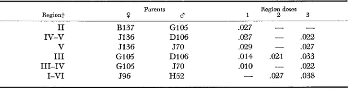
FIGURE 1 .-Genetic dissection of 25A-27E with segmental aneuploids of 2L for localization of aGpdh-I. The translocation parents (in parentheses) and their breakpoints are listed. The six regions between the translocations are indicated as I-VI. The shorter horizontal lines represent the limits of the aneuploid region of the crosses which generated them. aGPDH activity of these offspring are presented in Table 2.
I n order to localize the locus more specifically, the region was dissected by the
six crosses listed in Figure 1. The enzyme activities of flies containing various
doses of the six regions are presented in Table 2. From this table it can be seen
that the dosage-dependent region falls between 25F and 26B, in agreement with
GRELL’S
(1967) localization. From Table 2 it is also evident that none of the
neighboring sub-regions exhibits any control over aGPDH activity. Furthermore,
eleven other large regions were tested (Table 1) for dosage effects on (YGPDH
and no significant elevations in duplications were observed.
Adh,
the gene for alcohol dehydrogenase, has been mapped genetically
to
11-50.1 and cytologically between 34E3 and 35D1 (GRELL,
JACOBSON
and
MURPHY
1965; LINDSLEY
and GRELL
1968). The duplication 30F-35BC listed
in Table 1 was tested
for
dosage control of alcohol dehydrogenase. The dupli-
cation had 5.8 units of activity (expressed as nmoles/min/fly), as compared to
4.0 units of activity for the euploid sibs. The duplication/euploid ratio was 1.45.
Hence, with two known gene-enzyme systems, the segmental aneuploid analysis
was successful in detecting structural genes.
TABLE 2
aGPDH activity
in
segmental aneuploidsin
the 25A-27E region of 2LtParents
Region+ ? d Region
doses
1 2 3
-
I1 B137 GI 05 .027 -
IV-v J136 D106 .027
-
,022V 5136 570 ,029 - ,027
I11 G105 D106 ,014 ,021 ,033
111-IV GI 05 570 ,010
-
.022I-VI J96 H52
-
.027 .038*
The aGPDH activity is expressed in pmoles/min/fly. Each measurement represents theA N E U P L O I D Y A N D S T R U C T U R A L G E N E S
163
Succinic dehydrogenase:
The search for chromosomal regions affecting SDH
activity was complicated by very low levels of detectable SDH activity in Dro-
sophila. The background levels of succinate-independent “nothing” dehydro-
genase were as great as,
if
not greater than, the succinate-dependent activity.
Moreover, activity fluctuations from genotype to genotype were significant with
both background and SDH activity. Hence, a single no-substrate control for all
the genotypes would not suffice even for the initial screen. This necessitated the
retesting of any significant elevation ( l o % + ) of the duplication over the euploid
sibling.
Table 1 lists the ratios of activity of duplication/activity of sibling in the 31
crosses. Only one region was dosage-sensitive, 27%30F o n 2L. This region was
dissected by
further crosses between translocations with included breakpoints.
The dissection (Table 3) localized the region responsible f o r enzyme elevation
to be between 28D and 29F.
Since SDH has been localized to the inner membrane of mitochondria
(SCHNAITMAN
and
GREENAWALT
1968;
REED
and
SACKTOR
1971; and
S. J.
O’BRIEN, unpublished)
,
isolated mitochondrial membranes were assayed for
SDH. Two observations suggested that the elevated oxidation reaction observed
in
the duplication was not SDH but a background “nothing” dehydrogenase:
(1) The elevation did not localize to mitochondria, rather to crude supernatants.
(2) Elevated dehydrogenase levels, albeit much lower than succinate-dependent,
were observed in duplications in the absence
of
succinate. Genetically, malate
dehydrogenase has been localized to this region (map locus=40), and the struc-
tural similarity between malic acid and succinic acid suggests that the increase in
activity could be due to MDH rather than SDH. Hence, both cytoplasmic and
mitochondrial MDH were extracted from the aneuploids and euploids and
examined for dosage dependency. Dosage dependency has been observed in the
segmental aneuploids between 28D-29F with cytoplasmic preparations for
malate oxidation as well as for succinate and for no-substrate controls (Table 3
TABLE 3
Succinic-mulate dehydrogenase activity in segmental aneuploids
in
2L+Region ? Parents 0“ Sex Dp
Region doses
3 2 DP/ Ratio
+
27E 28B 27E 28B 28B 28C 28B 28C 28C 28D 28C 28D 28D 29F 28D 29F 29F 30F 29F 30F 27E 3UF
R50 H52
H52 R50
B65 R50
R50 B66
A l l l B66 B66 A l l l A145 A l l l A l l l Ale5 L52 A145 Ale5 L52
H52 L52
6
0
8
P
6
0
8
0
6
0
0
8.0 7.0 11.9 9.8 12.0 8.8 11.9 12.6
8.8 9.5
13.3 18.3 11.2 5.3 13.0 8.3 9.1 10.6 12.5 10.5 15.3 6.7
1.1 1.2 1.3 0.94
0.93
0.73 2.1 1.6 0.86 1.2 2.3
164
s.
J.O’BRIEN
ANDR. c.
GETHMANNand O’BRIEN 1973). Thus it is suggested that
Mdh-l
is cytologically localized
between 28D and 29F with the reservations stated below.
DR.
E. H.
GRELL
(personal communication) has recently induced a deficiency
for
Jammed
and
Mdh-2
(GRELL
1969) which is cytologically localized between
30D
and 32A on
2L. These breakpoints overlap our large duplication (27E-30F)
,
but do not include the smaller region (28D-29F) which exhibits dosage depen-
dency (Table
3 ) .
Furthermore, the overlapping region (29F-30F) does not
exhibit dosage dependency in our hands. The possibility that the published break-
point of A145 (29F) was incorrect was graciously reexamined by A. ATTALLAH
and W.
NASH.
They confirmed the breakpoint of A145 to 29F which is to the
left of GRELL’S
deficiency. I t is not clear from the available data if
Mdh-1
and
Mdh-2
are the same locus and, if
so, why there is the apparent discrepancy in
the independently determined cytological localization. The 28D-29F locus is
probably not the mitochondrial
M d h
in light of lack of dosage dependency for
that enzyme in these same aneuploid individuals (O’BRIEN 1973). Further
investigation of this region is certainly necessary to unequivocally resolve this
dilemma.
aGPO:
The screen for
&GPO (Table
1) detected one region which possessed
elevated aGPO in duplications, region 5OC-52E in 2R. Mitochondria were pre-
pared from duplications and euploids of this region and aGPO was extracted from
the inner membrane. The elevation does reside in the inner membrane, and the
activity is a-glycerophosphate-dependent. Unfortunately, this region could not
be further dissected genetically because there are no other translocations with
breakpoints within this region. Thus we were unable to construct and test a
deficiency for this region at this writing. Nevertheless, this is the first case, to our
knowledge, of a nuclear gene’s affecting the activity of a mitochondrial inner
membrane enzyme in a dose-response manner.
The
duplications, general characteristics:
As previously stated, there was no
obvious correlation between the
R.V.
and the length of the duplications. We also
compared the duplication: euploid enzyme activity ratio of a G P 0
us.duplication
size and found no correlation. Likewise, no apparent relationship existed between
R.V.
and aGPO ratio or between
R.V.
and
SDH
ratio. There was, however, a
striking increase in the variance of all the enzyme ratios in crosses in which the
R.V.
was less than 50%. For the most part, we rejected those translocation crosses
which resulted in low aGPO or
SDH
ratios or a low
R.V.
DISCUSSION
ANEUPLOIDY A N D STRUCTURAL GENES
165
duplications, and can be used to more specifically localize the dosage-sensitive
locus. Although strict dosage dependency
(1
:2: 3 in monosomics, euploids, and
trisomics, respectively) is a characteristic only of
structural genes, it is not con-
clusive evidence for the structural gene identity. Further credence can be
obtained, as it has in other systems, by analysis of electrophoretic variation, by
the demonstration of interallelic complementation of recessive “null” alleles
(O’BRIEN and MACINTYRE
197213; BELL,MACINTYRE
and
OLIVIERI
1972), and
by the presence of immunologically crossreacting material
in
“null” alleles
(
GLASSMAN
1965; BELL
and MACINTYRE
1973). Hence, the segmental aneuploid
approach can only indicate probable structural gene loci; it cannot confirm the
nature of the locus. It is important, however, to realize that the analysis of aneu-
ploids gives two kinds of evidence pertinent to the identity of
a structural gene:
enzyme elevation in duplications and enzyme diminution in deficiencies. There
are few, if
any, other types of genes which are capable of
producing both effects
on the product of a second gene. It is unfortunate that in the case of
(uGPO,
only
one of these results (an elevation in duplications) could be examined.
The data obtained with the three known gene-enzyme loci emphasizes the
feasibility of the aneuploid technique within the limits o i the three assumptions
discussed in the introduction: The enzymes are (1) nuclear in their inheritance,
(2) located on the autosomes, and
(3)
dependent upon gene dosage for their level
of
activity. Any
of
these assumptions could be wrong with respect to the mito-
chondrial enzymes.
In Neurospora and in rat liver, there is strong evidence for mitochondrial-
specific ribosomal synthesis of mitochondrial inner membrane components. These
data show chloramphenicol-sensitive incorporation of radioactive amino acids
into the inner membrane and into subunits of
cytochrome oxidase (
WEISS,
SEBOLD
and
BUCHER
1971; NEUPERT,
BRDICZKU
and
BUCHER
1967;
COOTE
and WORK
1971). Thus, it is possible that the mitochondrial enzymes examined in this study
might be coded by mitochondrial DNA.
It is also possible that these genes might be sex-linked. Since the
X
chromo-
some comprises
20%
of
the Drosophila genome, and since genes appear to be
randomly distributed with respect to chromosome location, it would be expected
that a number of enzyme loci would be sex-linked. Such genes would not be
detected in these experiments, although they could
be
recognized through the
construction of
X
chromosome aneuploids in a manner similar to the construction
of
the autosomal aneuploids.
Finally, for mitochondrial enzymes, the rate-limiting step in enzyme activity
might not be gene dosage, but rather, the number
of
mitochondrial sites or seats
of
enzyme action on the inner membrane. If
this is
true,
one would expect to find
no response to duplication aneuploidy, although it is possible that such enzymes
might be sensitive
to
deficiency aneuploidy. In these experiments, only dupli-
cation aneuploids were examined in the initial screen, thus the structural genes
would not have been detected.
166
s.
s.
O'BRIEN AND R.c.
GETHMANNcould have been f o r any one of
the above three reasons. However, since most, if
not all, nuclear enzyme loci do show dosage sensitivity, the use of these Y-auto-
some translocations to produce segmental aneuploids should prove to be a useful
and relatively simple technique for localizing new genes, as well as being useful
in studies
of regulation of these genes.
W e would like to thank DR. E. H. GRELL for information and permission to cite cytological breakpoints of Df(2L)Mdh. We are also grateful to ABDELFATTAH
M. ATTALLAH
and Dr. WIL-LIAM NASH for confirming the autosomal breakpoint of stock A145 and DR. Ross J. MACINTYRE
for critically reviewing the manuscript.
LITERATURE CITED
BAHN, E., 1971 Position-effect variegation for an isoamylase in Drosophila melanogaster.
BAKER, W. K., 1968 Position-effect variegation. Advan. Genet. 14: 133-169.
BELL, J. B. and R. J. MACINTYRE, 1973 Characterization of Acid phosphatase-1 null activity mutants of Drosophila mlanogasfer. Biochem. Genet. 10 (In press).
BELL, J. B.,
R.
J. MACINTYRE and A. P. OLIVIERI, 1972 Induction of null activity mutants for the Acid phosphatase-1 gene in Drosophila melanogaster. Biochem. Genet. 6: 205-216. COOTE, J. L. and T. S. WORK, 1971 Proteins coded by mitochondrial DNA of mammalian cells.Eur. J. Biochem. 23: 564-574.
FARNSWORTH, M. W., 1965 Oxidative phosphorylation in the Minute mutants of Drosophila. J. Exp Zool. 160: 363-368.
GLASSMAN,
E., 1965 Genetic regulation
of xanthine dehydrogenase in Drosophila melanogaster. Federation Proc. 24: 1243-1251.GRELL, E. H., 1962 The dose effect of ma-Z+ and ry+ on xanthine dehydrogenase activity in Drosophila melanogaster. Z . Vererbl. 93 : 371-377. __
,
1967 Electrophoretic variants of a-glycerophosphate dehydrogenase in Drosophila melanogaster. Science 158: 1319-1320.Alcohol dehydrogenase in Drosophila melanogaster: isozymes and genetic variants. Science 149 : 80-82.
A molecular approach to the study of heterozygosity in natural populations. 11. Amount of variation and degree of heterozygosity in natural populations of Drosophila pseudoobscura. Genetics 54: 595-609.
LINDSLEY,
D. L. and E. H. GRELL, 1968 Genetic uariations of Drosophila melanogaster. Carnegie Inst. Wash. Publ. No. 627.LINDSLEY,
D.L.,
1,.
SANDLER, B. S. BAKER, A. T. C. CARPENTER, R. E. DENELL, J. C. HALL,P. A.
JACOBS, G. L.G. MIKLOS,
B. K. DAVIS, R. C. GETHMANN, R. W.HARDY,
A. HESSLER, S. M. MILLER, H. NOZAWA, D. M. PARRY and M. GOULD-SOMERO, 1972 Segmental aneuploidy and the genetic gross structure of the Drosophila genome. Genetics 71 : 157-184.MCREYNOLDS, M. S. and G. B. KITTO, 3970 Purification and properties of Drosophila malate dehydrogenases. Biochim. Biophys. Acta. 198: 165-1 75.
NASI, D. and J. B. BEI.I., 1968 Larval age and the pattern of DN-4 synthesis in polytene chro- mosomes. Can. J. Genet. Cytol. 10: 82-90.
NEUPERT, W., D. BRDICZKU and TH. BUCHER, 1967 Incorporation of amino acids into the outer and inner membranes of isolated rat liver mitochondria. Biochem. Biophys. Res. Comm. 27:
Comparative analysis of malate dehydrogenase in Drosophila melanogaster. Hereditas 67: 79-82.
GRELr., E. H
,
K. B. JACOBSON and J. B. MURPHY, 1965LEWONTIN, R. C. and J. L. HUBBY, 1966
488-493. O'BRIEN, S. J., 1973
ANEUPLOIDY A N D STRUCTURAL GENES
167
O'BRIEN, S. J. and R.
J.
MACINTYRE, 19@3 An analysis of gene enzyme variability in natural populations of Drosophila melanogmter andD.
simulans. Am. Natur.103:
97-113.-,
1971 A biological map of D. melanogaster. Drosophila Inform. Serv.46:
89-93.-,
1972a The a-glycerophosphate cycle in Drosophila melanogaster.I.
Biochemical and devel- opmental aspects. Biochem. Genet.7:
141-161.-
,
1972b The a-glycerophosphate cycle in Drosophila melanogmter. 11. Genetics aspects. Genetics71
: 127-138.Localization of trehalase in flight: muscle of the blowfly Phormia regina. Arch. Biochem. Biophys. 145: 392-401.
Enzymatic properties of the inner and outer membranes of rat liver mitochondria. J. Cell Biol. 38: 158-175.
Drosophila alcohol dehydrogenase: Purification and par- tial characterization. J. Biol. Chem. 243: 3110-3115.
Alcohol dehydrogenase: A polymorphism in Drosophila melunogaster. J. Exptl. Zool. 160: 147-154.
Incorporation of amino acids into a polypeptide of Neurospora crassu. Euro. J. Biochem. 22: 19-26.
Corresponding Editor: A. CHOVNICK
REED,
W. D. and B. SACTOR, 1971SCHNAITMAN,
c.
and J.w.
GREENAWALT 1968SOFER, W. H. and H. URSPRUNG, 1968
URSPRUNG,