Scholarship@Western
Scholarship@Western
Electronic Thesis and Dissertation Repository
11-15-2013 12:00 AM
Development of Biodegradable and Stimuli-Responsive
Development of Biodegradable and Stimuli-Responsive
Macromolescules and Their Assemblies
Macromolescules and Their Assemblies
Ali Nazemi
The University of Western Ontario
Supervisor
Dr. Elizabeth R. Gillies
The University of Western Ontario Graduate Program in Chemistry
A thesis submitted in partial fulfillment of the requirements for the degree in Doctor of Philosophy
© Ali Nazemi 2013
Follow this and additional works at: https://ir.lib.uwo.ca/etd
Part of the Materials Chemistry Commons, Organic Chemistry Commons, and the Polymer Chemistry Commons
Recommended Citation Recommended Citation
Nazemi, Ali, "Development of Biodegradable and Stimuli-Responsive Macromolescules and Their Assemblies" (2013). Electronic Thesis and Dissertation Repository. 1715.
https://ir.lib.uwo.ca/etd/1715
This Dissertation/Thesis is brought to you for free and open access by Scholarship@Western. It has been accepted for inclusion in Electronic Thesis and Dissertation Repository by an authorized administrator of
DEVELOPMENT OF BIODEGRADABLE AND STIMULI-RESPONSIVE
MACROMOLECULES AND THEIR ASSEMBLIES
(Thesis format: Integrated Article)
by
Ali Nazemi
Graduate Program in Chemistry
A thesis submitted in partial fulfillment
of the requirements for the degree of
Doctor of Philosophy
The School of Graduate and Postdoctoral Studies
The University of Western Ontario
London, Ontario, Canada
ii
Polymersomes are potentially multifunctional soft materials constructed by the
self-assembly of amphiphilic block copolymers in aqueous medium. While much research has
focused on controlling the assembly and encapsulation properties of polymersomes, their
surface functionalization has been relatively unexplored. This is important because it
plays a critical role in determining their properties such as toxicity and biodistribution
behavior. The work described in this thesis involves the development of a biocompatible
and biodegradable polymersome systems based on poly(ethylene
oxide)-b-polycaprolactone (PEO-PCL) block copolymers with azide surface groups as a novel
scaffold for various biomedical applications. The surface functionalization of these
polymersomes with polyester dendrons bearing alkyne focal points with different
peripheral groups, such as amines and guanidines, as well as a small molecule rhodamine
dye is accomplished and their conjugation yields are compared to each other. Moreover,
dendritic and non-dendritic polymersome-based MRI contrast agents, with the highest
currently reported longitudinal relaxivity for a polymersome system, are developed by
decorating PEO-PCL polymerosomes' surfaces with both non-dendritic and dendritic
Gd(III)-based contrast agents. In addition, PEO-PCL polymersomes were employed to
develop a multifunctional system with the potential to interfere with the viral infection
process at two levels. In addition to their use as materials for functionalizing the surfaces
of nanomaterials, dendrimers and their assemblies have been widely used as drug
delivery vehicles. In order to enable a new level of control over drug release, backbone
photodegradable dendrimers and dendrons are synthesized by incorporation of a
monomer unit based on o-nitrobenzyl esters and 2,2-bis(hydroxymethyl)propionic acid. It
is shown that these dendrimers undergo effective photolysis to release only small
molecules upon irradiation with UV light. Finally, these dendrons are incorporated into
amphiphilic Janus dendrimer structures and their self-assembly to dendrimersomes
followed by their photodegradation are discussed.
Keywords
Polymersome, biodegradable, dendrimer, dendron, MRI contrast agent, influenza virus,
iii
The research discussed in this thesis is a result of contributions from the author as well as
coworkers from Western University and supervisor Dr. Elizabeth Gillies whose exact
contributions are described here.
Chapter 1 contains materials that are submitted for publication in a journal article and
a book chapter. Subsection "Dendrimer Conjugates with Imaging Agents", is a part of the
submitted book chapter, and "Surface Functionalization of Polymersomes", is a part of
the submitted journal article. These sections were written collaboratively by the author
and supervisor Dr. Elizabeth Gillies.
Chapter 2 describes a project proposed by Dr. Elizabeth Gillies and Dr. Colin
Bonduelle. Dendrons used for this work were supplied by Ryan Amos, a graduate student
in the Gillies group at the time. All other experimental work was carried out by the author
under the supervision of Dr. Gillies. The manuscript was initially drafted by the author
and Dr. Gillies provided assistance with editing and final preparation.
Chapter 3 comprises work proposed by Dr. Gillies and the author. Relaxivity
measurements of the developed contrast agents were performed by Dr. Francisco
Martinez and Dr. Timothy Scholl at the Robarts Research Institute at Western. All the
other experimental work were accomplished by the author under the supervision of Dr.
Gillies. The manuscript was initially drafted by the author and Dr. Gillies provided
assistance with editing and final preparation.
Chapter 4 describes a project proposed by Dr. Gillies. Dr. S. M. Mansour Haeryfar's
research laboratory in the Department of Microbiology and Immunology at Western
University provided input on the antiviral potential of these molecules and reviewed the
manuscript. All the experimental work as well as the biological assay were carried out by
the author. The manuscript was initially drafted by the author and Dr. Gillies provided
iv
dendrons were synthesized by Tylor Schon, a fourth year undergraduate student
supervised by the author. All the other dendrimer synthesis experiments and
photodegradation studies were carried by the author under the supervision of Dr. Gillies.
The manuscript was initially drafted by the author and Dr. Gillies provided assistance
with editing and final preparation.
Chapter 6 describes a project proposed by the author. All the experimental work was
accomplished by the author under the supervision Dr. Gillies. This chapter was initially
drafted by the author and Dr. Gillies provided assistance with editing and final
v
I would like to give my sincere thanks to my supervisor, Dr. Beth Gillies, for giving me
the opportunity to join her research team and for supporting me throughout my time in
her group. Beth, you are a great supervisor. Under your supervision, I not only trained in
many experimental skills, but I also learned how to think critically and develop scientific
ideas.
I would also like to thank all the past and present members of the Gillies group for all
their help and for creating such an excellent environment in the lab, that leaves me with
many unforgettable memories. Many thanks to all the staff at UWO without whom this
thesis would not have been possible: Mat Willans, Doug Hairsine, Darlene McDonald,
the main office crew, all the ChemBio Store and Electronics Shop staff, and the
undergraduate lab technicians for all their support during TAing. Furthermore, I am
thankful to my thesis examiners Dr. Guthrie, Dr. Hudson, Dr. Min, and Dr. Yousaf for
reading through my thesis.
Special thanks to my wife, Mahboubeh, who has been by my side since the
undergraduate level. Mahboubeh, I have no doubt that I could not have come this far if it
was without you, and I cannot tell you how grateful I am for all the unconditional support
you have had for me. We all know that research does not go well most of the time and
this leaves us with frustration. You have always cheered me up during such times and
celebrated with me for my successes. I cannot wait to celebrate your successful defense
in the near future.
Last but definitely not least, I am deeply grateful to my father, mother, and brothers
for all their encouragement, support, and unconditional love throughout my life. You
guys have always been there for me. Mom and Dad, I know how difficult it was for you
to live in a foreign country while you had your whole life back in Iran. You forfeited all
you had back there and came this far just to provide a better life for us. I know it is
impossible to make up for all you have done; however, to show my sincere appreciation,
I would like to dedicate this thesis to you, my dear Mom and Dad.
vi
Abstract ... ii
Co-Authorship Statement... iii
Acknowledgments... v
List of Tables ... x
List of Figures ... xi
List of Schemes ... xvi
List of Abbreviations ... xvii
Chapter 1 ... 1
1 Biodegradable Polymersomes and Dendrimers in Biomedical Applications ... 1
1.1 Introduction to Macromolecules ... 1
1.1.1 Biodegradable Polymers ... 3
1.1.2 Block Copolymers ... 3
1.1.3 Block Copolymer Self-Assembly in Solution... 4
1.1.4 Other Macromolecular Architectures: Dendrimers ... 6
1.1.5 Amphiphilic Janus Dendrimers and Their Assemblies ... 10
1.2 Macromolecules for Biomedical Applications ... 12
1.2.1 Block Copolymer Assemblies for Drug Delivery ... 13
1.2.1.1 Polymeric Micelles for Drug Delivery ... 15
1.2.1.2 Polymersomes for Drug Delivery ... 16
1.2.2 Dendrimers for Biomedical Applications ... 17
1.2.2.1 Dendrimer-Drug Conjugates ... 18
1.2.2.2 Dendrimer-Carbohydrate Conjugates ... 19
1.2.2.2.1 Dendrimer-N-Acetylneuraminic Acid Conjugates ... 21
1.2.2.3 Dendrimer Conjugates with Imaging Agents ... 24
1.2.2.3.1 Dendrimer Conjugates for MRI ... 24
1.3 Surface Functionalization of Polymersomes ... 29
1.3.1 Dendritic Surface Functionalization of Polymersomes ... 31
vii
1.5 Scope of This Thesis ... 42
1.6 References ... 44
Chapter 2 ... 53
2 Dendritic Surface Functionalization of Biodegradable Polymer Assemblies ... 53
2.1 Introduction ... 53
2.2 Results and Discussion ... 55
2.2.1 Synthesis of Block Copolymers ... 55
2.2.2 Synthesis of Alkyne-Functionalized Dendrons ... 58
2.2.3 Formation of Nanoassemblies and Surface Functionalization Reactions ... 60
2.2.4 Effects of Surface Functionalization on the Nanoassemblies ... 65
2.2.5 Cellular Uptake of the Guanidine Dendron-Functionalized Micelles ... 68
2.3 Conclusion ... 69
2.4 Experimental ... 70
2.5 References ... 76
Chapter 3 ... 81
3 Biodegradable Dendritic Polymersomes as Modular, High Relaxivity MRI Contrast Agents ... 81
3.1 Introduction ... 81
3.2 Results and Discussion ... 82
3.2.1 Design and Synthesis ... 82
3.2.2 Functionalization of Polymersome Surfaces with Dendritic and Non-Dendritic Contrast Agents ... 85
3.2.3 Evaluation of the Relaxivity Properties of the Contrast Agents ... 86
3.3 Conclusion ... 88
3.4 Experimental ... 88
3.5 References ... 92
Chapter 4 ... 95
4 Multifunctional Dendritic Sialopolymersomes as Potential Antiviral Agents: Their Lectin Binding and Drug Release Properties ... 95
viii
4.2.2 Functionalization of Polymersomes with Sialodendrons ... 99
4.2.3 Evaluation of Inhibitory Potencies Using an Enzyme-Linked Lectin Inhibition Assay ... 101
4.2.4 Encapsulation and Release of Zanamivir by Naked Polymersomes and Dendritic Sialopolymersomes ... 104
4.3 Conclusion ... 107
4.4 Experimental ... 107
4.5 References ... 113
Chapter 5 ... 117
5 Synthesis and Degradation of Backbone Photodegradable Polyester Dendrimers . 117 5.1 Introduction ... 117
5.2 Results and Discussion ... 118
5.2.1 Design and Synthesis ... 118
5.2.2 Photodegradation Study of the Dendrimers ... 122
5.3 Conclusion ... 124
5.4 Experimental ... 124
5.5 References ... 133
Chapter 6 ... 136
6 Photodegradable Amphiphilic Janus Dendrimers and Dendrimersomes as Potential Smart Drug Delivery Vehicles ... 136
6.1 Introduction ... 136
6.2 Results and Discussion ... 139
6.2.1 Synthesis of Photodegradable Amphiphilic Janus Dendrimers ... 139
6.2.2 Self-Assembly of Amphiphilic Janus Dendrimers in Aqueous Media ... 141
6.2.3 Photodegradation Study of the Dendrimersomes ... 143
6.3 Conclusion ... 147
6.4 Experimental ... 147
6.5 References ... 152
ix
Appendix 1: Permission to Reuse Copyrighted Material ... 160
Appendix 2: Supporting Information for Chapter 2 ... 167
Appendix 3: Supporting Information for Chapter 3 ... 174
Appendix 4: Supporting Information for Chapter 4 ... 181
Appendix 5: Supporting Information for Chapter 5 ... 187
Appendix 6: Supporting Information for Chapter 6 ... 205
x
xi
Figure 1.1. Mathematical equations for Mw and Mn... 2
Figure 1.2. Chemical structures of the most common polyesters. ... 3
Figure 1.3. Representation of various BCP architectures. ... 4
Figure 1.4. Cartoon representation of a) polymer micelles, b) polymersomes. ... 5
Figure 1.5. Schematics of a) a hyperbranched polymer; b) a dendrimer; c) a dendron. .... 7
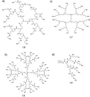
Figure 1.6. Readily accessible dendrimer backbones: a) PAMAM; b) PE; c) PPI; d) PLL. ... 9
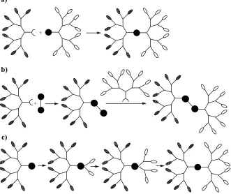
Figure 1.7. Schematic representation of main methods for the synthesis of Janus dendrimers... 10
Figure 1.8. Examples of Janus dendrimers based on a) ether/amide linkages; b) benzyl ether and PAMAM dendrons. ... 11
Figure 1.9. Schematic representation of a dendrimersome. ... 12
Figure 1.10. PAMAM-DOX conjugates with a) amide and b) hydrazone linkages ... 19
Figure 1.11. Conjugation strategies for Neu5Ac. ... 21
Figure 1.12. Chemical structures of: a) the clinical agent Gd(III)-DTPA (Magnevist) and dendrimer conjugates of DTPA derivatives containing b) an aromatic isothiocyanate and c) a more flexible aliphatic isocyanate. ... 26
Figure 1.13. Chemical structures of: a) the clinical agent Gd(III)-DOTA (Dotarem) and dendrimer conjugates of DOTA derivatives containing b) an aromatic isothiocyanate linker; c) a a phosphinic acid linker; d) an amino acid-based linker. ... 27
Figure 1.14. Dendrimer conjugates of HOPO derivatives using a) a rigid amide linkage
xii
acids. ... 29
Figure 1.15. Schematic showing the functionalization of polymersomes bearing
peripheral azide groups with dendrons having focal point alkynes. ... 32
Figure 1.16. Schematic for the preparation of a) dendritic mannose polymersomes; b)
non-dendritic mannose polymersomes. ... 33
Figure 1.17. Schematic representation of sequential photodegradation of dendrimers
functionalized with three o-nitrobenzyl ester groups at their core. ... 39
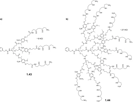
Figure 1.18. Chemical structures of a) first generation and b) second generation
photodegradable amphiphilic dendrons used for DNA complexation and release. ... 40
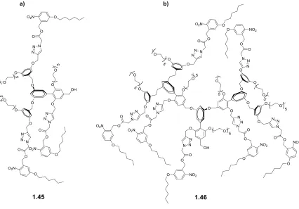
Figure 1.19. Chemical structures of a) first generation and b) second generation
amphiphilic photodegradable dendrons used for encapsulation and release of Nile Red. 41
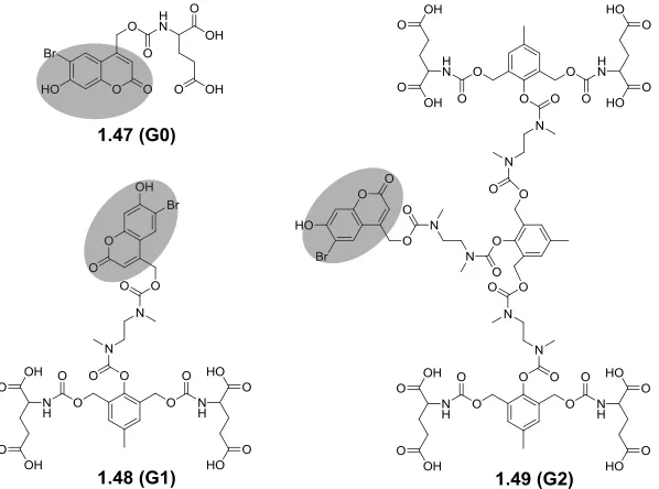
Figure 1.20. Structures of photodegradable dendrimers containing coumarin as NIR
light-degrading groups. ... 42
Figure 2.1. SEC traces for copolymers: (a) 2.3 and 2.4 and (b) 2.5 and 2.6. Detection was
based on light scattering (90 trace shown). ... 58
Figure 2.2. Size distribution profiles measured by DLS for: a) micelles prepared from
copolymer 2.3; b) vesicles prepared from copolymer 2.5; c) extruded vesicles prepared
from copolymer 2.5. ... 61
Figure 2.3. TEM images of: a) micelles prepared from copolymer 2.3; b) vesicles
prepared from copolymer 2.5. ... 61
Figure 2.4. Click reaction yields as a function of azide loading on: a) vesicles (remaining
copolymer is 2.5); b) micelles (remaining copolymer is 2.3). ... 63
Figure 2.5. TEM images of: a) vesicles with 2% azide loading, following conjugation of
xiii
azide loading, following conjugation of dendron 2.8 and dialysis. ... 65
Figure 2.6. Size distribution profiles following click reactions and dialysis, measured by
DLS for: a) vesicles and b) micelles. ... 66
Figure 2.7. Preparation of PEO-PCL micelles functionalized with dendrons having
peripheral guanidines, and their uptake into HeLa cells as visualized by fluorescence
microscopy (detection of the rhodamine label). In contrast, micelles bearing the
rhodamine label, but no dendron exhibited no detectable uptake. ... 69
Figure 3.1. Schematic for the preparation of dendritic and non-dendritic
Gd(III)-functionalized polymersomes. ... 83
Figure 3.2. Size distribution profiles for: a) naked polymersome; non-dendritic
polymersome 3.4; c) dendritic polymersome 3.3. ... 86
Figure 3.3. TEM images of (a) naked polymersome; (b) dendritic Gd(III)-functionalized
polymersomes 3.3; (c) non-dendritic Gd(III)-functionalized polymersomes 3.4. ... 86
Figure 3.4. Longitudinal relaxivity (r1) of dendron 3.1, polymersome 3.3, and
polymersome 3.4 in phosphate buffer (0.1 M, pH 7.4) as a function of field strength at
298 K. ... 87
Figure 4.1. Yields for the azide + alkyne “click” conjugation reaction between dendron
4.4 and PEO-PCL polymersomes having varying percentages of azide-terminated
copolymer 2.6 (remaining percentage is methoxy-terminated PEO-PCL 2.5). Note that
the error bar on the 20 wt% copolymer 2.6 measurement represents the standard deviation
of triplicate experiments designed to assess the reproducibility of the conjugation. ... 101
Figure 4.2. a) Size distribution profiles for naked polymersomes and polymersomes
composed of different percentages of azide-functionalized copolymer 2.6 following
“click” conjugation of dendron 4.3; b) TEM image of polymersomes prepared from 40
wt% copolymer 2.6 following conjugation of dendron 4.3; c) TEM image of naked
xiv
monovalent Neu5Ac derivative 4.5; b) sialodendron 4.3; c) dendritic sialopolymersomes
prepared from the conjugation of sialodendron 4.3 to polymersomes prepared from 20
wt% and 40 wt% azide-functionalized copolymer 2.6. Note that in b) and c) the inhibitor
concentration corresponds to the dendron concentration... 104
Figure 4.4. Release profiles of zanamivir from a) naked PEO-PCL polymersomes and b)
dendritic sialopolymersomes. All experiments were performed in triplicate. ... 106
Figure 5.1. SEC traces of G1 (5.14), G2 (5.15), and G3 (5.16) dendrimers with their
corresponding PDIs. ... 122
Figure 5.2. a) UV-visible spectra for G3 dendrimer (5.16) upon irradiation with UV light
for 60 min. Inset shows the expanded region between 285-450 nm. b) Evolution of 1H
NMR spectra during the photolysis of a 10 mg/mL sample of G3 dendrimer (5.16) in
(CD3)2SO... 123
Figure 6.1. a) SEC traces and b) MW and PDI characteristics of AJDs 6.2, 6.4, and 6.6. a
Molecular weight calculated based on chemical structures of the dendrimers, b Mn
obtained from SEC, and c PDI was determined from SEC. ... 141
Figure 6.2. Size distribution profiles measured by DLS for assemblies formed by a) 2nd
generation AJD 6.4 and b) 3rd generation AJD 6.6. ... 142
Figure 6.3. TEM images of a) particles prepared from AJD 6.4 and b) dendrimersomes
formed by AJD 6.6. ... 143
Figure 6.4. UV-visible spectra for G3 AJD 6.6 a) as THF solution and b) as
self-assembled dendrimersomes in water upon irradiation with UV light for 30 min. Inset
shows the expanded region between 285-415 nm. ... 144
Figure 6.5. DLS measurements for the photolysis of dendrimersomes: a) plot of mean
count rate versus irradiation time and b) size distribution profile of the dendrimersome
xvi
Scheme 1.1. Photoisomerization mechanism for o-nitrobenzyl ester derivatives. ... 38
Scheme 2.1. Synthesis of HO-PEO-N3 (2.2). ... 56
Scheme 2.2. Synthesis of PEO-PCL BCPs. ... 57
Scheme 2.3. Synthesis of rhodamine-labeled guanidine dendron 2.8. ... 59
Scheme 2.4. Synthesis of alkyne-functionalized rhodamine 2.10. ... 59
Scheme 2.5. Synthesis of rhodamine-labeled PEO-PCL block copolymers 11 and 12. .. 60
Scheme 2.6. Preparation of functionalized a) vesicles and b) micelles... 62
Scheme 2.7. Synthesis of dendron 2.14. ... 67
Scheme 3.1. Synthesis of Gd(III)-functionalized dendron 3.1. ... 84
Scheme 3.2. Synthesis of Gd(III) complex 3.2. ... 84
Scheme 4.1. Synthesis of sialodendron 4.3. ... 98
Scheme 4.2. Synthesis of rhodamine-labeled sialodendron 4.4. ... 99
Scheme 4.3. Preparation of dendritic sialopolymersomes... 100
Scheme 5.1. Synthesis of monomer 5.5 and G1 dendron 5.8. ... 119
Scheme 5.2. Synthesis of G2 dendron 5.10 and G3 dendron 5.12. ... 120
Scheme 5.3. Synthesis of G1-G3 dendrimers 5.14 - 5.16. ... 121
xvii A Ac AJD aq ATRP BCP bis-MPA Boc BOP br BSA CL Con A CT d DAB DCC DIPEA DLS DMAP DMEM DMF DNA DOTA DOX DTPA EDC EI ELLA EPR equiv ESI Et3N EtOAc fhydrophilic FBS G h HA HABA HBP HBTU HCl absorption acetyl
amphiphilic Janus dendrimer aqueous
atom-transfer radical polymerization block copolymer
2,2-bis(hydroxymethyl) propionic acid tert-butoxycarbonyl
(benzotriazol-1-yloxy)tris(dimethylamino)phosphonium hexafluorophosphate
broad
bovine serum albumin
-caprolactone Concanavalin A computed tomography doublet 3,3'-diaminobenzidine N,N'-dicyclohexylcarbodiimide N,N-diisopropylethylamine dynamic light scattering 4-dimethylaminopyridine
dulbecco’s modified eagle medium N,N-dimethylformamide deoxyribonucleic acid 1,4,7,10-tetraazacyclododecane-1,4,7,10-tetraacetic acid doxorubicin diethylenetriaminepentaacetic acid N-(3-dimethylaminopropyl)-N’-ethylcarbodiimide electron impact
enzyme-linked lectin inhibition assay enhanced permeability and retention equivalent
electrospray mass spectrometry N,N,N-triethylamine
ethyl acetate
hydrophilic volume fraction fetal bovine serum
xviii HOPO HPLC HRMS HRP-LFA Hz IC50 ICP-MS IR IUPAC J LCST LFA LSM m MALDI-MS MALS MeOH min Mn mp MRI MSA Mw MW MWCO NA NaAsc NaOMe Neu5Ac NIPAAm NIR NMR PAMAM PBD PBD-PEO PBS PBST PCL PDI PDLLA PE PEG PEO PEO-PCL hydroxypyridinone
high-performance liquid chromatography high-resolution mass spectrometry
horseradish peroxidase-labeled LFA hertz
inhibitory concentration-50
inductively coupled plasma mass spectrometry infrared
International Union of Pure and Applied Chemistry coupling constant
lower critical solution temperature Limax flavus agglutinin
laser scanning microscope multiplet
matrix-assisted laser desorption/ionization mass spectroscopy multi-angle light scattering
methanol minute(s)
number average molecular weight melting point
magnetic resonance imaging methanesulfonic acid
weight average molecular weight molecular weight
molecular weight cutoff neuraminidase sodium ascorbate sodium methoxide N-acetylneuraminic acid N-isopropylacrylamide near infrared
nuclear magnetic resonance poly(amido amine)
polybutadiene
poly(butadiene-b-ethylene oxide) phosphate buffered saline
xix PGA PLLA PMOXA PPh3 PPI quant r1 RAFT RES RI ROMP ROP rpm rt SEC SPECT t TAX TEM TEG TFA THF TLC TLQ UV ε µwave R poly(glycolic acid) poly(L-lactic acid) poly(2-methyl-2-oxazoline) triphenylphosphine poly(propylene imine) quantitative longitudinal relaxivity
reversible addition-fragmentation chain-transfer polymerization reticuloendothelial system
refractive index
ring-opening metathesis polymerization ring-opening polymerization
revolutions per minute room temperature
size exclusion chromatography
single photon emission computed tomography triplet
paclitaxel
transmision electron microscopy triethylene glycol
trifluoroacetic acid tetrahydrofuran
thin layer chromatography trimethyl-locked quinone ultraviolet
molar extinction coefficient microwave
Chapter 1
1 Biodegradable Polymersomes and Dendrimers in
Biomedical Applications
*1.1 Introduction to Macromolecules
The word macromolecule is a Greek-Latin hybrid word that contains two contradictory
terms. It refers to a small mass (Greek: molecula, diminutive of moles = mass) that is
large (Large: makros).1 Thus, they are simply large molecules. According to the
International Union of Pure and Applied Chemistry (IUPAC), a macromolecule is
defined as
"a molecule of high relative molecular mass, the structure of which essentially
comprises the multiple repetition of units derived, actually or conceptually, from
molecules of low relative molecular mass."
Macromolecules are either natural, such as proteins, DNA, and polysaccharides, or
synthetic, such as synthetic rubbers, fibers and dendrimers, with molecular weights
(MWs) of several thousands to millions. Humankind has used naturally occurring
macromolecules since the early days of civilization. For instance, proteins in meat and
polysaccharides in grain are essential constituents of food, and a high MW resin called
Amber was used in old Greece as jewelry. On the other hand, the first synthetic and
semisynthetic macromolecules, such as nitrocellulose in 1869, were prepared without any
insight to their chemical structure. It was not until 1920s when scientists began to obtain
knowledge about the structures of macromolecules, and soon after, fully synthetic
macromolecules such as polychloroprene, polystyrene, and nylon 6.6 were discovered
and commercialized.1
*
Compared to traditional small molecules with single molar masses, synthetic
macromolecules display molar mass distributions. Among the different techniques
developed to measure these molar mass distributions,1 mass spectroscopy techniques
such as matrix-assisted laser desorption/ionization mass spectroscopy (MALDI-MS) and
size exclusion chromatography (SEC) have found widespread applications in chemistry
and materials science laboratories. Compared to mass spectroscopy, which is an absolute
method for molar mass determination, most SEC requires a correlation of the measured
properties of standards with molar masses that have been independently determined with
those of the sample. Because of the presence of the above-mentioned molar mass
distribution in most synthetic macromolecules, their molar mass is often calculated
around an average value. Depending on the statistical method that is applied to calculate
the average molar mass, different average values can be defined, among which number
average molar mass, Mn, and weight average molar mass, Mw, are most commonly used.
Mathematical expressions for Mn and Mw are shown in Figure 1.1. In these equations, Ni
is defined as the number of moles of each macromolecule species and Mi as the molar
mass of that species. Another term widely used in macromolecular science is
polydispersity index (PDI), which is calculated as Mw/Mn. PDI indicates the distribution
of individual molar masses in a batch of polymer sample. It has a value of greater than 1.
However, as the polymer chain lengths become more uniform, the PDI approaches 1.
Figure 1.1. Mathematical equations for Mw and Mn.
Among various natural and synthetic macromolecules, of particular interest to this
thesis are biodegradable polymers, block copolymers (BCPs), and dendritic architectures.
In the following sections, these families of macromolecules will be briefly introduced and
recent advancements in their self-assembly behaviors and their use in biomedical
1.1.1 Biodegradable Polymers
During the past few decades, the field of synthetic polymers has progressed to such an
extent that synthetic polymers are essential in daily life. This mainly stems from their low
cost, reproducibility in production, and their resistance to physical aging.2 However,
when they are intended to be used for a limited period of time, such as in surgery,
pharmacology, or agriculture, such resistance becomes problematic. In all these
time-limited applications, elimination of the artificial materials after use is desirable. For such
applications, biodegradable polymers have been emerged as an important class of
materials. These are defined as materials that can degrade by the action of living
organisms. Biodegradable polymers have found use in applications ranging from bulk
commercial materials such as biodegradable plastics to highly specialized drug delivery
vehicles. Mainly, they include polyesters such as polycaprolactone (PCL),
poly(D/L-lactic acid) (PDLLA), poly(L-poly(D/L-lactic acid) (PLLA), and poly(glycolic acid) (PGA) (Figure
1.2). In addition to these commonly used biodegradable polymers, other backbones such
as polyamides, polyanhydrides, polyphosphazenes, polydisulfides, polyacetals,
poly(ortho ester)s, and other polyesters derived from diacids and diols have been used as
biodegradable polymers. These biodegradable polymers have found widespread
biomedical applications in materials such as stents and sutures,3 tissue engineering,4-9 and
drug delivery vehicles.10,11
Figure 1.2. Chemical structures of the most common polyesters.
1.1.2 Block Copolymers
BCPs are macromolecules containing two or more chemically distinct homopolymer
blocks that are linked together. As shown in Figure 1.3, BCPs can be classified into a
number of architecturally different categories. Linear BCPs contain two or more polymer
BCPs attached at a common branch point. Architecturally similar to star BCPs, when
polymers containing at least three homopolymers are attached at a common branching
point, are called mixed-arm star BCPs.
Figure 1.3. Representation of various BCP architectures.
BCPs exhibit many interesting properties, one of which is their ability to phase
separate both in thin films and in solution. This property stems from the inherent
immiscibility of the chemically different polymer blocks. As a consequence of phase
separation, BCPs form nanoscopic patterns in thin films,12 while self-assembling into a
wide range of morphologies in solution.13 To better control these processes, BCPs with
well-defined structures, specific chain lengths, and low PDIs are required. A great deal of
control over these parameters has been achieved by the development of various living
polymerization techniques including certain classes of ionic polymerization,14-16
atom-transfer radical polymerization (ATRP),17,18 reversible addition-fragmentation
chain-transfer polymerization (RAFT),19 nitroxide-mediated polymerization,20 and ring-opening
metathesis polymerization (ROMP). 21,22 These advanced techniques allow for the precise
tailoring of BCPs architecture and composition.
1.1.3 Block Copolymer Self-Assembly in Solution
As described above, one of interesting properties of BCPs is their ability to undergo
self-assembly in solution as a result of the inherent immiscibility of the polymer blocks.
Amphiphilic BCPs are composed of both hydrophilic and hydrophobic polymer blocks.
In aqueous solution, well-defined amphiphilic BCPs undergo self-assembly in order to
morphologies obtained from self-assembly include spherical micelles,23 helical rods,24
toroids,25 vesicles,26,27 macroscopic tubes,28 and multicompartment cylinders.29 These
morphologies are a result of the inherent molecular curvature of the BCPs.30 More
specifically, for an amphiphilic diBCP suspended in aqueous solution it's been shown that
the resulting self-assembled morphology is dictated by the hydrophilic volume fraction of
the BCP (fhydrophilic).31 In aqueous medium, polymers with fhydrophilic between 20 % and 42
% are expected to form vesicles. BCPs with fhydrophilic between 42 % and 50 % are
expected to form worm-like assemblies while ones with fhydrophilic > 50 % are expected to
form spherical micelles.
Two morphologies that have been extensively studied are polymeric micelles and
vesicles. In micelles (Figure 1.4a), the hydrophobic portions of the BCP aggregate with
each other to avoid contact with water, while the hydrophilic portions are directed
towards water. When compared to micelles formed by surfactants, micelles formed by
BCPs show significant improvements in their thermodynamic stability with a lower
critical micelle concentration.32 The diameters of polymeric micelles typically fall in the
range of 10-100 nm.33 In addition to polymeric micelles, another morphology that has
received great interest in recent years is BCP vesicles, often called “polymersomes”
(Figure 1.4b).
Figure 1.4. Cartoon representation of a) polymer micelles, b) polymersomes.
Polymersomes are morphologies with membranes that resemble those of liposomes,
vesicles obtained from phospholipids. This self-assembled structure consists of
hydrophilic blocks directed towards the external and internal aqueous solution, and
comparison with phospholipid vesicles, polymersomes have been shown to have several
improved properties. Based on the fact that the MWs of the polymers are usually several
times greater than those of phospholipids, polymersome membranes are thicker, which
results in higher stability and lower permeability than common phospholipid bilayers.34
In addition, chemical versatility of the BCP syntheses creates endless opportunities to
tune the polymersome properties. While micelles can only encapsulate hydrophobic
drugs in the core, polymersomes are capable of entrapping hydrophobic drugs within
their membrane as well as encapsulating hydrophilic species in their aqueous core.
Polymeric micelles and vesicles have been prepared by a variety of different methods.
The method of preparation often depends on solubility and other properties of the
constituent BCPs. The easiest method for the preparation BCP micelles and vesicles is
the direct dissolution of BCPs in water.34,35 In addition, film rehydration methods have
also been widely used for assembly formation.36,37 In this method, the BCP is first
dissolved in a volatile organic solvent. The solvent is then removed under a stream of air
or nitrogen. After subjecting to vacuum to remove most of the organic solvent, the
resulting film is hydrated by pure water or buffer solution. The assemblies are normally
formed upon stirring/sonication. In a method known as "solvent switch", "phase
inversion", or "nanoprecipitation", a solution of polymer in an organic solvent which is
miscible with water (such as ethanol or tetrahydrofuran) and is a good solvent for both
blocks, is diluted or injected into water or buffer solution. The organic solvent is then
normally removed by dialysis.38,39 Alternatively, these assemblies can be formed by oil in
water emulsion procedures.40,41 In this approach, the BCP is dissolved in a volatile
organic solvent that is immiscible with water, and this solution is then injected into a
rapidly stirring aqueous media. The organic solvent is then left to evaporate. Solvent-free
techniques such as electroformation have also been employed for the preparation of
assemblies.42
1.1.4 Other Macromolecular Architectures: Dendrimers
In addition to the above-mentioned polymers and BCPs, dendritic architectures including
hyperbranched polymers (HBPs) and dendrimers are another major class of
three-dimensional globular architecture. Structurally, HBPs are comprised of dendritic units,
linear units, and terminal groups (Figure 1.5a). An important characteristic of HBPs is
that these structural units are randomly distributed along their backbone.43 In other words,
they possess an irregular dendritic structure. Compared to HBPs with irregular structures,
and linear polymers and BCPs with molar weight distributions, dendrimers are
structurally perfect dendritic structures with a single or very narrow molar weight
distribution (Figure 1.5b). Dendrimers comprise three structural regions: a) a core, b)
layers of branching repeat units comprising the backbone, where each layer typically
results from one stage of growth and is termed a “generation”, and c) end groups on the
peripheral layer. Alternatively, when dendrimers are prepared from a monovalent core
moiety (focal point), a wedge-like structure typically called a “dendron” results (Figure
1.5c).
Figure 1.5. Schematics of a) a hyperbranched polymer; b) a dendrimer; c) a dendron.
The iterative synthesis of dendrimers can generally be categorized into two strategies,
the divergent approach and the convergent approach. In the divergent approach,44-48 the
dendrimer is grown outwards from the core by the repetition of coupling and activation
steps. This approach is the preferred one for the large scale preparation of dendrimers
because the quantity of dendrimer sample increases with each generation and the removal
of excess reagents by techniques such as precipitation, distillation, or ultrafiltration is
facilitated by their differences in mass. However, the exponentially increasing number of
reactions or incomplete couplings also increases, ultimately leading to incomplete
branching and flawed structures that are nearly impossible to separate from the target
molecule.
In the convergent approach,49 growth initiates from what will become the dendrimer
periphery and progresses towards the core. When the desired generation is reached, the
resulting “dendrons” are coupled to a core molecule. As this approach only involves a
small number of coupling reactions at each generation, the molecules that result from
incomplete couplings can often be separated from the desired molecules as they are
sufficiently different in structure. This affords dendrimers with higher structural
homogeneity and monodispersity than the divergent approach. Nevertheless, the
couplings become increasingly challenging due to steric hindrance as the dendrons
approach higher generations. Furthermore, although the molar mass increases with each
generation, the excesses of dendrons used in the couplings, incomplete couplings and
losses associated with the purification generally result in a decrease in the overall mass of
material at each step, making this approach less attractive on a large or industrial scale.
Due to their iterative syntheses and highly branched structures, dendrimers and
dendrons possess several properties that are unique relative to traditional polymers. As
mentioned above, while most syntheses of linear and HBPs lead to a range of molecules
differing in MWs, the iterative syntheses of dendrimers leads to molecules with a single
or very narrow range of MWs. Furthermore, while linear or HBPs can theoretically be
grown infinitely, the growth of dendrimers is mathematically limited. This is due to the
exponential increase in the number of monomer units with each generation, while the
volume available for these units increases with the cube of the dendrimer radius. Finally,
one of the most important differences in the context of bioconjugate chemistry is that
while linear polymers have only two end groups, dendrimers have an exponentially
increasing number of end groups. This results in the properties of dendrimers being
dominated by these end groups at high generations, and also provides many sites for the
conjugation of functional moieties. Based on these unique properties, dendrimers and
Over the past few decades, tremendous progress has been made in the optimization of
dendrimer syntheses and a diverse array of backbones are now readily accessible. Some
of the more commonly used backbones include the poly(amido amine) (PAMAM)
“Starburst” (Figure 1.6a), polyester (PE) dendrimers based on 2,2-bis(methylol)propionic
acid (Figure 1.6b), poly(propylene imine) (PPI) (Figure 1.6c), and poly(L-lysine) (PLL)
(Figure 1.6d).
Figure 1.6. Readily accessible dendrimer backbones: a) PAMAM; b) PE; c) PPI; d) PLL.
1.1.5 Amphiphilic Janus Dendrimers and Their Assemblies
In addition to conventional dendrimers with uniform compositions as shown above, there
also exists a unique class of dendrimers known as "Janus dendrimers". These are
dendrimers with well-defined but asymmetric architectures of two chemically distinct
dendrons on opposite sides with different chemical compositions, peripheral groups, or
polarities. They are also known as surface-block dendrimers, diblock dendrimers,
codendrimers, diblock co-dendrimers, or bow-tie dendrimers.54
As shown in Figure 1.7, three main approaches have been proposed for the synthesis
of Janus dendrimers.54
Figure 1.7. Schematic representation of main methods for the synthesis of Janus
dendrimers.
In the simplest approach, two dendrons with complementary functional groups at their
focal points are reacted with each other to obtain the desired Janus dendrimer (Figure
1.7a). In the second method, one of the dendrons is first reacted with a multifunctional
(Figure 1.7b). In the final approach, the focal point of one of the dendrons is used for the
divergent growth of the second dendron (Figure 1.7c). This method has not been found as
popular as the previous two approaches. It should be noted that as the purification of
macromolecules is often a difficult and tedious task to perform, highly efficient reactions
need to be employed for the synthesis of Janus dendrimers. Despite the difficulty in the
synthesis of Janus dendrimers compared to symmetrical dendrimers, Janus dendrimers
with various backbones such as benzyl ether, phenylene, phosphorous, PAMAM,
3,3'-diaminobenzidine (DAB), lysine, ester, etc., have been synthesized via the
above-mentioned methods (Figure 1.8).54 The difficulties in their syntheses can certainly
account for the relatively limited number of examples of Janus dendrimers in comparison
to conventional symmetrical dendrimers.
Figure 1.8. Examples of Janus dendrimers based on a) ether/amide linkages; b) benzyl
ether and PAMAM dendrons.
Similar to amphiphilic BCPs, when constituents of Janus dendrimers are hydrophilic
and hydrophobic dendrons, they are called "amphiphilic Janus dendrimers (AJDs)". To
date, AJDs with a variety of dendritic backbones have been synthesized54 and their
self-assembly behaviours have been studied, resulting in the formation of different
morphologies ranging from nano-aggregates55 to vesicles,56,57 multilamellar aggregates,57
button structures,58 and ribbons.59,60 More recently, Percec and coworkers have
synthesized a total number of 107 AJDs, with different backbones and generation
macromolecules were able to form structures including vesicles (named as
dendrimersomes), cubosomes, disks, tubular vesicles, and helical ribbons. The authors
concluded that dendrimersomes not only exhibit stability and mechanical strength of
polymersomes, but also have the advantages of superior size uniformity, ease of
formation, and chemical modification.61 A cartoon representation of a dendrimersome is
shown in Figure 1.9.
Figure 1.9. Schematic representation of a dendrimersome.
In a follow up study by the same group, it was observed that dendrimersome size and
stability were inversely proportional to the membrane thickness, meaning that
dendrimersomes with thinner membrane were larger and more stable. They attributed this
observation to the increased degree of interdigitation of the membrane-forming
hydrophobic dendron, which resulted in the shrinkage of the membrane thickness and its
higher stability.
1.2 Macromolecules for Biomedical Applications
For living cells to function, nature employs macromolecules and finds intelligent ways of
regulating their self-assembly behaviour to impart specific chemical and structural
functions. To mimic such systems, improve the biological functions of organs and
tissues, and cure diseases, researchers have prepared a wide range of synthetic
macromolecules with similar/improved features and established the field of
nanobiotechnology. Synthetic macromolecules, both in their molecular and
self-assembled structures, have been the focus of intense research for a wide range of
in tissue engineering,4-9 sutures,3 bone fixation devices and vascular grafts,66 drug
delivery systems,10,11,67,68 and diagnostics.69-71 In the following sections, recent advances
made in the biomedical applications of BCPs and their assemblies, and dendrimers will
be highlighted.
1.2.1 Block Copolymer Assemblies for Drug Delivery
Over the past several decades, many advances have been made in the development of
therapeutics to treat human diseases. However, many current drugs and new drug
candidates still suffer from significant limitations. For example, the low aqueous
solubilities of hydrophobic drugs are major obstacles for their administration. One of the
ways to overcome solubility problems is the use of excipients. However this can result in
undesirable side effects, such as when Cremophor EL or ethanol are used for the
solubilization of paclitaxel (TAX).72 An additional challenge encountered is the rapid
elimination of drug molecules from the blood stream, which limits their therapeutic
efficacy and increases the required dose. Moreover, many drugs exhibit a lack of
specificity for their therapeutic target. For example, many anti-cancer drugs not only
affect cancer cells but also kill non-cancerous and healthy cells causing severe side
effects. These challenges have motivated significant interest in the development of drug
delivery systems, where the incorporation of a drug into a polymeric system can enhance
its solubility, prolong its circulation time, and enhance its specificity for its target.
Among the various structures obtained by engineering BCP self assembly, which were
described earlier, polymeric micelles and vesicles are among the extensively investigated
systems. Therefore, the recent advances made in their applications as drug careers will be
discussed in this section.
There are several considerations that need to be taken to an account in the application
of self-assembled materials for in vivo drug delivery. Generally, delivery materials need
to avoid uptake by reticuloendothelial system (RES) and have prolonged circulation time
in the blood.73 One of the factors to control this property is the corona-forming
hydrophilic polymer block composition. A few examples of synthetic hydrophilic blocks
that have been used to achieve this property include poly(ethylene oxide) (PEO),74 poly
and polyvinylpyrrolidone.75 Although these hydrophilic blocks give so called "stealth"
properties to the assemblies, PEO has been found to be the most effective candidate due
to its excellent biocompatibility and minimal protein adsorption.75,77-79 In addition, the
size of the delivery vehicle also plays an important role in determining its circulation
time. It's been found that particles with diameters less than 200 nm can overcome the
clearance by RES.80 Furthermore, the lower size limit for the particles in order to avoid
renal clearance and thus rapid urinary excretion has been shown to be 5.5 nm.81 Particles
with diameters larger than 100 nm are found to accumulate mostly in liver and spleen.82
To minimize the side effects of drugs, it is preferable that drug delivery vehicles
selectively accumulate in the therapeutic sites. This is often achieved through selective
targeting. For cancerous tumors, there are two main targeting mechanisms, namely
passive and active targeting. Due to the tendency of tumor cells for rapid growth, it has
been shown that solid tumor tissues generally possess unusual characteristics such as
hypervasculature and incomplete vascular architecture which result in their leaky
behaviour.83 Because of these properties, tumor blood vessels show high permeability to
macromolecules and nano-sized particles.83 Additionally, because of the immature
lymphatic capillaries in cancer tissues, their lymphatic drainage system fails to operate as
in normal tissue.83 As a result, the uptaken nanoparticles are retained for prolonged
periods of time in tumor cells. This effect, known as the enhanced permeability and
retention (EPR) effect, is the basis for the passive tumor targeting by polymeric systems.
On the other hand, in active targeting, biologically specific interactions between the
diseased cells and the delivery vehicles are sought. This includes interactions such as
antigen-antibody binding. In this case, because of the overexpression of tumor-associated
antigens on tumor cells, specific antibodies that interact with those antigens can be
chosen and engineered onto the surface of the assembled materials.84 Other active
targeting mechanisms involving the binding of small molecules such as folate85 or
peptides such as RGD86 to receptors overexpressed in cancerous tissue have also been
1.2.1.1
Polymeric Micelles for Drug Delivery
Polymeric micelles have been prepared using a wide range of BCP compositions.
Readers are referred to several comprehensive review articles published in this
context.11,83,87-89 With the advances made in this field, several BCP micellar drug delivery
systems have reached clinical trials. For instance, doxorubicin (DOX)-BCP micelles,
TAX-BCP micelles, and cisplatin BCP micelles are all in the Phase II of clinical trials.88
It should be noted that hydrophobic drugs can either be physically entrapped within the
hydrophobic core of BCP micelles or chemically conjugated to the hydrophobic polymer
block and the drug release mechanism is mainly dependent on the type of encapsulation.
It has been proposed that in the case of covalently bound drug, bulk degradation of
polymer matrix or surface degradation is the main pathway of release, while for the
physically entrapped drug, diffusion plays the main role for drug release.83 Kataoka and
coworkers90 incorporated DOX at the core of BCP micelles composed of poly(ethylene
oxide-b-benzyl L-aspartate) by physical entrapment and showed that drug release occurs
very slowly from the micelles. In fact, it was found that even after 100 hours (h) only a
small percentage of the encapsulated DOX was released from the micelles. This was
suggestive of the stability of such micelle-drug complex. In an example of covalently
linked micelle-drug conjugate, Hruby and coworkers91 prepared micelle-DOX
bioconjugates via a pH-sensitive hydrazone linkage. In this study, poly(ethylene
oxide-b-allyl glycidyl ether) BCP was first functionalized with hydrazide groups by first reacting
the allyl side chains of the polymer with methyl sulfanylacetate and then treating the
resulting product with hydrazine hydrate. The resulting hydrazide groups of the polymer
were then reacted with the ketone moiety of DOX, to provide a hydrazone linkage and
yield a drug-containing BCP with approximately 3 wt.% drug loading. It was shown that
micelles formed from this BCP were able to release 43% of their drug upon incubation at
pH 5.0 for 24 h, while incubation at pH 7.4 resulted in 16% drug release. This illustrates
the importance of a labile linkage between drugs and micelles for the release of
1.2.1.2
Polymersomes for Drug Delivery
Unlike BCP micelles, owing to their aqueous core and hydrophobic membrane,
polymersomes can potentially be multifunctional. They are capable of encapsulating
hydrophilic drugs in their cores and entrapping hydrophobic species within their
membranes. Given the multifunctional capabilities of polymersomes and advances made
in the area of polymer synthesis, polymersomes composed of a wide range of BCPs have
been prepared and studied. These include polymer-polymer, polymer-polypeptide,
polymer-polysaccharide, and polypeptide-polysaccharide BCPs. Readers are referred to
several review articles published in this context for more details.30,79,92-95 Owing to their
aqueous core, they have not only been investigated as drug careers, but have also been
used as vehicles for proteins, DNA, and imaging agents. For the sake of space, only a few
examples of these systems will be discussed in this section.
Discher and coworkers have elegantly used polymersomes composed of a blend of
PEO-b-poly(lactic acid) and PEO-polybutadiene (PBD) to encapsulate both DOX and
TAX. In this system, DOX was loaded into the aqueous cores of the polymersomes while
TAX was entrapped within their hydrophobic membranes. The authors showed that when
used in vivo, this system demonstrated a higher maximum tolerated dose and increased
tumor shrinkage and maintenance compared to the case when both drugs were
administered as free drugs.96 In another example, Zhong and coworkers used a dually
responsive polymersome for protein delivery.97 Polymersomes were comprised of
poly(ethylene glycol)-S-S-poly(2-(diethyl amino)ethyl methacrylate) diBCP. In this BCP,
the poly(2-(diethyl amino)ethyl methacrylate block is the hydrophobic block that can be
protonated under mildly acidic conditions, resulting in disintegration of polymersomes.
Moreover, the disulfide bond ensures the responsiveness of polymersomes under
intracellular-mimicking reductive environments. This BCP self-assembled into
polymersomes of 55-67 nm, which were able to efficiently encapsulate proteins such as
bovine serum albumin (BSA) and cytochrome C. The authors showed that while protein
release was minimal at neutral pH and 37 °C, the release rate was significantly enhanced
at pH 6.0 due to disintegration of the polymersomes. Interestingly, it was found that the
mM dithiothreitol, pH 7.4). These polymersomes were able to efficiently deliver
cytochrome C protein to MCF-7 cells upon hours of incubation with the cells and induced
increased apoptosis of the cells. Thus, polymersomes are promising delivery vehicles for
future protein therapies, which currently suffer from delivery difficulties.
To demonstrate the potential of polymersomes for gene delivery, Li and coworkers98
synthesized poly[(n-butyl methacrylate)-b-(N-acryloylmorpholine)] amphiphilic BCPs,
self-assembled them into polymersomes, and used them for DNA delivery in gene
therapy. Compared to the traditional polyethylenimine as a DNA complexing agent,
these polymersomes exhibited improved plasmid DNA condensing efficiency, DNase I
degradation protection, and cellular uptake by renal tubular epithelial and human
hepatocellular carcinoma cell lines. Moreover, compared to polyethylenimine, these
polymersomes were not cytotoxic and showed high serum stability, making them
promising candidates for DNA delivery. In addition to their use for drug delivery
purposes, polymersomes have also been employed for the encapsulation of various types
of imaging agents. Some examples include the encapsulation of near infrared
(NIR)-emissive porphyrin-based fluorophores,99 hydrophilic lanthanide complexes,100 and
membrane-entrapped superparamagnetic iron oxide nanoparticles.101
1.2.2 Dendrimers for Biomedical Applications
Whether prepared by a convergent or divergent approach, dendrimers are still much more
costly than conventional polymers. This means that over the longer term, the applications
of dendrimers will almost certainly be limited to high value added products. One area that
meets this criterion is biomedical materials, where the cost of a material is less important
than its performance. In addition, very well-defined materials are typically required by
regulatory agencies to approve their use in the human body. For this, the structural
homogeneity of dendrimers that results from their iterative syntheses is a distinct
advantage over other classes of synthetic macromolecules. As a result, the biomedical
applications of dendrimers are starting to be widely investigated. The following
subsections will discuss recent advances made in applications of dendrimers as drug
careers, imaging agents, and multivalent carbohydrate scaffolds (glycodendrimers). A
biologically relevant molecules to dendrimers. It will explore how the specific
conjugation chemistries are determined based on the application, the chemical
functionalities available on the molecules of interest and those on the dendrimer’s focal
point or periphery.
1.2.2.1
Dendrimer-Drug Conjugates
Among the currently studied drug delivery systems, dendrimers have emerged as an
attractive class of materials, mainly because of their well-defined structures. In addition,
they possess many peripheral groups for drug conjugation and their nanoscale sizes can
lead to enhanced blood circulation times and selective accumulation in tumors via the
EPR effect.
As reviewed recently in the context of chemotherapeutics,51,52 drugs can be
incorporated into dendrimers either by covalent conjugation to the periphery or by
noncovalent encapsulation within the backbone of the dendrimer. Both classes of delivery
systems have been demonstrated to be more effective than the free drug in certain
laboratory studies and each approach is associated with its own advantages and
disadvantages. However, control over the drug:dendrimer ratio in a noncovalent system
can present challenges and noncovalently incorporated drugs are often released too
rapidly under physiological conditions. This has limited their in vivo efficacy thus far.
With the use of optimized chemical reactions, the covalent attachment of drugs to
dendrimers benefits from superior control over the ratio of drug:dendrimer in the
resulting conjugate. Moreover, the problem of the burst release observed for physically
entrapped drugs upon injection can be mitigated to a great extent by covalently attaching
drugs to dendrimers. In selecting the appropriate bioconjugation chemistry for
dendrimer-drug conjugates, there are some important considerations. In order to achieve a controlled
release of the drug from a dendrimer-drug bioconjugate, the linker stability under various
physiological conditions is crucial. The lability of a given linker in a specific
microenvironment plays an important role in the specificity and the rate of drug release.
For example, to obtain selective and controlled release of drug in cancerous tissues or
within the endosomes and lysosomes of cells, which are known to be more acidic than
effective.102 In addition, the released drug derivative needs to be identical to, or as active
as the original drug in order to be effective.103
The review of various dendrimer-drug conjugates is out of the scope of this thesis. For
this purpose, readers are directed to several comprehensive reviews published in the
literature.52,104 Here, to illustrate the importance of the linkage type in pharmacological
behavior of drugs, one example will be discussed. In this example, Kono and coworkers
prepared PAMAM-DOX-PEO conjugates via both amide (Figure 1.10a) and hydrazone
(Figure 1.10b) linkages and observed that the conjugates containing hydrazone linkages
exhibited seven times higher cytotoxicity to HeLa cancer cells than the conjugates
containing amides. This result highlights the importance of the more labile hydrazone
linkage for the treatment of the cancerous cells. However, both conjugates showed lower
cytotoxicity to the same cell line when compared to free DOX.105
Figure 1.10. PAMAM-DOX conjugates with a) amide and b) hydrazone linkages
1.2.2.2
Dendrimer-Carbohydrate Conjugates
Carbohydrates are the most abundant group of natural products found on the earth. Aside
from their important roles in supplying energy to cells and structural support to plants,
carbohydrates are implicated in a vast array of biological processes. These include
hormonal activities, fertilization, embryogenesis, neural development, and many other
cellular processes such as cell-cell recognition, cell proliferation, cellular transport, viral
the efficient synthesis of saccharides and their incorporation into various systems to
obtain specific biological effects has attracted much attention in the past few decades.
One such example is the tremendous effort that has been devoted to enhancing the
multivalency of carbohydrates by incorporating them onto multivalent architectures such
as dendrimers. It is known that multivalent interactions are prevalent in biology, such as
in the adhesion of viruses and bacteria to cell surfaces and in the binding of cells to other
cells.107 Many of these processes involve the interactions of carbohydrates with protein
receptors called lectins. While the interactions of individual carbohydrate ligands with
lectins is often weak, multivalency provides a means of significantly increasing the
strength of the interaction.107
A wide variety of nanoscale materials such as linear and HBPs, nanoparticles, and
polymer assemblies can be used as backbones to present carbohydrates in a multivalent
manner,108-111 but the well-defined nature of the dendrimer backbone provides
advantages. For example, the number of carbohydrate ligands present on a given
molecule can be precisely determined, allowing advancements in the fundamental
understanding of carbohydrate-lectin interactions. In addition, the product monodispersity
and reproducibility in its synthesis is advantageous for the development of a clinical
therapeutic. A wide range of saccharides including mannose, galactose, glucose, lactose,
maltose, xylose, N-acetylneuraminic acid (Neu5Ac) (sialic acid), and other
oligosaccharides have been conjugated to various dendrimer peripheries via different
linkages such as amide, hydrazide, amine, thioether, thriourea, and triazole linkages.112
Unlike dendrimer-drug conjugates, in which fine-tuned lability of the linkage is essential
for controlled release of the drug, carbohydrates typically do not need to be released from
the dendrimer periphery in order to exhibit activity. Thus, although the linkages can have
modest effects on the binding affinities of multivalent carbohydrates, the choice of
linkage is determined primarily by synthetic requirements. In this section, the conjugation
of Neu5Ac, which is relevant to this thesis, to the peripheries of various dendrimer
backbones will be discussed and the biological properties of the resulting bioconjugates
will be briefly introduced. This discussion provides representative examples of the
different conjugation chemistries that can be for coupling of dendrimers with
1.2.2.2.1
Dendrimer-
N
-Acetylneuraminic Acid Conjugates
Neu5Ac is the most abundant sialic acid found in mammalian cells. This negatively
charged molecule is found in complicated glycans on mucin and in glycoproteins that are
embedded in cell membrane. It is known that all types of influenza viruses interact with
Neu5Ac residues on the host cell surface through their trimeric lectin hemagglutinin
(HA), and this is followed by endocytosis of the virus into the cell.107 Monovalent
Neu5Ac can inhibit this interaction at millimolar concentrations, but there is significant
interest in the development of multivalent Neu5Ac derivatives in order to obtain higher
binding affinity. For this purpose, various dendrimer-Neu5Ac conjugates have been
developed. Different strategies to construct such bioconjugates is shown in Figure 1.11.