Western University Western University
Scholarship@Western
Scholarship@Western
Electronic Thesis and Dissertation Repository
7-20-2015 12:00 AM
Complementary Mass Spectrometry Methods for Characterizing
Complementary Mass Spectrometry Methods for Characterizing
Protein Folding, Structure, and Dynamics
Protein Folding, Structure, and Dynamics
Siavash Vahidi
The University of Western Ontario
Supervisor
Dr. Lars Konermann
The University of Western Ontario Graduate Program in Chemistry
A thesis submitted in partial fulfillment of the requirements for the degree in Doctor of Philosophy
© Siavash Vahidi 2015
Follow this and additional works at: https://ir.lib.uwo.ca/etd
Part of the Analytical Chemistry Commons, Biochemistry, Biophysics, and Structural Biology Commons, and the Physical Chemistry Commons
Recommended Citation Recommended Citation
Vahidi, Siavash, "Complementary Mass Spectrometry Methods for Characterizing Protein Folding, Structure, and Dynamics" (2015). Electronic Thesis and Dissertation Repository. 3011.
https://ir.lib.uwo.ca/etd/3011
This Dissertation/Thesis is brought to you for free and open access by Scholarship@Western. It has been accepted for inclusion in Electronic Thesis and Dissertation Repository by an authorized administrator of
i
COMPLEMENTARY MASS SPECTROMETRY METHODS FOR CHARACTERIZING PROTEIN FOLDING, STRUCTURE, AND DYNAMICS
(Thesis format: Integrated Article)
by
Siavash Vahidi
Graduate Program in Chemistry
A thesis submitted in partial fulfillment of the requirements for the degree of
Doctor of Philosophy
The School of Graduate and Postdoctoral Studies The University of Western Ontario
London, Ontario, Canada
© Siavash Vahidi 2015
ii
Abstract
Proteins are involved in virtually every biochemical process. A comprehensive
characterization of factors that govern protein function is essential for understanding the
biomedical aspects of human health. This dissertation aims to develop complementary
mass spectrometry-based methods and apply them to solve problems pertaining to the
area of protein structure, folding, and dynamics.
Chapter 2 uses fast photochemical oxidation of proteins (FPOP) to characterize
partially disordered conformers populated under semi-denaturing conditions at
equilibrium. In FPOP, ·OH generated by laser photolysis of H2O2 introduces oxidative
modifications at solvent accessible side chains. By contrast, buried sites are protected
from radical attack. Using apomyoglobin (aMb), it was demonstrated that under
optimized conditions undesired effects can be almost completely eliminated and detailed
structural information can be obtained.
Chapter 3 combines FPOP with submillisecond mixing to enable studying early
events in protein folding in a kinetic fashion. aMb served as a model system for these
measurements. Spatially-resolved changes in solvent accessibility follow the folding
process. Data revealed that early aMb folding events are driven by both local and
sequence-remote docking of hydrophobic side chains. Assembly of a partially formed
scaffold after 0.2 ms of folding is followed by stepwise consolidation that ultimately
yields the native state. The mixer used improved the time resolution by a factor of 50
compared to earlier FPOP experiments. In conjunction with slower mixing techniques,
iii
Chapter 4 uses ion mobility mass spectrometry (IM-MS) to explore the structural
relationship between semi folded solution and gas phase protein conformers. Collision
cross sections (CCSs) provide a measure of analyte size. Mb was used as model system
because it follows a sequential unfolding pathway that comprises two partially disordered
states. IM-MS data showed that the degree of gas phase unfolding is not strongly
correlated with the corresponding solution. Gas phase unfolding as well as collapse
events can lead to disparities between gaseous and solution structures for partially
unfolded proteins. IM-MS data on non-native conformers should therefore be interpreted
with caution.
Chapter 5 uses HDX-MS to examine the role of conformational dynamics for the
function of multi-protein molecular machines such as FoF1 ATP synthase. HDX-MS
monitors backbone deuteration kinetics in the presence of D2O. Disordered segments
exchange more rapidly than those in tightly folded regions. Measurements of
spatially-resolved deuterium are performed using LC-MS. It was found that the H-bonding
network of key power transmission elements is insensitive to PMF-induced mechanical
stress. Unexpectedly, HDX-MS reveals a pronounced destabilization of the C-terminus
during rotational catalysis under proton motive force (PMF). The behavior of is
attributed to kinetic friction within the apical rotorbearing.
Keywords: protein structure | folding intermediates | conformational dynamics | mass
spectrometry | kinetic | rapid mixing | covalent labeling | hydroxyl radicals |
iv
Co-Authorship Statement
The work in Chapters 2, 3 and 4 were published in the following articles respectively:
Vahidi S, Stocks BB, Liaghati-Mobarhan Y, & Konermann L (2012) Mapping
pH-Induced Protein Structural Changes Under Equilibrium Conditions by Pulsed
Oxidative Labeling and Mass Spectrometry. Anal. Chem. 84:9124-9130.
Reproduced with permission. ©American Chemical Society
Vahidi S, Stocks BB, Liaghati-Mobarhan Y, & Konermann L (2013)
Submillisecond Protein Folding Events Monitored by Rapid Mixing and Mass
Spectrometry-Based Oxidative Labeling. Anal. Chem. 85:8618-8625.
Reproduced with permission. ©American Chemical Society
Vahidi S, Stocks BB, & Konermann L (2013) Partially Disordered Proteins
Studied by Ion Mobility-Mass Spectrometry: Implications for the Preservation
of Solution Phase Structure in the Gas Phase. Anal. Chem. 85:10471-10478.
Reproduced with permission. ©American Chemical Society
The work in Chapter 5 has been incorporated into the following article:
Vahidi S, Bi Y, Dunn SD, & Konermann L (2015). H/D Exchange Mass
Spectrometry Reveals Friction-Mediated Torsional Stress During FOF1 ATP
Synthase Operation. In preparation.
The original draft for each of the above articles was prepared by the author. Subsequent
revisions were by the author and Dr. Lars Konermann. The computer code used in
Chapter 4 for generating random protein structures was written by Dr. Konermann.
Protein expression/purification and functional assays appearing in Chapters 5 were
completed by the laboratory of Dr. Stanley D. Dunn. All other experimental work and
v
Acknowledgments
My advisor Prof. Lars Konermann is a fantastic teacher, a gentleman, and a true scholar
with an endless passion for science. Working with Lars was the main driving force
behind my decision to come to Western. He deserves full credit for inconspicuously
instilling in me a rational framework within which I could address scientific questions.
Lars has managed to establish a brand of science that is distinctly his and a research
program that deserved every bit of my best effort. Part of my immense respect for Lars
comes from neither of us needing to go easy on the other's opinion to have a good
relationship. I am incredibly grateful for the position I held for five years in the group and
sad to give up my desk. I take solace in the permanent nature of being a former
Konermann Lab member.
I am thankful to all 27 members of the Lab whose time overlapped with mine. In
particular, the senior PhD students and postdocs who populated the Lab upon my arrival
had a profound impact on me. I was lucky to have a desk strategically positioned next to
Bradley Stocks’ and pick his brain on daily basis. It was not a coincidence that the
beginning of our collaboration and friendship marked the end of my struggles with
oxidative labeling of proteins. Thanks for taking me under your wings and teaching me
the nuances of maintaining and troubleshooting lab equipment and the art of performing
robust experiments.
The Biochemistry Department has been a great source of support. Many thanks to
Drs. Gary Shaw and James Choy for devoting many hours of their time to me for career
advice and unselfishly supporting my future career direction. I would like to extend my
sincere thanks to Prof. Stanley Dunn and Yumin Bi for their friendship, biochemical
extraordinaire, seemingly endless supply of exotic proteins, and Stan’s many teachings of
Americanisms.
My efforts at Western were actively supported by my father, mother and brother.
Thank you for filling me up with encouragement, affection, and vision. Finally, a big
thank you to my better half, Sara, for taking me as I am, keeping me sane throughout my
vi
Table of Contents
Title... i
Abstract ... ii
Co-Authorship Statement ... iv
Acknowledgments ... v
Table of Contents ... vi
List of Symbols and Abbreviations ... x
Chapter 1: Introduction ... 1
1.1 The protein structure-function paradigm ... 1
1.1.1 The Basics of Protein Structure and Folding ... 1
1.1.2 Protein Structural Hierarchy ... 2
1.1.3 Factors Contributing to Protein Stability ... 3
1.1.4 Protein Folding Mechanisms ... 4
1.1.5 Protein Dynamics... 8
1.1.6 Protein Misfolding ... 9
1.2 Experimental Methods to Study Protein Structural and Motional Properties ... 9
1.2.1 X-ray Crystallography ... 9
1.2.2 Cryogenic Electron Microscopy (Cryo-EM) ... 11
1.2.3 Nuclear Magnetic Resonance (NMR) Spectroscopy ... 12
1.2.4 Optical spectroscopy ... 14
1.2.5 Computational Methods ... 17
1.3 Fundamentals of Mass Spectrometry ... 18
1.3.1 Electrospray Ionization ... 18
1.3.2 Mass Analyzers ... 20
1.4 Structural Mass Spectrometry of Proteins ... 23
1.4.1 Hydrogen/Deuterium Exchange (HDX) ... 24
1.4.2 Covalent Labeling ... 29
1.4.3 Crosslinking ... 32
vii
1.4.5 Ion Mobility Spectrometry (IMS) ... 34
1.5 Integrative Structural Biology ... 37
1.6 Scope of Thesis ... 38
1.7 References ... 39
Chapter 2: Mapping pH-Induced Protein Structural Changes under Equilibrium Conditions by Pulsed Oxidative Labeling and Mass Spectrometry ... 62
2.1 Introduction ... 62
2.2 Experimental Section ... 65
2.2.1 Materials ... 65
2.2.2 Optical Spectroscopy ... 66
2.2.3 Oxidative Labeling ... 66
2.2.4 LC/ESI-MS ... 68
2.2.5 Data Analysis ... 69
2.3 Results and Discussion ... 69
2.3.1 Optical Spectroscopy ... 69
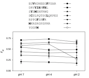
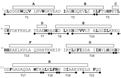
2.3.2 Validation of FPOP Strategy Using Test Peptides ... 71
2.3.3 Intact Protein Analyses ... 74
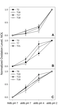
2.3.4 Solvent Accessibility Measurements Using Normalized Oxidation Levels ... 76
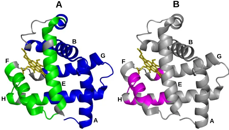
2.3.5 Structure of Native aMb ... 78
2.3.6 The pH 4 Intermediate ... 82
2.4 Conclusions ... 83
2.5 References ... 85
Chapter 3: Submillisecond Protein Folding Events Monitored by Rapid Mixing and Mass Spectrometry-Based Oxidative Labeling ... 91
3.1 Introduction ... 91
3.2 Experimental Section ... 95
3.2.1 Materials ... 95
3.2.2 Submillisecond Mixer Design ... 96
3.2.3 Mixer Characterization ... 97
viii
3.2.5 LC/ESI-MS and Data Analysis... 102
3.3 Results and Discussion ... 103
3.3.1 Global Solvent Accessibility Changes ... 104
3.3.2 Solvent Accessibility of Individual Protein Segments ... 107
3.3.3 Submillisecond Events ... 107
3.3.4 Folding Events Beyond 1 ms ... 112
3.4 Conclusions ... 113
3.5 References ... 115
Chapter 4: Partially Disordered Proteins Studied by Ion Mobility-Mass Spectrometry: Implications for the Preservation of Solution Phase Structure in the Gas Phase ... 122
4.1 Introduction ... 122
4.2 Experimental Section ... 126
4.2.1 Materials and Sample Preparation ... 126
4.2.2 Ion Mobility-Mass Spectrometry ... 126
4.2.3 Modeling ... 129
4.3 Results and Discussion ... 129
4.3.1 Mb Unfolding in Solution Monitored by Optical Spectroscopy... 129
4.3.2 Mb Unfolding Probed Via ESI Charge State Distributions ... 130
4.3.3 Ion Mobility Results ... 132
4.3.4 Collision Cross Sections of Model Structures ... 136
4.3.5 Effects of Solution Phase Structure on Gas Phase Conformation ... 137
4.3.6 Intensity-Weighted CCS Distributions ... 141
4.4 Conclusions ... 143
4.5 References ... 144
Chapter 5: H/D Exchange Mass Spectrometry Reveals Friction-Mediated Torsional Stress During FOF1 ATP Synthase Operation ... 153
5.1 Introduction ... 153
5.2 Experimental Section ... 155
5.2.1 Materials ... 155
5.2.2 Preparation of inside-out membrane vesicles... 155
ix
5.2.4 Hydrogen/Deuterium Exchange... 157
5.2.5 Liquid Chromatography-Mass Spectrometry ... 159
5.2.6 Peptide Mapping ... 161
5.2.7 Sequence coverage and proteomic profile of membranes ... 161
5.2.8 HDX Data Analysis ... 162
5.3 Results and Discussion ... 163
5.4 References ... 173
Chapter 6: Conclusions ... 177
6.1 Summary ... 177
6.2 Future Directions ... 180
6.2.1 Characterization of the Labeling Pulse in FPOP ... 180
6.2.2 Pushing the HDX-MS Envelope ... 182
6.3 References ... 183
Appendix I-Permissions ... 187
x
List of Symbols and Abbreviations
ADP - adenosine diphosphate
AFM - atomic force microscopy
AMP-PNP - adenosine5′-(β,γ-imido)triphosphate
ATP - adenosine triphosphate
CCS - collision cross section
CD - circular dichroism
CEM - chain ejection model
CEST - chemical exchange dependent saturation transfer
CID - collision-induced dissociation
CPMG - Carr-Purcell-Meiboom-Gill
CRM - charge residue model
Cryo-EM - cryogenic electron microscopy
cyt c - cytochrome c
DC - direct current
DESI - desorption electrospray ionization
E. coli - Escherichia coli
ECD - electron capture dissociation
ECD - electron capture dissociation
ESI - electrospray ionization
ETD - electron transfer dissociation
FCCP - carbonyl cyanide 4-(trifluoromethoxy)phenylhydrazone
xi
FRET - Förster resonant energy transfer
FT-ICR - Fourier transform ion cyclotron resonance
GEE - glycine ethyl ester
HDX - hydrogen/deuterium exchange
IDP - intrinsically disordered proteins
IEM - ion evaporation model
IMS - ion mobility spectrometry
LC - liquid chromatography
MAIV - matrix assisted ionization vacuum
MD - molecular dynamics
MS - mass spectrometry
Mb - myoglobin
N - native state
NMR - nuclear magnetic resonance
NOESY - nuclear Overhauser effect spectroscopy
·OH - hydroxyl radical
PDB - protein data bank
PEP - phospho(enol)pyruvic acid
PMF - proton motive force
PTM - post translational modifications
RF - radio frequency
xii
SDS PAGE - sodium dodecyl sulfate polyacrylamide gel electrophoresis
τ - radical lifetime
[] - mean residue ellipticity
t1/2 - half-life
TEMPO - 2,2,6,6-Tetramethylpiperidin-1-yl)oxyl
tricine - N-(2-hydroxy-1,1-bis(hydroxymethyl)ethyl)glycine
TROSY - transverse relaxation optimized spectroscopy
TWIM - travelling-wave ion mobility
UPLC – ultra performance liquid chromatography
XFEL - X-ray free electron lasers
1
Chapter 1
1
Chapter 1: Introduction
1.1
The protein structure-function paradigm
1.1.1
The Basics of Protein Structure and Folding
Proteins are the molecular devices through which the genetic information encoded in the
DNA is put into action. They are highly abundant and occur in all cells and come in a
wide variety of sizes; from relatively small proteins to huge macromolecules with
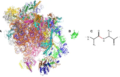
molecular weights in the MDa range (Figure 1.1A&B).
Figure 1.1 X-ray structures of (A) the 2.8 MDa human mitochondrial ribosome (PDB ID: 3J9M),
and (B) the 8.5 kDa human ubiquitin (PDB ID: 1UBQ). The ribosome is a ribonucleoprotein
complex. The structures are depicted to scale. (C) Structure of a two-residue polypeptide
segment. The peptide bond is drawn in red.
A B
… N
H N
H O
Rn O
Rn+1
2
Proteins exhibit astonishing diversity in terms of their biological function. Tasks
such as cellular respiration, enzyme catalysis, signal transduction, protein synthesis and
degradation are performed by proteins (1). Despite their structural and functional
diversity, all proteins are made up of the same ubiquitous 20 naturally occurring amino
acids (Figure 1.1C). The protein structure-function paradigm holds that proteins need to
fold into a specific three-dimensional structure to carry out their functions. How proteins
are able to find this structure, called the native state (N), remains an active area of
research (2).
1.1.2
Protein Structural Hierarchy
The native structure of proteins can be split into four categories. The primary structure of
the protein refers to the amino acid sequence of the polypeptide chain (Figure 1.1C). The
amide and carbonyl groups are the same for all amino acids. Therefore the side chains (R
groups) embody key properties such as size, electric charge, and hydrophobicity (1). The
many amino acids comprising a protein are connected via peptide bonds (Figure 1.1C -
highlighted in red), which exhibits partial double bond character. Except for the peptide
bond, which almost always adopts a trans configuration, free rotation around other bonds
along the protein backbone paves the way for the existence of countless
three-dimensional structures. In addition to the choice of amino acids, the cellular machinery is
armed with post translational modifications (PTMs) to further regulate structure and
function (3). Secondary structure refers to local structural arrangements such as α-helices,
β-sheets, etc. (1). Tertiary structure of proteins defines the association of the secondary
3
complexes, quaternary structure determines the overall geometry and arrangement of
individual subunits relative to one another within protein complexes (1).
1.1.3
Factors Contributing to Protein Stability
What are the driving forces behind protein folding? The second law of
thermodynamics posits that the native state of protein is the conformation with the lowest
free energy (4). At first glance, protein folding might seem to contradict this because
conversion of an unfolded chain to a compact structure is concomitant with a significant
loss of conformational entropy. The following factors contribute to the stability of the
folded state of proteins.
The hydrophobic effect and interactions are widely considered to be the most
important factor contributing to the stability of native proteins (5, 6) Six of the twenty
naturally occurring amino acids are highly hydrophobic, with another eight having
intermediate hydrophobicity. Globular proteins form tightly packed structures, within the
cores of which the hydrophobic amino acids side chains are buried and sequestered from
the surrounding water. By contrast, amino acids with charged and polar side chains are
often found on the protein’s surface where they interact with the surrounding water.
Theoretical studies suggest that hydrophobic forces alone are adequate to cause the
folding of proteins with ca. 200 amino acids (7).
Hydrogen bonding is the hallmark of the secondary structural elements of proteins
(8). In -helices the amide hydrogen (N-H) is hydrogen bonded to the oxygen of the
carbonyl group of the amino acid four residues earlier. β-sheets also participate in
4
bonding because these moieties can form hydrogen bonds with water even in the
unfolded state (9).
van der Waals interactions or“self-solvation”isoftencitedasanimportantfactor
(10). The tight packing of atoms within the compact structure of folded protein implies
the importance of close-range interactions. These include forces between pairs of
permanent and induced dipoles.
Salt bridges originate from electrostatic interactions between the negatively
charged carboxylate (RCOO−) of Asp and Glu residues and the cationic side chains of
Lys and Arg (11). Other residues with ionizable side chains such as His can also
participate, depending on solution pH and their pKa. Salt bridges on the surface of
proteins do not contribute much to stability. This is because they are heavily solvated by
water. Shielding of salt bridges from water and burying them in the interior is
energetically unfavorable. These factors severely limit the ability of salt bridges to
stabilize the native state. Some proposals even outline a destabilizing role for salt bridges
(12).
Conformational (chain) entropy is by far the strongest force opposing the
formation of the folded protein (13). Protein folding is concomitant with a large loss in
chain entropy as the polypeptide coils up into its compact native state from its many open
denatured configurations.
1.1.4
Protein Folding Mechanisms
In a series of elaborate experiments that culminated in the 1972 Nobel Prize, Christian
5
sequence (14). The Anfinsen dogma postulates that the native structure is a unique, stable
and kinetically accessible structure that represents the minimum free energy of the
protein and its surrounding solvent. This finding gave birth to protein folding research.
The astounding speed of protein folding has been of special interest since the
early days. Using conservative estimates, a relatively small protein with 100 amino acids
can fold in ~10150 different ways. What is the mechanism via which disordered
polypeptide chains avoid the numerous unproductive avenues and find their way to the
native states (15, 16)? Do proteins fold by pre-determined “folding pathways” (17)? In
1969 Cyrus Levinthal pointed out that protein folding does not take place based on a trial
and error conformational search (18). Even in the unrealistic scenario where a protein
could try one different conformation every 10-50 seconds, folding would still take 10100
seconds. This value is 80 orders of magnitude longer than the age of the universe and
contradicts the experimentally observed ms-s folding times. This disparity in the
experimental and theoretical folding times represents one facet of the so-called “protein
foldingproblem”.
Statistical thermodynamics work from the Dill (19) and Wolynes (20) groups in
the late 80s addressedLevinthal’sparadox by introducing the energy landscapes concept.
Hyperdimensional free energy maps were conceptualized to display the energetics
preferences of polymer chains. For foldable polymer models, it was shown that a small
number of compact, low-energy conformational ensembles populate the bottom of
funnel-shaped energy landscapes. By contrast, there are numerous high energy unfolded
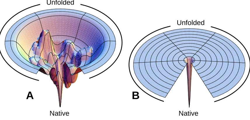
conformers that correspond to the rim of the funnel (Figure 1.2) (19). Protein folding
6
conformational search ensures that structures with increasingly lower free energies are
adopted. Calculations have shown that steps favourable by as little as kBT ensure rapid
folding times, consistent with experimental observations (21).
Figure 1.2 Protein folding landscapes. (A) Free energy landscape with multiple folding pathways
with a surface biased towards the native state (N). The ruggedness of the funnel leads to the
formation of transient protein folding intermediates (B) Folding on the Levinthal “golf course”
entails an unbiased and random search for the native state. Images taken from
http://dillgroup.stonybrook.edu and reproduced under a Creative Commons Attribution 4.0
International License.
While this framework addressed critical questions such as the speed of folding, a
molecular picture of how the disordered protein transitions into the native state was
lacking. A popular approach to remedy this is characterizing the properties of the
transient folding intermediates (local minima on the bumpy funnel) which are populated
during the folding process (22, 23). This approach relies on the tenet that snapshots of the
chain during folding would give insights into a general rule-book based on which
proteins fold. The combination of hydrogen/deuterium exchange and quench flow mixing
A
B
Native Native
Unfolded
7
(see Section 1.4.1) has proven to be highly effective (23). In the case of some proteins,
conformers that highly resemble those found in kinetic experiments can be populated
under semi-denaturing solution composition (24). Once the similarity of the structures is
validated, this approach allows detailed structural characterization under equilibrium
conditions (25).
Several protein folding models have been proposed that attempt to capture the
structural transitions that ultimately yield the native state (26). The diffusion-collision
model divides the protein into several microdomains small enough for all conformations
to be searched through rapidly (27). Coalescence of these unstable microdomains yields
the native state. The framework model assigns a dominant role to local forces (28). This
model predicts that the formation of secondary structure elements precedes chain collapse
and tertiary interactions. The nucleation-condensation model proposes that the folding
process initiates from a nucleus that consists primarily of adjacent residues (22). Folding
proceeds because this nucleus is not stable unless it makes tertiary interactions with the
surrounding residues. Secondary and tertiary native state contacts grow concurrently as
the nucleus grows rapidly. Fersht and coworkers have proposed a unifying folding
mechanism that envisages the framework and nucleation-condensation models to be
different manifestations of an underlying common mechanism (29). Proteins may appear
to fold via either model depending on the stability of folding intermediates and the height
of the transition barriers. The hydrophobic collapse model posits that hydrophobic forces
result in polypeptide chain collapse early in the folding process and long-range
interactions precede or are concomitant with the formation of local contacts such as
8
1.1.5
Protein Dynamics
The advent of high resolution structural methods over the past half-century have led to a
great many atomic-resolution models of proteins essential to life. Although studies of
static three-dimensional structures determined via crystallography and other methods are
crucial (see Section 1.2), proteins in solution are highly dynamic. It is becoming
increasingly clear that a complete description of biological function must take into
account conformational motions that can manifest over a broad range of time and length
scales. Hence, the structure-function paradigm has to be extended to include dynamics
(32, 33). This is akin to the situation where a few photographs snapped during a hockey
game do not reflect the entire process of scoring goals. Proteins undergo a variety of
conformational changes that enable them to function as catalysts (34), ion transporters
(35), signaling switches (36), etc. Homologous protein structures exhibit disparate
dynamics, spanning order of magnitude in time, thereby underscoring the importance of
this aspect (37).
The concept of energy landscapes can also be applied within the context of the
native protein. Although the energy well corresponding to the native state in Figure 1.2
looks deep, ruggedness within this energy well gives rise to substates that are separated
by barriers with varying heights. Similar to protein folding, the energy difference
between the states and the height of the barriers separating them gives rise to protein
dynamics on various time scales (38). Slow dynamics correspond to μs-ms timescale
interconversion between substates separated by energy barriers of several kBT.
9
milliseconds. Proteins are not static within these substates. They undergo numerous
small-amplitude fluctuations around the average structure on the ps-ns timescale. The
energy to cross the barrier for these faster dynamics is readily available under ambient
conditions as the interconverting states are separated by less than 1 kBT. This gives rise to
loop motions and sidechain rotations.
1.1.6
Protein Misfolding
Studying the structural and motional properties of proteins goes beyond a
curiosity-driven scientific endeavour. Only proteins that fold properly possess the stability to
function robustly in the cellular environment. Close to 50 disorders such as Alzheimer’s
disease, spongiform encephalopathies, Parkinson’sdisease, Senile systemic amyloidosis,
and type II diabetes have been linked with the misfolding of normally soluble, peptides
and proteins, and their subsequent conversion into insoluble toxic aggregates (39). While
protein folding started as curious scientific discovery, contemporary protein research is
motivated by this biomedical aspect and its implications for human health (40).
Understanding of the fundamentals that govern biomolecular structure, folding, and
interactions will aid in the development of therapeutics.
1.2
Experimental Methods to Study Protein Structural and
Motional Properties
1.2.1
X-ray Crystallography
X-ray crystallography is the foremost technique for determining the atomic structure of
proteins. The importance of this method cannot be overstated: crystallographic projects
10
first protein structure in 1958 (42). The Protein Data Bank (PDB) currently contains more
than 100,000 structures, of which ca. 90% were determined by X-ray crystallography.
In the beginning of the 20th century, Max von Laue and the Braggs worked out
much of the theory behind the interaction of X-ray beams and material (43). X-rays that
pass through a crystal scatter off the electrons in the crystal lattice and then interfere with
each other. In some instances, the electromagnetic waves undergo constructive
interference; in others, they cancel each other out. The result is a diffraction pattern from
which the electron density that scattered the original beam can be calculated. This is
followed by model building and structure refinement. The average structure resolution in
the PDB is around 2 Å. Distinguishing individual atoms within the sample is possible for
resolutions of ca. 1 Å (41). Resolution suffers in images of complex samples due to
imperfections and incoherent motions within crystals (43, 44).
X-ray crystallography requires several milligrams of relatively pure material.
Some proteins, in particular those containing dynamic regions, are not amenable to X-ray
diffraction methods because they do not form crystals. The excision of such domains
gives truncated constructs that sometimes form crystals more readily. However, this
strategy may perturb the protein structure. Another source of artefacts comes from within
the crystals. Protein-protein contacts in the tightly packed environment of crystals have
been shown to differ from those in solution (45). Crystallography of membrane proteins
continues to lag far behind that of soluble targets (41). X-ray data provide snapshots of
the protein ground state, and generally do not report on dynamics. An interesting
development has been synergistic utilization of NMR-based order parameters and X-ray
11
Some of the limitations mentioned are expected to be overcome with the more
widespread implementation of X-ray free electron lasers (XFEL) (46, 47). Some early
signs of success have already emerged. A notable example is the determination of the
photosystem II structure at 1.95 Å using serial femtosecond crystallography (48, 49).
1.2.2
Cryogenic Electron Microscopy (Cryo-EM)
Cryo-EM has been revolutionized over the past decade and holds great potential as a
method for the structure determination of large biomolecular assemblies (50). The advent
of electron guns with enhanced brightness and coherence, along with advances in
specimen handling and processing methods have improved resolution from several
nanometers to the sub-nanometer regime (51). The biggest advantage of cryo-EM over
X-ray crystallography is that structural information is obtained from small quantities of
noncrystalline material. It is best suited for probing large (>200-300 kDa), rigid protein
complexes that possess some form of symmetry. Several studies have revealed the
architecture of systems that have long been intractable by X-ray crystallography. These
include 6.9 Å structure of V-type ATP synthase from Saccharomyces cerevisiae (52), 6.2
Å structure of the a-subunit of F-type ATPase, (53) and 11 Å structure of chromatin
fibers (54). A hybrid approach that is being increasingly applied entails fitting
atomic-level models of domains and smaller fragments obtained from other methods into
cryo-EM density maps. This methodology marries the best attributes of the two methods and
has proven to be highly valuable for studying a number of challenging targets. A pair of
ground-breaking studies from the Baumeister (55) and the Martin (56) groups focusing
12
Despite these notable examples, studying smaller and flexible targets by cryo-EM
remains challenging. Degradation of the structural integrity of the specimen during
analysis is one of the biggest hurdles the method faces. Similar to X-ray crystallography,
information about protein conformational dynamics are currently unavailable via
cryo-EM. Continued improvement in the areas described above promises that cryo-EM will
play an increasingly prominent role in structural biology (57).
1.2.3
Nuclear Magnetic Resonance (NMR) Spectroscopy
NMR spectroscopy is uniquely suited for characterizing biomolecular structure and
dynamics (58). NMR employs exploits high magnetic fields and radio frequency to probe
NMR active nuclei such as 1H, 13C, and 15N within a macromolecule as “spies” of protein
structure and motion. Unlike X-ray crystallography (Section 1.2.1) and cryo-EM
(Section 1.2.2), NMR offers the advantage of studying biomolecules under physiological
conditions (59) or under favorable conditions in living cells (60).
NMR has played a significant role in protein folding research as the detection
method for hydrogen/deuterium exchange measurement of pulsed-labeled kinetic protein
folding intermediates (23). The development of three-dimensional triple-resonance
experiments (61) and transverse relaxation optimized spectroscopy (TROSY)
experiments (62) mitigated spectral overlap and enabled studying of large proteins. The
advent of Carr-Purcell-Meiboom-Gill (CPMG) relaxation dispersion (63) and chemical
exchange dependent saturation transfer (CEST) (64) experiments have enabled detailed
characterization of energetic of the excited states of protein that are weakly populated
13
advanced computational modeling produced atomically-resolved models of folding
intermediates that have shaped our understanding of protein folding and misfolding (65,
66).
Wüthrich and coworkers (Nobel Prize Laureate 2002) developed the Nuclear
Overhauser effect spectroscopy (NOESY) experiments that yield inter-nuclear distance
constraints and 3D structures of proteins up to ca. 50 kDa (67). In addition, an extensive
array of NMR experiments has been developed that quantify biomolecular dynamics over
a wide range of timescales, from picoseconds to days. R1ρ, ZZ exchange, and relaxation
dispersionexperimentsaresuitedtoprobetheμs-ms regime. R1, R2 pulse sequences are
used to investigate faster dynamic events. Studying the dynamics of large protein
complexes was recently made possible via the application of methyl-TROSY experiments
and isotopic labeling of methyl-containing side chains (68). High quality 13C-1H
correlation maps for all six methyl-containing amino acids (Ile, Leu, Val, Ala, Thr, and
Met) of systems with masses exceeding 1 MDa have been recorded (69). Notable
examples include the interrogation of the archaeal proteasome in complex with various
activators (70) and activation mechanism of the Hsp70 molecular chaperones (71).
Despite these advances, NMR still struggles with large and asymmetric
multi-subunit complexes. Spectra from large asymmetric systems suffer from extensive
overlap. in vitro reconstitution of the complex from separately expressed NMR active and
NMR inactive subunits can be applied in a limited number of cases. First steps to remedy
these shortcomings have already been taken (72). The ever-increasing magnetic field
strengths, more elaborate sequential protein co-expression methodologies and NMR pulse
14
1.2.4
Optical spectroscopy
The vast majority of early protein folding experiments that elucidated the kinetics of the
folding process were performed using optical methods. These include UV-Vis
absorbance, far and near UV-Vis, circular dichroism (CD), fluorescence and Förster
resonance energy transfer (FRET) spectroscopy, infrared (IR) and Raman spectroscopy,
among other methods.
1.2.4.1
Circular dichroism (CD) spectroscopy
Far-UV (190-260 nm) and near-UV (250-350 nm) CD spectroscopy provide a measure of
average secondary and tertiary structure content of proteins, respectively. The
combination of this methodology with stopped-flow mixing and rapid-mixing devices has
been a valuable source of information about the kinetics of protein folding (73).
Early stopped-flow data on model proteins showed that a substantial fraction of
the overall change in the far-UV CD signal resulting from refolding falls within the
instrumental dead time of approximately 1 ms. Meanwhile, the near-UV spectroscopic
signature did not reach native levels until the later stages of the folding process (74).
These results were generally interpreted as formation of intermediates with substantial
secondary structure but lacking native-like tertiary structure packing. Later on the advent
of faster turbulent- and continues-mixing methods (75) and temperature-jump triggering
helped resolve the kinetics down to the ns/μs time scale (76). However, the details of
structural reorganization corresponding to the early folding events remained nebulous for
15
1.2.4.2
Fluorescence
The fluorescence emission of Trp, and to a lesser extent Tyr, side chains can serve as a
valuable reporter of the local environment of these moieties (73). Although complex
photophysical phenomena complicate data interpretation, generally a solvent-exposed
tryptophan in the unfolded state of a protein shows a broad emission spectrum similar to
that of free tryptophan or its derivative N-acetyl-L-tryptophanamide (NATA) (λmax 350
nm). Shielding of the tryptophan side chain within the hydrophobic core of native
proteins or in a compact folding intermediate typically results in a major blue shift of the
emission maximum and enhanced fluorescence yield. Within the native state of proteins,
amino acid side chains of Lys, Tyr, Gln, Asn, Glu, Asp, Cys, and His quench
fluorescence by one of intersystem crossing, solvent quenching, excited-state proton and
electron transfer mechanisms (78). The presence of multiple fluorophores can be a source
of complication. Site-directed mutagenesis can be used to remove multiple fluorophores.
A Trp-scanning strategy has been devised to characterize the structure of transient folding
intermediates (79).
Similar to CD spectroscopy, monitoring fluorescence intensity as a function of
folding or unfolding time can provide useful information on solvent accessibility and
proximity to quenchers of an individual fluorescence probe. It serves as an exquisite
method to study early folding events. Classic examples include misfolding of cytochrome
c due to initial misligation of the heme iron by His33 (80), and implications of proline
16
1.2.4.3
Förster resonant energy transfer (FRET)
In FRET a donor fluorescent dye is excited by a light source. This energy can
subsequently be transferred nonradiatively to an acceptor dye. One of the requirements of
FRET is that measurements should be performed between a pair of fluorophores where
the emission of the donor dye overlaps with the absorption of the acceptor. As the two
molecules come closer together, the donor emission decreases in intensity while the
acceptor emission increases. FRET efficiency depends on the donor-to-acceptor distance
withaninverse6thpower,andisthusreferredtoas”molecularruler”. It is most sensitive
to measurement between 2-8 nm range (73).
Both fluorescence and FRET are valuable methods for characterizing the
conformation of gas phase molecules (82, 83). Comparison of solution phase data to
those obtained in the gas phase can reveal the nature of structural changes of gas phase
proteins. The first steps towards this direction have been taken (84).
1.2.4.4
Single-Molecule Methods
Single-molecule experiments report on the motions of individual protein
molecules and help to understand how this translates into an ensemble signal reported by
other methods. The applications of single-molecule fluorescence spectroscopy and FRET
have been particularly useful in this regard (85, 86). This approach was used to measure
the length of protein folding barrier-crossing events (87). Perhaps one of the most
mesmerizing applications of this methodology was direct observation of the operation of
F1 ATPase rotary molecular machine. Afluorescentactinfilamentwasattachedtotheγ
17
therotationofthemotionof“shaft”andtheattachedfilament.Thismotioncanbe“seen”
directly using a fluorescence microscope (88). FRET-based measurements with a donor
ontheγsubunitandanacceptoronthestationaryperipheralstalkhave revealed a great
deal regarding the torque, speed, force generation, efficiency of this system (89). The
rapid nature and specificity of these optical methods allow observation of individual
turnover events. This technology has also enabled temporal mapping of protein energy
landscape in vivo (90). Single-molecule force spectroscopy techniques such optical trap
force spectroscopy and atomic force microscopy monitor the response of proteins to force
(91). Recent examples utilize this technology to unravel the mechanism of action of
chaperones on substrate proteins (92), and the importance of nascent protein folding (93).
1.2.5
Computational Methods
Despite the success of experimental techniques in providing information on structure and
dynamics of proteins, they are generally limited in their spatial and temporal resolution.
With the exception of single-molecule measurements (Section 1.2.4), most methods
report ensemble-average properties rather than individual molecules. An alternative is
computational modeling of biomolecules.
All-atom molecular dynamics (MD) simulation have become the method of
choice for simulating the motions of biomolecules (94). In MD simulations the positions
and velocities of every atom in the system is evolved according to first-principles laws of
physics. The forces acting upon particles representing atoms are calculated using a model
known as a force field (95). Dramatic improvements in computation power and parallel
18
biochemist, David E. Shaw, have pushed the length of these simulation onto the
millisecond time scale (96). The combination of these have shed new light on protein
folding (97-99), drug binding (100, 101), protein design (102), and the conformational
changes critical to enzyme function (103). The faithfulness of this methodology promises
to increase as accuracy of force fields improves (104).
1.3
Fundamentals of Mass Spectrometry
The origins of mass spectrometry (MS) go back to Sir Joseph Thomson more than a
century ago (105). Today MS is among the most widely used analytical techniques. An
ion source and a mass analyzer are the two main elements of each mass spectrometer. A
mass spectrometer reports the mass-to-charge ratio (m/z) of gaseous ions produced by the
ion source.
1.3.1
Electrospray Ionization
MS of biological macromolecules (e.g. proteins and DNA) was not possible prior to the
late 1980s. Electron impact (EI) and other traditional ionization methods were ineffective
for these analytes (106). Two ground-breaking inventions that culminated in a Nobel
Prize completely changed this picture. John Fenn and Koichi Tanaka shared the 2002
Chemistry Nobel Prize for inventing electrospray ionization (ESI) (107) and
matrix-assisted laser desorption/ionization (MALDI) (108), respectively. More than two decades
later, a suite of “soft” ionization methods such as desorption electrospray ionization
(DESI) (109) and matrix assisted ionization vacuum (MAIV) (110) have been developed.
19
without the breakage of any covalent bonds. ESI boasts the advantage of facile online
coupling with liquid chromatography (LC) for separation of complex mixtures prior to
MS. Because this thesis exclusively employed ESI, other ionization methods will not be
discussed.
ESI generates multiply charged [M + zH]z+ ions. No practical size limit has been
found for analytes compatible with ESI. The current record represents a 18 megadalton
virus assembly (111) Multiple charging lowers the m/z of these large entities to a range
where most mass spectrometers can operate. High charge states are also desirable for
applications involving collision induced dissociation (CID) (112), and electron-mediated
dissociation (113). The ESI entails infusion of the analyte solution through a capillary
held at an electric potential of several kV. The Taylor cone that forms at the tip of this
capillary emits charged droplets that undergo multiple rounds of solvent evaporation and
jet fission. These yield nanodroplets that are charged close to the Rayleigh limit. The
mechanism via which these nanodroplets generate gaseous ions has been a hot research
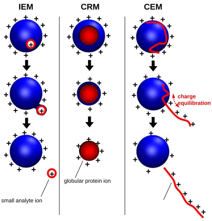
topic since the inception of ESI. The current consensus (114, 115) is that
low-molecular-weight analytes follow the ion evaporation model (IEM) (Figure 1.3, first column)
whereby gaseous ions are ejected from the nanodroplet surface by field emission (116).
By contrast, large globular species such as folded proteins follow the charge residue
model (CRM) and are produced by evaporation of droplets to dryness (Figure 1.3, second
column) (117). It has been proposed that disordered polymers and unfolded proteins are
20
Figure 1.3 Summary of ESI mechanisms. IEM: Small ion ejection from a charged droplet. CRM:
Release of a globular protein into the gas phase. CEM: Ejection of an unfolded protein chain.
Charge equilibration is indicated by red arrows. Figure reproduced with permission from
©American Chemical Society (114).
1.3.2
Mass Analyzers
The mass analyzer is tasked with measuring the m/z and relative abundance of gaseous
ions produced by the ion source. There are various types of mass analyzers including
+
+
+
+
+
+
+
+
+
+
+
+
IEM
CRM
CEM
+
+
+
+
+
+
+
+
+
+
+
+
+
+
+
+
+
+
+
+
+
+
+
+
+
+
+
+
+
+
+
+
+
+
+
+
+
+
+
+
+
+
+
+
+
+
+
+
+
+
+
+
+
+
+
+
+
+
+
+
+
+
+
+
+
+
+
+
+
+
+
+
+
+
+
+
+
+
+
+
+
+
+
+
+
+
small analyte ion
globular protein ion
charge equilibration
21
quadrupole, time-of-flight (TOF), ion traps, Orbitraps, and Fourier transform ion
cyclotron resonance (FT-ICR) mass analyzers. Because this work exclusively employed
quadrupole-TOF instruments, only these two mass analyzers will be discussed in detail.
1.3.2.1
Quadrupole Mass Analyzer
A quadrupole mass analyzer is composed of four parallel cylindrical rods. A radio
frequency (RF) and a direct current (DC) are applied to opposing rod pairs. For a
combination of RF and DC voltages, only ions with a certain m/z have the proper
trajectory to traverse the quadrupole and reach the detector. By contrast, ions with
unstable trajectories undergo collisions with the rods and get neutralized. The stability of
an ion trajectory for any given RF and DC voltage is governed by the Mathieu equation.
The RF and DC voltages can be scanned such that the desired m/z range is covered. In
the absence of a DC voltage (RF-only mode), quadrupoles act as ion guides. Thus
quadrupoles are routinely used as versatile ion transmission and selection devices for
MS/MS applications (106).
Quadrupoles generally offer a limited resolution (R≃5000) and limited m/z range.
But modern devices can resolve up to 32,000 m/z and transmit ions up to 100,000 m/z in
RF-only mode. Despite their limited resolution and slow scanning times, quadrupoles are
very robust instruments and have remained a fixture in the pharmaceutical and
environmental labs (119). Triple-quadrupole instruments excel at targeted quantitative
22
1.3.2.2
Time of Flight (TOF) Mass Analyzer
A TOF mass analyzer separates ions based on the time it takes them to traverse a fight
tube. An ion pusher accelerates an ion with a mass to charge ratio of m/z in a flight tube
by application of a voltage, U. This leads to an ion velocity v according to
2
2 1
mv
zU 1.1
Rearrangement yields
(2 )1/2 m zU
v 1.2
The time t it takes an ion to traverse a flight tube with length l is given by
)1/2 (2 ) 1/2( / )1/2 2
( l U m z
zU m l v l
t 1.3
Equation 1.1 dictates that time of flight depends on the m/z of each ion. Ions with
different m/z have different drift times and hence are separated in the flight tube. Ions
with lower m/z will reach the detector first followed by their “heavier” counterparts.
Modern mass spectrometers use a time-to-digital device to convert flight times to m/z.
Reflectrons in orthogonal flight tubes are utilized to correct for energy dispersions in the
original ion packet (119).
The reflectron uses a constant electrostatic field to reverse the trajectory of ion
beam toward the detector. The more energetic ions penetrate deeper into the reflectron
and dwell longer before their trajectory is reflected towards the detector. The ion of the
same m/z and with lower initial energy will penetrate the reflectron later but will spend
less time changing direction within reflectron. This mechanism corrects for differences in
23
energy reach the detector at the same time and record the same flight time. Without a
reflectron, the TOF resolution is no better than R≃5000. Modern orthogonal TOF devices
offer superb sensitivity at R≃40,000 and sub ppm mass accuracy. The combination of the
quadrupole and TOF yields one of the most popular instrument designs for biomolecular
analysis known as Q-TOF (Figure 1.4).
Figure 1.4 Schematic of the Q-TOF instrument used in this thesis with components marked. The
red line indicates the ion trajectory.
1.4
Structural Mass Spectrometry of Proteins
At first glance the choice of MS as a tool for structural interrogation of proteins might
seem peculiar. Protein structural changes generally do not result in any change their
molecular weight, implying no change in the principle property measured by the mass
spectrometer, m/z. In addition, the vacuum of the mass spectrometer seems like an
unsuitable environment for proteins which have evolved to function in an aqueous
environment. Unlike NMR, MS was not initially developed as a structural biology tool.
The methodologies discussed in the following sections illustrate how the MS community
7.5 cm
Turbomolecular Pumps Scroll
Pump
Quadrupole Source
TWIG
Trap TWIG
IMS TWIG
Transfer
TWIG Pusher Detector Sampling Cone
Extraction Cone “Backing”
Analyte
24
has collectively defied all odds in building a vast arsenal of techniques to report on
protein structure, folding and dynamics (121-123).
1.4.1
Hydrogen/Deuterium Exchange (HDX)
The concept of HDX was introduced more than six decades ago by Linderstrøm-Lang
(124). HDX exploits the incessant exchange of labile hydrogens within the protein with
those of the solvent. Labile sites include the amide backbone hydrogen and hydrogens
with sufficient acidity on the amino acid side chains. Disordered and solvent-exposed
segments of the protein readily exchange their hydrogens upon exposure to D2O. By
contrast, this process is orders of magnitude slower for regions involved in
hydrogen-bonding networks (e.g.α-helicesandβ-sheets) or those occluded from the solvent (125).
Measurement in the early days used scintillation counting (126) and
UV-spectrophotometry (127). HDX enjoyed great success when NMR-based methods were
employed (128). MS-based measurement were first introduced in the early 1990s (129,
130). HDX is an indispensable tool for monitoring the structure and dynamics of proteins
(131, 132).
Mass spectrometry-based measurements exploit the mass difference between
hydrogen (H) and deuterium (D). In the typical bottom-up experiments, the exchange
process is initiated by diluting the protein into a D2O-based buffer. HDX is then allowed
to proceed for different incubation time periods (continuous-labeling). Subsequently,
exchange is quenched by lowering the pH and temperature. This is followed by digestion
by an acid-active protease (e.g. pepsin), and LC-MS analysis. HDX and other labeling
25
arise from analysis of proteins in the gas phase. MS simply serves as a sensitive tool for
monitoring the amide deuteration pattern imprinted on the protein backbone in solution.
The elegance of HDX lies in the benign nature of the label and its relatively
simple workflow. One of the biggest hurdles experimentalists have to deal with is “back
exchange” during post-labeling sample handling. This spurious effect tends to degrade
the structurally informative deuteration pattern (133). The half-life (t1/2) of an
unstructured typical amide at pH 7.0, 25 °C is under 1 second. This imposes limitation on
all down-stream sample handling protocols. LC-MS separation and digestions are
required to be carried out near 0 °C and at pH ~2.7 where t1/2 is on the order of ~1 hour,
thereby providing enough time to carry out the MS measurement. Short LC gradients on
the order of 10-15 minutes are often employed. Despite these efforts back exchange is not
completely suppressed. The side chain deuterium is entirely lost due to back exchange,
and backbone amides’ deuterium content can be measured with a 30-40% back exchange.
The use of anti-freeze compounds in LC solvents enables sub-zero LC and improved
deuterium retention (134, 135) The application of rapid capillary electrophoresis
separation is a new method to shorten the peptide separation step (136).
The spatial resolution of bottom-up HDX-MS depends on the length of the
generated peptides, typically around 10 residues. Middle-down and top-down
methodologies employing electron capture dissociation (ECD) or electron transfer
dissociation (ETD) have been described that enable measurements down to the
single-residue level (137). The application of ultraviolet-photodissociation for single-single-residue
HDX measurements is an exciting but unexplored area (138). An alternative to the
26
generates a great many overlapping peptides (139-142) However, occurrence of
differential back exchange for overlapping peptides and error propagation complicate its
application (143).
Backbone amide HDX can proceed via acid- or based-catalyzed mechanisms
(131). The intrinsic rate of exchange (kch) for a completely unstructured polypeptide can
be described according to:
kch kacid[H]kbase[OH] 1.4
where kacid and kbase are the rate constants for the acid- and base-catalyzed reactions,
respectively. Base catalysis is dominant under physiological conditions suitable for
studying proteins.
The currently accepted model for HDX divides amide hydrogens into two
categories, “open” and “closed”, in dynamic equilibrium with each other with rates of
interconversion kop and kcl according to:
D O open
k open k
k
closed N H N D
H
N ch
cl op
2 1.5
This model posits that exchange can only occur for “open” hydrogen amides. These
correspond to solvent-accessible amide hydrogens that have become disengaged from
hydrogen bonding due to the conformational fluctuations described in Section 1.1.5. By
contrast, “closed” amide hydrogens are exchange-incompetent because they are tightly
hydrogen-bonded or not within the reach of the solvent. Once the “open” state is
populated, deuteration proceeds with the intrinsic rate constant kch. Kinetic treatment of
27
[NH]exp(kHDXt) 1.6
where [N-H] refers to the concentration of a single undeuterated amide after exposure
time t and
ch cl op ch op HDX k k k k k k 1.7
Stably hydrogen-bonded amides with transient opening events typical of thermal
fluctuations give rise to EX2 kinetic (kcl >> kch). In this scenario eq 1.7 simplifies to
ch cl op HDX k k k
k 1.8
Under these conditions a large number of opening events is required for backbone amides
to exchange. This results in isotopic distributions that shift to higher m/z over time as the
protein undergoes more and more opening events. By contrast, long-lived global
unfolding events lead to the less common EX1 kinetics (kcl << kch) that manifest as
bimodal isotopic envelops.
The model described above invokes a combination of hydrogen-bonding and
solvent accessibility as the main determinants of exchange events (125). A few studies
employing MD simulations have appeared in the literature that attempt to improve upon
this rather simplistic model (144). Deeper insights into the exact nature of the
conformational excursions that give rise to HDX should become available as the time
scale accessible by MD simulations approaches experimental HDX timescale (145).
Although HDX does not routinely deliver the quantitative information that NMR
provides, it is an excellent probe of protein structure and dynamics (131, 132).
28
of studies found in the literature (146). Here the deuteration pattern of the protein of
interest is compared in the bound and unbound forms. Binding often results a pronounced
reduction in deuterium uptake in the vicinity of the binding site (131, 147), although
other scenarios are also possible (148). This methodology has been applied to map the
interaction of agonists with receptor proteins. Another area of significant interest is HDX
of proteins implicated in neurodegeneration such as the amyloid β peptide (149),
α-synuclein (150), prion protein (151). HDX is also starting to gain a foothold in the
pharmaceutical industry for assessing the structural integrity of monoclonal antibodies
(152) and development of therapeutics (153, 154). HDX is also an exquisite tool for
monitoring light-induced conformational changes (155, 156). Despite these advances,
studies of membrane proteins continue to lag far behind soluble systems (157).
The examples described above correspond to studies performed under equilibrium
conditions. From eq 1.4 it is obvious that pH affects the labeling rate (158, 159). This
property is exploited in pulsed-labeling studies of kinetic events such as protein folding.
After triggering folding in a rapid mixing device, the protein is pulsed for a brief time
period (often a few ms at pD 8-10), followed by quenching and LC-MS. The
discriminating labeling HDX rate under these conditions (103 s-1 at pH 9) provides a
binaryreadoutoffolded(“closed”)andunfolded(“open”)elementsastheproteinfolds.
The folding of α1-antitrypsin to its metastable structure (160) and ribonuclease H (161),
and the effect of the GroEL/ES chaperone system on the folding mechanism of proteins