1071-412X/96/$04.0010
Copyrightq1996, American Society for Microbiology
Silicate Antibodies in Women with Silicone Breast Implants:
Development of an Assay for Detection
of Humoral Immunity
GUO-QIU SHEN,* EMMANUEL A. OJO-AMAIZE, MELKON S. AGOPIAN,
AND
JAMES B. PETER
Specialty Laboratories, Inc., Santa Monica, California 90404-3900
Received 28 September 1995/Returned for modification 14 December 1995/Accepted 4 January 1996
Silicon, in the form of sodium silicate (Na
2SiO
3), adsorbed onto bovine serum albumin (BSA)-precoated
plates served as the solid-phase antigen in an enzyme immunoassay to detect silicate-reactive antibodies in the
plasma of 40 symptomatic women with silicone breast implants, 91 asymptomatic women with silicone breast
implants, 50 healthy control women, and 52 women with rheumatic diseases and without silicone breast
implants. Silicate-reactive antibodies of immunoglobulin G (IgG) or IgM isotypes were detected in the plasma
of 30% (12 of 40) of the symptomatic women with silicone breast implants; 9% (8 of 91) of the asymptomatic
women with silicone breast implants; 5% (1 of 20) of the women without implants who had systemic lupus
erythematosus; and 0% (0 of 32) of the women without implants who had either Sjo
¨gren syndrome,
sclero-derma, or rheumatoid arthritis. Only 2% (1 of 50) of the sera from the healthy control women contained
silicate-reactive antibodies. Preincubation of sera with silicate and eight other metal compounds (including
SiO
2) demonstrated that the IgG and IgM antibodies bound specifically to silicate, because preincubation with
Na
2SiO
3inhibited more than 90% of the activity, whereas CrO
3, Li
2SO
4, MgSO
4, NiSO
4, HgCl
2, ZrOCl
2,
BeSO
4, and SiO
2failed to inhibit the IgG or IgM antibody binding to the silicate-BSA plates. Furthermore, the
F(ab
*
)
2portion and not the Fc portion of the silicate-reactive IgG was reactive with BSA-bound silicate in the
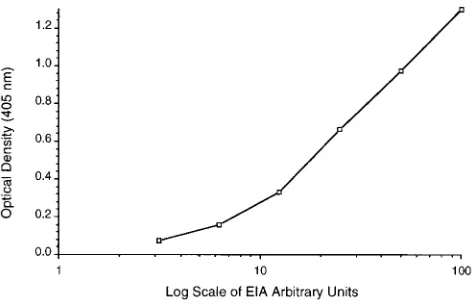
enzyme immunoassay. The assay for silicate-reactive antibodies was quantified by assigning arbitrary units to
a standard curve composed of serial twofold dilutions of high-positive (ten times higher than the cutoff) silicate
antibody sera. This novel assay is a useful method for detecting and quantifying humoral immune response to
silicate.
Organic compounds of silicon (Si) compose the silicone
tet-ramer (polydimethylsiloxane), which is used in breast implant
prostheses (2, 3). Although it was once thought that products
composed of silicon in various forms were inert and
nonim-munogenic, recent studies show that specific immune
re-sponses can be generated against silicone
(polydimethylsilox-ane) solutions (8, 9, 11, 12, 16, 20). Although there are no solid
cause-and-effect data as yet, several reports of different
auto-immune conditions in women with silicone breast implants
(17–19) suggest that silicone may have an adverse effect on the
body’s immune system. To this end, certain studies have
re-cently focused on developing laboratory methods for detecting
cellular (16) and humoral (8, 12, 20) immune responses to Si
compounds, including SiO
2, silicate, and silicone, herein
col-lectively referred to as silicon(e). Of the tests for assessment of
humoral immunity to silicon(e) previously reported (8, 12, 20),
none used silicate (Na
2SiO
3) and none were quantitative. In
this study, we developed an enzyme immunoassay (EIA) for
detecting and quantitating specific immunoglobulin G (IgG)
and IgM antibodies to bovine serum albumin (BSA)-bound
silicate in the plasma of women with silicone breast implants
and compared the results with those for healthy women and
women with rheumatic diseases who did not have silicone
breast implants.
MATERIALS AND METHODS
Reagents.Lithium sulfate (Li2SO4), nickel sulfate hexahydrate (NiSO4z6H2O), chromic trioxide (CrO3), magnesium sulfate (MgSO4), and silicon dioxide (SiO2) were purchased from Sigma Chemical Company (St. Louis, Mo.). Other reagents and equipment included beryllium sulfate tetrahydrate (BeSO4z4H2O) (Aldrich Chemical Company, Inc., Milwaukee, Wis.), silicon in the form of sodium silicate (Na2SiO3) (Spex Industries, Inc., Edison, N.J.), magnesium chloride (MgCl2z 6H2O), sodium azide (NaN3), sodium carbonate (Na2CO3), sodium chloride (NaCl), sodium phosphate dibasic (Na2HPO4), sodium phosphate monobasic (NaH2PO4zH2O) (Spectrum Chemical Company, Gardena, Calif.), a microtiter plate reader and shaking platform (Tecan U.S., Hillsborough, N.C.), alkaline phosphatase-conjugated goat anti-human IgG and anti-human IgM (American Qualex, La Mirada, Calif.), Immulon 1 flat-bottom microtiter plates (Dynatech, Chantilly, Va.), fat-free BSA, Tween 20 (polyoxyethylene sorbitan monolaurate), and p-nitrophenyl phosphate disodium substrate (Sigma Chemical Company).
Blood donors: breast implant patients and controls.EDTA plasma was ob-tained by venipuncture from 50 healthy control women who did not have breast implants, 40 symptomatic women with silicone breast implants, and 91 asymp-tomatic women with silicone breast implants. Of the 40 sympasymp-tomatic women’s plasma samples, 31 were remnant samples from a previous study (16). These women had various symptoms, including fatigue, fibromyalgia, insomnia, skin disorders, joint pains, muscle cramps, arthritis, allergies, and arrhythmia. The other nine samples were sent to Specialty Laboratories for routine clinical test-ing. The samples from 91 asymptomatic women were obtained from Y. Shoen-feld (Tel Aviv, Israel) as controls used in a different study for patients with a variety of autoimmune diseases. All assays were done in a blinded fashion.
Plasma samples from 52 women without silicone implants but with different autoimmune disorders were also tested by EIA for the presence of silicate-reactive antibodies of either the IgG or the IgM isotype. Of the women without implants, 20 had systemic lupus erythematosus, 10 had Sjo¨gren syndrome, 10 had scleroderma, and 12 had rheumatoid arthritis.
Assay for silicate-specific antibodies.In preliminary experiments in which silicate was first used to coat plates and then subjected to blocking with BSA, a negative signal was obtained. Thus, it was determined that plates should be precoated with BSA before the addition of silicate. Antibodies to silicate (ad-sorbed to BSA) were assayed by EIA. Briefly, Immulon 1 microtiter plates were precoated with 200ml of 0.5% fat-free BSA in distilled water (dH2O) per well. After incubation at room temperature with shaking for 2 h, the plates were * Corresponding author. Mailing address: Specialty Laboratories,
Inc., 2211 Michigan Ave., Santa Monica, CA 90404-3900. Phone: (310) 828-6543, ext. 217. Fax: (310) 828-6634.
162
on August 17, 2020 by guest
http://cvi.asm.org/
washed three times with dH2O. One half of each plate was coated with 200ml of a solution containing 1 mg of Na2SiO3per ml of dH2O (optimal concentration; data not shown), and the other half (left uncoated and washed with dH2O) served as control wells without added silicate. After overnight incubation at 378C in zip-lock bags, the plates were washed three times with dH2O, air dried, wrapped in Parafilm, and stored at2208C until used. Portions (200ml) of plasma, diluted 1:100 in 0.5% BSA in phosphate-buffered saline (PBS)-Tween (a dilution found to be optimal and uniformly used in subsequent assays), were added to the appropriate wells; the plates were incubated overnight with shaking at room temperature. Following incubation, the plates were washed three times with PBS-Tween (0.05% Tween 20). Portions (200ml) of alkaline phosphatase-conjugated goat anti-human IgG or goat anti-human IgM (diluted 1:2,000 and 1:4,000, respectively) were added to the plates. After room temperature incuba-tion for 1 h with shaking, the plates were washed three times with PBS-Tween, blot dried, and, after addition of 200ml of substrate buffer containing 1 mg of p-nitrophenyl phosphate disodium per ml to all wells, shaken at 300 rpm for 10 to 15 min at room temperature. The plates were read at 405 nm on a Molecular Devices reader linked to the Softmax computer program. The calibration curve was constructed by plotting the optical density (OD) of the standards at 405 nm (y axis) versus arbitrary EIA units (x axis).
Na2SiO3titration.Various concentrations of Na2SiO3, ranging from 0.039 to 10 mg/ml, were added to the BSA-precoated plates. Two silicate antibody-positive plasma samples and one silicate antibody-negative control plasma sam-ple were used to establish an optimal concentration of silicate.
Antibody titration.Three silicate antibody-positive plasma samples (high [80 to 100 units], medium [30 to 40 units], and low [15 to 20 units]) and three negative control plasma samples were serially diluted from 1:50 to 1:400 to find an optimal dilution of plasma. The diluted plasma samples were added to plates coated with a solution containing 1 mg of Na2SiO3per ml and assayed in the EIA.
Conjugate titration.Optimal concentrations of antibody and antigen were used to establish the optimal conjugate concentration. Various dilutions (1:1,000 to 1:8,000) of alkaline phosphatase-conjugated goat anti-human IgG or IgM were tested.
Determination of the interassay and intra-assay variation.The interassay and intra-assay coefficients of variation were determined by running 20 replicates of high-, medium-, and low-positive and negative plasma samples in one experiment on a given day to determine the intra-assay variation and on 10 different days to determine the interassay variation.
Establishment of reference range and standard curve.The standard curves for silicate-specific IgG or IgM antibodies were established from OD values ob-tained from twofold serial dilutions of a high-positive plasma sample ranging from 1:50 to 1:1,600. An arbitrary value of 100 U was assigned to the highest dilution (1:50), and a value of 3 U was assigned to the lowest dilution (1:1,600). All subsequent OD values were transformed to units from the standard curve.
Determination of antigen specificity.In order to assess whether the antibody reactivity detected in the patient plasma samples was specifically directed to silicate, seven different metal salts (as well as SiO2and Na2SiO3) were used at different concentrations for absorption of a highly positive silicate-reactive serum sample. BeSO4, CrO3, Li2SO4, MgSO4, NiSO4, HgCl2, ZrOCl2, SiO2, and Na2SiO3were incubated in plastic tubes for 4 h at 378C in mixtures with con-centrations ranging from 0 to 2.5 mg of a 1:100 dilution of silicate-reactive plasma per ml. After incubation, the mixtures were dispensed into plates coated with BSA or BSA-silicate and the plates were incubated at room temperature overnight with gentle shaking (300 rpm). Following incubation, the plates were washed three times with PBS-Tween and treated thereafter as described for the
assay of silicate-reactive antibodies. The percent inhibition was calculated by the following formula:
percent inhibition5ODwithout absorbent2ODwith absorbent ODwithout absorbent 3
100
Preparation of F(ab*)2and Fc from silicate-specific IgG.An Affi-Gel protein
A-agarose column (Bio-Rad) was used to purify IgG anti-silicate antibodies from a positive serum sample (13). The IgG fraction neutralized with 1 M Tris-HCl, pH 8.0, was digested with immobilized pepsin (14). Briefly, the IgG was digested with saturated immobilized pepsin in a tube. The tube was incubated in a shaking water bath at 378C for 4 h. Separation was accomplished by centrifugation at 1,0003g for 5 min. The supernatant containing the fragments was removed and was separated into F(ab9)2and Fc by using an immobilized protein A column. The Fc fractions eluted with 0.1 M glycine (pH 3.0) were neutralized with 1 M Tris-HCl (pH 8.0). Undigested IgG, Fc, and F(ab9)2fractions were assayed for reactivity with silicate by the EIA described herein.
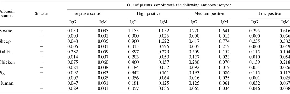
Effects of serum albumin source on binding of anti-silicate IgG and IgM antibodies to silicate-albumin complex.Six different serum albumins from bo-vines, sheep, rabbits, chickens, pigs, and humans were used to precoat the plates, which were then coated with silicate. The positive and negative controls were assayed for reactivity with silicate by the EIA described herein.
RESULTS
Antigen concentration curve.
Concentration-reactivity curves
show that the plates coated with the solution containing 1 mg
of Na
2SiO
3per ml produced optimal reactivity regardless of
whether a high or low silicate antibody-positive plasma sample
was used or whether the silicate-reactive antibodies were of the
IgG or IgM isotype (Fig. 1). The data shown represent the
silicate-reactive IgM antibodies. The IgG reaction is similar to
the IgM reaction (data not shown).
Silicate antibody titration.
Each of the silicate-reactive sera
was proportionally positive at dilutions ranging from 1:50 to
1:400 (data not shown). The 1:100 dilution was optimal, and
subsequent assays were performed with this dilution.
Conjugate titration.
The best results were obtained with a
1:2,000 dilution of the conjugate for IgG and with a 1:4,000
dilution for IgM (data not shown).
Establishment of standard curve.
The linearity of the
sili-cate reactivity of the positive control sera allowed arbitrary
EIA units to be assigned to increasing dilutions of antiserum
ranging from 1:50 to 1:1,600 (Fig. 2).
Determination of specificity of the silicate-reactive
antibod-ies in the sera of some women with silicone breast implants.
Of
the eight compounds tested, only Na
2SiO
3inhibited more than
90% of the IgG or IgM antibody binding to Na
2SiO
3(Fig. 3).
Determination of reference range.
On the basis of the mean
number of EIA units plus 3 standard deviations for 50 healthy
control women, a cutoff of 10 U (OD of 0.18) was established
FIG. 1. Silicate concentration-reactivity curves.h, high-positive serum sam-ple for anti-silicate IgG;E, low-positive serum sample for anti-silicate IgG;F, normal serum sample from a healthy control woman.
FIG. 2. Establishment of standard curve. The standard curve for detection of silicate IgG antibodies in a high-positive serum is shown.
on August 17, 2020 by guest
http://cvi.asm.org/
for both IgG and IgM silicate reactivities. The result for one of
the healthy control women was over this range (0.18 standard
deviations above the mean). For reasons of confidentiality, no
detailed clinical information concerning this patient is
avail-able. The frequency distribution curve, based on silicate
reac-tivity observed in the sera of these 50 healthy control women,
shows that all of the healthy controls had between 2 and 10
EIA units of silicate-specific IgG or IgM antibodies (Fig. 4).
Determination of interassay and intra-assay variation.
The
interassay and intra-assay coefficients of variation for detection
of silicate-reactive IgG antibodies ranged between 6% and
12%, whereas those for silicate-specific IgM were between 5%
and 13% (data not shown). These low coefficients of variation
demonstrate the reproducibility of this EIA for the detection
of silicate-reactive antibodies.
Presence of silicate-reactive IgG and IgM antibodies in the
sera of women with silicone breast implants or women without
implants but with different autoimmune diseases.
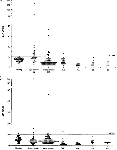
Twelve of
the 40 symptomatic women tested (30%) had silicate-reactive
antibodies restricted to the IgG isotype and 4 (10%) had
an-tibodies of both the IgG and IgM isotypes. None had
antibod-ies restricted to the IgM isotype (Fig. 5). In comparison, of the
91 asymptomatic women with silicone breast implants, only 8
(9%) had IgG anti-silicate antibodies (P
,
0.001) and 2 (2%)
had antibodies of both the IgG and IgM isotypes. None of the
women with Sjo
¨gren syndrome, scleroderma, or rheumatoid
arthritis had silicate-reactive antibodies of either the IgG or
the IgM isotype (Fig. 5). Only one of 20 systemic lupus
ery-thematosus patients tested had low levels of both IgG and IgM
silicate-reactive antibodies (Fig. 5).
FIG. 3. Determination of silicate specificity by a competitive inhibition assay with different metal salts.f----f, Na2SiO3;f——f, Li2SO4;Ç, MgSO4;E, CrO3;å, NiSO4;3, HgCl2;v----v, ZrOCl2;h, SiO2;v——v, BeSO4.
FIG. 4. Normal range frequency distribution curve for silicate IgG antibodies in 50 healthy control women.
FIG. 5. Demonstration of the relationship between silicate-reactive antibod-ies in women with silicone breast implants (SBI) and various autoimmune dis-eases. (A) Silicate-reactive IgG antibodies in healthy women, symptomatic women with breast implants, asymptomatic women with breast implants, and women without implants but with different autoimmune diseases. SLE, systemic lupus erythematosus; SS, Sjo¨gren syndrome; Scl, scleroderma; RA, rheumatoid arthritis. (B) Silicate-specific IgM antibodies in the different groups of women as described for panel A.
FIG. 6. Effect of pepsin digest on silicate-reactive IgG antibodies in a high-positive plasma sample.F, F(ab9)2fragment;h, Fc fragment;■, purified IgG (undigested).
on August 17, 2020 by guest
http://cvi.asm.org/
Evidence that the silicate activity is mediated by the F(ab
*
)
2portion of the silicate-reactive IgG molecule.
The results
de-picted in Fig. 6 clearly demonstrate that the silicate-reactive
IgG in the plasma sample of one of the women with silicone
breast implants who was highly positive for silicate
anti-bodies did interact with silicate through its F(ab
9
)
2fragment.
As expected for an antigen-antibody reaction, no antisilicate
activity was mediated by the Fc portion of the silicate-reactive
IgG (Fig. 6).
BSA as a carrier for silicate antigen.
Table 1 shows that
silicate bound the BSA, which acted as a carrier. Silicate could
not directly bind to the plates without BSA precoating. The
anti-silicate antibody was detected only on the BSA-silicate
plates.
Determination that BSA was the best carrier for silicate.
Detection of silicate antibodies was performed on the plates
which were precoated with serum albumins from different
an-imal species. The best reaction of anti-silicate antibodies was
demonstrated by BSA-precoated plates (Table 2). The serum
albumins from humans and pigs were not good carriers for
silicate antibody detection.
DISCUSSION
Evidence from human and animal models suggests that
poly-dimethylsiloxanes (silicone), the analogs of the silicon-oxygen
ions found in silicates, are not immunologically inert but can
elicit specific humoral and cell-mediated immune reactions (8,
9, 11, 12, 16, 20). In this report, we describe an EIA method for
detection and quantitation of silicate-reactive antibodies in the
sera of women with silicone breast implants. Although elevated
silicon concentrations are found in sera from some women
with silicone breast implants (16), in uremic and dialysis
pa-tients (6, 10), in the brains of papa-tients with senile dementia,
and in neurofibrillary tangles of patients with Alzheimer’s
dis-ease (4, 7), no reports of silicate-reactive antibodies (as
op-posed to silicone antibodies) in humans or animals are
avail-able. Herein, we show that 30% of the 40 symptomatic women
with silicone breast implants produced silicate-reactive IgG
antibodies and 10% had both silicate-reactive IgM antibodies
and silicate-reactive IgG antibodies. Nine percent of 91
asymp-tomatic women with silicone breast implants had IgG
anti-silicate antibodies, a percentage which is significantly lower
than that observed for symptomatic women. Only 2% (1 of 50)
of the 50 healthy control women and 0% (0 of 32) of the
women with rheumatic diseases without silicone implants were
positive for silicate-specific antibodies. One of 20 women (5%)
without implants who had lupus erythematosus had low levels
of silicate antibodies. Because false-positive results for IgM
isotypes can occur with sera positive for IgM rheumatoid factor
(1) in the presence of specific IgG, the four plasma samples
with silicate-reactive IgM (plus silicate-reactive IgG) were
treated with goat anti-human IgG to rule out potential false
positives for silicate-reactive IgM. The treatment failed to
re-move the reactive IgM, thus confirming the
silicate-reactive IgM positivity of those sera.
The data showed that BSA is the best carrier for silicate
binding to microtiter plates to detect anti-silicate antibodies.
The reason why human and pig serum albumins could not bind
to silicate as carriers (Table 2) is unknown. Further studies are
needed to determine the mechanism of silicate-albumin
bind-ing.
Previous reports focused on polydimethylsiloxane
(silicone)-specific antibodies in women with silicone breast implants (8,
12, 20). However, the nature of the silicon(e) eliciting the
specific response is unknown. The fact that the present studies
and our previous report (16) show that specific humoral and
cell-mediated immune responses can be detected by using
in-organic Si compounds (silicon dioxide and sodium silicate)
suggests that immune responses elicited by
polydimethylsilox-ane might in part reflect reaction with one or another
com-pound of silicon. Indeed, the frequency of antibodies detected
by our method (30%) is far greater than that (1.7% to 3%)
reported by Kossovsky et al., who employed silicone gel
ad-sorbed to a variety of proteins (12).
The mechanism of induction of antibodies or cell-mediated
immunity by silicon(e) is unknown. Because silicate is known
TABLE 1. Effects of order of addition of BSA on binding of anti-silicate antibodies to the silicate-albumin complex
Ig isotype Protocol
OD of plasma sample
Negative control
High positive
Medium positive
Low positive
IgG BSA/Silicatea 0.062 1.105 0.678 0.110
Silicate/BSAb 0.044 0.062 0.047 0.025
IgM BSA/Silicatea 0.104 0.974 0.498 0.246
Silicate/BSAb 0.089 0.103 0.076 0.068
aBSA precoated. bSilicate precoated.
TABLE 2. Effect of albumin source on binding of anti-silicate antibodies to the silicate-albumin complex
Albumin
source Silicate
OD of plasma sample with the following antibody isotype:
Negative control High positive Medium positive Low positive
IgG IgM IgG IgM IgG IgM IgG IgM
Bovine 1 0.050 0.035 1.155 1.052 0.720 0.641 0.295 0.616
2 0.000 0.001 0.000 0.026 0.000 0.013 0.000 0.036
Sheep 1 0.040 0.035 0.960 1.222 0.617 0.774 0.255 0.582
2 0.006 0.001 0.015 0.596 0.005 0.219 0.000 0.049
Rabbit 1 0.282 0.059 0.897 0.279 0.509 0.152 0.115 0.104
2 0.014 0.007 0.203 0.050 0.127 0.023 0.010 0.054
Chicken 1 0.075 0.060 0.460 0.157 0.280 0.070 0.139 0.218
2 0.024 0.038 0.184 0.052 0.092 0.019 0.051 0.026
Pig 1 0.092 0.083 0.342 0.161 0.193 0.086 0.115 0.117
2 0.007 0.035 0.056 0.064 0.016 0.025 0.001 0.025
Human 1 0.047 0.031 0.181 0.125 0.125 0.061 0.052 0.067
2 0.029 0.001 0.057 0.036 0.065 0.034 0.046 0.038
on August 17, 2020 by guest
http://cvi.asm.org/
to induce interleukin-1 production in antigen-presenting
mono-cytes and macrophages (5), it is tempting to postulate that silicate
can interact directly or indirectly (by binding to exogenous or
endogenous peptides) with antigen-presenting monocytes and
macrophages and can then be presented to B cells and T cells in
conjunction with major histocompatibility complex molecules for
induction of a specific immune response. A similar hypothesis has
been proposed for Be
21(15).
Because silicone-reactive antibodies are reported to be
present in the circulation of about only 1 to 3% of all women
with silicone breast implants (12), a subject with an implant
might test negative for silicone antibodies but test positive for
silicate antibodies (positive rate
5
30%) or vice versa (or both
could be positive or negative). Thus, addition of a new test for
silicate-reactive antibodies could prove valuable for the
accu-rate assessment of effects of silicon(e) on the immune system.
Results of preliminary studies in our laboratories (unpublished
data) indicate that there is no close correlation between the
presence of silicate-reactive antibodies described herein and
the T-cell responses to silicate, SiO
2, and silicone gel described
elsewhere (16). Of 300 silicone breast implant patients tested
in our laboratories for T-cell reactivity with silicate, 140 (47%)
were reactive with either SiO
2, silicate, or silicone (or a
com-bination of these antigens) in the T-cell proliferation assay, but
only 10% of the 140 patients had silicate-specific IgG
antibod-ies (15a). Therefore, a combined cellular-humoral test for
sil-icon(e) reactivity is likely to increase the sensitivity of
labora-tory tests for silicon(e) reactivity. The clinical relevance of the
presence of silicate antibodies or silicate- and/or
silicon(e)-specific T cells in women with silicone breast implants is yet to
be determined. Studies designed to answer these questions are
currently under way.
ACKNOWLEDGMENTS
We thank Navanit Lal for technical assistance, Sharon Hunt Ger-ardo for critical review, and Mindy Shaffer for assistance in preparing the manuscript.
REFERENCES
1. Agopian, M. S., F. M. Boctor, and J. B. Peter. 1988. False-positive test result for IgM anticardiolipin antibody due to IgM rheumatoid factor. Arthritis Rheum. 31:1212.
2. Arkles, B., and P. Redinger. 1983. Silicones in biomedical applications, p.
749–768. In M. Szycher (ed.), Biocompatible polymers, metals, and compos-ites. Technomic Publishing, Lancaster, Pa.
3. Blais, P. 1983. Silicone breast implant: technological and physiochemical aspects, p. 544–551. In L. R. Rubin (ed.), Biomaterials in reconstructive surgery. C.V. Mosby, St. Louis.
4. Candy, J. M., A. E. Oakley, and J. Klinowski. 1986. Aluminosilicates and senile plaque formation in Alzheimer’s disease. Lancet i:354–357. 5. Dinarello, C. A. 1978. Interleukin-1. Rev. Infect. Dis. 6:51–95.
6. Dobbie, J. W., and M. J. B. Smith. 1986. Urinary and serum silicon in normal and uraemic individuals in silicon biochemistry. Ciba Found. Symp. 121: 194–213.
7. Edwardson, J. A., J. Klinowski, A. E. Oakley, R. H. Perry, and J. M. Candy. 1986. Aluminosilicate and the aging brain: implications for the pathogenesis of Alzheimer’s disease in silicon biochemistry. Ciba Found. Symp. 121: 160–179.
8. Goldblum, R. M., R. P. Relley, A. A. O’Donell, D. Pyron, and J. P. Heggers. 1992. Antibodies to silicone elastomers and reactions to ventriculoperitoneal shunts. Lancet 340:510–513.
9. Heggers, J. P., N. Kossovsky, R. W. Parson, M. C. Robson, R. C. Pelley, and
R. J. Talmage.1983. Biocompatibility of silicone implants. Ann. Plast. Surg.
11:38–45.
10. Indrapraset, S., V. Alexander, and H. S. Gonick. 1974. Tissue composition of major and trace elements in uremia and hypertension. J. Chronic Dis. 27: 155–161.
11. Kossovsky, N., J. P. Heggers, and M. C. Robson. 1987. Experimental dem-onstration of the immunogenicity of silicone-protein complexes. J. Biomed. Mater. Res. 21:1125–1133.
12. Kossovsky, N., M. Zeidler, G. Chun, H. Papasian, A. Nguyen, S. Rajguru, J.
Stassi, J. Gelman, and E. Sponsier.1993. Surface dependent antigens iden-tified by high binding avidity of serum antibodies in a subpopulation of patients with breast prosthesis. J. Appl. Biomater. 4:281–288.
13. Lindmark, R., K. Thoren-Tolling, and J. Sjoquist. 1983. Binding of immu-noglobulins to protein A and immunoglobulin levels in mammalian sera. J. Immunol. Methods 62:1–13.
14. Mage, M. G., and E. Lamoy. 1987. Preparation of Fab and F(ab9)2fragments from monoclonal antibodies. Immunol. Ser. 33:79–97.
15. Newman, L. S. 1993. To Be21or not to Be21: immunogenetics and occupa-tional exposure. Science 262:197–198.
15a.Ojo-Amaize, E. A., et al. Unpublished data.
16. Ojo-Amaize, E. A., V. Conte, H.-C. Lin, R. F. Brucker, M. S. Agopian, and
J. B. Peter.1994. Silicone-specific blood lymphocyte response in women with silicone breast implants. Clin. Diagn. Lab. Immunol. 1:689–695.
17. Sergott, T. J., J. P. Limoli, C. M. Baldwin, and D. R. Lamb. 1986. Human adjuvant disease, possible autoimmune disease after silicone implantation: a review of the literature, case studies, and speculation for the future. Plast. Reconstr. Surg. 78:104–114.
18. Shons, A. R., and W. Schubert. 1992. Silicone breast implants and immune disease. Ann. Plast. Surg. 28:491–499.
19. Spierra, H. 1988. Scleroderma after silicone augmentation mammoplasty. JAMA 260:236–238.
20. Wolf, L. E., M. Lappe, R. D. Peterson, and E. G. Ezrailson. 1993. Human immune response to polydimethylsiloxane (silicone): screening studies in a breast implant population. FASEB J. 7:1265–1268.