M E T H O D O L O G Y
Open Access
A robust and cost-effective approach to
sequence and analyze complete genomes
of small RNA viruses
Kiril M. Dimitrov
1†, Poonam Sharma
1†, Jeremy D. Volkening
2, Iryna V. Goraichuk
1,3, Abdul Wajid
4,5,
Shafqat Fatima Rehmani
4, Asma Basharat
4, Ismaila Shittu
6, Tony M. Joannis
6, Patti J. Miller
1and Claudio L. Afonso
1*Abstract
Background:Next-generation sequencing (NGS) allows ultra-deep sequencing of nucleic acids. The use of
sequence-independent amplification of viral nucleic acids without utilization of target-specific primers provides advantages over traditional sequencing methods and allows detection of unsuspected variants and co-infecting agents. However, NGS is not widely used for small RNA viruses because of incorrectly perceived cost estimates and inefficient utilization of freely available bioinformatics tools.
Methods:In this study, we have utilized NGS-based random sequencing of total RNA combined with barcode
multiplexing of libraries to quickly, effectively and simultaneously characterize the genomic sequences of multiple avian paramyxoviruses. Thirty libraries were prepared from diagnostic samples amplified in allantoic fluids and their total RNAs were sequenced in a single flow cell on an Illumina MiSeq instrument. After digital normalization, data were assembled using the MIRA assembler within a customized workflow on the Galaxy platform.
Results:Twenty-eight avian paramyxovirus 1 (APMV-1), one APMV-13, four avian influenza and two infectious
bronchitis virus complete or nearly complete genome sequences were obtained from the single run.
The 29 avian paramyxovirus genomes displayed 99.6% mean coverage based on bases with Phred quality scores of 30 or more. The lower and upper quartiles of sample median depth per position for those 29 samples were 2984 and 6894, respectively, indicating coverage across samples sufficient for deep variant analysis. Sample processing and library preparation took approximately 25–30 h, the sequencing run took 39 h, and processing through the Galaxy workflow took approximately 2–3 h. The cost of all steps, excluding labor, was estimated to be 106 USD per sample.
Conclusions: This work describes an efficient multiplexing NGS approach, a detailed analysis workflow, and
customized tools for the characterization of the genomes of RNA viruses. The combination of multiplexing NGS technology with the Galaxy workflow platform resulted in a fast, user-friendly, and cost-efficient protocol for the simultaneous characterization of multiple full-length viral genomes. Twenty-nine full-length or near-full-length APMV genomes with a high median depth were successfully sequenced out of 30 samples. The applied de novo assembly approach also allowed identification of mixed viral populations in some of the samples.
Keywords: Newcastle disease virus, Next-generation sequencing, Multiplexing, Galaxy, De novo assembly,
Multiplexing, Complete genomes, Mixed infection, Avian paramyxovirus
* Correspondence:[email protected] †Equal contributors
1Exotic and Emerging Avian Viral Diseases Research Unit, Southeast Poultry Research Laboratory, US National Poultry Research Center, Agricultural Research Service, USDA, 934 College Station Road, Athens, GA 30605, USA Full list of author information is available at the end of the article
Background
Conventional laboratory methods like enzyme-linked immunosorbent assay, nucleic acid hybridization tech-nique, and polymerase chain reaction (PCR) are all common and inexpensive diagnostic and research tools utilized in virology [1]. However, as these assays are highly dependent on reagents (primers, probes, anti-bodies) developed from previously known and character-ized viruses, they might be ineffective for the identification of new viral variants, new pathogens or a mixed popula-tion of pathogens that have high genetic divergence from those described previously [1, 2]. Next-generation sequen-cing (NGS) technologies enable large numbers of samples to undergo parallel sequencing, and can be used for the detection and characterization of multiple agents from one sample. The use of sequence-independent amplifica-tion of viral nucleic acids eliminates the need for prior knowledge of genomic sequences and provides advantages over traditional methods such as PCR amplification or microarray hybridization dependent on target-specific primers [2, 3]. NGS technologies allow screening of clinical and environmental samples for the presence of viral pathogens, including previously unknown viruses [4]. This has led to the discovery of numerous viral pathogens [4–6], including 2009 pandemic influenza A, a novel pegi-virus, Canine bocavirus 3, and a novel hepacivirus [7–10].
The majority of previously employed techniques used virus enrichment prior to cDNA synthesis and library preparation, or used specific primers for amplification. Virion enrichment steps such as centrifugation, poly-ethylene glycol precipitation, ultrafiltration, chloroform treatment or nuclease treatment have been used for RNA viruses [3, 11, 12]. Other techniques, such as gDNA depletion and host RNA depletion to enrich for viral RNA [13] and DNase pretreatment of the allantoic fluid to enrich for viral particles, have also been described [14]. Different methods have been employed for producing cDNA following enrichment, including
se-quence-independent single primer amplification
(SISPA) and universal primers which have been used for sequencing RNA viruses [4, 15] including Newcas-tle disease virus (NDV) [11] and avian paramyxovirus (APMV) 4 and 6 [16].
The genus Avulavirus of the family Paramyxoviridae, orderMononegavirales, consists of 14 known avian para-myxovirus serotypes (APMV 1 –14) [17–19]. Of these, APMV-1, synonymous with Newcastle disease virus, is the most widely characterized and studied due to the economic importance of Newcastle disease (ND) caused by virulent strains of the virus. Newcastle disease is one of the most significant poultry diseases and infects both wild and domestic avian hosts. NDV has a single-stranded, non-segmented, negative-sense RNA genome consisting of six genes in order of 3’to 5’: nucleocapsid
(NP), phosphoprotein (P), matrix (M), fusion (F), hemagglutinin-neuraminidase (HN), and polymerase (L), coding for these six structural proteins and at least one additional V protein [20–22]. Newcastle disease viruses have three genome sizes–15186, 15192 and 15198 nucle-otides, and are genetically grouped into two divergent classes that are further classified in genotypes [23–25].
Newcastle disease viruses are constantly evolving and different genetic groups undergo simultaneous evolutionary changes in different geographical loca-tions [22, 26] making the available genetic makeup information outdated. These evolutionary changes present challenges for prompt diagnosis. Some currently validated methods are target-oriented and might fail to de-tect new viral genetic variants [27–29]. Lack of complete genetic information for many NDV isolates further ham-pers the better understanding of Newcastle disease evolu-tion and epidemiology. Furthermore, mixed viral infecevolu-tion are not uncommon in animals, and in the case of poultry, they are quite frequent [30]. Efficient and accurate identi-fication of these pathogens is essential for the develop-ment of adequate disease control strategies. These challenges require an approach that provides de novo, rapid and high-quality genetic characterization of full-length viral genomes.
Until recently, genome sequencing of small RNA viruses, including NDV, has been performed using over-lapping genome amplification with primer pairs. This approach is laborious, depends on preexisting informa-tion, and produces very low depth. NGS advances pro-vide tools for deep sequencing of multiple viral strains in a short time. However, the simultaneous cost- and time-effective sequencing and characterization of a large number of NDV genomes has not yet been reported. The aim of the current study was to utilize sequence-independent NGS technologies applied to viral nucleic acids for the simultaneous and rapid characterization of multiple NDV genomes. We demonstrate a straightfor-ward, efficient protocol for multiplexed sequencing using a single flow cell on the Illumina MiSeq platform coupled with a detailed customized Galaxy workflow for
de novo assembly that allows for quick and accurate
generation of near-full-length, or full-length, genome se-quences of dozens of isolates, simultaneously. Further-more, we report the efficient detection and complete sequencing of contaminant RNA viruses.
Methods
Virus propagation
in 9-to-11-day-old specific-pathogen-free (SPF) embryo-nating chicken eggs [31]. The background information of the 30 isolates used in the study is summarized in Additional file 1: Table S1.
RNA isolation
RNA from each sample was extracted from allantoic fluids. Two milliliters (equal volumes of 0.25 ml) of each sample were aliquoted into 8 microtubes, each contain-ing 0.75 ml of TRIZOL LS (Invitrogen, USA). After 5 min of incubation, 0.2 ml of chloroform was added to each tube and shaken vigorously. After 10 min of additional incubation at room temperature, tubes were centrifuged at 12000 × g for 15 min at 4 °C. The aqueous phase from all eight tubes was removed and pooled for each sample. Two milliliters of aqueous layer of each sample were treated with 4 μl Turbo DNase 2U/μl (Ambion, USA) for 15 min at 37 °C and then placed on ice. The extraction proceeded using the QIAamp® Viral RNA Mini Kit (Qiagen, USA) according to the manufac-turer’s instructions. Briefly, DNase-treated aqueous phase was passed through a spin column for RNA ab-sorption on the QIAamp silica membrane followed by washing with 2 ml of provided buffers AW1 and AW2 using the QIAvac 24 Plus vacuum manifold (Qiagen, USA). The RNA was eluted in 50μl buffer AVE. Eluted RNA was quantified using a Qubit® RNA HS Assay Kit in a Qubit® fluorometer (ThermoFisher Scientific, USA) and stored at -20 °C until further use.
NDV RNA capture
A set of three biotinylated oligonucleotides designed from consensus of alignment of 330 available NDV genomes were used for NDV RNA capture (Oligo 1 –
5’- AGA GAA TCT GTG AGG TAC GA/3Bio -3’ at
nucleotide position 8; Oligo 2 – 5’ -TTC TCA AGT CAT CGT GAC AG/3Bio -3’at position 5905; Oligo 3– 5’- CCC TGC ATC TCT CTA CAG/3Bio -3’at position 12226) (GenBank accession number AF431744). RNA capture reactions were performed using 50μl RNA incu-bated with 167 μl 6X saline-sodium phosphate-EDTA
buffer (900 mM NaCl, 60 mM NaH2PO4, 60 mM
Na2EDTA), 2 μl RNaseOUT 40 U/μl (Invitrogen, USA)
and 1.5 μl 100 μM mix of the three primers. Reactions were performed at 70 °C for 5 min, followed by 15 min at 55 °C. Two hundred fifty microliters of 1X binding and wash buffer solution (2 M NaCl) containing 12.5μl (0.05%) Sera-Mag beads (magnetic streptavidin-coated beads, GE Healthcare Life Sciences, USA) was prepared for each sample and mixed with the capture reaction products for binding. Washing was done on a magnetic stand with 500 μl of 0.5X binding and wash buffer one time and twice with bead wash buffer (5 M NaCl, 1 M Tris-HCl [pH 7.5], 0.5 M EDTA, and 0.01% Tween® 20)
to remove unbound RNA. Viral RNA was recovered by adding 19 μl of 10 mM Tris-HCl (pH 7.5) to the bead mixture and incubating at 65 °C for 5 min followed by cooling on ice until the next step. In a separate experi-ment, a comparison with three known NDV was per-formed and the libraries were prepared side-by-side with and without the capture step, while all remaining steps of the library preparation were identical.
Reverse transcription
Reverse transcription reactions were performed using the M-MLV Reverse Transcriptase (Moloney Murine Leukemia Virus Reverse Transcriptase, Invitrogen, USA)
and 10 μl RNA (concentration varied from below
250 pg/μl to 55 ng/μl), 1.0 μl Random Primers mix (3μg/μl) (Invitrogen, USA) and following manufacturer’s instruction. The cDNA products were purified using
60 μl Agencourt® RNAClean® XP beads (Beckman
Coulter, USA) as per manufacturer’s instructions. Puri-fied cDNA were recovered in 15 μl of 10 mM Tris-Cl (pH 7.5) and quantified using a Qubit® ssDNA Assay Kit
(ThermoFisher Scientific, USA) on the Qubit®
fluorometer.
Library preparation, quality and quantity assessment
DNA libraries (n= 30, one library for each sample), were prepared for deep sequencing using 1 ng purified cDNA (0.2 ng/μl in molecular grade water) and the Nextera XT DNA Library Preparation Kit (Illumina, USA) following the manufacturer's protocol. The resulting dsDNA prod-ucts were purified with 30 μl Agencourt® AMPure® XP beads (Beckman Coulter, USA) by incubating at room temperature for 5 min followed by two washes with 200μl 80% ethanol on a magnetic stand. The tubes with the beads were air dried for 10 to 15 min at room temperature and the library products were recovered in 52.5μl of Resuspension Buffer. The quality and fragment length distribution for each library was assessed using the Agilent High Sensitivity DNA Kit (Agilent Technolo-gies, USA) on the Agilent 2100 Bioanalyzer (Agilent Technologies, Germany). The Qubit® fluorometer and the Qubit®dsDNA HS Assay Kit were used for measuring the concentration of the libraries.
Equimolar dilution and pooling of the NGS libraries
the library pool was diluted to 10 pM. Control library (3% PhiX library, Illumina, USA) was added and the pool was snap-chilled on ice. The library pool (600 μl) was loaded in the flow cell of the 500 cycle MiSeq Reagent Kit v2 (Illumina, USA) and pair-end sequencing (2 × 250 bp) was performed on the Illumina MiSeq instru-ment (Illumina, USA). After automated cluster gener-ation in MiSeq, the sequencing reads were processed and all statistical data generated by the instrument were collected and summarized.
Genome assembly
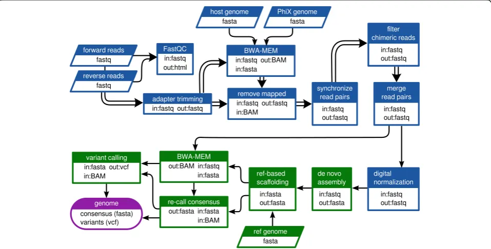
A workflow was designed to perform pre-processing and assembly of the raw sequencing data as diagrammed in Fig. 1 using the Galaxy platform interface [32]. Process-ing was carried out via Galaxy and PBS/Torque on a local dual node 128-core cluster. Briefly, raw read quality was assessed using FastQC [33] and residual adapter sequences were trimmed using Cutadapt v1.6 [34]. Trimmed reads were mapped against the Gallus and PhiX174 reference genomes using BWA-MEM v0.2.1 in order to identify host and control library read contamin-ation [35, 36]. Host and control library reads were fil-tered using the Filter sequences by mapping v0.0.4 tool in Galaxy [37]. The forward and reverse files, which were no longer synchronized due to adapter trimming and filtering, were re-synchronized using in-house tool. Overlapping read pairs were joined with PEAR v0.9.6.0
[38]. Chimeric Nextera reads were removed by an in-house tool which discarded single reads with partial mappings in opposite orientations. Digital normalization via median k-mer abundance was performed using the Khmer package v1.1-1 (cutoff = 100, kmer size = 20, number of tables to use = 4, table size = 1e9) [39, 40].De novo assembly was performed using the MIRA assem-bler v3.4.1 [41]. The following parameters and settings were specified for the assembly step: assembly method = de novo, assembly quality grade = accurate, use read extension = yes, minimum reads per contig = 100, mini-mum overlap = 16, mark repeats = yes, maximini-mum mega-hub ratio = 0.2, spoiler detection = yes, with default settings for the rest of the parameters. Reference-based orientation and scaffolding of the contigs produced by the assembler were performed using V-FAT v1.0.0 (Broad Institute, Cambridge, MA, USA). The consensus sequence was then re-called based on BWA-MEM map-ping of trimmed but un-normalized read data to the genome scaffold and parsing of the mpileup alignment using in-house software. As a final step, LoFreq [42] was used to estimate variant frequencies in the obtained gen-omic data. A graphic representation of all major steps included in the sample preparation and analyses is pro-vided in Additional file 2: Figure S1. The obtained se-quences were phylogenetically analyzed with closely related sequences of isolates deposited in GenBank using MEGA6 [43], as previously described [25].
Fig. 1Customized Galaxy workflow used in the current study.Double arrowsindicate steps where the read pairs were processed in parallel.
Results
Nucleic acids quantification and libraries fragment size
The nucleic acid concentrations obtained at different steps throughout the preparation of the libraries for se-quencing are summarized in Additional file 3: Table S2. The lowest detected RNA concentration was 2 ng/μl and the maximum was 55 ng/μl. After RNA purification, the RNA concentrations of five samples were below the de-tection limit of Qubit (250 pg/μl); however, these sam-ples resulted in sufficient cDNA quantity to be further processed in library preparation. The generated libraries had a relatively narrow combined distribution of mean fragment lengths (mean 351 bp, standard deviation 30 bp, with 26 of 30 libraries within the range of 334 to 371 bp) (see Additional file 3: Table S2). It was observed that the true fragment length distributions observed post-sequencing were shorter than expected based on Bioanalyzer reports, even after counting for adapter length (Table S2). As a result, a large proportion (more than 90% in nearly all libraries) of read pairs overlapped at the ends. The source of the discrepancy with the Bioa-nalyzer estimates is still unclear.
Summarized statistics of the sequencing run
A summary of the sequencing run statistics as estimated by the MiSeq instrument is provided in Table 1. A clus-ter density of 917 +/- 19 K/mm2and 92.34% of the clus-ters passing the chastity filter yielded a total of 8.4 Gigabases of data. Of 17.7 million total reads, 96.31% passed the instrument quality control filter. Almost 80% of the bases were assigned Phred quality scores equal or greater to Q30 (Q30 score is equivalent to an expected error rate of 0.001). The fraction of reads in the pool
assigned to each sample varied from 0.0007 to 7.16% (mean 3.2 ± 1.4%).
Optimization of the assembly/analysis workflow
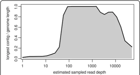
In order to take advantage of the overlapping reads, a merging step was introduced to produce longer pseudo-reads and to reduce complexity of the assembly task. An essential optimization was made by reducing the esti-mated coverage depth to a level that would still produce optimal assemblies. Two techniques for data reduction were investigated. Random sub-sampling resulted in loss of specific regions in the genome with reproducibly low coverage (data not shown). Digital normalization, which aims to down-sample high-coverage regions while pre-serving reads from low-coverage areas, provided means for decreasing the number of used reads to an optimal level without loss of data, and thus, was incorporated into the customized Galaxy workflow prior to assembly. In order to determine an optimal target depth for bly, preliminary test assemblies using the Velvet assem-bler v1.2.10 [44] were performed on a geometric progression of sampling depths from 10x to 10000x (the approximate depth of the raw data) with an additional optimization of the velvetg “cov_cutoff” parameter for each depth (parameter used to low coverage nodes). The results indicated that optimal (in this case, full-length) assembly occurred over a range of approximately one order of magnitude (100x to 1000x). Below and above this range, fragmentation began to occur (Fig. 2).
Data analysis
The final outputs of the analysis workflow for each sam-ple included a consensus genome scaffold (.fasta), a file of all assembled contigs (.fasta), a variant frequency call file (variant call format or .vcf ) and a set of summary
Table 1Statistics of next-generation sequencing of 30 avian paramyxovirus isolates in a single run
Data Results
Cluster density (K/mm2)a 917 +/- 19
Clusters passing filterb 92.34%
Total number of reads 17762176
Pass-filter readsc 16403251
Percentage of reads passing filter 96.31%
≥Q30d 77.9%
Lowest representation for any indexe 0.0007%
Highest representation for any indexe 7.16%
a
shows number of clusters per square millimeter (optimal cluster density is 1000–1200, can vary with chemistry)
b
indicates the purity of the signals detected from the clusters (i.e. signals passing chastity filter that is the ratio of the brightest base intensity divided by the sum of the brightest and second brightest base intensities and the filtration process removes the least reliable clusters from the image analysis results)
c
reads passing filter (about 15 million reads are expected from an optimally clustered flow cell)
d
percentage of bases with Phred quality score equal or greater to 30
e
percentage of pass-filter reads assigned to any index
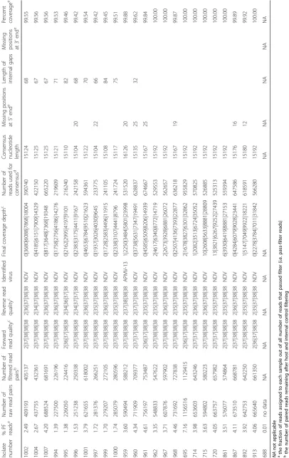
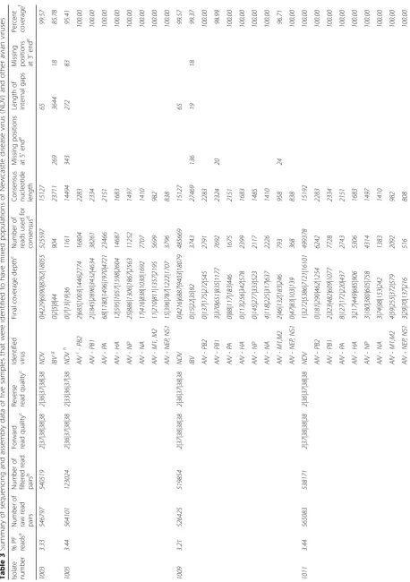
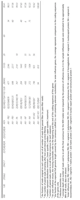
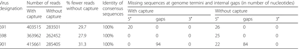
statistics on the run and the assembly. An in-depth sum-mary of the outputs from all samples is presented in Tables 2 and 3, including detailed information on read quality and depth distributions and genome coverage per sample. A total of 29 full-length or near-full-length APMV genomes (99.56% mean genome coverage) were obtained from 30 libraries with only one sample (#1005) having coverage below 99% and nine samples having 100% coverage (Tables 2 and 3). The lower and upper quartiles of median depth per position of the sequencing results were 2984 and 6894 respectively, allowing for ac-curate detection of low-frequency single nucleotide vari-ants. In fact, all but one NDV samples had a median read depth of at least 2583 (the exception, sample 1005, was found to consist of approximately 98% avian influ-enza virus reads after host filtering). In addition to NDV genome assembly, the de novo strategy allowed for the detection of full-length and near-full-length genomes of avian influenza virus (AIV) in libraries of isolates 998, 1005, 1009 and 1011 [45], as well as infectious bronchitis virus (IBV) in samples 1003 and 1009. The coverage of the two obtained IBV genomes was 85.78 and 99.37%, while the sequenced AIV genes had coverages ranging between 92.23 and 100%, and two complete AIV ge-nomes were sequenced (see Table 3). The estimated me-dian depths for the IBV (5 and 22) and AIV (from 35 to 1274) isolates were lower (Table 3), most likely reflecting the lower titer of these viruses in the samples. Sample 959 was identified as a member of the novel APMV serotype 13 and the median depth for this sample was 3484. The host reads were between 0.1 and 5.4% (aver-age 1.3%) of all reads per sample. No data was obtained from the library of sample 688 (only 0.0007% of the raw reads were assigned to this sample). The results from the comparison of the three NDV libraries prepared side-by-side with and without the capture step showed identical accuracy, not significantly affected overall coverage and near full-length and full-length genomes were obtained using both approaches. However, the number of NDV-specific reads decreased by approximately 30% when the capture step was not performed (see Table 4).
While the high-throughput workflow sometimes
resulted in short segments of missing data at the gen-ome termini and/or at one short internal gap, complete sequences for all coding regions of the 29 APMV posi-tive samples were obtained directly from the workflow. Nearly all of the short missing regions occurred at either the termini (a common issue in viral NGS sequencing) [46] or at one specific intergenic location in the genome between genes N and P which displayed extremely low coverage in all analyzed samples (possibly as a result of high GC content – 76%). For the purpose of sub-mitting full-length NDV sequences to GenBank, we sequenced the termini using a previously described
protocol [47] and primers designed for the current study (see Additional file 4: Table S3). The internal gaps, where necessary, were sequenced using PCR and Sanger sequencing (for primers sequences see Additional file 4: Table S3). This additional work was not included in the time/cost estimates, as it was per-formed to submit complete NDV sequences to Gen-Bank and would not be necessary for a full analysis of the coding regions.
Time and cost estimates
The time and cost estimates for all steps are summarized in Additional file 5: Table S4. Assuming the addition of the first reagent as the start and the final dilutions of the samples as the end of the procedure, the approximate time taken for preparing 30 samples was 25 to 30 person-hours. The sequencing run (500 cycle kit) lasted 39 h. Submission of the raw data to the customized Galaxy workflow and data analysis on the cluster took an additional 2 to 3 h. The average cost of all steps, including all reagents but excluding labor, depreciation and maintenance of equipment, was estimated to be approximately 106 USD per sample.
Discussion
Next-generation sequencing has been previously de-scribed for whole-genome sequencing of NDV by our team and others [48–55]; however, this study is the first report that demonstrates robust simultaneous genomic characterization of multiple NDV viruses in a single NGS run. The study further demonstrates the added benefit of conducting random non-targeted sequencing with an optimizedde novoassembly workflow for identi-fication of mixed viral infections. In contrast to previous work, here an optimized and customized workflow that employs publically available tools and produced consist-ently high quality assemblies of complete genomes is described in details. This study also provides detailed statistical and sequencing information that allows quality and quantity assessment of the obtained results.
The avian influenza viruses belong to the family
Ortho-myxoviridae and have genomes containing eight
seg-ments of single-stranded, negative-sense RNA that code for 10 or 11 proteins, depending on the strain [57]. Despite the diverse nature of the RNA present in sam-ples with mixed populations, the procedure described here successfully produced complete genomes of these viruses.
Our results also demonstrate the capability of the methodology to produce quality libraries and assem-blies without any physical or mechanical enrichment. The cDNA and dsDNA concentrations were not found to be proportional to the initial total RNA con-centrations. The introduced nuclease step aided diges-tion of host nucleic acids resulting in low average number (see Tables 2 and 3) of host-associated reads per sample. The abundance of host nucleic acids may pose a problem in obtaining sufficient numbers of viral reads for optimal viral genome assembly [4]. To avoid or decrease problems caused by contamination with host sequences others have developed method-ologies for enrichment of target viral RNAs. We have not utilized any pretreatment or purification; however, a target-specific capture step with biotinylated oligos designed from three different conservative regions of the NDV genome was tested. The comparison of re-sults from samples with and without the RNA capture step presented here demonstrates that the primary tradeoff comes in the form of approximately 30% re-duced depth of coverage, although the coverage was still sufficient for proper consensus re-calling (see Table 4). The ability of the capture step to reduce host sequences and other non-target RNA and to im-prove downstream assembly and analysis should be further assessed on different sample types (e.g. clinical samples, formalin-fixed paraffin-embedded samples) that may contain less amounts of viral RNA. For egg-grown viruses with high viral titers the observed de-crease of reads without the RNA capture step was not essential for obtaining complete coverage with sufficient depth. In clinical diagnostic samples, how-ever, the number of NDV sequencing reads is often significantly lower, and introducing the RNA capture step could improve the final results.
There is a clear difference between the presented application of NGS and the use of this approach in diag-nostics. Here we describe the use of high-titer egg-grown viruses for production of high quality and deep data useful for detailed genomic characterization and rare variant analyses. However, the use of this NGS tech-nology for diagnostics is more complex. It requires to clearly establish sensitivity, specificity and limit of detec-tion based on the nature of samples and these are be-yond the scope of the current work. The described methodology has been successfully transferred for use with clinical samples and optimization studies are in progress in our lab.
Prior to the production run, considerable time was spent optimizing the assembly and analysis workflow for the task at hand. While some of the steps in the work-flow are fairly standard procedures in NGS analyses (QC summarization, adapter trimming, contaminant read fil-tering), others were tailored to the specific characteris-tics of the data being generated. The most critical optimization, however, was reducing estimated coverage depth to a level that would produce optimal assemblies. It has previously been shown that, past a certain level, increasing read depth can decrease de novo assembly quality [58]. This effect can have significant conse-quences when working with massively deep sequencing data such as viral population studies that can easily ex-ceed 10000x sample coverage. Digital normalization has been included in similar workflows by others [59, 60] but is often overlooked in naïve approaches to high-coverage de novo assembly. As the assembler used in our workflow (MIRA) is relatively resource-intensive overlap-layout-consensus (OLC) assembler, we chose a target (100x) at the lower end of the empirically deter-mined optimal range to incorporate as a cut-off into the customized production workflow. Graph-based assem-blers such as Velvet utilize de Bruijn graph algorithms and assemble data by representing the genome by a set of short k-mer sequences [44]. Notably, graph-based assemblers are less resource-demanding and can be suc-cessfully utilized with limited computational resources. However, for graph-based assemblers, the k-mer size is an essential parameter [44, 59, 60] and the optimal value has to be determined depending on the characteristics of
Table 4Comparison of differences in number of reads and genome coverage of three samples prepared with and without capture of NDV RNA
Virus designation
Number of reads % fewer reads without capture
Identity of consensus sequences
Missing sequences at genome termini and internal gaps (in number of nucleotides)
With capture
Without capture
With capture Without capture
5′ gaps 3′ 5′ gaps 3′
691 403515 283501 29.7 100% 20 0 0 26 0 0
698 363962 262452 27.9 100% 0 0 0 25 0 0
the sequence reads, while this is avoided using an OLC assembler. In our hands, MIRA consistently produces quality assemblies with minimal tuning needed. Add-itionally, due to the potential skewing effects of digital normalization and V-FAT scaffolding on the proportion of nucleotide variant frequencies, the post-assembly step to re-map the un-normalized data to the genome scaf-fold allowed for proper consensus re-calling and precise variant analysis.
One important aspect to the use of NGS approaches in mainstream viral sequencing studies is the capacity to multiplex samples in order to reduce costs. The time and cost summary for all steps (summarized in Additional file 5: Table S4) demonstrate that the simul-taneous processing of 30 samples requires approximate one hour of operator time per sample with a cost of ap-proximately 106 USD per sample. Those values, although still high for diagnostics purposes, are 15 to 20 times lower (based on internal estimates) compared to the cost of primer-based sequencing with Sanger technologies. Furthermore, the demonstrated lower and upper quartiles of median depth per position (2984 and 6894, respectively), allow for accurate consensus re-calling and rare variant analysis. The final output not only includes the consensus genome sequence but also produced a variant call format file (https://samtools.github.io/hts-specs/VCFv4.2.pdf) and demonstrate that the protocol could be used for research on viral quasispecies and evolutionary studies (Dimitrov et al., in preparation). As previously reported by Gould et al., the 1998 Newcastle disease outbreak in Australia was pre-ceded by several months of circulation of mutant quasispe-cies of the virulent cleavage site [61]. These viral variants were undetected in the Sanger consensus sequences but could have been detected and properly quantified if the methodology described here was available at that time. The obtained genomic coverages (see Tables 2 and 3) illustrate the ability of the described protocol in generating full-length or near-full-full-length RNA virus genomes. Al-though very short internal gaps were present due to complete absence of coverage in the raw data and short sequences at the genome termini were missing, all coding sequences (commonly used in genetic stud-ies) were obtained.
The total turnaround time for the entire testing (sam-ple preparation, sequencing and analyses) was approxi-mately 72 h, of which most of the time consisted of library preparation and the sequence run. The duration of the sequencing run could be reduced by approxi-mately 15 h by using a 300-cycle configuration without any anticipated drop-off in assembly quality based on our observed fragment length distributions, although this assumption is untested. The time taken for data analysis may be expected to vary somewhat based on the available computational resources in a lab, although in
our protocol this represents a small fraction of the total turnaround time to begin with.
The obtained results were phylogenetically consistent with preliminary studies of the tested viruses (data not shown) and expectations based on previous research. Almost all of the samples from Pakistan were of sub-genotype VIIi which is currently circulating in Pakistan and may be causing a new panzootic [25, 62]. Ukrainian samples were of different sub-genotypes (II, VIg and VIId) that have been reported to be isolated from pigeons in Ukraine [25] and also in Europe [63]. The Nigerian samples were of genotypes XIVb and XVIIa which have been reported to circulate in Nigeria since 2006 [64] and previously un-sequenced full-length genomes of these sub-genotypes have been reported by us [48, 49]. In addition, and demonstrating the broad applicability and the advantages of thede novoapproach described here, the first complete APMV-13 genome was obtained [17] and avian influenza and infectious bronchitis viruses populations were identified. Phylogen-etic analyses of the obtained NDV and IBV sequences are presented in Additional file 6: Figures S2 and S3).
Conclusion
In summary, a robust chemistry and bioinformatics protocol utilizing publicly available tools to sequence and analyze complete genomes from small RNA viruses is described. Thirty-five full-length or near-full-length avian RNA viral genomes with a high median coverage depth were successfully sequenced out of 30 samples. The applied de novo approach allowed identification of mixed viral populations in some of the samples. The combination of multiplexing NGS technology with the customized Galaxy workflow platform enabled a quick turnaround time and cost-efficient methodology for sim-ultaneous characterization of multiple viral genomes.
Additional files
Additional file 1: Table S1.Background information of the avian paramyxovirus isolates used in this study. (DOCX 15 kb)
Additional file 2: Figure S1.Major processing steps used in the current study (PDF 222 kb)
Additional file 3: Table S2.Nucleic acid concentrations and library fragment size distributions of thirty virus isolates used in the study. (DOCX 18 kb)
Additional file 4: Table S3.Sequences of primers used for sequencing internal gaps and missing termini. (DOCX 13 kb)
Additional file 5: Table S4.Time and cost analysis of next-generation sequencing of thirty avian paramyxovirus isolates. (DOCX 15 kb) Additional file 6: Figure S2.Phylogenetic analysis based on the complete genome coding sequence of Newcastle disease virus isolates studied here and selected closely related sequences from GenBank.
Acknowledgements
The authors gratefully acknowledge Dawn Williams-Coplin and Tim Olivier for their technical assistance. We appreciate critical and constructive reviews provided by three anonymous reviewers. The mention of trade names or commercial products in this publication is solely for the purpose of providing specific information and does not imply recommendation or endorsement by the U.S. Department of Agriculture. The USDA is an equal opportunity provider and employer.
Funding
This work was supported by USDA/ARS CRIS 6040-32000-072, U.S. Defense Threat Reduction Agency BAA projects FRCALL12-6-2-0005 and ARS#715 FRCALL 12-6-1-0046, and U.S. Department of State Biosecurity Engagement Program NDV 31063.
Availability of data and materials
The complete genome sequences (n= 28) of the Newcastle disease virus isolates obtained in this study were submitted to GenBank and are available under the accession numbers KY171989 to KY171995, KY076030 to KY076039, KY076043, KU295453 to KU295455, KX496962 to KX496964, KY042127, KX236101, KU133362, and KT948996. The sequence of the APMV-13 is available under accession number KX119151. The nucleotide sequences of the four H9N2 AIVs have been deposited in GenBank under accession numbers KU042891 to KU042922. The sequences of the two IBV are available under accession numbers KY588134 and KY588135. All software tools utilized in the workflow that are not described else-where, along with their Galaxy wrappers, can be found online at https:// github.com/jvolkening/b2b-utils.
Authors’contributions
CLA, PS, KMD, and PJM conceived this project. AW, SFR, IS, and TMJ coordinated field sampling efforts and isolated viruses. PS, IVG, and AW prepared and sequenced the viral isolates. JDV developed the Galaxy workflow and associated software tools. PS, KMD, JDV, and CLA conducted analyses of the data. PS, KMD, JDV, PJM, and CLA wrote the manuscript. All authors read and approved the final manuscript.
Competing interests
The authors declare that they have no competing interests.
Consent for publication
Not applicable.
Ethics approval and consent to participate
Not applicable.
Publisher’s Note
Springer Nature remains neutral with regard to jurisdictional claims in published maps and institutional affiliations.
Author details
1Exotic and Emerging Avian Viral Diseases Research Unit, Southeast Poultry Research Laboratory, US National Poultry Research Center, Agricultural Research Service, USDA, 934 College Station Road, Athens, GA 30605, USA. 2BASE2BIO, 1945 Arlington Drive, Oshkosh, WI 54904, USA.3National Scientific Center Institute of Experimental and Clinical Veterinary Medicine, 83 Pushkinskaya Street, Kharkiv 61023, Ukraine.4Quality Operations Laboratory (QOL), University of Veterinary and Animal Sciences, Syed Abdul Qadir Jilani Road, Lahore 54000, Pakistan.5Institute of Biochemistry and Biotechnology, University of Veterinary and Animal Sciences, Syed Abdul Qadir Jilani Road, Lahore 54000, Pakistan.6Regional Laboratory for Animal Influenza and other Transboundary Animal Diseases, National Veterinary Research Institute, PMB01, Vom 930010, Plateau State, Nigeria.
Received: 16 February 2017 Accepted: 29 March 2017
References
1. Wu Q, Ding SW, Zhang Y, Zhu S. Identification of viruses and viroids by next-generation sequencing and dependent and homology-independent algorithms. Annu Rev Phytopathol. 2015;53:425–44. http://doi.org/10.1146/annurev-phyto-080614-120030.
2. Victoria JG, Kapoor A, Li L, Blinkova O, Slikas B, Wang C, Naeem A, Zaidi S, Delwart E. Metagenomic analyses of viruses in stool samples from children with acute flaccid paralysis. J Virol. 2009;83:4642–51. http://doi.org/10.1128/ jvi.02301-08.
3. Allander T, Emerson SU, Engle RE, Purcell RH, Bukh J. A virus discovery method incorporating DNase treatment and its application to the identification of two bovine parvovirus species. Proc Natl Acad Sci U S A. 2001;98:11609–14. http://dx.doi.org/10.1073/pnas.211424698.
4. Neill JD, Bayles DO, Ridpath JF. Simultaneous rapid sequencing of multiple RNA virus genomes. J Virol Methods. 2014;201:68–72. http://doi.org/10.1016/ j.jviromet.2014.02.016.
5. Reuter G, Pankovics P, Boros A. Identification of a novel astrovirus in a domestic pig in Hungary. Arch Virol. 2011;156:125–8. http://doi.org/10.1007/ s00705-010-0827-5.
6. Cholleti H, Hayer J, Abilio AP, Mulandane FC, Verner-Carlsson J, Falk KI, Fafetine JM, Berg M, Blomstrom AL. Discovery of novel viruses in mosquitoes from the Zambezi valley of Mozambique. PLoS One. 2016;11: e0162751. http://doi.org/10.1371/journal.pone.0162751.
7. Chiu CY. Viral pathogen discovery. Curr Opin Microbiol. 2013;16:468–78. http://dx.doi.org/10.1016/j.mib.2013.05.001.
8. Chandriani S, Skewes-Cox P, Zhong W, Ganem DE, Divers TJ, Van Blaricum AJ, Tennant BC, Kistler AL. Identification of a previously undescribed divergent virus from the Flaviviridae family in an outbreak of equine serum hepatitis. Proc Natl Acad Sci U S A. 2013;110:E1407–15. http://dx.doi.org/10. 1073/pnas.1219217110.
9. Li L, Pesavento PA, Leutenegger CM, Estrada M, Coffey LL, Naccache SN, Samayoa E, Chiu C, Qiu J, Wang C, et al. A novel bocavirus in canine liver. Virol J. 2013;10:54. http://dx.doi.org/10.1186/1743-422x-10-54.
10. Lauck M, Sibley SD, Lara J, Purdy MA, Khudyakov Y, Hyeroba D, Tumukunde A, Weny G, Switzer WM, Chapman CA, et al. A novel hepacivirus with an unusually long and intrinsically disordered NS5A protein in a wild Old World primate. J Virol. 2013;87:8971–81. http://dx.doi.org/10.1128/jvi.00888-13. 11. Djikeng A, Halpin R, Kuzmickas R, Depasse J, Feldblyum J, Sengamalay N,
Afonso C, Zhang X, Anderson NG, Ghedin E, Spiro DJ. Viral genome sequencing by random priming methods. BMC Genomics. 2008;9:5. http://dx.doi.org/10.1186/1471-2164-9-5.
12. Thurber RV, Haynes M, Breitbart M, Wegley L, Rohwer F. Laboratory procedures to generate viral metagenomes. Nat Protoc. 2009;4:470–83. http://dx.doi.org/10.1038/nprot.2009.10.
13. Marston DA, McElhinney LM, Ellis RJ, Horton DL, Wise EL, Leech SL, David D, de Lamballerie X, Fooks AR. Next generation sequencing of viral RNA genomes. BMC Genomics. 2013;14:444. http://dx.doi.org/10.1186/1471-2164-14-444.
14. Rosseel T, Ozhelvaci O, Freimanis G, Van Borm S. Evaluation of convenient pretreatment protocols for RNA virus metagenomics in serum and tissue samples. J Virol Methods. 2015;222:72–80. http://dx.doi.org/10.1016/j. jviromet.2015.05.010.
15. Zhao J, Liu J, Vemula SV, Lin C, Tan J, Ragupathy V, Wang X, Mbondji-Wonje C, Ye Z, Landry ML, Hewlett I. Sensitive detection and simultaneous discrimination of influenza a and B viruses in nasopharyngeal swabs in a single assay using next-generation sequencing-based diagnostics. PLoS One. 2016;11:e0163175. http://doi.org/10.1371/journal.pone.0163175. 16. Rosseel T, Lambrecht B, Vandenbussche F, van den Berg T, Van Borm S.
Identification and complete genome sequencing of paramyxoviruses in mallard ducks (Anas platyrhynchos) using random access amplification and next generation sequencing technologies. Virol J. 2011;8:463. http://dx.doi. org/10.1186/1743-422x-8-463.
17. Goraichuk I, Sharma P, Stegniy B, Muzyka D, Pantin-Jackwood MJ, Gerilovych A, Solodiankin O, Bolotin V, Miller PJ, Dimitrov KM, Afonso CL. Complete genome sequence of an avian paramyxovirus representative of putative New serotype 13. Genome Announc. 2016;4:e00729–16. http://dx.doi.org/10. 1128/genomeA.00729-16.
18. Afonso CL, Amarasinghe GK, Banyai K, Bao Y, Basler CF, Bavari S, Bejerman N, Blasdell KR, Briand FX, Briese T, et al. Taxonomy of the order Mononegavirales: update 2016. Arch Virol. 2016;161:2351–60. http://dx.doi.org/10.1007/s00705-016-2880-1.
20. Chambers P, Millar NS, Bingham RW, Emmerson PT. Molecular cloning of complementary DNA to Newcastle disease virus, and nucleotide sequence analysis of the junction between the genes encoding the haemmaglutinin-neuraminidase and the large protein. J Gen Virol. 1986;67:475–86. http://dx. doi.org/10.1099/0022-1317-67-3-475.
21. Miller PJ, Koch G. Newcastle disease. In: Swayne DE, Glisson JR, McDougald LR, Nolan LK, Suarez DL, Nair V, editors. Diseases of poultry. 13th ed. Hoboken: Wiley-Blackwell; 2013. p. 89–138.
22. Miller PJ, Decanini EL, Afonso CL. Newcastle disease: evolution of genotypes and the related diagnostic challenges. Infect Genet Evol. 2010;10:26–35. http://dx.doi.org/10.1016/j.meegid.2009.09.012.
23. Czeglédi A, Ujvári D, Somogyi E, Wehmann E, Werner O, Lomniczi B. Third genome size category of avian paramyxovirus serotype 1 (Newcastle disease virus) and evolutionary implications. Virus Res. 2006;120:36–48. http://dx.doi.org/10.1016/j.virusres.2005.11.009.
24. Diel DG, da Silva LH, Liu H, Wang Z, Miller PJ, Afonso CL. Genetic diversity of avian paramyxovirus type 1: proposal for a unified nomenclature and classification system of Newcastle disease virus genotypes. Infect Genet Evol. 2012;12:1770–9. http://dx.doi.org/10.1016/j.meegid.2012.07.012. 25. Dimitrov KM, Ramey AM, Qiu X, Bahl J, Afonso CL. Temporal, geographic,
and host distribution of avian paramyxovirus 1 (Newcastle disease virus). Infect Genet Evol. 2016;39:22–34. http://dx.doi.org/10.1016/j.meegid. 2016.01.008.
26. Miller PJ, Kim LM, Ip HS, Afonso CL. Evolutionary dynamics of Newcastle disease virus. Virology. 2009;391:64–72. http://dx.doi.org/10.1016/j.virol.2009. 05.033.
27. Rue CA, Susta L, Brown CC, Pasick JM, Swafford SR, Wolf PC, Killian ML, Pedersen JC, Miller PJ, Afonso CL. Evolutionary Changes Affecting Rapid Diagnostic of 2008 Newcastle Disease Viruses Isolated from Double-Crested Cormorants. J Clin Microbiol. 2010. http://dx.doi.org/10.1128/JCM.02213-09 28. Kim LM, Afonso CL, Suarez DL. Effect of probe-site mismatches on detection
of virulent Newcastle disease viruses using a fusion-gene real-time reverse transcription polymerase chain reaction test. J Vet Diagn Invest. 2006;18: 519–28. PMID:17121078.
29. Khan TA, Rue CA, Rehmani SF, Ahmed A, Wasilenko JL, Miller PJ, Afonso CL. Phylogenetic and biological characterization of Newcastle disease virus isolates from Pakistan. J Clin Microbiol. 2010;48:1892–4. http://dx.doi.org/10. 1128/JCM.00148-10.
30. Allison AB, Gottdenker NL, Stallknecht DE. Wintering of neurotropic velogenic Newcastle disease virus and West Nile virus in double-crested cormorants (Phalacrocorax auritus) from the Florida Keys. Avian Dis. 2005;49: 292–7. http://dx.doi.org/10.1637/7278-091304R.
31. Alexander DJ, Swayne DE. Newcastle disease virus and other avian paramyxoviruses. In: Swayne DE, Glisson JR, Jackwood MW, Pearson JE, Reed WM, editors. A Laboratory Manual for the Isolation and Identification of Avian Pathogens. 4th ed. Kennett Square: The American Association of Avian Pathologists; 1998. p. 156–63.
32. Afgan E, Baker D, Van den Beek M, Blankenberg D, Bouvier D,Čech M, Chilton J, Clements D, Coraor N, Eberhard C. The Galaxy platform for accessible, reproducible and collaborative biomedical analyses: 2016 update. Nucleic Acids Res. 2016;gkw343. http://doi.org/10.1093/nar/gkw343 33. Andrews S. FastQC A Quality Control tool for High Throughput Sequence
Data. http://www.bioinformatics.babraham.ac.uk/projects/fastqc/. Accessed 1 Feb 2017.
34. Martin M. Cutadapt removes adapter sequences from high-throughput sequencing reads. EMBnet Journal. 2011;17:10–2. http://dx.doi.org/10.14806/ ej.17.1.200.
35. Li H, Durbin R. Fast and accurate long-read alignment with Burrows-Wheeler transform. Bioinformatics. 2010;26:589–95. http://dx.doi.org/10. 1093/bioinformatics/btp698.
36. Li H. Aligning sequence reads, clone sequences and assembly contigs with BWA-MEM. arXiv preprint arXiv:13033997. 2013.
37. Cock PJ. Galaxy tool for filtering reads by mapping. http://toolshed.g2.bx. psu.edu/view/peterjc/seq_filter_by_mapping. Accessed 1 Feb 2017. 38. Zhang J, Kobert K, Flouri T, Stamatakis A. PEAR: a fast and accurate Illumina
Paired-End reAd mergeR. Bioinformatics. 2014;30:614–20. http://dx.doi.org/ 10.1093/bioinformatics/btt593.
39. Crusoe MR, Alameldin HF, Awad S, Boucher E, Caldwell A, Cartwright R, Charbonneau A, Constantinides B, Edvenson G, Fay S, et al. The khmer software package: enabling efficient nucleotide sequence analysis. F1000Res. 2015;4:900. http://dx.doi.org/10.12688/f1000research.6924.1.
40. Brown CT, Howe A, Zhang Q, Pyrkosz AB, Brom TH. A reference-free algorithm for computational normalization of shotgun sequencing data. arXiv preprint arXiv:12034802. 2012.
41. Chevreux B, Wetter T, Suhai S. Genome sequence assembly using trace signals and additional sequence information. In: computer science and biology; Hanover. Germany. 1999;99:45–56.
42. Wilm A, Aw PP, Bertrand D, Yeo GH, Ong SH, Wong CH, Khor CC, Petric R, Hibberd ML, Nagarajan N. LoFreq: a sequence-quality aware, ultra-sensitive variant caller for uncovering cell-population heterogeneity from high-throughput sequencing datasets. Nucleic Acids Res. 2012;40:11189–201. http://dx.doi.org/10.1093/nar/gks918.
43. Tamura K, Stecher G, Peterson D, Filipski A, Kumar S. MEGA6: molecular evolutionary genetics analysis version 6.0. Mol Biol Evol. 2013;30:2725–9. http://dx.doi.org/10.1093/molbev/mst197.
44. Zerbino DR, Birney E. Velvet: algorithms for de novo short read assembly using de Bruijn graphs. Genome Res. 2008;18:821–9. http://doi.org/10.1101/ gr.074492.107.
45. Lee DH, Swayne DE, Sharma P, Rehmani SF, Wajid A, Suarez DL, Afonso C. H9N2 low pathogenic avian influenza in Pakistan (2012-2015). Vet Rec Open. 2016;3:e000171. http://doi.org/10.1136/vetreco-2016-000171. 46. Alfson KJ, Beadles MW, Griffiths A. A new approach to determining
whole viral genomic sequences including termini using a single deep sequencing run. J Virol Methods. 2014;208:1–5. http://doi.org/10.1016/j. jviromet.2014.07.023.
47. Brown PA, Briand F-X, Guionie O, Lemaitre E, Courtillon C, Henry A, Jestin V, Eterradossi N. An alternative method to determine the 5’extremities of non-segmented, negative sense RNA viral genomes using positive replication intermediate 3’tailing: Application to two members of the Paramyxoviridae family. J Virol Methods. 2013;193:121–7. http://dx.doi.org/ 10.1016/j.jviromet.2013.05.007.
48. Shittu I, Sharma P, Joannis TM, Volkening JD, Odaibo GN, Olaleye DO, Williams-Coplin D, Solomon P, Abolnik C, Miller PJ, et al. Complete genome sequence of a genotype XVII Newcastle disease virus, isolated from an apparently healthy domestic duck in Nigeria. Genome Announc. 2016;4: e01716–15. http://dx.doi.org/10.1128/genomeA.01716-15.
49. Shittu I, Sharma P, Volkening JD, Solomon P, Sulaiman LK, Joannis TM, Williams-Coplin D, Miller PJ, Dimitrov KM, Afonso CL. Identification and complete genome sequence analysis of a genotype XIV Newcastle disease virus from Nigeria. Genome Announc. 2016;4:e01581–15. http://dx.doi.org/ 10.1128/genomeA.01581-15.
50. Yurchenko KS, Sivay MV, Glushchenko AV, Alkhovsky SV, Shchetinin AM, Shchelkanov MY, Shestopalov AM. Complete Genome Sequence of a Newcastle Disease Virus Isolated from a Rock Dove (Columba livia) in the Russian Federation. Genome Announc. 2015; 3e01514-14. http://dx.doi.org/ 10.1128/genomeA.01514-14.
51. Yurchenko KS, Sobolev IA, Glushchenko AV, Shestopalov AM. Complete genome sequence of genotype Ib Newcastle disease virus isolated from a mallard (anas platyrhynchos) in Russia. Genome Announc. 2015;3. http://dx. doi.org/10.1128/genomeA.01414-15.
52. Van Borm S, Rosseel T, Steensels M, van den Berg T, Lambrecht B. What’s in a strain? Viral metagenomics identifies genetic variation and contaminating circoviruses in laboratory isolates of pigeon paramyxovirus type 1. Virus Res. 2013;171:186–93. http://doi.org/10.1016/j.virusres.2012.11.017.
53. Shabbir MZ, Akhtar S, Tang Y, Yaqub T, Ahmad A, Mustafa G, Alam MA, Santhakumar D, Nair V, Munir M. Infectivity of wild bird-origin avian paramyxovirus serotype 1 and vaccine effectiveness in chickens. J Gen Virol. 2016;97:3161–73. http://doi.org/10.1099/jgv.0.000618.
54. Satharasinghe DA, Murulitharan K, Tan SW, Yeap SK, Munir M, Ideris A, Omar AR. Detection of inter-lineage natural recombination in avian paramyxovirus serotype 1 using simplified deep sequencing platform. Front Microbiol. 2016;7:1907. http://dx.doi.org/10.3389/fmicb.2016.01907.
55. Van Borm S, Rizotto LS, Ullmann LS, Scagion GP, Malossi CD, Simao RM, Araujo Jr JP, Cordeiro IM, Keid LB, Oliveira TM, et al. Complete genome sequence of a vaccinal Newcastle disease virus strain isolated from an Owl (Rhinoptynx clamator). Genome Announc. 2016;4. http://dx.doi.org/10.1128/genomeA.01243-16. 56. Jackwood MW, de Wit S. Infectious bronchitis. In: Swayne DE, Glisson JR,
McDougald LR, Nolan LK, Suarez DL, Nair V, editors. Diseases of poultry. 13th ed. Hoboken: Wiley-Blackwell; 2013. p. 139–59.
58. Junemann S, Prior K, Albersmeier A, Albaum S, Kalinowski J, Goesmann A, Stoye J, Harmsen D. GABenchToB: a genome assembly benchmark tuned on bacteria and benchtop sequencers. PLoS One. 2014;9:e107014. http://doi.org/10.1371/journal.pone.0107014.
59. Wan Y, Renner DW, Albert I, Szpara ML. VirAmp: a galaxy-based viral genome assembly pipeline. Gigascience. 2015;4:19. http://doi.org/10.1186/ s13742-015-0060-y.
60. Zheng Y, Gao S, Padmanabhan C, Li R, Galvez M, Gutierrez D, Fuentes S, Ling KS, Kreuze J, Fei Z. VirusDetect: An automated pipeline for efficient virus discovery using deep sequencing of small RNAs. Virology. 2017;500: 130–8. http://dx.doi.org/10.1016/j.virol.2016.10.017.
61. Gould AR, Kattenbelt JA, Selleck P, Hansson E, La-Porta A, Westbury HA. Virulent Newcastle disease in Australia: molecular epidemiological analysis of viruses isolated prior to and during the outbreaks of 1998-2000. Virus Res. 2001;77:51–60. http://dx.doi.org/10.1016/S0168-1702(01)00265-9.
62. Miller PJ, Haddas R, Simanov L, Lublin A, Rehmani SF, Wajid A, Bibi T, Khan TA, Yaqub T, Setiyaningsih S, Afonso CL. Identification of new sub-genotypes of virulent Newcastle disease virus with potential panzootic features. Infect Genet Evol. 2015;29:216–29. http://dx.doi.org/10.1016/j. meegid.2014.10.032.
63. Alexander DJ. Newcastle disease in the European Union 2000 to 2009. Avian Pathol. 2011;40:547–58. http://dx.doi.org/10.1080/03079457.2011. 618823.
64. Snoeck CJ, Owoade AA, Couacy-Hymann E, Alkali BR, Okwen MP, Adeyanju AT, Komoyo GF, Nakouné E, Le Faou A, Muller CP. High genetic diversity of Newcastle disease virus in poultry in West and Central Africa: cocirculation of genotype XIV and newly defined genotypes XVII and XVIII. J Clin Microbiol. 2013;51:2250–60. http://dx.doi.org/10.1128/JCM.00684-13.
• We accept pre-submission inquiries
• Our selector tool helps you to find the most relevant journal • We provide round the clock customer support
• Convenient online submission • Thorough peer review
• Inclusion in PubMed and all major indexing services • Maximum visibility for your research
Submit your manuscript at www.biomedcentral.com/submit