R E S E A R C H
Open Access
Antibodies against Lewis antigens inhibit
the binding of human norovirus GII.4
virus-like particles to saliva but not to
intestinal Caco-2 cells
Noelia Carmona-Vicente
1, David J. Allen
2,4, Jesús Rodríguez-Díaz
1, Miren Iturriza-Gómara
3,4and Javier Buesa
1*Abstract
Background:Human noroviruses (NoVs) are the main cause of gastroenteritis worldwide. The most commonly
detected NoV strains belong to the genetically diverse GII.4 genotype, with new pandemic variants emerging periodically. Despite extensive efforts, NoV investigation has been hampered by the lack of an effective in vitro cell culture system. However, NoV-derived recombinant virus-like particles (VLPs) resembling empty capsids are good surrogates for analysing NoV antigenicity and virus-ligand interactions. NoV VLPs have been reported to bind to histo-blood group antigens (HBGAs). We have analysed the ability of NoV VLPs derived from GI.1 genotype and from three GII.4 genotype variants, GII.4-1999, GII.4-2004 and GII.4-2006b, to bind to porcine gastric mucin (PGM), human saliva and differentiated human intestinal Caco-2 cells (D-Caco-2 cells).
Results:Distinct patterns of saliva binding with the NoV GII.4 variant VLPs were observed, although they bound to D-Caco-2 cells independently of the expression of HBGAs. Monoclonal antibodies against Lewis antigens were able to block the binding of NoV VLPs to saliva, but not to D-Caco-2 cells. Blocking HBGAs on the surface of D-Caco-2 cells with specific monoclonal antibodies did not affect NoV VLP binding to cellular membranes. Co-localisation of Lewis y (Ley) and H-type 2 antigens with NoV VLPs was not observed by immunofluorescence assays.
Conclusion:Although the binding of NoV VLPs of GII.4 genotype variants to human saliva samples occur with distinct HBGA binding patterns and can be blocked by antibodies against Lewis antigens, their attachment to D-Caco-2 cells can be mediated by other receptors, which still need further investigation.
Keywords:Human norovirus (NoV), Virus-like particles (VLPs), Caco-2 cells, GII.4 genotype, Histo-blood group antigens (HBGAs), Receptor binding
Background
Noroviruses (NoVs) are the main cause of sporadic cases and outbreaks of acute gastroenteritis and are associated with a large burden of disease globally [1–3]. NoVs are small, non-enveloped, icosahedral viruses with a positive single-stranded RNA genome; they belong to the Calici-viridae family and are genetically classified into 6 gen-ogroups (GI-GVI) with a recently proposed genogroup VII [4], although genogroup I (GI) and GII cause most
human NoV infections. Despite this diversity over the past two decades most reported NoV outbreaks and epidemics have been caused by NoV GII.4 genotype. Phylogenetic analyses of the GII.4 strains circulating in the last 20 years have shown that this genotype can be divided into distinct variants, which peak and wane over time in a similar pattern to that described for influenza viruses [5–7]. Several studies have linked NoV suscepti-bility to histo-blood group antigens (HBGAs), namely with the secretor status associated with the presence of at least one functionalFUT2allele, and with Lewis anti-gens (Leaand Leb), determined by theFUT3gene [8, 9]. The HBGAs, including the ABO, secretor and Lewis
* Correspondence:Javier.Buesa@uv.es
1Department of Microbiology, School of Medicine, University of Valencia,
Avda. Blasco Ibáñez, 17, 46010 Valencia, Spain
Full list of author information is available at the end of the article
families, are distributed on cell membranes and mucosal epithelia with high polymorphism. HBGAs are synthe-sized from various disaccharide precursors through sequential additions of monosaccharides with specific linkages catalysed by different glycosyltransferases [10]. The syntheses of the secretor, Lewis and ABO antigens are catalyzed by an α-1,2 fucosyltransferase (FUT2), an α-1,3 or α-1,4 fucosyltransferase (FUT3) and two glyco-syltransferases (A and B enzymes), respectively. Homo-zygote carriers of inactive FUT3 alleles essentially lack Lea and Leb antigens; such individuals are denoted Lewis-negative and constitute about 5 % of the Cauca-sian population. Secretor-positive individuals express Leb antigen, while secretor-negative individuals express Lea antigen [11]. Human NoVs are known to recognize HBGAs as attachment factors, with different NoV strains showing different properties regarding the ability to bind to different antigens [8, 10].
The NoV genome is organized in three open reading frames (ORFs). The VP1, encoded by ORF2, is the major capsid protein, which is further organized into the N-terminal (N), the shell (S), and the protruding (P) domains. The P domain is divided into two subdo-mains: P1 and P2 [12]. The P1 subdomain forms the anchoring portion of the P dimer connecting it to the S domain, while the P2 subdomain is exposed on the surface of the capsid protein and is the most variable region of the virus. The main epitopes for immunorecog-nition and the histo-blood group antigen (HBGA) binding domains reside within this P2 subdomain. The emergence and accumulation of mutations along the P2 subdomain is the main driver of evolution for GII.4 strains, which results in epidemic strains with altered antigenicity and HBGA binding properties [13–16]. It has been reported that NoVs attach to either HBGA expressed on the gastro-duodenal epithelial cells of secretor-positive individuals [17, 18]. Human secretor positive saliva and synthetic HBGAs have been used in VLP binding and/or blocking assays in different studies [19–21]. However, it has also been shown that NoV can bind to enterocytes independ-ently of HBGAs [22]. Human NoVs have for long time been elusive to propagation in cell cultures [23, 24], although it has been recently reported that human NoVs can infect B lymphocytes in the presence of HBGA-expressing bacteria [25]. Caco-2 cells, originally derived from a human colonic adenocarcinoma, show morpho-logic and physiomorpho-logic markers of differentiation character-istic of the mature small intestine enterocytes, express carbohydrates of the histo-blood group family on their surface, and allow significant attachment of norovirus VLPs mainly when these cells are differentiated [26].
In this study we have assessed the binding properties of VLPs of different variants of NoV GII.4 genotype to Caco-2 cells. Moreover, blockade activity of the VLP
binding by porcine gastric mucin (PGM) and monoclo-nal antibodies (mAbs) against NoV VLPs, anti-Lewis an-tigens (Lea, Leb, Lexand Ley) or anti-H antigens (H1 and H2) mAbs was investigated in order to better under-stand the interactions between NoVs and the cellular surface of intestinal cells.
Results
Distribution of Lewis antigens, secretor status and ABO types among the saliva donors
[image:2.595.305.538.410.733.2]Lewis antigens, secretor status (FUT2) and ABO blood group of the 22 saliva donors were determined to analyse the VLP binding patterns. By ABO typing, 10 subjects were type A, 5 type B, 7 type O and none of them were type AB. According to the secretor status, 86.4 % (19/22) were secretors, 22.7 % (5/22) were secretor homozygous (SeSe), 63.6 % (14/22) were secretor heterozygous (Sese), and the remaining 13.6 % (3/22), were non-secretors (sese). The anti-Lewis antigen mAb-based EIA classified the saliva samples into four groups according to Lewis an-tigens patterns: Lea+ b+/Lex- y+(77.3 %), Lea+ b+/Lex+ y+ (4.5 %), Lea+ b-/Lex+ y-(13.6 %) and Lea- b-/Lex- y+(4.5 %) (Table 1).
Table 1Distribution of ABO types, secretor status (FUT2), and Lewis antigens among 22 saliva donors
Saliva sample Sex Age ABO types FUT2 Lewis antigens
1 M 64 A se-se- Lea+Leb-/Lex+Le
y-2 F 8 A se-se- Lea+Leb-/Lex+Le
y-3 F 31 B se-se- Lea+Leb-/Lex+Le
y-4 F 63 A Se + Se+ Lea+Leb+/Lex-Ley+
5 F 39 A Se + se- Lea+Leb+/Lex-Ley+
6 M 37 A Se + se- Lea+Leb+/Lex-Ley+
7 F 32 A Se + se- Lea+Leb+/Lex-Ley+
8 F 27 A Se + se- Lea+Leb+/Lex-Ley+
9 F 43 A Se + se- Lea+Leb+/Lex-Ley+
10 F 48 A Se + se- Lea-Leb-/Lex-Le
y-11 M 38 A Se + se- Lea+Leb+/Lex-Ley+
12 F 47 B Se + Se+ Lea+Leb+/Lex-Ley+
13 M 58 B Se + se- Lea+Leb+/Lex-Ley+
14 M 31 B Se + Se+ Lea+Leb+/Lex-Ley+
15 M 26 B Se + se- Lea+Leb+/Lex-Ley+
16 M 34 O Se + se- Lea+Leb+/Lex-Ley+
17 F 28 O Se + se- Lea+Leb+/Lex-Ley+
18 M 49 O Se + se- Lea+Leb+/Lex+Ley+
19 F 47 O Se + se- Lea+Leb+/Lex-Ley+
20 M 30 O Se + se- Lea+Leb+/Lex-Ley+
21 F 27 O Se + Se+ Lea+Leb+/Lex-Ley+
NoV VLP binding to porcine gastric mucin (PGM)
All VLPs (GII.4-v0, GII.4-v2 and GII.4-2006b) bound to PGM. Binding was detected with the anti-NoV rabbit antiserum (pAb) and no binding differences were observed among the three variants tested. VLP binding to PGM was specific, since no reactivity was detected in the control wells (Fig. 1).
NoV VLP binding to saliva: different strains show distinct binding patterns determined by HBGAs
Binding assays to saliva were performed to analyse the NoV VLP binding patterns using saliva from non-secretors (n= 3) and secretors (n= 19) that were also divided in 3 groups depending on their blood group types (A, B and O). Four recombinant NoV VLP variants representing genogroups I and II were analysed: GI.1 (Norwalk virus), GII.4-1999 (v0), GII.4-2004 (v2) and GII.4-2006b. Although a limited number of samples were tested, a clear distinction between secretors and secretors was observed, since the saliva from non-secretors bound very inefficiently any of the VLPs (Fig. 2). The results of the binding assay to the saliva from secretor positive donors indicated that human NoV genogroups I and II exhibit distinct HBGA binding patterns (Fig. 2). GI.1 bound more efficiently to saliva from blood type A and O donors than to type B saliva
samples, GII.4-v0 and v2 variants recognized samples from all blood groups (A, B and O) and GII.4-2006b VLPs recognized more efficiently saliva samples from blood group B donors.
NoV VLP binding to saliva is inhibited by pre-incubation with anti-Lewis and anti-NoV mAbs
Incubation of secretor (SeSe, O-type, Lea+Leb+/Lex-Ley+) saliva-coated wells with anti-Ley or anti-Leb resulted in blocking of the binding of all GII.4 VLP variants to saliva, with an additive effect observed when anti-Leb and anti Ley were used in combination, resulting in 60 to 75 % reduction in the binding of the different NoV VLP variants when compared to untreated wells (Fig. 3). Anti-Lea or anti-Lex alone or in combination did not strongly block the binding to secretor saliva samples.
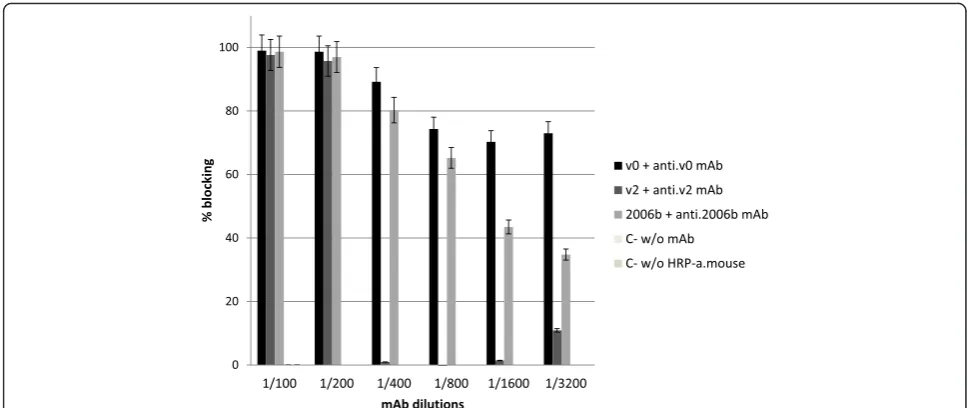
Anti-NoV mAbs tested prevented the binding of their homologous NoV VLPs to the saliva from an O blood group individual. Complete binding inhibition was seen when the mAbs were used up to a 1:200 dilution, whereas the blocking activity of anti-v2 mAb sharply drop out at 1:400 dilution and the anti-v0 and anti-2006b mAbs blocked the binding till higher dilutions (Fig. 4).
NoV VLP binding to D-Caco-2 cells
VLPs of all three GII.4 variants bound to D-Caco-2 cells. Binding was not uniform across the entire cell prepar-ation, and VLPs could be detected only on certain cells or clusters of the cell layer. As an example, fluorescence microscopy images showing the attachment of 2004 vari-ant NoV VLPs (v2) to D-Caco-2 cells are shown in Fig. 5. No signals were detected in any of the negative controls without VLPs.
Fig. 1Binding of NoV VLPs (GII.4-1999 (v0) and GII.4-2004 (v2) and GII.4-2006b) to porcine gastric mucin (PGM). Microtiter plates coated with PGM (10μg/ml) or without PGM (PBS) were incubated with the different VLPs at 2μg/ml for 1 h and processed for detection of bound VLPs by ELISA, using an anti-NoV VLP rabbit polyclonal antiserum (pAb). Binding is expressed as percentages referred to the highest OD 450 nm value obtained in duplicate assays. Error bars indicate standard deviations
[image:3.595.306.538.86.239.2] [image:3.595.57.291.409.644.2]Blockade of VLP binding to D-Caco-2 cells
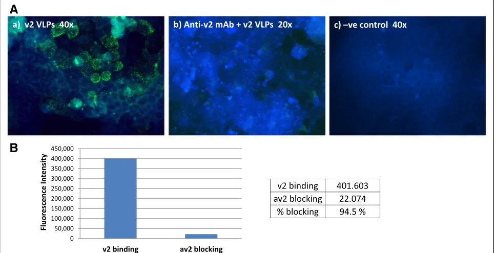
Anti-NoV mAbs used in this study strongly blocked the attachment of NoV VLPs to the surface of D-Caco-2 cells, and with the same strain-specificity as that ob-served in the salivary assays. Consistent with the ELISA results, a 74 % binding reduction was observed when VLPs were pre-incubated with the av2.mAb prior to adding to the cells (Fig. 6a). The blocking activity of the av0.mAb was of 82 %, whereas anti-2006b mAb also blocked the VLP binding (54.5 % reduction) (Table 2). When the VLPs were incubated with different PGM con-centrations, blockade of NoV VLPs binding to the D-Caco-2 cells occurred in a dose-dependent manner. At a concentration of 10 μg/ml, PGM completely abolished the ability of NoV VLPs to bind to D-Caco-2 cells. PGM at a concentration of 1 μg/ml caused a 50 % reduction of VLPs bound to the cells, and no blocking effect was seen at a concentration of 0.1 μg/ml (data not shown). Figure 6b shows the blockade by anti-v2.mAb of VLPs binding to D-Caco-2 cells. Fluorescence intensity was
measured using the ImageJ software program, which demonstrated a 94.5 % blocking activity.
Analysis of D-Caco-2 cells HBGA diversity reveals high expression of H-type 2, Leyand Lexantigens
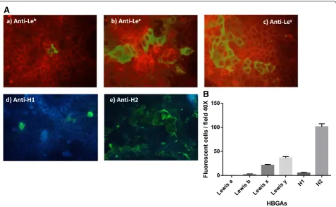
To determine if anti-Lewis (anti-Lea, Leb, Lex and Ley) and/or anti-type H (H1 and H2) antigen mAbs could in-fluence and block the VLP binding to D-Caco-2 cells, the expression and distribution of these antigens on the surface of the D-Caco-2 cells were investigated. The majority of D-Caco-2 cells expressed H-type 2 antigen on their surface. High expression of Leyand Lexantigens was also observed, whereas Leb and H-type 1 antigens were sparsely expressed (Fig. 7).
NoV VLPs bind to D-Caco-2 cells independently of Ley and H-type 2 antigen expression
Preincubation of D-Caco-2 cells with anti-Lewis Leyand anti-H-type 2 mAbs did not result in any relevant block-ing of GII.4 bindblock-ing to the D-Caco-2 cells. Blockade by
[image:4.595.58.541.90.416.2]these antibodies was tested because of the high expres-sion of Ley and H-type 2 antigens on the surface of D-Caco-2 cells. Furthermore, HBGA expression did not co-localise with the VLP binding, as it is shown as an example in Fig. 8 with GII.4-2004 (v2) VLPs and the H-type 2 antigen.
Discussion
HBGAs are neutral carbohydrates which are present on the surface of erythrocytes and mucosal epithelial cells, or as free oligosaccharides in milk, saliva and intestinal fluid of secretor individuals [27]. They are synthesized by sequential enzymatic transfer of single carbohydrate residues to specific precursor carbohydrate substrates, with very high genetic polymorphism [28]. Previous stud-ies suggested that HBGAs are likely to play an important role as cell receptors for NoV attachment [29, 30], although the molecular mechanism leading to cell entry and infection are to date largely unknown.
NoV binding assays using salivary EIAs have in recent years become the technique of choice for the study of the binding specificities of NoVs to HBGAs, since saliva samples containing different HBGA expression profiles are easy to collect, and the methods are relatively simple and fast to perform. Moreover, assays using synthetic HBGA have proven to be challenging in terms of repro-ducibility, but also troublesome due to the unavailability or difficulties in obtaining such reagents. In the absence of a reliable cell culture system or an appropriate animal model for human noroviruses, the NoV VLPs and HBGA binding assays have contributed significantly to our understanding of host susceptibility to NoV infection and
have also allowed the mapping of regions responsible for the interaction between HBGAs and the P2 sub-domain [10, 31, 32].
In this study we have identified the HBGA binding profiles of four different NoV VLPs representing gen-ogroups I (GI.1) and II (GII.4-1999 (v0), GII.4-2004 (v2) and GII.4-2006b) using saliva samples from different individuals. Four different binding patterns based on the secretor status, blood type and Lewis phenotype have previously been described [9, 30]. The VLPs used in this study exhibited distinct HBGA binding patterns: GI.1 VLPs bound more efficiently to saliva type A and O than to saliva type B, GII.4-v0 and v2 variant VLPs bound to all types A, B and O, and GII.4-2006b VLPs bound most efficiently to saliva type B. These results are in agree-ment with those reported by Uusi-Kerttula et al. [21], who analysed the binding of different NoV genotypes and GII.4 variants to salivary and synthetic HBGAs.
While many studies have compared results of binding assays to saliva and to synthetic HBGAs, the present study also compares NoV VLP binding patterns to saliva and to the human intestinal cell line Caco-2. Polarized or D-Caco-2 cells are also known to be a useful tool for the study of NoV binding, as both native NoVs and VLPs bind and penetrate the cell surface despite their inability to lead to productive infection [22, 33–35]. We have used Caco-2 cells to understand host-virus interac-tions because it has been demonstrated that they can differentiate in culture into cells with high homology to mature enterocytes in the intestinal epithelium [36, 37]. Moreover, they express HBGAs, like Lewis or H-type antigens [17, 22, 38].
[image:5.595.55.540.87.291.2]Our experiments with D-Caco-2 cells clearly demon-strated that VLPs bound to certain subpopulations of cells, and anti-NoV antibodies abolished binding to these cells. We performed the VLP binding assays to D-Caco-2 cells at 37 °C, likely to represent physiological conditions more accurately. Previous work has been performed at 4 °C to prevent internalization which was reported to occur in 5-7 % of the cells when incubating at 37 °C [35]. However, similar internalisation has also been reported even when incubation is performed at 4 °C [22].
Using HBGA-specific mAbs to localise HBGAs on the surface of D-Caco-2 cells showed widespread expression of H2 and Ley, and more diffuse or clustered expression of H1, Leband Lex. Our observations are similar to those previously reported by Murakami et al. [22], and con-trast with others reporting a high type H1 antigen ex-pression [38]. However, Caco-2 are a heterogeneous cell population and the expression of morphological and
functional characteristics depend on the degree of differ-entiation, which may explain the different results ob-served in relation to HBGA antigen expression, making it difficult to compare results obtained by different la-boratories. Blocking HBGA on the surface of D-Caco-2 cells with specific mAbs did not affect NoV VLP bind-ing, furthermore, no co-localisation of HBGA and NoV VLPs was observed by immunofluorescence. These re-sults therefore suggest that binding to Caco-2 cells could be mediated by receptors other than HBGAs, in agree-ment with a recent report by Murakami et al. [22]. Some viruses require interactions with more than one cellular surface molecule to initiate their replication cycle, for example bovine herpesvirus [39], and adenovirus [40]. Furthermore, some NoV strains, such as GII.4-2004, fail to bind efficiently to any HBGA suggesting they may bind other carbohydrates [41, 42], e.g. negatively charged sugars, similar to feline calicivirus (FCV) [43] or murine
A
B
[image:6.595.59.538.87.433.2]norovirus (MNV) [44]. Specifically, FCV attach to aα2, 6-linked sialic acid residue, but uses the junctional adhe-sion molecule 1 (JAM-1) for internalization into host cells [45]. Murine norovirus, in contrast, binds to a ganglioside GD1a present on the surface of murine mac-rophages [44]. In support of these hypotheses, some studies have demonstrated binding of GII noroviruses to negatively charged heparan sulfate [34], sialylated Lewisx carbohydrate [46], H-type 3 antigen [21] and ganglio-sides [47]. Furthermore, studies have shown that HBGA association is not sufficient to overcome the failure to propagate these viruses in vitro, as exemplified by the
resistance to infections of cell lines expressing HBGAs [23, 48]. It must be acknowledged that the expression and potential role of other HBGAs such as H3 and H4 was not investigated here, and this requires fur-ther study.
We propose that the D-Caco-2 cell binding assays may be more suitable for studying in vitro human NoV at-tachment and virus-host interactions than the salivary assays, which may only be considering the HBGA-NoV interactions, and are also likely to provide a more reliable surrogate for neutralising assays.
Conclusions
The interaction of NoV GII.4 genotype variants VLPs with human secretor positive saliva samples and with D-Caco-2 cells is mediated by different attaching receptors. Although their binding to saliva occurs according to distinct HBGA binding patterns and can be blocked by antibodies against Lewis antigens, their attachment to D-Caco-2 cells may be dependent on other receptors, which still need further investigation.
Methods
Recombinant NoV VLP expression
Copy DNA derived from capsid genes of NoV strains GI.1 and GII.4-Den Haag_2006b were cloned and expressed by the Bac-to-Bac baculovirus expression system (Invitrogen,
A
B
[image:7.595.57.541.92.339.2]Fig. 6Blockade of binding of NoV VLPs to D-Caco-2 cells by anti-NoV mAbs.a(a) D-Caco-2 cells were incubated with GII.4-2004 variant (v2) VLPs. (b) Same concentration of VLPs was pre-incubated with the homologous variant-specific mAb (at 1:100 dilution of the antibody) and added to D-Caco-2 cells; (c) negative control incubated with PBS.bMeasurement using the ImageJ software program of the fluorescence intensity revealing NoV VLPs (v2) bound to D-Caco-2 cells and its blockade by anti-v2.mAb
Table 2VLP binding blockade by NoV GII.4 variant-specific mAbs
NoV GII.4 variant VLP binding to D-Caco-2 cellsa
mAb GII.4 1999 (v2) GII.4-2004 (v0) GII.4-2006b
none 23 ± 5 17 ± 2 22 ± 3
av2 mAb 6 ± 2 (74 %) -
-av0 mAb - 3 ± 1 (82 %)
-3C3G3 mAb - - 10 ± 2 (54.5 %)
a
[image:7.595.55.292.606.689.2]Paisley, UK), and recombinant NoV VLPs were pro-duced in Sf9 insect cells following described procedures [49]. VLPs were purified from the cellular fraction lysed with Triton X-100 and centrifuged through a 40 % (w/ v) sucrose cushion. VLPs of strains GII.4-1999 (v0) and GII.4-2004 (v2) were also expressed by recombinant
baculoviruses as previously described [13]. The three GII.4 variants were chosen by their reported distinct binding properties [15, 50]. The different NoV VLP preparations were examined by electron microscopy (EM) after negative staining with 2 % phosphotungstic acid for purity, morphology and integrity appraisal.
Fig. 8NoV VLPs bind to D-Caco-2 cells independently of H type-2 antigen expression. GII.4-2004 (v2) VLPs were added to D-Caco-2 cells after adding anti-H2 antigen. After 1 h incubation at 37 °C with an anti-H2 mAb, the cells were fixed and incubated with secondary FITC-conjugated anti-rabbit IgG (to detect VLP binding) or Texas red-conjugated anti-mouse IgG (to detect anti-H2) antibodies. No co-localization of H2 antigen expression and VLP binding was observed
A
B
[image:8.595.60.538.87.381.2] [image:8.595.63.537.563.684.2]Saliva samples
Saliva samples were collected from 22 healthy adult volunteers, none of whom had taken any medication at the time or around the time of sample collection. To minimize the effects of the circadian rhythm, saliva samples were consistently collected in the morning hours (between 8 and 10 a.m.). Also, participants were instructed not to smoke, eat, drink, or brush their teeth in the 2 h before saliva collection. Freshly collected saliva samples were centrifuged at 10,000 × g for 5 min to remove particulate material, host and microbial cells and boiled at 100 °C to inactivate antibodies. Superna-tants were collected, divided into several aliquots and stored at -80 °C until their use. Blood ABO typing of the saliva donors was performed by hemagglutination assays (ALBAclone® Monoclonal ABO Antisera, Alpha Labora-tories, Eastleigh, England). Lewis antigens were pheno-typed in the saliva samples by ELISA with monoclonal antibodies against Lea, Leb, Lex and Ley(Covance, Ded-ham, MA, USA). Briefly, plates were coated with saliva samples at 1:1000 dilution in carbonate/bicarbonate buf-fer (pH 9.6) at 4 °C overnight. After blocking with PBS containing 3 % (w/v) bovine seroalbumin (PBS-BSA), plates were incubated with the different Lewis anti-gen monoclonal antibodies (anti-Lea BG-5, Leb BG-6, Lex BG-7 or Ley BG-8, Covance) at 1:100 during 1 h at 37 °C and detected by a secondary mouse antibody mix (anti-IgG, anti-IgM and anti-IgA) (Sigma) conjugated to horseradish peroxidase (HRP). After each step, plates were washed with PBS containing 0.05 % of Tween-20 (PBS-T). The reaction was developed by the addition of OPD Fast (Sigma, Dorset, England) and stopped after 10 min incubation with 3 M H2SO4. Absorbance was measured at 492 nm in a microplate reader (Multiskan FC, Thermo Scientific, Vantaa, Finland).
The secretor (FUT2+) and non-secretor (FUT2-) status was investigated by genotyping the FUT2 gene. Genomic DNA was extracted from saliva samples using the Qiagen QIAamp DNA Mini Kit. Genotyping for FUT2 was performed by PCR-RFLP as described previously [51].
Monoclonal (mAb) and polyclonal (pAb) antibody production
BALB/c mice were immunized by intraperitoneal (IP) inoculation of three doses of GII.4-2006b VLPs with Freund’s adjuvant at 15-day intervals. Splenocytes were fused with Sp2/0-Ag14 mieloma cells as described previ-ously [52] and hybridomas were screened by ELISA using the same NoV VLP genotype as the antigen. An anti-GII.4-2006b was obtained (3C3G3) and used along the present study. The anti-v0.mAb and anti-v2.mAb were previously obtained as described [13]. A polyclonal antiserum (pAb) against NoV VLPs was obtained at
the Public Health England (PHE, London) by immunizing rabbits with a mixture of VLPs (GII.4-v0, GII.4-v2 and GII.3).
Enzyme-linked immunoassays (EIAs)
VLP binding to porcine gastric mucin (PGM)
Microtiter plates were coated with PGM at 10 μg/ml in carbonate-bicarbonate buffer (pH 9.6) at 37 °C over-night. Plates were washed three times with PBS-T (PBS containing 0.05 % Tween-20), blocked with PBS contain-ing 1 % skimmed milk and incubated 30 min at 37 °C. VLPs were added at 2μg/ml in PBS and incubated 1 h at 37 °C. After three washes, the anti-VLPs rabbit polyclonal antiserum (pAb) was added at 1:1000 dilu-tion. Binding was detected with HRP-conjugated anti-rabbit IgG at 1:10,000 dilution (Bioss, Woburn, MA). The reaction was developed by the addition of TMB (3,3’,5,5’-tetramethyl-benzidine, Sigma) and stopped after 10 min incubation with 4 N H2SO4. Absorbance was measured at 450 nm in a microplate reader Multiskan FC (Thermo Scientific). Negative and blank controls were included in all assays.
Blocking assays
The same EIA protocol used to measure NoV VLP bind-ing to HBGAs in saliva was followed as described [9], adding an incubation step with the different mAbs before the addition of the VLPs, and/or preincubating the VLPs in the presence of the NoV-specific antibodies. Monoclonal antibodies against Lewis antigens were added to the plate at a dilution of 1:10. NoV VLPs (GI.1, GII.4-2006b, GII.4-v0 and GII.4-v2) were added at 2-10μg/ml depending of the VLP variant, and their detec-tion was performed with the anti-NoV rabbit polyclonal serum followed by a secondary HRP-conjugated anti-rabbit IgG antibody diluted at 1:10,000 (Bioss, Woburn, MA). The ability of anti-NoV mAbs to block the binding of VLPs to saliva was tested using serial 2-fold dilutions of each mAb from 1:100 until 1:3200. The VLPs (2 μg/ml in PBS) were pre-incubated with the assayed antibodies, or with PBS in the negative control, for 1 h at 37 °C. The mixtures were then added to the saliva-coated plates and incubated for 1 h at 37 °C. The binding of VLPs was determined by incubation with HRP-conjugated anti rabbit or anti-mouse IgG antibodies (1:2000 dilution), as appropriate, followed by the peroxidase substrate TMB step.
Caco-2 cell binding assays
Caco-2 cell culture
Caco-2 cells were grown in Dulbecco’s modified Eagle’s minimum essential medium (DMEM) (Invitrogen) sup-plemented with 4.5 % glucose, 10 % (v/v) fetal bovine serum (Invitrogen), non-essential amino acids, 100 I.U./ ml penicillin, 100 mg/ml streptomycin, 2 mM glutam-ine and 1 mM sodium pyruvate, at 37 °C under a humidified atmosphere with 5 % CO2. Binding medium (BM), used when the cells were incubated with the VLPs, contained all of the above except fetal bovine serum.
Cell binding assays
Caco-2 cell binding assays were perfomed as previously described [17] with modifications. Caco-2 cells were seeded at 2 × 10⁵cells/ml on culture slides (BD Falcon, Bedford, MA). Cells were cultured for 9-10 days to allow cell differentiation (D-Caco-2). D-Caco-2 cells were washed twice with PBS and incubated with VLPs (5μg/ ml) in 200μl BM at 37 °C for 1 h, under a humidified at-mosphere. Negative controls consisted of cells incubated without VLPs. After 3 washes with PBS, cells were fixed with methanol for 15 min at room temperature and then blocked with PBS containing 1 % skimmed milk (PBS-milk 1 %) for 30 min at 37 °C. Fixed cells were subse-quently incubated at 37 °C for 1 h with polyclonal rabbit IgG anti-NoV VLPs (PHE) antibody at 1:4000 dilution in PBS. After washing the cells with PBS-0.05 % Tween-20, they were incubated with a goat FITC-conjugated anti-rabbit IgG antibody (Abcam, Cambridge, UK) at 1:1000 dilution for 1 h at 37 °C. Finally, slides were mounted with 10 % glycerol in saline and preparations were observed under a Nikon Eclipse 80i fluorescence micro-scope equipped with a digital camera and the Hamamatsu camera controller C4742-95 (Hamamatsu City, Japan).
Expression of HBGAs
Immunofluorescence assays were performed as descr-ibed above in order to analyse the expression of Lewis antigens, using anti-Lea BG-5, Leb BG-6, Lex BG-7 and Ley BG-3 monoclonal antibodies (Covance) and anti-H antigens (mAb to blood group H1(O) and anti-blood group H2 antigen, Abcam). Anti-Lewis antigen mAbs were used diluted to 1:100 and anti-H antigens mAbs to 1:200. For visualization, a secondary antibody FITC-conjugated anti-mouse IgG antibody (Santa Cruz Biotechnology) diluted 1:400 was used. HBGA expression in D-Caco-2 cells was evaluated by counting immuno-fluorescent cells in 10 microscopic fields (40X). A semi-quantitative four stage scoring system was applied with the following ranges: negative (-); < 50 fluorescent cells (1+); 51-199 fluorescent cells (2+), and >200 fluorescent cells (3+).
VLP-cell binding blocking assays
To assess the ability of anti-HBGA-specific antibodies to block the binding of VLPs to D-Caco-2 cells, cell chambers were incubated with anti-Ley or anti-H-type 2 mAbs (as described for the HBGA expression assay) prior to incubation with the NoV GII.4-v2 VLPs. In order to study the ability of the anti-NoV antibodies to block the binding of the NoV VLPs to D-Caco-2 cells, VLPs were incubated with their homologous antibody using the same conditions as in the salivary blocking assays, but with BM as the diluent. Blocking of VLPs binding to D-Caco-2 cells by PGM was performed by incubating the GII.4-v2 VLPs for 1 h at 37 °C with PGM at a concentration of 0, 10 and 50 μg/ml. The rest of the procedure was described in the binding assays section. Control cells were incu-bated with buffer instead of mAbs/VLP, and VLPs in the absence of mAbs or PGM were used as positive binding controls. Staining specificity was assessed in negative controls by (1) omission of the VLP step and (2) replacement of primary antibodies with 3 % BSA/ PBS. Immunofluorescence was assessed on a quantita-tive scale by calculating the number of posiquantita-tive cells on each well chamber in 10 microscopic fields (40X). Fluorescence intensity of the VLP-cell binding and blocking assays was measured using the Java-based image processing ImageJ 1.49 program [53].
Statistical analysis
The nonparametric Mann-Whitney test was applied to determine the presence or absence of significant differ-ences. In all cases p-values below 0.05 were considered statistically significant. The analyses were performed by using the IBM SPSS software vs. 22.0 (IBM Corp.).
Ethics statement
Appropriate informed written consent was obtained from all four volunteers and the saliva samples were analysed anonymously. The study protocol and consent forms were approved by the Human Research Ethics Committee of the University of Valencia. The animal studies were evaluated and approved by the Animal Experimentation and Welfare Ethics Committee of the University of Valencia. JB possesses the accreditation by the Conselleria de Agricultura, Generalitat Valenciana, to design and perform experiments with laboratory animals.
Competing interests
The authors declare that they have no competing interests.
Authors’contributions
Acknowledgements
This work was supported by the Spanish Ministry of Economy and Competitiveness (SAF2012-38368). Noelia Carmona-Vicente was recipient of a“V Segles”fellowship from the University of Valencia and Jesús Rodríguez-Díaz was recipient of a“Ramón y Cajal”contract from the Spanish Ministry of Economy and Competitiveness (RYC-2013-12442). Miren Iturriza-Gómara receives support form the Wellcome Trust ISSF awarded to the University of Liverpool, by the National Institute for Health Research (Grant number NIHR HPRU 2012-10038).
Disclaimer
The views expressed are those of the authors and not necessarily those of the NHS, the NIHR, the Department of Health or Public Health England.
Author details
1Department of Microbiology, School of Medicine, University of Valencia,
Avda. Blasco Ibáñez, 17, 46010 Valencia, Spain.2Virus Reference Department,
Public Health England, London, UK.3CIMI, Institute of Infection and Global
Health, University of Liverpool, Liverpool, UK.4NIHR Health Protection Research Unit in Gastrointestinal Infections, University of Liverpool, Liverpool, UK.
Received: 11 March 2016 Accepted: 12 May 2016
References
1. Tam CC, Rodrigues LC, Viviani L, Dodds JP, Evans MR, Hunter PR, Gray JJ, Letley LH, Rait G, Tompkins DS, O'Brien SJ. Longitudinal study of infectious intestinal disease in the UK (IID2 study): incidence in the community and presenting to general practice. Gut. 2012;61:69–77. doi:10.1136/gut.2011. 238386.
2. Gastanaduy PA, Hall AJ, Curns AT, Parashar UD, Lopman BA. Burden of norovirus gastroenteritis in the ambulatory setting–United States, 2001-2009. J Infect Dis. 2013;207:1058–65. doi:10.1093/infdis/jis942.
3. Patel MM, Widdowson MA, Glass RI, Akazawa K, Vinje J, Parashar UD. Systematic literature review of role of noroviruses in sporadic gastroenteritis. Emerg Infect Dis. 2008;14:1224–31. doi:10.3201/eid1408.071114.
4. Vinje J. Advances in laboratory methods for detection and typing of norovirus. J Clin Microbiol. 2015;53:373–81. doi:10.1128/JCM.01535-14. 5. Buesa J, Montava R, Abu-Mallouh R, Fos M, Ribes JM, Bartolome R,
Vanaclocha H, Torner N, Dominguez A. Sequential evolution of genotype GII.4 norovirus variants causing gastroenteritis outbreaks from 2001 to 2006 in Eastern Spain. J Med Virol. 2008;80:1288–95. doi:10.1002/jmv.21182. 6. Siebenga JJ, Vennema H, Zheng DP, Vinje J, Lee BE, Pang XL, Ho EC, Lim W,
Choudekar A, Broor S, et al. Norovirus illness is a global problem: emergence and spread of norovirus GII.4 variants, 2001-2007. J Infect Dis. 2009;200:802–12. doi:10.1086/605127.
7. Koelle K, Cobey S, Grenfell B, Pascual M. Epochal evolution shapes the phylodynamics of interpandemic influenza A (H3N2) in humans. Science. 2006;314:1898–903. doi:10.1126/science.1132745.
8. Hutson AM, Atmar RL, Estes MK. Norovirus disease: changing epidemiology and host susceptibility factors. Trends Microbiol. 2004;12:279–87. http://dx.doi.org/10.1016/j.tim.2004.04.005.
9. Huang P, Farkas T, Marionneau S, Zhong W, Ruvoen-Clouet N, Morrow AL, Altaye M, Pickering LK, Newburg DS, LePendu J, Jiang X. Noroviruses bind to human ABO, Lewis, and secretor histo-blood group antigens: identification of 4 distinct strain-specific patterns. J Infect Dis. 2003;188:19–31. doi:10.1086/375742.
10. Cao S, Lou Z, Tan M, Chen Y, Liu Y, Zhang Z, Zhang XC, Jiang X, Li X, Rao Z. Structural basis for the recognition of blood group trisaccharides by norovirus. J Virol. 2007;81:5949–57. doi:10.1128/JVI.00219-07.
11. Koda Y, Soejima M, Kimura H. The polymorphisms of fucosyltransferases. Leg Med (Tokyo). 2001;3:2–14. doi: S1344622301000050.
12. Prasad BV, Hardy ME, Dokland T, Bella J, Rossmann MG, Estes MK. X-ray crystallographic structure of the Norwalk virus capsid. Science. 1999;286: 287–90. doi:10.1126/science.286.5438.287.
13. Allen DJ, Noad R, Samuel D, Gray JJ, Roy P, Iturriza-Gomara M. Characterisation of a GII-4 norovirus variant-specific surface-exposed site involved in antibody binding. Virol J. 2009;6:150. doi:10.1186/1743-422X-6-150.
14. Allen DJ, Gray JJ, Gallimore CI, Xerry J, Iturriza-Gomara M. Analysis of amino acid variation in the P2 domain of the GII-4 norovirus VP1 protein reveals
putative variant-specific epitopes. PLoS ONE. 2008;3, e1485. doi:10.1371/ journal.pone.0001485.
15. Debbink K, Donaldson EF, Lindesmith LC, Baric RS. Genetic mapping of a highly variable norovirus GII.4 blockade epitope: potential role in escape from human herd immunity. J Virol. 2012;86:1214–26. doi:10.1128/JVI.06189-11. 16. Shanker S, Choi JM, Sankaran B, Atmar RL, Estes MK, Prasad BV. Structural
analysis of histo-blood group antigen binding specificity in a norovirus GII.4 epidemic variant: implications for epochal evolution. J Virol. 2011;85:8635–45. doi:10.1128/JVI.00848-11.
17. Marionneau S, Ruvoen N, Le Moullac-Vaidye B, Clement M, Cailleau-Thomas A, Ruiz-Palacois G, Huang P, Jiang X, Le Pendu J. Norwalk virus binds to histo-blood group antigens present on gastroduodenal epithelial cells of secretor individuals. Gastroenterology. 2002;122:1967–77. doi:10.1053/gast.2002.33661. 18. Hutson AM, Atmar RL, Graham DY, Estes MK. Norwalk virus infection and
disease is associated with ABO histo-blood group type. J Infect Dis. 2002; 185:1335–7. doi: 10.1016/j.tim.2004.04.005.
19. Tan M, Jiang X. Norovirus and its histo-blood group antigen receptors: an answer to a historical puzzle. Trends Microbiol. 2005;13:285–93. doi:10.1016/ j.tim.2005.04.004.
20. Frenck R, Bernstein DI, Xia M, Huang P, Zhong W, Parker S, Dickey M, McNeal M, Jiang X. Predicting susceptibility to norovirus GII.4 by use of a challenge model involving humans. J Infect Dis. 2012;206:1386–93. doi:10.1093/infdis/jis514. 21. Uusi-Kerttula H, Tamminen K, Malm M, Vesikari T, Blazevic V. Comparison of
human saliva and synthetic histo-blood group antigens usage as ligands in norovirus-like particle binding and blocking assays. Microbes Infect. 2014;16: 472–80. doi:10.1016/j.micinf.2014.02.010.
22. Murakami K, Kurihara C, Oka T, Shimoike T, Fujii Y, Takai-Todaka R, Park Y, Wakita T, Matsuda T, Hokari R, et al. Norovirus binding to intestinal epithelial cells is independent of histo-blood group antigens. PLoS ONE. 2013;8, e66534. doi:10.1371/journal.pone.0066534.
23. Duizer E, Schwab KJ, Neill FH, Atmar RL, Koopmans MPG, Estes MK. Laboratory efforts to cultivate noroviruses. J Gen Virol. 2004;85:79–87. doi:10.1099/vir.0.19478-0.
24. Herbst-Kralovetz MM, Radtke AL, Lay MK, Hjelm BE, Bolick AN, Sarker SS, Atmar RL, Kingsley DH, Arntzen CJ, Estes MK, Nickerson CA. Lack of norovirus replication and histo-blood group antigen expression in 3-dimensional intestinal epithelial cells. Emerg Infect Dis. 2013;19:431–8. doi:10.3201/eid1903.121029.
25. Jones MK, Watanabe M, Zhu S, Graves CL, Keyes LR, Grau KR, Gonzalez-Hernandez MB, Iovine NM, Wobus CE, Vinje J, et al. Enteric bacteria promote human and mouse norovirus infection of B cells. Science. 2014;346: 755–9. doi:10.1126/science.1257147.
26. White LJ, Ball JM, Hardy ME, Tanaka TN, Kitamoto N, Estes MK. Attachment and entry of recombinant Norwalk virus capsids to cultured human and animal cell lines. J Virol. 1996;70:6589–97.
27. Marionneau S, Cailleau-Thomas A, Rocher J, Le Moullac-Vaidye B, Ruvoen N, Clement M, Le Pendu J. ABH and Lewis histo-blood group antigens, a model for the meaning of oligosaccharide diversity in the face of a changing world. Biochimie. 2001;83:565–73. doi: S0300-9084(01)01321-9. 28. Clausen H, Hakomori S. ABH and related histo-blood group antigens;
immunochemical differences in carrier isotypes and their distribution. Vox Sang. 1989;56:1–20.
29. Tan M, Jiang X. Norovirus-host interaction: multiselections by human histo-blood group antigens. Trends Microbiol 2011; 19:382–8. doi:10.1016/j.tim.2011.05.007. 30. Huang P, Farkas T, Zhong W, Tan M, Thornton S, Morrow AL, Jiang X. Norovirus
and histo-blood group antigens: demonstration of a wide spectrum of strain specificities and classification of two major binding groups among multiple binding patterns. J Virol. 2005;79:6714–22. doi:10.1086/375742.
31. Tan M, Huang P, Xia M, Fang PA, Zhong W, McNeal M, Wei C, Jiang W, Jiang X. Norovirus P particle, a novel platform for vaccine development and antibody production. J Virol. 2011;85:753–64. doi:10.1128/JVI.01835-10. 32. Tan M, Huang P, Meller J, Zhong W, Farkas T, Jiang X. Mutations within the
P2 domain of norovirus capsid affect binding to human histo-blood group antigens: evidence for a binding pocket. J Virol. 2003;77:12562–71. doi:10.1128/JVI.77.23.12562-12571.
33. Takanashi S, Saif LJ, Hughes JH, Meulia T, Jung K, Scheuer KA, Wang Q. Failure of propagation of human norovirus in intestinal epithelial cells with microvilli grown in three-dimensional cultures. Arch Virol 2014;159:257–66. doi:10.1007/s00705-013-1806-4.
the cellular membrane. J Virol. 2004;78:3817–26. doi:10.1128/JVI.78.8. 3817-3826.
35. Tamura M, Natori K, Kobayashi M, Miyamura T, Takeda N. Interaction of recombinant Norwalk virus particles with the 105-kilodalton cellular binding protein, a candidate receptor molecule for virus attachment. J Virol. 2000;74:11589–97. doi:10.1128/JVI.74.24.11589-11597.
36. Pinto M, Robine-Léon S, Appay MD, Kedinger M, Triadou N, Dussaulx E, Lacroix B, Simon-Assmann P, Haffen K, Fogh J, Zweibaum A. Enterocyte-like differentiation and polarization of the human colon carcinoma cell line Caco-2 in culture. Biol Cell. 1983;47:323–30.
37. Rousset M. The human colon carcinoma cell lines HT-29 and Caco-2: two in vitro models for the study of intestinal differentiation. Biochimie. 1986; 68:1035–40.
38. Amano J, Oshima M. Expression of the H type 1 blood group antigen during enterocytic differentiation of Caco-2 cells. J Biol Chem. 1999;274: 21209–16.
39. Li Y, van Drunen Littel-van den Hurk S, Babiuk LA, Liang X. Characterization of cell-binding properties of bovine herpesvirus 1 glycoproteins B, C, and D: identification of a dual cell-binding function of gB. J Virol. 1995;69:4758–68. 40. Wickham TJ, Mathias P, Cheresh DA, Nemerow GR. Integrins alpha v beta 3
and alpha v beta 5 promote adenovirus internalization but not virus attachment. Cell. 1993;73:309–19. doi: 0092-8674(93)90231-E.
41. Bok K, Abente EJ, Realpe-Quintero M, Mitra T, Sosnovtsev SV, Kapikian AZ, Green KY. Evolutionary dynamics of GII.4 noroviruses over a 34-year period. J Virol. 2009;83:11890–901. doi:10.1128/JVI.00864-09.
42. Lindesmith LC, Donaldson EF, Lobue AD, Cannon JL, Zheng DP, Vinje J, Baric RS. Mechanisms of GII.4 norovirus persistence in human populations. PLoS Med. 2008;5:e31. doi:10.1371/journal.pmed.0050031.
43. Stuart AD, Brown TD. Alpha2,6-linked sialic acid acts as a receptor for Feline calicivirus. J Gen Virol. 2007;88:177–86. doi:10.1099/vir.0.82158-0.
44. Taube S, Perry JW, Yetming K, Patel SP, Auble H, Shu L, Nawar HF, Lee CH, Connell TD, Shayman JA, Wobus CE. Ganglioside-linked terminal sialic acid moieties on murine macrophages function as attachment receptors for murine noroviruses. J Virol. 2009;83:4092–101. doi:10.1128/JVI.02245-08. 45. Makino A, Shimojima M, Miyazawa T, Kato K, Tohya Y, Akashi H. Junctional
adhesion molecule 1 is a functional receptor for feline calicivirus. J Virol. 2006;80:4482–90. doi:10.1128/JVI.80.9.4482-4490.2006.
46. Rydell GE, Nilsson J, Rodriguez-Diaz J, Ruvoen-Clouet N, Svensson L, Le Pendu J, Larson G. Human noroviruses recognize sialyl Lewis x neoglycoprotein. Glycobiology. 2009;19:309–20. doi:10.1093/glycob/cwn139.
47. Han L, Tan M, Xia M, Kitova EN, Jiang X, Klassen JS. Gangliosides are ligands for human noroviruses. J Am Chem Soc. 2014;136:12631–7. doi:10.1021/ ja505272n.
48. Guix S, Asanaka M, Katayama K, Crawford SE, Neill FH, Atmar RL, Estes MK. Norwalk virus RNA is infectious in mammalian cells. J Virol. 2007;81: 12238–48. doi:10.1128/JVI.01489-07.
49. Nicollier-Jamot B, Pico V, Pothier P, Kohli E. Molecular cloning, expression, self-assembly, antigenicity, and seroepidemiology of a genogroup II norovirus isolated in France. J Clin Microbiol. 2003;41:3901–4. doi:10.1128/JCM.41.8.3901-3904.
50. de Rougemont A, Ruvoen-Clouet N, Simon B, Estienney M, Elie-Caille C, Aho S, Pothier P, Le Pendu J, Boireau W, Belliot G. Qualitative and quantitative analysis of the binding of GII.4 norovirus variants onto human blood group antigens. J Virol. 2011;85:4057–70. doi:10.1128/JVI.02077-10.
51. Marionneau S, Airaud F, Bovin NV, Le Pendu J, Ruvoen-Clouet N. Influence of the combined ABO, FUT2, and FUT3 polymorphism on susceptibility to Norwalk virus attachment. J Infect Dis. 2005;192:1071–7. doi:10.1086/432546. 52. Harlow E, Lane D. Antibodies: a laboratory manual. New York: Cold Spring
Harbor Laboratory; 1988.
53. Schneider CA, Rasband WS, Eliceiri KW. NIH Image to ImageJ: 25 years of image analysis. Nat Methods. 2012;9:671–5. doi:10.1038/nmeth.2089.
• We accept pre-submission inquiries
• Our selector tool helps you to find the most relevant journal
• We provide round the clock customer support
• Convenient online submission
• Thorough peer review
• Inclusion in PubMed and all major indexing services
• Maximum visibility for your research
Submit your manuscript at www.biomedcentral.com/submit