Neural Prosthetics and Parietal Cortex
Thesis by Boris Revechkis
In Partial Fulfillment of the Requirements for the Degree of Doctor of Philosophy
California Institute of Technology Pasadena, California
2015
Acknowledgements
I’d like to thank my advisor, Richard Andersen, for giving me the rare privilege of listening to neurons both primate and human, Tyson Aflalo for persistent advice and guidance, Bijan Pesaran for giving me my first glimpse of real neuroscience when I was just a curious undergraduate, and our clinical study subject for having the courage and fortitude to turn her profound hardships into learning opportunities for all of us.
I’d also like to thank Dr. Nader Pouratian for his critical surgical expertise and enthusiasm, Ueli Rutishauser, Chess Stetson, Eunjung Hwang, Markus Hauschild, helpful for scientific discussion, Tessa Yao for administrative omnipresence, Kelsie Pejsa for animal care, and Viktor Shcherbatyuk and Spencer Kellis for technical assistance. I thank the National Institutes of Health for providing the funding that allowed this thesis to be undertaken.
Dedication
I dedicate this thesis to my family:
To my parents and grandparents for abandoning their former lives, friends, and homes, and traveling halfway across the planet so that their children could live better.
To my brother, for asking my parents for a little brother.
Abstract
In the last decade, research efforts into directly interfacing with the neurons of individuals with motor deficits have increased. The goal of such research is clear: Enable individuals affected by paralysis or amputation to regain control of their environments by manipulating external devices with thought alone. Though the motor cortices are the usual brain areas upon which neural prosthetics depend, research into the parietal lobe and its subregions, primarily in non-human primates, has uncovered alternative areas that could also benefit neural interfaces. Similar to the motor cortical areas, parietal regions can supply information about the trajectories of movements. In addition, the parietal lobe also contains cognitive signals like movement goals and intentions. But, these areas are also known to be tuned to saccadic eye movements, which could interfere with the function of a prosthetic designed to capture motor intentions only. In this thesis, we develop and examine the functionality of a neural prosthetic with a non-human primate model using the superior parietal lobe to examine the effectiveness of such an interface and the effects of unconstrained eye movements in a task that more closely simulates clinical applications. Additionally, we examine methods for improving usability of such interfaces.
Table of Contents
Acknowledgements ... ii
Abstract ... iv
List of Abbreviations ... ix
List of Illustrations and Tables ... x
1. Introduction ... 1
2. Background ... 3
2.1 Brain Machine Interfaces ... 5
2.2 Parietal Cortex ... 7
2.3 References ... 10
3. Non-Human Primate Parietal Cortex and Neural Prosthetics ... 13
3.1 Parietal Neural Prosthetic Control of a Computer Cursor in a Graphical-User-Interface Task ... 13
3.1.1 Introduction ... 13
3.1.2 Methods... 15
3.1.2.1 Behavioral Setup ... 16
3.1.2.2 Neural Recordings ... 16
3.1.2.3 Behavioral Task ... 18
3.1.2.4 Performance Measures ... 20
3.1.2.5 Decoder Training ... 22
3.1.2.6 Decoder Calculation ... 24
3.1.2.7 Saccade Task ... 25
3.1.3 Results ... 26
3.1.3.1 Saccade Task ... 26
3.1.3.2 Behavioral Task – Manual Control ... 27
3.1.3.3 Behavioral Task – Brain Control ... 30
3.1.4 Discussion ... 36
3.1.5 References ... 39
3.2 State Decoding Improves Use of a Computer Cursor for Neuroprosthetic Applications ... 41
3.2.1 Introduction ... 41
3.2.2 Methods... 42
3.2.2.1 Behavioral Setup ... 42
3.2.2.2 Neural Recordings ... 43
3.2.2.4 Performance Measures ... 46
3.2.2.5 Statistical Tests ... 47
3.2.2.6 Decoder Training Procedure ... 47
3.2.2.7 Velocity Decoder Computation ... 48
3.2.2.8 State Decoder Computation ... 50
3.2.3 Results ... 51
3.2.4 Discussion ... 55
3.2.5 References ... 57
4. Human Parietal Cortex ... 58
4.1 Selectivity for Hand Movement Execution and Feedback in Human Parietal Neurons and Local Fields ... 58
4.1.1 Introduction ... 58
4.1.2 Methods... 60
4.1.2.1 Human Subject Recruitment ... 60
4.1.2.2 Experimental Setup ... 61
4.1.2.3 Behavioral Tasks ... 62
4.1.2.3.1 Effector Specificity in Decoding Delayed Reaches ... 62
4.1.2.3.2 Limb Tuning Task ... 66
4.1.2.3.3 Effector Specificity in Hand Gestures ... 69
4.1.2.3.4 Online Gesture Control ... 72
4.1.3 Results ... 76
4.1.3.1 Delayed Reach Task ... 76
4.1.3.2 Limb Tuning Task... 81
4.1.3.3 Gesture Task ... 83
4.1.3.3.1 LFP Response ... 83
4.1.3.3.2 Single/Multi-unit Response ... 86
4.1.3.4 Online Gesture Control Task ... 89
4.1.4 Discussion ... 93
4.1.5 References ... 98
Appendices ... 100
A. Tuning and Decoding Properties of Neural Populations ... 100
1. Human Neural Population ... 100
List of Abbreviations
AIP Anterior Intraparietal Area
BA Brodmann Area
BOLD Blood-oxygen-level Dependent
BMI Brain Machine Interface
FDR False Discovery Rate
FMRI Functional Magnetic Resonance Imaging
FOV Field of View
HMD Head-Mounted Display
IQR Inter-Quartile Range
ITI Inter-Trial Interval
LFP Local Field Potential
LIP Lateral Intraparietal Area
LOOCV Leave-One-Out Cross Validation
NHP Non-Human Primate
PCA Principal Component Analysis
PPC Posterior Parietal Cortex
PRR Parietal Reach Region
PSTH Peristimulus Time Histogram
TTA Time To Acquire
TTH Time To Hold
TTR Time To Reward
List of Illustrations and Tables
Figure 3.1.2.3-1 Face in the Crowd Task Trial Structure. ... 18
Figure 3.1.3.1-1 Manual Control Performance. ... 27
Figure 3.1.3.2-1 Effect of the Crowd on Gaze During Manual Control. ... 28
Figure 3.1.3.2-2 Gaze with Crowd on during Manual vs Brain Control.. ... 29
Figure 3.1.3.3-1 Performance Measures Across Training and Assessment Conditions (Brain Control) ... 31
Table 1 Statistics for All Combinations of Conditions and Measures ... 32
Figure 3.1.3.3-2 Hand and Cursor Position During Decoding ... 35
Figure 3.2.2.3-1 Face in a Crowd Task Structure. ... 44
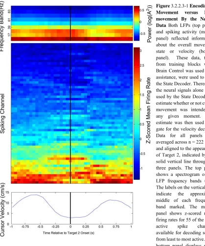
Figure 3.2.2.3-1 Encoding of Movement versus Non-movement By the Neural Data .... 46
Figure 3.2.3-1 Summary State Decoder Performance.. ... 52
Figure 3.2.3-2 Effect of State Decoding on TTH Per Decoder ... 54
Figure 4.1.2.3.1-1 Example Frame from Stereoscopic Display. ... 62
Figure 4.1.2.3.1-2 Reaching Task Progression.. ... 65
Figure 4.1.2.3.2-1 Limb Tuning Task Diagram. ... 67
Figure 4.1.2.3.2-1 Gesture Task Progression ... 69
Figure 4.1.2.3.4-1 Online Gesture Control Task And Training Task. ... 73
Figure 4.1.3.1-1 Delayed Reaching Task Offline Decoding Performance. ... 77
Figure 4.1.3.1-2 Time Course of Decoding Performance Throughout the Trial. ... 79
Figure 4.1.3.1-3 Limb Tuning Task Results. ... 81
Figure 4.1.3.3.1-1 Responses in the Local Field Potentials of Array 1 to Task Variables. ... 84
Figure 4.1.3.3.2-1 Example Neural Responses during the Gesture Task.. ... 88
Figure 4.1.3.4-1 Online Gesture Control Performance Summary. ... 90
Figure 4.1.3.4-2 Change in Activity of Units Used For Decoding.. ... 93
Figure A-1 PSTHs of Tuned Units Used for Offline Decoding of Delayed Reach Task.. ... 100
Figure A-1 Neuron Dropping Curves for Delayed Reach Task By Effector.. ... 101
Figure A-2 PSTHs of Tuned Neurons Recorded during a Delayed Reach Center-out Task. ... 102
Figure B-1 Targeting Task 3 Schematic.. ... 105
Figure B-2 fMRI Results and Array Implant Locations.. ... 106
Figure C-1 Local Field Potential Power for Array 1 During the Gesture Task, Collapsed in Time. ... 108
Figure C-2 Local Field Potentials of Array 2 During the Gesture Task.. ... 109
1.
IntroductionEver since it was discovered that the activity of individual neurons in the mammalian brain correlate with specific sensory stimuli, decisions, and, movements, a tantalizing and profound question has loomed large. Is it possible to predict thoughts and intentions from neural activity? Can those predictions be used to connect brains and their owners directly with the world through artificial devices, bypassing the body? Though many envisioned incarnations of such technology remain outside the scope of modern neuroscience, one subset has proven an active and fruitful area of research over the last 20 years. A small number of paralysis patients have successfully been able to manipulate computer cursors and robotic limbs based on the activity of tens to a few hundreds of neurons in their primary motor cortices. However, many technical and scientific challenges remain to be conquered before these technologies can be applied beyond the proof of concept.
prosthetics in both non-human primates and humans. In applying the neural signals located in the parietal lobe, we improve upon the technology as a whole while also learning more about how this part of the primate brain contributes to motor behavior.
In Chapter 2, we give an overview of the history of recording of neural activity at the single-unit level, the properties of parietal cortex, as well as an overview of neural prosthetics research to date. In Chapter 3, we present two studies of neural prosthetics carried out with a non-human primate subject. In the first, we examine the effect of eye movements on the ability of a prosthetic user to perform neural control of a cursor. Because some areas in parietal cortex are sensitive to gaze direction and movements of the eye, it is important to determine whether or not record from hand-related areas can be done without interference of eye-related signals. In the second study, we describe a paired decoder system, one state-based and one continuous, that reduces unwanted noise in a neurally controlled cursor and improves upon the state of the art.
2.
BackgroundEver since the link between the brain and mind has been considered, generations of natural philosophers, scientists, and even pseudoscientists have been examining and investigating the workings of this organ. But, it was not until Luigi Galvani’s experiments at end of the 18th century that the association between the nervous system and electricity was observed. Galvani determined that injecting current into the severed frog leg caused its muscles to contract (Piccolino 1997). With this, modern electrophysiology was born.
However, its infancy was long. Little was known about the brain during Galvani’s time. Over the next century, biologists and anatomists steadily investigated the brain and its various structures at progressively smaller scales. In the late 1880s, Santiago Ramόn y Cajal transformed neuroscience as a whole through detailed drawings and descriptions of what he deemed the functional units of the brain: the neuron. But scientists still lacked the technology to investigate such tiny structures and what their function might be.
communication between neurons in the periphery as well as in the brain. Adrian was the first to record them directly, albeit from muscle fibers.
Not long after, Adrian was able to make similar recordings from the optic nerve of the toad. During those experiments, Adrian played the electrical recordings of the nerve over a loudspeaker so that he could listen to the impulses while working with the preparation in a nearly dark room. Eventually, he noticed that the intensity of impulses played over the loudspeaker varied with his own movements around the room. He realized that he was in the field of view of the toad’s eye, and that the rate of activity along the optic nerve was responding to his movement. This and related work garnered Adrian and his colleague Sir Charles Scott Sherrington the Nobel prize in Physiology or Medicine in 1932 (Hodgkin 1979).
By the late 1960s, it was well established through lesion and stimulation of the precentral gyrus that this area was critically important for generating movements, garnering it the title “motor cortex” (Kandel 2013). However, it was not until that time that techniques were developed to record individual neurons in the cortices of awake, behaving animals such as the macaque monkey (Evarts 1968, 1966). Studies in this era observed the close relationship between direction of motion of an animal’s arm and the discharge rates of the neurons of its motor cortex.
2.1 Brain Machine Interfaces
implanting electrodes for long periods of time in order to record from such neurons and allow primates and eventually humans to manipulate external devices (Schmidt 1980).
By the late 1990s, the improvements in neural recording technology and computer hardware made this possible. Arrays of 16 or more electrodes could now be implanted chronically in the mammalian brain. Computers were fast and cheap enough to allow detection, analysis, decoding, and transmission of neural control signals in real-time. Though it was not known for certain how many individual neurons would need to be recorded simultaneously to allow useful decoding, Nicolelis and colleagues demonstrated this capability in several landmark studies using only a few dozen: first in the rat (Chapin et al. 1999) and shortly thereafter in non-human primates (Wessberg et al. 2000). These studies involved so-called “open-loop” control, wherein the animal generating the neural signals did not get direct feedback about the prosthetic device being manipulated. Shortly thereafter, 3D “closed-loop” control was achieved in primates where in the animals received real-time visual feedback regarding the effector being controlled via neural signals (Taylor, Tillery, and Schwartz 2002).
even when recording or distracted by other stimuli. Rapid improvements followed. By 2013, another human subject was able to manipulate a sophisticated robot arm with 17 degrees of freedom to feed herself and even shake hands with others (Collinger et al. 2013).
However, many challenges remain. The recording technology used in the human clinical trials and in this thesis only maintains the ability to record action potentials for 6 months to a few years. It requires connecting hardware that must permanently break the skin, providing a potential root for infection. Furthermore, the number of neurons recorded on a single array is still low enough that the loss of only a few can lead to significant degradation of decoding capabilities. And, slight disruptions can significantly impact recording quality on the arrays as a whole. Though these challenges are significant, further improvements in recording and implantation technologies could eventually lead to practical and transformative prosthetic technologies.
2.2 Parietal Cortex
on behavioral goals and not “the details of muscular contraction during execution”, which could be processed by downstream motor areas. Parietal regions receive “signals that describe the position and movement of the body in space”, making it ideally situated between sensory and motor areas to carry out the hypothesized function (Mountcastle et al. 1975).
In the years that followed, numerous other groups investigated the parietal lobe. Kalaska and colleagues echoed many of these observations of primate PPC and Broadmann’s area 5 in particular (Kalaska, Caminiti, and Georgopoulos 1983, Scott, Sergio, and Kalaska 1997), noting that area 5 neurons had similar properties to motor cortical neurons. But, area 5 neurons were less sensitive to movement properties like dynamics but retained tuning to kinematics (Cohen et al. 1990).
2.3 References
Aflalo, T. N. S., R. A. Andersen, S. Kellis, and C. Klaes. in press. "Decoding Motor Imagery from the Posterior Parietal Cortex of a Tetraplegic Human." Science. Andersen, Richard A., and He Cui. 2009. "Intention, Action Planning, and Decision
Making in Parietal-Frontal Circuits." Neuron 63 (5):568--583.
Chapin, J. K., K. A. Moxon, R. S. Markowitz, and M. A. Nicolelis. 1999. "Real-time control of a robot arm using simultaneously recorded neurons in the motor cortex." Nat Neurosci 2 (7):664-70. doi: 10.1038/10223.
Cohen, M., Kalaska Jf, Prud'homme M, and Hyde Ml. 1990. "Parietal area 5 neuronal activity encodes movement kinematics, not movement dynamics." Experimental Brain.
Collinger, J. L., B. Wodlinger, J. E. Downey, W. Wang, E. C. Tyler-Kabara, D. J. Weber, A. J. McMorland, M. Velliste, M. L. Boninger, and A. B. Schwartz. 2013. "High-performance neuroprosthetic control by an individual with tetraplegia." Lancet 381 (9866):557-64. doi: 10.1016/S0140-6736(12)61816-9.
Cui, H., and R. A. Andersen. 2007. "Posterior parietal cortex encodes autonomously selected motor plans." Neuron 56 (3):552-9. doi: 10.1016/j.neuron.2007.09.031. Cui, H., and R. A. Andersen. 2011. "Different representations of potential and selected
motor plans by distinct parietal areas." J Neurosci 31 (49):18130-6. doi: 10.1523/JNEUROSCI.6247-10.2011.
Desiraju, T. 1972. "Discharge properties of neurons of the parietal association cortex during states of sleep and wakefulness in the monkey." Brain Res 47 (1):69-75. Engel, A. K., and P. Fries. 2010. "Beta-band oscillations -- signalling the status quo?"
Current opinion in neurobiology.
Evarts, E. V. 1966. "Pyramidal tract activity associated with a conditioned hand movement in the monkey." J Neurophysiol 29 (6):1011-27.
Evarts, E. V. 1968. "A technique for recording activity of subcortical neurons in moving animals." Electroencephalogr Clin Neurophysiol 24 (1):83-6.
Fetz, E. E. 1969. "Operant conditioning of cortical unit activity." Science 163 (3870):955-8.
Georgopoulos, A. P., J. F. Kalaska, R. Caminiti, and J. T. Massey. 1982. "On the relations between the direction of two-dimensional arm movements and cell discharge in primate motor cortex." J Neurosci 2 (11):1527-37.
Hartje, W., and G. Ettlinger. 1973. "Reaching in light and dark after unilateral posterior parietal ablations in the monkey." Cortex 9 (4):346-54.
Hecaen, H., W. Penfield, C. Bertrand, and R. Malmo. 1956. "The syndrome of apractognosia due to lesions of the minor cerebral hemisphere." AMA Arch Neurol Psychiatry 75 (4):400-34.
Hochberg, Leigh R., Mijail D. Serruya, Gerhard M. Friehs, Jon A. Mukand, Maryam Saleh, Abraham H. Caplan, Almut Branner, David Chen, Richard D. Penn, and John P. Donoghue. 2006. "Neuronal ensemble control of prosthetic devices by a human with tetraplegia." Nature 442 (7099):164--171.
Hodgkin, A. 1979. "Edgar Douglas Adrian, Baron Adrian of Cambridge, 30 November 1889 - 4 August 1977." Biogr Mem Fellows R Soc 25:1-73.
Hubel, D. H., and T. N. Wiesel. 1959. "Receptive fields of single neurones in the cat's striate cortex." J Physiol 148:574-91.
Hubel, D. H., and T. N. Wiesel. 1962. "Receptive fields, binocular interaction and functional architecture in the cat's visual cortex." J Physiol 160:106-54.
Hyvarinen, J., and A. Poranen. 1974. "Function of the parietal associative area 7 as revealed from cellular discharges in alert monkeys." Brain 97 (4):673-92.
Kalaska, J. F., R. Caminiti, and A. P. Georgopoulos. 1983. "Cortical mechanisms related to the direction of two-dimensional arm movements: relations in parietal area 5 and comparison with motor cortex." Exp Brain Res 51 (2):247-60.
Kandel, Eric R. 2013. Principles of neural science. 5th ed. New York: McGraw-Hill. Mitzdorf, U. 1985. "Current source-density method and application in cat cerebral cortex:
investigation of evoked potentials and EEG phenomena." Physiol Rev 65 (1):37-100.
Mountcastle, V.B., J. C. Lynch, A. Georgopoulos, H. Sakata, and C. Acuna. 1975. "Posterior parietal association cortex of the monkey: command functions for operations within extrapersonal space." Journal of Neurophysiology 38 (4):871-908.
Mulliken, G. H., S. Musallam, and R. A. Andersen. 2008a. "Decoding Trajectories from Posterior Parietal Cortex Ensembles." Journal of Neuroscience 28 (48):12913--12926.
Musallam, S., B. D. Corneil, B. Greger, H. Scherberger, and R. A. Andersen. 2004. "Cognitive control signals for neural prosthetics." Science 305 (5681):258-62. doi: 10.1126/science.1097938.
Pesaran, B., S. Musallam, and R. A. Andersen. 2006. "Cognitive neural prosthetics." Curr Biol 16 (3):R77-80. doi: 10.1016/j.cub.2006.01.043.
Pesaran, B., J. S. Pezaris, M. Sahani, and P. P. Mitra. 2002. "Temporal structure in neuronal activity during working memory in macaque parietal cortex." Nature \ldots. Piccolino, M. 1997. "Luigi Galvani and animal electricity: two centuries after the
foundation of electrophysiology." Trends Neurosci 20 (10):443-8.
Quian Quiroga, R., L. H. Snyder, A. P. Batista, H. Cui, and R. A. Andersen. 2006. "Movement intention is better predicted than attention in the posterior parietal cortex." J Neurosci 26 (13):3615-20. doi: 10.1523/JNEUROSCI.3468-05.2006. Sakata, H., Y. Takaoka, A. Kawarasaki, and H. Shibutani. 1973. "Somatosensory
Schmidt, E. M. 1980. "Single neuron recording from motor cortex as a possible source of signals for control of external devices." Ann Biomed Eng 8 (4-6):339-49.
Scott, S. H., Sergio, and J. F. Kalaska. 1997. "Reaching movements with similar hand paths but different arm orientations. II. Activity of individual cells in dorsal premotor cortex and parietal area 5." Journal of Neurophysiology.
Snyder, L. H., A. P. Batista, and R. A. Andersen. 1997. "Coding of intention in the posterior parietal cortex." Nature 386 (6621):167-70. doi: 10.1038/386167a0.
Taylor, D. M., S. I. Tillery, and A. B. Schwartz. 2002. "Direct cortical control of 3D neuroprosthetic devices." Science 296 (5574):1829-32. doi: 10.1126/science.1070291.
3.
Non-Human Primate Parietal Cortex and Neural Prosthetics3.1 Parietal Neural Prosthetic Control of a Computer Cursor in a Graphical-User-Interface Task
Note: Section 3.1 is adapted from Revechkis et al., 2014, under the Creative Commons License (CC BY 3.0). Minor modifications have been to the text to correct typographical errors in the published text. Figure locations, formatting, references, and captions have been modified to accommodate this thesis.
3.1.1 Introduction
To this end, we designed a task for non-human primates (NHPs) that incorporated these behavioral elements and assessed its effect on an interface driven by neural activity in the PPC. The “Face in a Crowd” task required selecting a single, icon-like stimulus, the “face”, from a group, the “crowd”. The correct target was indicated by an initial sample face stimulus. The targets were refined so as to naturally require a visual search of several saccades to locate the matching stimulus without imposing any artificial constraints on eye movements, i.e., during free gaze. After visually locating the matching target, it was selected by manipulating a manually or neurally controlled computer cursor. This task created a NHP analog to human use of a GUI. In the “Crowd Off” task condition, no Crowd appeared, reducing behavior to a traditional center-out task.
therefore relevant to determine if these added task demands and their neural correlates interfere with the signals upon which a useful neural prosthetic would depend.
Area 5d, a subregion of PPC, was chosen as the substrate for Brain Control due to its selectivity for arm kinematics (Bremner and Andersen, 2012, Crammond and Kalaska, 1989, Cui and Andersen, 2011, Graziano et al., 2000). Neural decoders were repeatedly trained to transform neural activity from this region into cursor commands during both the Crowd On and Crowd Off task conditions to determine whether the Crowd’s presence during training or thereafter adversely affected decoding performance.
Some of the behavioral variables mentioned above, e.g., attention and working memory, are difficult to measure directly. Eye movements are closely related to them (Soto et al., 2005) and much more readily recorded. Therefore, we examined eye movements during the various phases of the Face in a Crowd task as well as during a saccade-only task to a) assess the degree of eye tuning in the recorded population of neurons, b) ensure the Face in a Crowd task required a visual search, and c) examine whether task performance under Brain Control was impaired as a result. Furthermore, we sought to determine if cursor movement under Brain Control could be dissociated from eye movements as during natural hand eye coordination.
A male rhesus monkey participated in this study. All procedures were approved by the California Institute of Technology Institutional Animal Care and Use Committee and were performed in accordance with NIH guidelines.
3.1.2.1Behavioral Setup
The monkey was seated in a chair and viewed all visual stimuli on a vertical LCD monitor placed about 40 cm from the eyes. The NHP’s head was held in place by a surgically implanted headpost. When Brain Control was performed, both arms were gently restrained to prevent large arm movements. Eye position was recorded using the ISCAN system (ISCAN Inc., Woburn, MA). Hand position was tracked at 120 Hz with a magnetic 6 degree of freedom trakStar sensor (Ascension Technology Corporation, Milton, VT) affixed to the hand. View of the hand was blocked by an opaque plate placed at neck height. Stimulus presentation was performed with the PsychoPy psychophysics library for Python (Peirce, 2007). Task control and recordings were performed with the Simulink real-time system (The MathWorks Inc., Boston, MA).
3.1.2.2Neural Recordings
3.1.2.3Behavioral Task
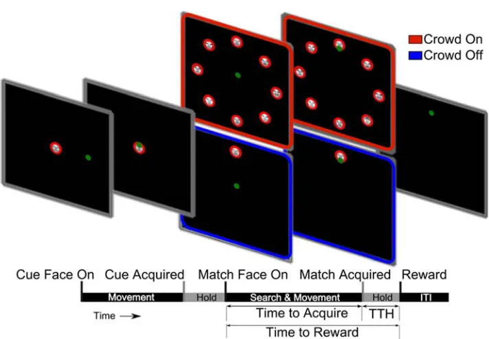
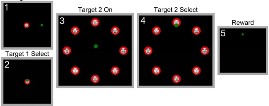
Figure 3.1.2.3-1 Face in the Crowd Task Trial Structure The timeline pictured schematizes the phases of the task and associated events. The start of each phase of the task is marked with a tick, labeled, and pictured above with a screenshot of the task display. The behavioral measures used and their corresponding temporal extents are also indicated below the timeline. Target 1 is the Cue Face, and Target 2 is the Match Face. In the Crowd On Condition, Target 2 is accompanied by 7 other faces of different individuals. In Crowd Off, it appears alone. The green dot represents the cursor.
A green cursor of radius 0.7cm was continuously presented on the screen. The cursor was controlled either by the monkey’s hand moving in the horizontal plane above a flat, table-top surface immediately in front of his body (Manual Control mode), or by the output of a neural decoder (Brain Control mode) with hands gently restrained on the table surface in a relaxed position with elbows bent at approximately 90 degrees.
visual stimuli/targets consisting of images of various human faces taken from the Psychological Image Collection at Stirling (PICS) database (http://pics.stir.ac.uk). Face targets consisted of a photographic head-on image of one of 3 facial expressions of 12 individuals. One individual was chosen for use as the “goal” face or individual for the current study. All faces were normalized for size with a red surrounding mask that obscured the overall shape of the head and hair. The faces were also normalized for total brightness. These manipulations made the stimuli subtle enough in their differences that they required fixation for correct identification of the goal individual. The goal individual’s expression varied from trial to trial but not within a trial. The outer diameter of all the face stimuli, red mask included, was 3 cm. Acceptance windows for all targets and the cursor were identical in size to their respective visual representations.
A trial began when a sample face cue (Target 1) of the goal individual appeared at the center of the screen (Figure 3.1.2.3-1). The subject moved the cursor to overlap the cue
for a contiguous Hold Period of 400ms. If overlap was broken during the Hold Period before 400ms elapsed, an entire new 400ms Hold Period would need to be performed. This rule was applied for all Hold Periods in the task. For the Crowd On condition, after the Hold Period, Target 1 disappeared, and a “crowd” of face stimuli of 8 individuals appeared. One of the 8 faces in the crowd (Target 2) was an identical match to the initial cue face, Target 1. Each face in the crowd was situated on a circle of radius 9cm centered on the middle of the screen and separated by 45 degrees on the circle (Figure 3.1.2.3-1). The
for another Hold Period of 400ms. After this second Hold Period, a juice reward was delivered via a tube placed in front of the monkey’s mouth. Simultaneously, all targets disappeared and a reward beep was sounded. A new trial began after an inter-trial interval (ITI) of 0.5s. Failure to locate, select, and Hold Target 2 within the 20s period resulted in termination of the trial: the disappearance of all targets, an auditory cue signifying trial failure, and a penalty ITI of 5-10s. Overlap with an incorrect target for 400ms or more also resulted in termination of the trial. An overlap of less than 400ms with an incorrect Target in the Crowd was permitted. The cursor was continuously controlled during the trials and ITI. In the “No Crowd” task condition, Target 2 appeared somewhere on the same circle described above, but with no other face stimuli present (Figure 3.1.2.3-1).
3.1.2.4Performance Measures
Task performance was assessed by the fraction of trials successfully completed and by measuring the time required to perform the various stages of the task (Figure 3.1.2.3-1).
In order to assess the effect of Brain Control on cursor control without the influence of task difficulty, we calculated the change in Time to Acquire, or ΔTTA, by subtracting average daily TTA in Manual Control from each subsequent Brain Control trial TTA, i.e.,
(1)
where superscripts MC or BC indicate Manual Control or Brain Control and k indicates Brain Control trial k. The average TTA in Manual Control, TTAMC, was computed per day
and per task condition (Crowd On or Off) and was only subtracted from Brain Control trials with the corresponding task condition on the same day. This calculation isolated the difference in TTA that was attributable solely to the use of Brain Control rather than Manual Control by eliminating time consumed by other aspects of the task, e.g., searching for and reacting to the presence of the correct target. This calculation thereby gave a direct indication of the effectiveness of Brain Control of the cursor independent of the influence of other, task-related factors. This measure was then examined as a function of Assessment condition and Training condition (described below) in subsequent analyses.
P values reported are the result of a non-parametric, two-sample Kolmogorov-Smirnov test
3.1.2.5Decoder Training
Each day began with the monkey performing 160 trials of both the Crowd and No Crowd task conditions alternating every 20 trials under Manual Control. This allowed assessment of daily variation in basic task performance without the influence of Brain Control quality. Next, the monkey’s hands were gently restrained.
A previously computed neural decoder, a Training Decoder, was used by the NHP to manipulate the cursor during an initial 250s Training Block in either the Crowd On or Crowd Off condition. The Training Decoder and computer assistance functioned like a set of training wheels on a bicycle, allowing the NHP to use neural activity to drive the cursor, though not fully independently.
Training Decoder during training. The next Training Decoder was used for 2 days, and the third for one day. All analyses described below were repeated on a restricted data set using only decoders trained with the first Training Decoder (days 1-4). The results of those analyses did not differ substantially from the results described below.
Furthermore, using a Training Decoder (itself trained on the Crowd Off task condition) to compute a new decoder with the Crowd On task condition could be considered a sort of worst-cast scenario in which the task type changes from one training block to the next. We reasoned that if we find no impairment of decoding function caused by the “switch” to the Crowd On condition, there is no reason to expect an impairment would arise if the two tasks were fully segregated with respect to training and assessment. It would be feasible that it might provide an advantage, but our main goal in the study was to examine whether or not these task contexts reduced decode performance.
When computing the new decoder after performing the Training Block, the noisy velocities of the cursor during the Training Block were reoriented to point towards the instantaneous goal to more accurately capture the assumed intentions of the subject (Gilja et al., 2012). For each time bin, the intention of the NHP was assumed to be either move the cursor towards the current correct face target or hold the cursor steady if the cursor already overlapped the correct target. Reaction times were accounted for by assuming an intention to hold the cursor steady until 200ms after Target 2 onset in the No Crowd condition and after initial cue onset and 400ms after onset of Target 2 and the other targets in the Crowd condition. These values were chosen based on average reaction times during Manual Control.
Once the new decoder was computed, it was then used for Brain Control by the NHP without assistance to perform the task in both Crowd On and Off conditions in 10 alternating blocks of 20 trials, yielding a total of 200 Assessment trials per decoder. This process of Training and Assessment of performance was repeated so that the effect on performance of the Crowd (during Training and/or Decoding) could be measured. Twenty-three decoders were trained and assessed across seven days. The task condition of the first Training Block on a given day was alternated to remove any order effects.
For transforming neural activity into cursor position and velocity, we used a linear decoding model coupled with a linear state space model of the cursor dynamics. (For further detail, see Section 3.2.2.7). The final decoder form closely resembled that described by Gilja and colleagues (Gilja et al., 2012).
3.1.2.7Saccade Task
To assess the correlation between eye kinematics and neural activity, the monkey was trained to perform a task in which a trial consisted of repeated fixation on a series of 4 yellow circular targets placed on a 2 by 2 equally spaced grid measuring 14cm square. After fixating a target for the required period of 500ms, the target would turn from yellow to grey. After successfully fixating on all 4 targets in any sequence, the targets would all disappear and juice reward would be delivered. An ITI of 0.5s followed. The position of the hand (which was not required to perform the task) was recorded along with neural signals during this task, though the hand rarely moved.
Two days, each consisting of approximately 1500 trials, were recorded. R-squared values between a) linear predictions of eye kinematics based on neuronal firing with b) actual eye kinematics were computed and validated using Leave One Out Cross-Validation (LOOCV) on 20 equally sized segments of the data.
ended on any of the other 3 targets were preselected from the data. Each saccade was labeled by the target at which the saccade ended. Observations were comprised of total number of spikes summed for each neural channel across a window beginning 0.150s before a saccade onset and 0.300s after. Linear discriminant analysis was used to classify the neural data into one of four possible targets/categories. LOOCV was used to obtain a measure of the performance of neural classification of saccade targets, whereby all observations save one were used as training data. The class of the “left out” trial was then predicted using the classifier. This was repeated using each available trial as the excluded trial. Performance was computed as the percentage of “left out” trials that were correctly classified. A permutation test, whereby target labels were randomly shuffled and LOOCV repeated, was used to generate a null distribution of performance in order to assess whether classification of the actual data exceeded chance levels (n = 103 permutations).
3.1.3 Results
3.1.3.1Saccade Task
R-squared values of 0.05 and 0.02, respectively, were obtained, indicating a measurable but small relationship with eye position. P values were not significant (>> 0.05) for eye velocity.
When trying to decode the goal of individual saccades from amongst the 4 possible targets in the saccade task based only on neural data, 41.54% correctness was achieved for n = 674 saccades. Though modest, this performance significantly exceeded chance level of 25% (p < 10-5, permutation test.) Thus, the neural activity correlated with saccades could not account for the 98% success rate achieved with the neural cursor during the Face in the Crowd task.
3.1.3.2Behavioral Task – Manual Control
During the Manual Control block of each day, the monkey was able to successfully complete > 99% of the trials correctly, i.e.,
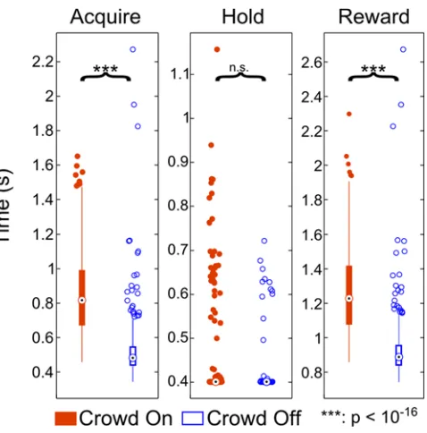
selecting the correct face before time ran out. This performance indicated that the animal had no difficulty in reliably finding the matching Face in the Crowd. Basic task performance statistics under Manual Control for all days (Crowd On n= 567 trials, Crowd Off n = 560 trials) revealed the desired effect (Figure 3.1.3.2-1). As expected, the presence
of the Crowd significantly increased TTA (Crowd On Median = 0.82, IQR = 0.32, Crowd Off Median = 0.48, IQR = 0.11, p < 10e-16) and TTR (Crowd On Median = 1.23, IQR = 0.34, Crowd Off Median = 0.89, IQR = 0.12, p < 10e-16), but not TTH (Crowd On Median = 0.40, IQR = 1.4e-14, Crowd Off Median = 0.40, IQR = 2.8e-14, p = 0.08). This is because the Crowd causes a visual search which delays Acquisition time, but not the time required to Hold the target once it has been initially contacted. The delay in Acquisition time of course results in slower overall task performance as measured by TTR.
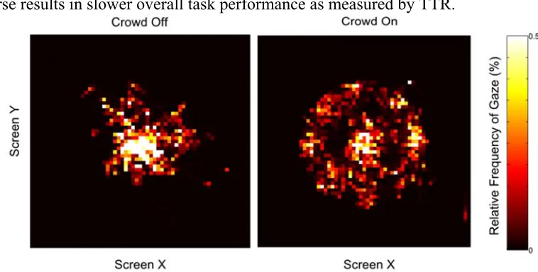
To verify that TTA was increased because the Crowd required the animal to search and identify the target face, we examined eye behavior prior to movement onset and between movement onset and target acquisition for each day. Eye positions for each trial were rotated to place Target 2 at the 3 o’clock position. Two-dimensional histograms of eye position for each task condition clearly reveal the monkey’s tendency to visually search though the faces in the Crowd On task condition before initiating his hand movement (Figure 3.1.3.2-1). Histograms for the period between Movement Onset and Target 2
acquisition looked similar, indicating that the monkey continued scanning the faces even during and after movement to select the correct target (Figure 3.1.3.2-2, left panel).
3.1.3.3Behavioral Task – Brain Control
During the Brain Control sessions, the monkey was able to successfully complete > 98% of the trials correctly, i.e., selecting the correct face before time ran out (Video 3.1-1). The fraction of trails that were completed successfully did not significantly differ between Manual and Brain Control blocks (p = 0.75).
Each decoder was trained during a Training Block either with the Crowd On or Crowd Off and then assessed for performance with the Crowd On or Off. This comprised a 2 x 2 factorial design with the “main effects” being Training Condition and Assessment Condition.
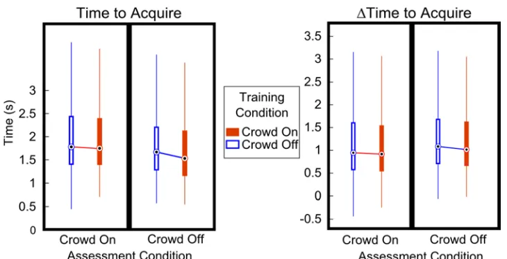
The task condition in which the decoders were trained, the Training Condition, did not significantly influence the TTA achieved (Crowd On Median = 1.65, IQR = 1.00, n = 1508 trials, Crowd Off Median = 1.69, IQR = 1.05, p = 0.22, n = 1871 trials, Figure 3.1.3.3-1,
Figure 3.1.3.3-1 Performance Measures Across Training and Assessment Conditions (Brain Control) Boxplots as in Figure 3.1.3.1-1 of performance measures. Outliers have been excluded for clarity. Assessment Condition, indicated by the label on the abscissa, denotes the task used during full Brain Control with no assistance. Training Condition, indicated by filled (Crowd On) or empty (Crowd Off) bands, denotes task used during Training of decoders . For Time to Acquire (left panel), only Assessment Condition reached significance (p < 10-16) . For ΔTime to Acquire, both the Training Condition (p = 0.03) and Assessment
Condition (p = 1.3e-6) were significantly better in the Crowd On conditions, though only by small margins.
No pairwise comparisons, indicated by lines joining adjacent medians, reached statistical significance. All group statistics are listed in Table 1.
Time to Acquire ΔTime to Acquire Assess
Condn
Crowd On Crowd Off Crowd On Crowd Off
Train Condition
Crowd Off Crowd On Crowd Off Crowd On Crowd Off Crowd On Crowd Off Crowd On
Median 1.779 1.748 1.597 1.531 0.969 0.940 1.106 1.038
IQR 1.062 1.01 1.000 0.984 1.055 1.012 0.999 0.978
n (Trials) 893 763 978 745 893 763 978 745
Table 1 Statistics for All Combinations of Conditions and Measures Table of data for all combinations of Training and Assessment conditions for both main measures. Layout of each group from left to right corresponds to layout in Figure 3.1.3.3-1. Note that both measures (Time to Acquire and ΔTime to Acquire) were computed using the same trials, thus the correspondence in number of trials between the left and right halves of the table.
The ΔTTA was computed for all trials to quantify how Brain Control affected the Acquisition of Target 2 relative to Manual Control in isolation from other factors. We then used the same statistical comparisons that were computed for the unadjusted TTA values to determine whether or not the Crowd’s presence during Training or Assessment influenced the ΔTTA. While the comparison revealed a significant effect of Training condition (Crowd On Median = 0.99, IQR = 1.00, n = 1508 trials; Crowd Off Median = 1.04, IQR = 1.03, n = 1871 trials; p = 0.03, Figure 3.1.3.3-1, right panel), and the trend
Taken together, these results indicated that the additional eye movements and various behavioral demands of the Crowd On task condition did not interfere directly with decoding of a neurally controlled cursor. The more complex task condition might have conferred a small albeit negligible advantage to performance of neural decoding.
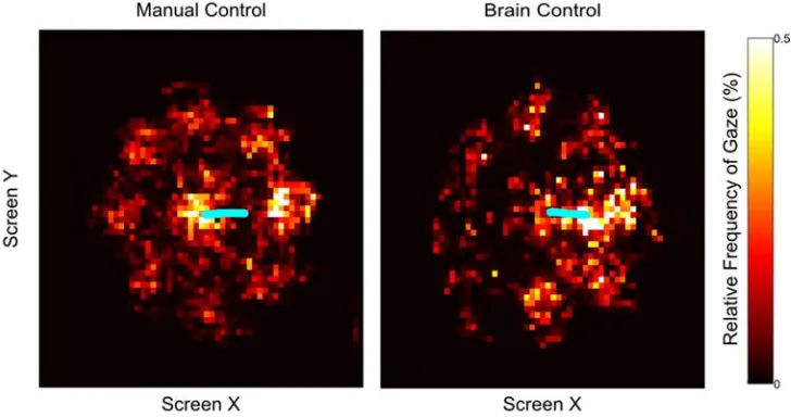
These results suggest the NHP was able to simultaneously gaze freely around the screen while independently controlling cursor position. However, an alternative hypothesis that saccades did negatively impact cursor control would also account for this result if the NHP simply learned to minimize extraneous saccades when the Crowd was present, To rule out this possibility, we once again examined 2D histograms of eye position during a phase in the trials when the cursor was actively being transported to the target (Figure, right panel).
Though the NHP was able to gaze around the screen with the Crowd On during Brain Control as well as in Manual Control, it should be noted that the overall number of saccades was reduced in Brain Control. We compared the number of saccades landing on a peripheral target (and not the correct target or the cursor) in a one second window ending on initial acquisition of Target 2 for each trial. We then compared the occurrence of these saccades in Manual (mean = 2.04 saccades, s.d. = 1.54) vs. Brain Control (mean = 0.82 saccades, std = 1.18) trials, revealing that there were significantly more in Manual Control (p < 10-16). One possible explanation for this outcome is the increased difficulty and imperfect accuracy of Brain Control, i.e., on trials where cursor control is worse, the subject would need to gaze at the cursor longer to maintain closed-loop control. An alternative explanation is extraneous saccades reduced decode accuracy, and the subject learned to make fewer saccades to maintain cursor control. To distinguish these possibilities, we computed the correlation between number of saccades to peripheral targets (as above) in each trial to the TTA across all n = 1718 trials. A correlation of r = -0.12 (p < 10e-7, T test) supports the former account and rules out the latter. Trials with many saccades to locations not occupied by the cursor were amongst the shortest, while the longest trials involved prolonged periods of gazing at the cursor, presumably to accommodate feedback control.
animal’s performance clearly demonstrated his ability to move the cursor back to the middle of the screen before Target 1 appeared in anticipation of the upcoming trial (Video 3.1-3). Additionally, in separate sessions, we confirmed that the decoders trained in this center-out style task could generalize to a 3x3 grid of the same face targets spaced evenly on an 18cm x 18cm square. While performance in terms of trial length was inherently slower than the circular, center-out task (p < 10-16) due to the longer cursor movements required (Time To Acquire: Median = 2.405s, IQR = 1.433, Time to Reward: Median = 3.226s IQR = 1.540), control of the cursor itself was qualitatively no different (Video 3.1-4).
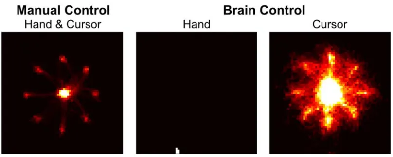
movements as measured by the tracking sensor did not directly influence cursor position. On most days both hands were observably and measurably still (Figure 3.1.3.3-2),
however on other days small, residual movements were made during performance of the task under Brain Control. We used the measured hand kinematics (in the horizontal plane that would typically be used to control the cursor in Manual Control) to predict the kinematics of the neural cursor. Cross-validated R-squared values never exceeded 0.03 for either dimension for any day, indicating little influence of residual hand movements on the decoded cursor.
3.1.4 Discussion
By targeting Area 5d for implant, we were able to obtain neural signals that reflected intended movements of the limb. Though residual eye-related signals were measurable in a control task, it was clear that they did not interfere with the functioning of the interface, whether during use of an existing decoder or during training To our knowledge, this study is the first confirmation that unconstrained gaze does not interfere with prosthetic control, even in a visually complex task environment. Furthermore, we demonstrated the ability of the subject to decouple gaze position, i.e., sensing, from control of the cursor, i.e., the motor intention. This is a crucial capability for providing natural, intuitive control.
This capability was further emphasized given the ability of the subject to manipulate the cursor even in the absence of overt visual targets during the trials with extended ITIs. This result suggests that neural activity in parietal cortex can capture motor intentions without the need for overt visual representations of movement goals.
3.1.5 References
Bremner, L.R. and Andersen, R.A. (2012).Coding of the Reach Vector in Parietal Area 5d. Neuron. 75, 342-351.
Buneo, C., & Andersen, R (2006). The posterior parietal cortex: sensorimotor interface for the planning and online control of visually guided movements. Neuropsychologia. 44, 2594-2606.
Colby, C., & Goldberg, M (1999). Space and attention in parietal cortex. Annual Review of Neuroscience. 22, 319-349.
Collinger JL, Wodlinger B, Downey JE, Wang W, Tyler-Kabara EC, Weber DJ, McMorland AJC, Velliste M, Boninger ML, Schwartz AB (2013). High-performance neuroprosthetic control by an individual with tetraplegia. Lancet 6736:61816-61819.
Crammond, D., & Kalaska, J (1989). Neuronal activity in primate parietal cortex area 5 varies with intended movement direction during an instructed-delay period. Experimental Brain Research. (1989) 76, 458-462.
Cui, H., & Andersen, R. A (2011). Different Representations of Potential and Selected Motor Plans by Distinct Parietal Areas. Journal of Neuroscience, 31(49), 18130-18136.
Franke F, Jackel D, Dragas J, Muller J, Radivojevic M, Bakkum D, Hierlemann A. (2012) High-density microelectrode array recordings and real-time spike sorting for closed-loop experiments: an emerging technology to study neural plasticity. Front Neural Circuits., 20;6:105.
Fraser G W, Chase S M, Whitford A and Schwartz A B (2009) Control of a brain–computer interface without spike sorting Journal of Neural Engineering, 6 1–8.
Gilja V, Nuyujukian P, Chestek CA, Cunningham JP, Yu BM, Fan JM, Churchland MM, Kaufman MT, Kao JC, Ryu SI, Shenoy KV., (2012). A high-performance neural prosthesis enabled by control algorithm design. Nature Neuroscience, 15(12), 1752-1757.
Graziano M S A, Cooke D F, Taylor C S R2000 Coding the Location of the Arm by Sight
Science 290 1782–6.
Hauschild, M., Mulliken, G., & Fineman, I (2012). Cognitive signals for brain–machine interfaces in posterior parietal cortex include continuous 3D trajectory commands. Proceedings of the National Academies of Science. 109(42), 17075–17080.
Hochberg, L. R., Serruya, M. D., Friehs, G. M., Mukand, J. A., Saleh, M., Caplan, A. H., et al (2006). Neuronal ensemble control of prosthetic devices by a human with tetraplegia. Nature, 442(7099), 164-171.
Ifft, P., Shokur, S., Li, Z., Lebedev, M., & Nicolelis, M (2013). A Brain-Machine Interface Enables Bimanual Arm Movements in Monkeys. Science Translational Medicine, 5(210), 210ra154-210ra154.
Louie, K., Grattan, L. E., & Glimcher, P. W (2011). Reward Value-Based Gain Control: Divisive Normalization in Parietal Cortex. Journal of Neuroscience, 31(29), 10627-10639.
Mulliken, G. H., Musallam, S., & Andersen, R. A (2008). Decoding Trajectories from Posterior Parietal Cortex Ensembles. Journal of Neuroscience, 28(48), 12913-12926.
Musallam, S., Corneil, B., Greger, B., & Scherberger, H (2004). Cognitive control signals for neural prosthetics. Science, 305, 258-262.
O'Doherty JE, Lebedev MA, Ifft PJ, Zhuang KZ, Shokur S, Bleuler H, Nicolelis MA. Active tactile exploration using a brain-machine-brain interface. Nature. 2011 Oct 5; 479(7372):228-31.
Peirce, JW (2007) PsychoPy - Psychophysics software in Python. Journal of Neuroscience Methods, 162(1-2), 8-13.
Pesaran, B., Nelson, M. J., & Andersen, R. A (2010). A Relative Position Code for Saccades in Dorsal Premotor Cortex. Journal of Neuroscience, 30(19), 6527-6537. Pesaran, B., Nelson, M. J., & Andersen, R. A (2008). Free choice activates a decision
circuit between frontal and parietal cortex. Nature, 453(7193), 406-409.
Rao, N., & Donoghue, J (2014). Cue to action processing in motor cortex populations. Journal of Neurophysiology, 111, 441-453.
Revechkis, B., Aflalo, T.N.S., Andersen, R., Use of a posterior parietal cortex brain-machine interface in a cognitive face-in-a-crowd task. Poster presented at: Society for Neuroscience 2013; November 13 2013; San Diego, CA.
Revechkis, B., T. N. Aflalo, S. Kellis, N. Pouratian, and R. A. Andersen. 2014. "Parietal neural prosthetic control of a computer cursor in a graphical-user-interface task." J Neural Eng 11 (6):066014. doi: 10.1088/1741-2560/11/6/066014.
Rothschild RM. Neuroengineering tools/applications for bidirectional interfaces, brain-computer interfaces, and neuroprosthetic implants - a review of recent progress. Front Neuroeng. 2010 ;15;3:112.
Shanechi, M. M., Hu, R. C., Powers, M., Wornell, G. W., Brown, E. N., & Williams, Z. M (2012). Neural population partitioning and a concurrent brain-machine interface for sequential motor function. Nat Neurosci, 15(12), 1715-1722.
Soto, D., Heinke, D., Humphreys, G. W., & Blanco, M. J. (2005). Early, Involuntary Top-Down Guidance of Attention From Working Memory. Journal of Experimental Psychology: Human Perception and Performance, 31(2), 248-261.
3.2 State Decoding Improves Use of a Computer Cursor for Neuroprosthetic Applications
3.2.1 Introduction
A common problem with computer-based neural prosthetics lies in the generally noisy output of the decoders (Wang et al., 2013, Ifft et al., 2013, Vargas-Irwin et al., 2010). This noise is present and particularly noticeable when the cursor or other effector is intended to be kept stationary, e.g., when attempting to select a target. Even if “clicking” is decoded separately for target selection (Kim et al., 2011), noisy positioning of the cursor would result in many wasted and potentially frustrating clicks if the cursor is not held in place at the correct time. Easily selecting targets is crucial for a neural prosthetic that utilizes a graphical user interface (GUI). Additionally, noisy cursor movement during other periods in which a stationary cursor is desired, e.g., during visual search of the screen or attentional disengagement from the interface altogether, would also make the interface more difficult to use. Additionally, it has not been demonstrated whether the intention to move at all can be decoded in the absence of limb movement, as would be relevant in a clinical setting.
Rather than simply relaxing the behavioral requirements of the task, we coupled the existing linear velocity decoder with a categorical state decoder. Such decoders have been used in studies involving parietal and motor cortex for offline reconstructions of behavior (Shenoy et al., 2003, Kemere et al., 2008, Aggarwal et al., 2013, Darmanjian et al., 2003), and for decoding “clicks” or selection in tetraplegic human subjects with motor cortical prosthetics (Kim et al., 2011). Additionally, previous work (Hwang and Andersen, 2009) has shown that local field potentials (LFPs) in the PPC can be used to detect the onset of an intended cursor movement even without production of limb movement. Thus, in the current study, we coupled state decoding with continuous control of a computer cursor in a GUI-like NHP task. We found that decoding of movement state conferred significantly better performance.
3.2.2 Methods
One male rhesus monkey participated in this study. All animal and surgical procedures were approved by the California Institute of Technology Institutional Animal Care and Use Committee and followed guidelines set forth by the National Institutes of Health.
3.2.2.1Behavioral Setup
VT). The PsychoPy psychophysics library was used for stimulus presentation (Peirce, 2007) and Simulink software (The MathWorks Inc., Boston, MA) for task control.
3.2.2.2Neural Recordings
Two 96-channel Cereport arrays (Blackrock Microsystems, Salt Lake City, UT), implanted on the superior parietal lobule, were used to record neural activity. Electrical potentials were amplified, digitized, and recorded with the Cerebus hardware and Central software suite (Blackrock Microsystems). Thresholds for action potential detection were set -4.5 times the RMS of the raw signal. Threshold crossing rates were calculated in non-overlapping 50ms bins by custom MATLAB software (The MathWorks Inc, Boston, MA). About 85 channels of discernible spike activity were used each day.
3.2.2.3Behavioral Task
The behavioral task (Figure 3.2.2.3-1) was designed to mimic human use of a GUI
(Revechkis et al., 2014). First, the primate was presented with a single, icon-like face image as a sample at the center of the screen (Target 1). After selecting it with the cursor, a matching target (Target 2) appeared at one of 8 possible positions. Target positions were radially distributed at 45 degrees 9cm from the center of the screen. In the Crowd On task condition (illustrated in Figure 3.2.2.3-1) seven non-matching face icons appeared at the
other target positions. In the Crowd Off condition (not illustrated in Figure 3.2.2.3-1) only
the matching face appeared somewhere on the circle. After Target 2 appeared (with or
without the Crowd), the primate had 20 seconds to select the matching face with the cursor. Selection of both Targets 1 and 2 was accomplished by visually overlapping the cursor with the target for a hold period of 0.5s.
3.2.2.4Performance Measures
duration between onset of Target 2 and the initial contact of the cursor with Target 2. Time to Hold (TTH), the primary measure addressed in the current study, measured the time between first contact with Target 2 and when reward was delivered, i.e., how long it took to successfully overlap Target 2 for the required hold period. This value could not be less than 0.5s by definition; however it could be greater if overlap of the cursor with the target was broken and reinitiated. Time to Reward (TTR) captured the time from Target 2 onset to completion of the trial and the concomitant juice reward.
3.2.2.5Statistical Tests
Two-sample Kolmogorov-Smirnov tests were used to detect differences between performance measures in various task conditions. All p values reported herein were outcomes of this test unless otherwise noted.
3.2.2.6Decoder Training Procedure
by 20cm region in the center of the display (the approximate extent of the task area on the display) for more than 3 seconds resulted in an abort of the current trial and a time out of 5-10s. In practice, this almost never occurred.
Neural data was recorded during a 250s Observation Block wherein the automatic controller performed the task in the Crowd Off condition. With the kinematics of the automatic cursor as responses and neural data as observations, a Velocity Decoder was computed (see below). This new decoder was then used as a Training Decoder during the subsequent Training Block as described in an earlier study (Revechkis et al., 2014).
After the Observation Block and the Training Block, a new Velocity Decoder was trained along with a State Decoder. They were then used by the NHP without assistance to perform the task in either the Crowd or No Crowd condition. The State Decoder was turned on and off in alternating blocks of 25-50 trials to assess its performance. This process of training both the Velocity and State Decoders and assessment of State Decoder performance constituted a single experimental session. One to three sessions were performed per day. Task condition did not vary within a session.
3.2.2.7Velocity Decoder Computation
For transforming neural activity into a cursor position, we used a linear predictor coupled with a linear model of the desired cursor dynamics,
where denotes the time step, ∈ is the predicted 4-dimensional kinematic state of the effector; ∈ is an N-dimensional list of features derived from neural recordings; ∈ is the state-space representation of the system dynamics of the effector motion; and ∈ addresses the influence of each feature upon each kinematic variable in .
The structure of the vector and matrix components of (2) is as follows for the th degree of freedom (DOF) at time step , , and its derivative :
1 0 ⋯ 0
, ⋯ , ⋮ (3)
In (3), is the step size of the discrete update equation in seconds, allows positional feedback onto velocity, is a smoothing parameter, , are the weights applied to the neural features for the th degree of freedom, and are the neural features at time step . The terms , were fit by regressing neural activity against cursor velocity using
3.2.2.8State Decoder Computation
The same threshold crossing data was supplied to the State Decoder as well as the Velocity Decoder. The State Decoder also used the channel-averaged power spectra as input (Figure 3.2.2.3-1). Principal component analysis was used to reduce the dimensionality
of the input data. The number of principal components required to capture at least 95% of the variance in the data were supplied to the classifier.
A naïve Bayesian classifier with uniform priors was trained on the neural data (observations) and movement state (responses). The response categories were determined using the same procedure described in previous work (Revechkis et al., 2014, Gilja et al., 2012) for interpreting the subject’s instantaneous intentions during the Training Block. If no target was one the screen the intended velocity was assumed to be 0, and the response was categorized as Rest. Otherwise, the response was Movement. Reaction times were taken into account by delaying changes in intended state after target onset according to reaction times measured during manual control of the cursor in preliminary training sessions. The classifier assumed normality of the observation data and therefore estimated only the mean and standard deviation for each element of the input data for each movement state.
linear, step-wise fashion. If in a given time bin, the State Decoder classified the current neural signals as Movement, the gain was increased by 0.4 to a maximum of 1. If Rest was decoded, the gain was decreased by 0.2 to a minimum of 0. The asymmetry in step size allowed fast initiation of movement but smooth, gradual deceleration. This way, noisy errors in the state classification would suddenly accelerate or halt movement less often than if the State Decoder output itself directly determined the gain.
3.2.3 Results
We examined whether the NHP was able to manipulate the cursor to perform the task during Brain Control without hand movement. Cross-validated R2 values between neural cursor kinematics and measured hand kinematics, though at times statistically significant, never exceeded 0.03 in any session for any dimension. We therefore concluded that the NHP was not able to affect the neural cursor using physical movement of the limb.
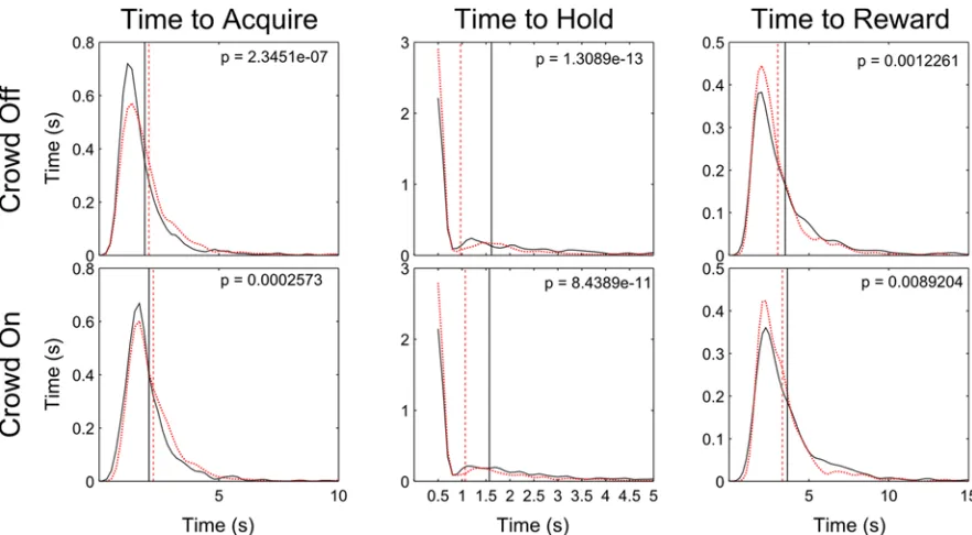
Sixteen total sessions were run. Half included assessment of the State Decoder in the Crowd On condition and half with the Crowd Off. The overall effect of the State Decoder on the various performance measures in each task condition are shown in Figure 3.2.3-1. The results are similar for both task conditions.
and from 3.65s to 3.33s with the Crowd On (p = 8.9e-3). The estimated probability distributions of Time to Reward (Figure 3.2.3-1, 3rd column) indicated that the State Decoder helped flatten the tails of the distributions, i.e., reduce the frequency of longer trials in the 5 and 6-second ranges in the Crowd Off and On conditions, respectively.
A small but significant increase in mean TTA was measured as a function of the State Decoder. With the Crowd Off, TTA increased by 0.178s (p = 2.3e-7). With the Crowd On, TTA increased by 0.190s (p = 2.6e-4). While this delay in the ability to acquire the target undesirably hampered performance, it was outweighed by the reduction in TTH.
While the overall effects were significant across all sessions, a more nuanced relationship was evident when observing the effects of the State Decoder on a session-wise basis (Figure 3.2.3-2, left panel). While the State Decoder significantly improved TTH for 12
out of 16 sessions (6 after Bonferonni correction), it seemed that the State Decoder only substantially improved TTH when the initial TTH without the State Decoder was poor. To quantify this relationship, we examined the session-wise improvement in TTH as a function of the initial TTH without the State Decoder (Figure 3.2.3-2, right panel). Linear
Example videos show the readily apparent improvement in performance caused by the State Decoder on a day of poor initial performance (Video 3.2-1). We also include video of task performance with State Decoding when initial performance was already strong for reference (Video 3.2-2).
To determine the relative effectiveness of the LFPs and spikes, we retrained decoders for each training set in the study using just Spikes or just LFP data. (The existing decoders used both Spikes and LFPs.) True Positive rate for the detection of movement in the training data was computed and compared across all three groups. State Decoders based
on LFPs alone (mean TP rate = 52.04%, S.D. = 6.04%) did not detect movement significantly better than the chance level of 50% (p = 0.26, T-test). Furthermore, the Spikes and LFPs group (mean = 68.40%, S.D. = 5.15%) did not perform significantly better than Spikes alone (mean = 68.15%, S.D. = 4.67%, p = 0.36, Two-sample KS test). This indicated that the spiking activity drove the state detection and that LFPs did not contribute.
3.2.4 Discussion
Use of the State Decoder benefitted performance of the neural interface. While it did not have a large effect in all sessions, it dramatically improved target selection when performance was initially poor. The time required to select targets averaged across all trials decreased from approximately 1.5s to one second in both the GUI-like Crowd On task condition and the more basic Crowd Off condition. This effect was strong enough to drive a decrease in overall trial times despite a slight penalty to the speed of initial target acquisition.
Decoding and training of both the velocity and state decoders were accomplished in the absence of physical movement. This is a likely clinical scenario, as patients who could benefit and have benefitted from neural prosthetics are those who lack any motor function. Furthermore, the task we designed involved operations similar to those employed in a modern GUI, e.g., browsing and selecting options from many possible alternatives with a cursor. The training, decoding, and task used in the current study strongly suggest that our results would likely be applicable and beneficial in a practical, clinical context.
3.2.5 References
Aggarwal V, Mollazadeh M, Davidson AG, Schieber MH, Thakor NV. “State-based decoding of hand and finger kinematics using neuronal ensemble and LFP activity during dexterous reach-to-grasp movements.” J Neurophysiol. 2013 Jun;109(12):3067-81. doi: 10.1152/jn.01038.2011.
Bremner, L.R. and Andersen, R.A. (2012).Coding of the Reach Vector in Parietal Area 5d. Neuron. 75, 342-351.
Darmanjian, S., Kim, S.P., Nechyba, M.C., Morrison, S., Principe, J., Wessberg, J., Nicolelis, M.A.L., “Bimodal brain-machine interfaces for motor control of robotic prosthetics,” in Proc. 2003 IEEE/RSJ Int’l Conf. on Intelligent Robots and Systems, vol. 4, Las Vegas, NV, pp. 3612-17.
Gilja, V., Nuyujukian, P., & Chestek, C (2012). A high-performance neural prosthesis enabled by control algorithm design. Nature, 15(12), 1752-1758.
Hwang, E. J., & Andersen, R. A (2009). Brain Control of Movement Execution Onset Using Local Field Potentials in Posterior Parietal Cortex. Journal Of Neuroscience, 29(45), 14363-14370. doi:10.1523/JNEUROSCI.2081-09.2009
Ifft, P., Shokur, S., Li, Z., Lebedev, M., & Nicolelis, M (2013). A Brain-Machine Interface Enables Bimanual Arm Movements in Monkeys. Science Translational Medicine, 5(210), 210ra154-210ra154.
Kemere, C., Santhanam, G., Yu, B. M., Afshar, A., Ryu, S. I., Meng, T. H., et al (2008). Detecting Neural-State Transitions Using Hidden Markov Models for Motor Cortical Prostheses. Journal Of Neurophysiology, 100(4), 2441-2452.
Kim SP, Simeral JD, Hochberg LR, Donoghue JP, Friehs GM, Black MJ. Point-and-click cursor control with an intracortical neural interface system by humans with tetraplegia. IEEE Trans Neural Syst Rehabil Eng. 2011 Apr;19(2):193-203.
Peirce, JW (2007) PsychoPy - Psychophysics software in Python. J Neurosci Methods, 162(1-2):8-13.
Revechkis, B., Aflalo, TNS, Kellis, S, Pouratian, N, & Andersen, RA (2014). Parietal neural prosthetic control of a computer cursor in a graphical-user-interface task. J Neural Engineering 11 066014.
Shenoy KV, Meeker D, Cao S, Kureshi SA, Pesaran B, Buneo CA, Batista AP, Mitra PP, Burdick JW, Andersen RA. Neural prosthetic control signals from plan activity. Neuroreport. 2003 Mar 24;14(4):591-6.
Vargas-Irwin CE, Shakhnarovich G, Yadollahpour P, Mislow JM, Black MJ, Donoghue JP. Decoding complete reach and grasp actions from local primary motor cortex populations. J Neurosci. 2010 Jul 21;30(29):9659-69.
4.
Human Parietal Cortex4.1 Selectivity for Hand Movement Execution and Feedback in Human Parietal Neurons and Local Fields
4.1.1 Introduction