Shell Morphology of the Egyptian Tortoise,
Testudo kleinmanni
Lortet,
1883, the Osteologically Least-Known
Testudo
Species
Massimo Delfino1,2,*, Francesco Chesi2, and Uwe Fritz3
1Paläontologisches Institut und Museum, Universität Zürich, Karl Schmid-Strasse 4, CH-8006 Zürich, Switzerland 2Dipartimento di Scienze della Terra, Università di Firenze, Via G. La Pira 4, I-50121, Firenze, Italy
3Museum of Zoology (Museum für Tierkunde), Senckenberg Dresden, A. B. Meyer Building, D-01109 Dresden, Germany (Accepted February 25, 2009)
Massimo Delfino, Francesco Chesi, and Uwe Fritz (2009) Shell morphology of the Egyptian tortoise, Testudo kleinmanni Lortet, 1883, the osteologically least-known Testudo species. Zoological Studies 48(6): 850-860. The present paper provides the first detailed description of the shell osteology of Testudo kleinmanni, a small
tortoise species that currently occurs only in a narrow, discontinuous strip along the southeastern Mediterranean coast, extending from Libya to Israel. Its bony shell differs from the partially codistributed species T. graeca in characters present on the nuchal, suprapygal, pygal, epiplastron, entoplastron, hyoplastron, and xiphiplastron. The other shell elements are very similar in both species. For such elements, only the smaller adult size of T. kleinmanni can be used for tentative species identification. Generally, shell osteology does not reflect the currently accepted sister-group relationship of T. kleinmanni and T. marginata (the only possible shared character may be the shape of the pectoro-abdominal sulcus). Until now, the species has never been identified from palaeontological assemblages and only very rarely in archaeological settings. The extreme rarity of fossil and subfossil records could be caused, at least in part, by the fact that diagnostic osteological characters were previously unknown. http://zoolstud.sinica.edu.tw/Journals/48.6/850.pdf
Key words: Osteology, Taxonomy, Mediterranean tortoises, Testudo graeca, Testudo marginata.
* To whom correspondence and reprint requests should be addressed. E-mail:massimo.delfino@pim.uzh.ch
C
helonians are by far the most abundantand best-studied reptiles from palaeontological and archaeological sites in the Mediterranean region. They are mostly represented by shell fragments or partially complete shells. Despite their abundance, the study and proper interpretation of the fossil record is impeded by the incomplete knowledge of the shell osteology of extant Mediterranean taxa. With respect to land tortoises (Testudinidae), the shell morphology of Testudo graeca Linnaeus, 1758 and T. hermanni Gmelin, 1789 is relatively well-studied, chiefly thanks to work by Staesche (1961), Cheylan (1981), Amiranashvili (2000), Hervet (2000), and Lapparent de Broin et al. (2006a b). The shell morphology of T. marginata Schoepff, 1793 was partially described by Młynarski (1980), Schleich (1982), and Lapparent de Broin et al.
(2006a b). The least-known species is the so-called Egyptian tortoise, T. kleinmanni Lortet, 1883, which is distributed along the Mediterranean coast from Libya to the Negev, southern Israel (Fritz and Havaš 2007). A few years ago, T. kleinmanni was split into 2 species (Perälä 2001),
T. kleinmanni sensu stricto (ranging west of the Nile) and T. werneri Perälä, 2001 (ranging east of the Nile). However, according to recent molecular and morphological investigations, recognition of T. werneri is not warranted (Attum et al. 2007, Široký and Fritz 2007).
Certain morphological characters of the shell of T. kleinmanni were described by some authors (among others, Günther 1869, Lortet 1887, Anderson 1898, Ernst and Barbour 1989, Lapparent de Broin et al. 2006a b), but a
comprehensive description was never published. Perhaps because of this, remains of T. kleinmanni
have never been identified in palaeontological assemblages (Lapparent de Broin 2000) and are reported only very rarely in archaeological settings (von den Driesch and Boessneck 1985, Boessneck 1988), even in sites within its current range. Interestingly, VM Chkhikvadze (in Gabashvili et al. 2000) suggested that T. burtschaki Chkhikvadze, 1975 from the Miocene of Georgia could be phylogenetically related to T. kleinmanni. This statement, which was not supported by any argument, is rather surprising because Gabashvili et al. (2000) proposed that T. burtschaki is
characterized by the absence of a hypo-xiphiplastral hinge, in contrast to T. kleinmanni. However, Danilov (2005) and Chkhikvadze (2006 2007) treated T. burtschaki as a representative of the Testudo sensu stricto group, which suggests the presence of a plastral hinge. This contradictory situation underlines the need for better knowledge of osteological features of the shells of all extant
Testudo species in order to assess relationships of fossil taxa.
The present study provides the first detailed osteological description of the shell of T. kleinmanni, which should allow the identification of palaeontological and archaeological remains. Particular emphasis is given to distinctions with
T. graeca, the only codistributed extant tortoise species (Schleich et al. 1996, Bouskila and Amitai 2001).
MATERIALS AND METHODS
Our osteological description of T. kleinmanni
is based on 8 shells in the collection of the Museum of Zoology Dresden (Museum für Tierkunde Dresden = MTD: MTD D 26762, 32832, 35692, 38650, 39221, 40289, 44284, and 44285; see Appendix for sex, shell lengths, and neural formulae). With 1 exception (MTD D 26762; with a small pleuro-peripheral fontanellae on 1 side only), all specimens were adults. In addition, the external shell morphology as well as the position and relationships of the scute sulci were studied in the following 41 alcohol-preserved specimens: MTD D 12469, 26016, 26017, 26850, 28286, 28287, 29119, 30855, 31125, 31598, 31803, 32434, 32435, 32710, 32742, 32798, 32831, 34857, 35074, 35075, 36797, 36897-36907, 37473, 39220, 40166, 42492, 45729, and 46938-46941. Osteological and alcohol-preserved specimens of
T. graeca, T. hermanni, and T. marginata, housed in the same and other collections were used for comparison. Anatomical nomenclature followed Lapparent de Broin (2001), while the taxonomy followed Fritz and Havaš (2007).
Description of the shell of T. kleinmanni
In order to provide characters suitable for the identification of palaeontological and archaeological remains, the following description is organized according to the bony shell elements that are usually preserved.
Carapace
(Figs. 1a, c, 2)
The carapace is highly domed and reaches a maximum length of 144 mm (Farkas et al. 1997). Males are smaller than females, and reach a maximum length of only 100-106 mm according to Schleich et al. (1996) or 130 mm according to Baha el Din (2006). Our largest males and females do not surpass these lengths. In comparison to other Testudo species, the carapacial and plastron elements of T. kleinmanni are quite thick; however, this character varies in different specimens.
Nuchal: The nuchal is generally wider than long and characteristically notched; the area covered by the cervical, usually triangular in shape, always distinctly protrudes from the anterior profile of the nuchal; it is noteworthy that this character already occurs in our smallest specimen (MTD D 26762; with a carapace length of 70 mm). Lapparent de Broin et al. (2006a: 292) stated that “the notch affects all the anterior border of the shell (nuchal and first peripherals) due to the apomorphic elongation of the peripherals adjacent to the nuchal: the cervical is present, triangular, posteriorly wide, sometimes narrowed but not absent … and protuberant in the middle of the notch”. The anterior edge of the nuchal is, therefore, W-shaped. The triple junction among the 1st marginal, vertebral, and pleural is always present on the nuchal and always relatively far from its lateral edge.
Neural series: Seven or, more rarely, 8 elements represent the neural series (Appendix). In a few specimens (MTD D 32832 and 40289), they are alternatively rectangular and octagonal from the 1st to the 6th, whereas the 7th is hexagonal. Irregularities frequently occur, mostly in the posterior sector of the series, and several
hexagonal elements with small posterolateral edges may be present (Appendix). The 1st, 3rd, 5th, and 7th neurals are encroached by the intervertebral sulci. The ventral surface of all neural elements bears traces of the connection
with the vertebrae.
Suprapygal: All available specimens possess only 1 suprapygal, which is approximately hexagonal (the posterolateral edges are very short), or in 1 case nearly trapezoidal (MTD D
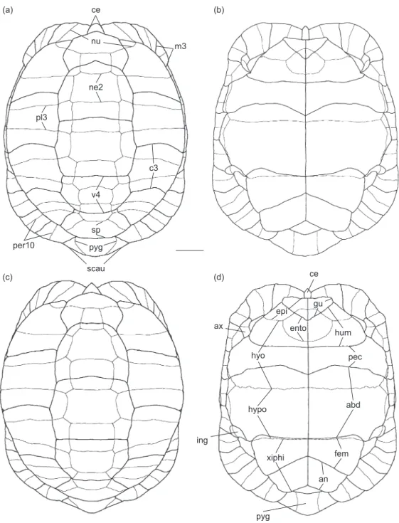
Fig. 1. Schematic drawings of the shell of Testudo kleinmanni Lortet, 1883. (a, b) male carapace in dorsal view and plastron in ventral view; (c, d) female carapace in dorsal view and plastron in ventral view. Thick lines represent scute sulci; thin lines, bony sutures. Bony plates: ento, entoplastron; epi, epiplastron; hyo, hyoplastron; hypo, hypoplastron; ne2, 2nd neural; nu, nuchal; per10, 10th peripheral; pl3, 3rd pleural; pyg, pygal; sp, suprapygal; xiphi, xiphiplastron. Horny shields: abd, abdominal; an, anal; ax, axillary; c3, 3rd costal; ce, cervical; fem, femoral; gu, gular; hum, humeral; ing, inguinal; m3, 3rd marginal; pec, pectoral; scau, supracaudal; v4, 4th vertebral. Scale bar = 10 mm. ce nu ne2 v4 sp pyg scau pl3 m3 per10 c3 ce hyo hypo xiphi pyg pec abd fem an hum gu epi ento ing ax (a) (b) (c) (d)
39221). The fusion of the suprapygals into a single element was also reported by Lapparent de Broin et al. (2006b).
Pleural series: The 8 pleurals are trapezoidal and show the typical Testudo alternation of the pleurals with a wide proximal edge and a narrow distal edge separated by pleurals in the opposite condition. There is some variation, however, as in MTD D 38650. In this specimen, the 2nd to 7th pleurals have approximately the same shape (note that the neural formula, given in Appendix, in this specimen is anomalous with 4 contiguous hexagonal elements). All even pleurals host the intercostal sulci. The visceral surface of the pleurals is characterized by an elongated convexity (transverse in strict anatomical terms, longitudinal if the main axis of the isolated pleural is considered). The proximal visceral area of the pleurals develops an irregular process contacting the vertebrae; in the 1st and last pleurals, the processes are more developed than in the others; the ones of the last pleurals show a flattened facet which contacts the dorsal edge of the ilium.
Peripheral series: The 3rd to 7th peripherals form the bridge, the bony junction of the carapace and plastron. In some cases, the external surface of the 1st (MTD D 39221) or the 1st 2 peripherals (MTD D 38650) is crossed by the costo-marginal sulcus which otherwise corresponds (along the entire series, including the pygal-suprapygal) to the pleuro-peripheral suture. The 3rd peripheral hosts the sulcus which corresponds to the horny axillary shield, whereas the 7th hosts the one corresponding to the inguinal shield (note that exceptionally up to 2 inguinal shields can occur; Schleich et al. 1996). All peripherals are crossed by 1 intermarginal sulcus. The visceral surface of the 3rd and 7th peripherals is characterized by the presence of an elongated scar of the dorsal projections of the hyo- and hypoplastron, respectively (the axillary and inguinal processes); the projection of the hyoplastron reaches the posterodorsal edge of the 2nd peripheral. The 2nd and 3rd peripherals as well as the 7th to 11th peripherals can have a slight dorsally raised edge; at the same height, corresponding to this edge, the peripherals involved in the bridge can develop a weak ridge; the entity of this raised edge and ridge is slightly amplified by the horny shields. The edge of the 11th peripheral regularly shows a notch corresponding to the lateral margin of the supracaudal shield; this change in orientation of the outline gives the ‘pointed’ appearance to the posterior edge of the carapace as described below.
Pygal: The pygal plate is generally trape-zoidal; in 1 case (MTD D 40289), it is hexagonal with short anterolateral sides. Its dorsal surface is moderately convex to nearly flat, whereas the ventral surface is regularly concave in both sexes with a gutter-like appearance (Fig. 2). On the pygal plate of all skeletonized specimens, no sagittal groove is present which should be expected when the horny supracaudal scute is divided. However, divided supracaudals occur in 15 of the 41 alcohol-preserved specimens (from both Egypt and Libya). In most cases, the division occurs only in the proximal sector of the external surface of the supracaudal, and very rarely on the internal one (as in MTD D 31125 and 32434, where just a hint of division is present). This character state was previously mentioned by Lortet (1887). Lapparent de Broin et al. (2006b) reported the presence of divided supracaudals in 14 of 67 specimens. It seems that in the cases in which the supracaudal is divided, the sulcus between the last vertebral and supracaudal is not approximately straight but anteriorly bi-convex. The lateral margins of the pygal are generally straight (slightly concave only in MTD D 44285). The posterior edge of this element is variably pointed (see below for the general shape of the posterior region of this area), and this character tends to develop with age. The pygal is not pointed in very small specimens, but weakly pointed in a specimen with a shell length of 70 mm (MTD D 26762), and markedly pointed in larger specimens (with the most pronounced condition in the largest specimen, MTD D 44285, a female with a shell length of 133 mm).
Plastron
(Figs. 1b, d, 3)
The plastral formula can be summarized as follows: abd >> an > hum >< pect ≥ gul > fem. This slightly differs from the formulae reported by Ernst and Barbour (1989; abd > an > pect >< hum > gul > fem) and Loveridge and Williams (1957; abd > [gu, hum, pec, an subequal] > fem) because our samples display some variability. In a few cases, the gulars can be nearly as long as the pectorals. The anterior lobe of the plastron is apically curved in the dorsal direction. In adults, a hypo-xiphiplastral hinge is present.
Epiplastron: This element is characteristically short in the anteroposterior direction. In most cases, the area covered by the gulars protrudes from the profile of the anterior plastral lobe. The
epiplastral dorsal pad (corresponding to the dorsal fold of the gular; Fig. 3) is approximately trapezoidal, as long as wide, or slightly longer than wide (as in MTD D 44285); it is moderately developed in the posterodorsal direction, so that it barely reaches the anterior edge of the entoplastron (as in MTD D 32832) or slightly overhangs it (as in MTD D 44285). Due to such modest development, in most cases, a small epiplastral pocket is present (particularly well developed in MTD D 44285), but just a shallow and wide depression is visible in others (as in MTD D 32832). The anterior edge of the pad generally develops a tubercle-like structure at its lateral edge, which is already developed in subadults (as in MTD D 26762). The dorsal surface between the pair of tubercles of the epiplastra is weakly concave, and its anterior margin is not rounded but acute (forming a ridge) and irregular in dorsal view. The ventral surface of the epiplastron (as well as of the entoplastron) covered by the gulars is not in relief. As noted by Günther (1869), the sum of the posterior angles of the gulars, when seen in ventral view, is nearly equal to a right angle.
Entoplastron: The entoplastron is rounded in external (ventral) view, but approximately triangular in visceral (dorsal) view. It is nearly entirely located in the anterior lobe of the plastron. It is characterized by being partially covered by the gulars (generally extending to the anterior 1/3 of the entoplastron). The entoplastron is not crossed by the humero-pectoral sulcus, but is slightly bent because it contributes, along with the hyoplastron, to the curvature to the anterior plastral lobe.
Hyoplastron: The anterolateral region of the
hyoplastron is markedly bent in the anterodorsal direction. This paired element hosts the humero-pectoral and the pectoro-abdominal sulci: the former is nearly straight whereas the latter is distinctly curved (with a wide anterior convexity) so that in some cases (MTD D 38650), it nearly reaches the hypo-hypoplastral suture, resembling the condition found in T. marginata. The anterolateral projection of the hyoplastron hosts the sulci, which delimits the axillary shield.
Hypoplastron: The hypoplastron is the largest plastral element. It is crossed by scute sulci only very close to its posterior border where the abdomino-femoral sulcus nearly coincides with the hyo-xiphiplastral hinge. It is noteworthy that the abdomino-femoral sulcus is laterally arched and therefore far from the hinge in the smallest skeleton available (MTD D 26762). The
Fig. 2. Posterior sector of the shell of Testudo kleinmanni
Lortet, 1883 (MTD D 40289) showing the distinctly concave ventral surface of the pygal. Note the presence of horny shields on the left 1/2 of shell (on the right in the picture). Scale bar = 10 mm.
Fig. 3. Schematic drawings of the morphology of the visceral surface of the plastron of Testudo kleinmanni Lortet, 1883. Note that in this case, the epiplastral pads do not overhang the entoplaston. ento, entoplastron; epi, epiplastron; epads, epiplastral pads; hyo, hyoplastron; hypo, hypoplastron; xiphi, xiphiplastron. Scale bar = 10 mm.
epads epi hyo hypo xiphi ento
posterolateral corner of the hypoplastron hosts the sulci indicating the presence of the inguinal shield. Due to the development of the hinge and not a suture between the hypo- and the xiphiplastron, the posterior edge of the former and the anterior edge of the latter are smoother than the sutures between other plastral elements; a ventrally sloping surface develops on the visceral surface along the hinge, mostly in the lateral sector of the hypoplastron and xiphiplastron.
Xiphiplastron: The ventral surface of the xiphiplastron is usually flat or nearly flat (see below under “Sexual dimorphism”). The dorsal area covered by the horny shields is moderately developed. A deep anal notch occurs in both sexes. The femoro-anal sulcus is generally straight, and due to the fact that the femorals are particularly short medially, it reaches the sagittal line quite near the anterior edge of the xiphiplastra, forming a markedly acute posterior angle with that line. In our sample, the anal suture usually is not “four times as long as the femoral suture” (as stated by Anderson 1898: 28; see also Lapparent de Broin et al. 2006a), but the area covered by the femorals is generally wider than that covered by the anal shield. The lateral margin of the xiphiplastra frequently has a distinct step corresponding to the posterolateral end of the femoro-anal sulcus.
Sexual dimorphism
The shape of the xiphiplastra is sexually dimorphic being posteriorly wider in males than in females. This difference is due to the medial edge of the xiphiplastra, which is longer in males than in females (compare Fig. 1b with 1d). The width of the anal notch is similar in both sexes (but usually slightly larger in males than in females). The posterior tips of the xiphiplastra are slightly directed downward in the male MTD D 38650. A similar, but weaker, character state occurs in the males MTD D 35692 and 39221, and perhaps is related to a weak, but evident, concavity located posteriorly to the femoro-anal sulcus along the lateral edge of the xiphiplastra (therefore between the sulcus and the posterior tip of the xiphiplastra). Le Berre (1989: 104) reported that “chez le femelles âgées, le lobe postérieur du plastron présente une charnière et est beaucoup plus mobile que chez les mâles”. This sentence was apparently misinterpreted by Schleich et al. (1996: 152) who, mentioning Le Berre (1989), wrote that the “plastral front lobe is movable” in very large
females. The anterior plastral lobe of the large female MTD D 44285 (shell 133 mm) is firmly attached to the rest of the plastron as in all other skeletonized and alcohol-preserved specimens at our disposal. Schleich et al. (1996: 152) reported that “males … show a prominently domed pygal with a visceral trough” and that “the pygal is viscerally prominently curved in males”; but again, according to the sample at our disposal, both the external curvature and depth of the internal groove (= trough) do not significantly differ between the 2 sexes (so that an isolated pygal cannot be referred to 1 sex or to the other). The plastron of male T. kleinmanni is quite flat and does not develop a concavity like in many other testudinid species (including T. graeca).
DISCUSSION Comparative morphology
According to our observations, neurals, pleurals, marginals, entoplastron, and hypoplastra of T. kleinmanni are not easily distinguishable from the only other species currently co-occurring with it, T. graeca. Extant specimens of both species differ in size in that T. graeca achieves a distinctly larger shell length, so that it could be speculated that isolated large shell elements, belonging to tortoises with shell lengths of > 14-15 cm, should represent T. graeca. However, except for the thickness and size, the following shell elements can be identified at the species level with various degrees of precision: the nuchal, suprapygal, pygal, epiplastra, hyoplastra, and xiphiplastra (see Table 1 for a summary of the major differences between T. kleinmanni and T. graeca). In T. kleinmanni, the nuchal is deeply notched and with an anterior W-shaped edge (due to the protruding cervical element); even if the anterior edge of the carapace is concave in some T. graeca (as
in MTD D 44856 from Tunisia and MTD D 11163 from Georgia), a W-shaped notch (with the medial triangular area covered by a distinctly protruding cervical) never occurs. Moreover, the lateral corner of the nuchal is largely covered by the 1st costal shield in T. kleinmanni but not in T. graeca,
where the triple junction among the 1st vertebral, costal, and marginal usually coincides (or nearly so) with the edge of the nuchal (compare Figs. 1 with 4a and 4b).
The fact that the 2 suprapygals are always fused into a single element in T. kleinmanni and
frequently separated in T. graeca (Lapparent de Broin et al. 2006a b; Fig. 4c) allows all separated suprapygals to be referred with certainty to the latter, but fused ones cannot unequivocally be referred to the former.
The pygal is always characteristically ventrally concave in T. kleinmanni, whereas it is flat in T. graeca (and other Testudo species; for T. graeca see Fig. 4d). Moreover, in dorsal view, the pygal is vaguely V-shaped and pointed in adults of T. kleinmanni (and therefore all of the posterior region of the carapace is typically convex) but more rectangular and never pointed in T. graeca
(Figs. 4d, e). Due to the possible division of the supracaudal shield, a sagittal sulcus should be present in some cases, but such morphology was not seen in our sample.
The epiplastra of T. kleinmanni and T. graeca
seem to slightly differ in that the epiplastron of T.
kleinmanni is anteroposteriorly shorter, and, above all, possesses pointed tubercles which delimit a slightly concave dorsal surface; dorsal pads are only moderately developed. In comparative specimens of T. graeca, the pads are more developed (Fig. 4f) and the tubercles, when present at all (as in MTD D 39616 and 44856), do not seem to be so pointed and do not delimit a concave surface.
The entoplastron of T. kleinmanni is not crossed by the humero-pectoral sulcus, whereas this may be the case in T. graeca (e.g., the juvenile MTD D 26763 from Libya).
The hyoplastron of T. kleinmanni is charac-terized by a nearly straight humero-pectoral sulcus and a deeply (anteriorly) convex pectoro-abdominal sulcus; in T. graeca, the humero-pectoral sulcus is anteriorly concave and the pectoro-abdominal sulcus is never so convex (Fig.
Table 1. Comparison of major differences in shell morphology of adult Testudo kleinmanni Lortet, 1883 and T. graeca Linnaeus, 1758 (size differences not mentioned)
T. kleinmanni T. graeca
Nuchal
- deeply notched
- anterior edge markedly W-shaped - covered by the 1st costal shield
- usually not deeply notched
- anterior edge not markedly W-shaped - not covered by the 1st costal shield Suprapygals
- always fused - frequently separated Pygal
- V-shaped
- posteriorly fairly pointed in dorsal view - always characteristically ventrally concave
- not V-shaped
- never posteriorly pointed in dorsal view - fairly flat ventrally
Epiplastron
- anteroposteriorly short
- moderately developed dorsal pads not delimiting a deep pocket and not significantly overhanging the entoplastron in dorsal view - presence of pointed tubercles usually delimiting a dorsal slightly
concave surface
- anteroposteriorly long
- well-developed dorsal pads delimiting a deep pocket and usually overhanging the entoplastron in dorsal view
- tubercles, if present, not so pointed and not delimiting a dorsal concave surface
Entoplastron
- nearly completely located in the anterior lobe of the plastron
- covered only by gular and humeral - not completely located in the anterior lobe of the plastron- covered by gular, humeral, and sometimes also by pectoral Hyoplastron
- humero-pectoral sulcus nearly straight
- deeply (anteriorly) convex pectoro-abdominal sulcus
- humero-pectoral sulcus anteriorly concave
- pectoro-abdominal sulcus not deeply (anteriorly) convex Xiphiplastron
- femoro-anal sulcus usually straight
- femoral scutes medially short (so that the femoro-anal sulcus medially approaches the anterior edge of the bone)
- femoro-anal sulcus usually sinuous
- femorals scutes medially long (femoro-anal sulcus not medially approaching the anterior edge of the bone)
4g).
The xiphiplastron of T. kleinmanni is crossed by the femoro-anal sulcus which is nearly straight and more anteromedially directed than in T. graeca
(Fig. 4d). The area covered by the femoral shield
is usually much shorter in T. kleinmanni than in T. graeca. It is noteworthy that the xiphiplastra are the only shell elements showing moderate sexual dimorphism.
According to Lortet (1887), the thickness of
Fig. 4. Details of the shell morphology of Testudo graeca Linnaeus, 1758 mentioned in the text: (a) anterior carapace of MTD D 3943 in dorsal view, note the absence of a medial notch; (b) same region of MTD D 11163, note the wide notch which is not W-shaped (because the area corresponding to the cervical shield does not protrude anteriorly); (c) posterior carapace of MTD D 11163 in posterior view, note the 2 suprapygal elements (sutures marked with a black line) and approximately rectangular pygal; (d) the same area in ventral view, note that the visceral side of the pygal is nearly flat and that the sinuous femoro-anal suture (marked with a black line) does not approach the anterior edge of the xiphiplastron (marked with a black line); (e) posterior carapace of MTD D 3943, note the absence of the posteriorly protruding triangular pygal which characterizes T. kleinmanni; (f) left lateral view of the carapace of MTD D 11163, note the well-developed epiplastral pads (much better developed than in other specimens of the same species); (g) ventral view of anterior plastron of same specimen, note the anteriorly concave humero-pectoral suture and weakly curved pectoro-abdominal suture (both marked with a black line). Bony plates and structures: epad, epiplastral pad; hyo, hyoplastron; nu, nuchal; pyg, pygal; sp I, suprapygal I; sp II, suprapygal II; xiphi, xiphiplastron. Horny shields: abd, abdominal; an, anal; ce, cervical; fem, femoral; hum, humeral; pec, pectoral. Specimens not to scale.
ce nu sp I sp II pyg ce nu xiphi pyg epad hyo fem an pec abd hum (a) (b) (c) (d) (e) (f) (g)
the shell of T. kleinmanni, if compared to its small size, is sufficiently diagnostic to distinguish it from all other Mediterranean Testudo species. However, considering that the thickness of the shell is quite variable in fossil populations (for T. hermanni, see
for example Lapparent de Broin et al. 2006b) and that this character is not always easily assessable on the basis of isolated shell fragments, the identification of shell remains should not be based on shell thickness alone.
Notes on the phylogenetic relationships of T. kleinmanni
Based mainly on craniological characters, Loveridge and Williams (1957) placed T. k l e i n m a n n i i n t h e m o n o t y p i c s u b g e n u s
Pseudotestudo, as opposed to the subgenus
Testudo comprising T. graeca, T. hermanni, T. horsfieldii, and T. marginata. However, according to both morphological and molecular data, this arrangement, which implies an isolated position for T. kleinmanni, is not warranted. Bour (1989) suggested that the taxon Pseudotestudo was based on ontogenetically variable morphological characters. A combined dataset of 3 mitochondrial and 2 nuclear genes provided evidence for a well-supported clade formed by (T. kleinmanni + T. marginata) + T. graeca, being sister to another clade comprising T. hermanni and T. horsfieldii
(Fritz and Bininda-Emonds 2007).
With respect to shell osteology, the close relationship of T. kleinmanni + T. marginata is
not confirmed by any significant synapomorphy. An exception could be the shape and position of the pectoro-abdominal sulcus, which is distinctly convex in the anterior direction in both species, and in some cases medially approaches the hyo-hypoplastral suture. On the other hand, the plastral color patterns of T. kleinmanni and T. marginata are supportive of their close relationship in that the plastra of both species bear triangular dark spots, a character which occurs in no other
Testudo species (Fritz and Cheylan 2001).
Remarkably, Lapparent de Broin et al. (2006b) stated that “the T. kleinmanni-werneri group is autapomorphic within the genus by its anteriorly notched dorsal shell and, convergently with Eurotestudo [read Testudo hermanni], by its potentially divided supracaudal and/or its fused suprapygal”. The nearly rectilinear humero-pectoral sulcus, a character not known in any other Testudo species, could represent a further autapomorphy of T. kleinmanni.
These few characters available on the shell should be taken into consideration for a future comprehensive analysis of the phylogenetic relationships of T. kleinmanni.
CONCLUSIONS
Testudo kleinmanni currently inhabits only a narrow, discontinuous coastal strip from Libya to southern Israel (Fritz and Havaš 2007), where it is “mostly found in areas between the 50-120 mm isohyets” (Baha el Din 2006: 304). According to Bonin et al. (2006), T. kleinmanni
occurs only 30-50 km inland. However, in the past, when aridification of northern Africa was less pronounced and anthropogenic impacts on the environment were less serious or absent, the species was more widely distributed: Perälä (2003a b) provided data for a formerly much-larger range extending up to 120 km inland. We suppose that the diagnostic osteological shell characters outlined in the present study will result in the identification of more fossil or subfossil remains of T. kleinmanni
that will shed new light on the range dynamics of this tortoise species. Such insights could have significant implications for conservation biology and wildlife management (see Lyman 1996 and Lyman et al. 2004 for the role of zooarchaeology in conservation biology and wildlife management).
Acknowledgments: Thanks go to H. Heidecke and colleagues (Museum of Zoology Dresden) for skeletonizing the specimens. I. Danilov (Saint Petersburg) provided information about the morphology of T. burtschaki and G. Bar-Oz (Haifa), S. Ikram (Cairo), and V. Linseele (Leuven) about archaeological remains of T. kleinmanni. S. Doglio (Roma), E. Razzetti (Pavia), and R. Sindaco (Torino) suggested or provided pertinent literature. T. Jashashvili (Zürich) translated Russian texts for us. The manuscript of this study profited from helpful comments by 2 anonymous reviewers. Francesco Chesi was partially supported by a DAAD scholarship.
REFERENCES
Amiranashvili NG. 2000. Differences in shell morphology of
Testudo graeca and Testudo hermanni, based on material from Bulgaria. Amphib.-Reptil. 21: 67-81.
Anderson J. 1898. Zoology of Egypt: Reptilia and Batrachia. London: Bernard Quaritch.
Kingsbury. 2007. An evaluation of the taxonomic validity of Testudo werneri. Amphib.-Reptil. 28: 393-401
Baha el Din S. 2006. A guide to the reptiles and amphibians of Egypt. New York: American Univ. in Cairo Press.
Boessneck J. 1988. Die Tierwelt des Alten Ägypten untersucht anhand kulturgeschichtlicher und zoologischer Quellen. München, Germany: CH Beck.
Bonin F, B Devaux, A Dupré. 2006. Turtles of the world. London: A & C Black.
Bour R. 1989. Caractères diagnostiques offerts par le crâne des tortues terrestres du genre Testudo. Mésogée 48: 13-19.
Bouskila A, P Amitai. 2001. Handbook of amphibians and reptiles of Israel. Jerusalem, Israel: Keter Publishing House.
Cheylan M. 1981. Biologie et écologie de la Tortue d'Hermann
(Testudo hermanni Gmelin 1789). Contribution de l'espéce à la connaissance des climats quaternaires de la France. Mém. Trav. EPHE Inst. Montpellier 13: 404. Chkhikvadze VM. 2006. Brief catalogue of recent and fossil
tortoises of North Eurasia. Periodn. nauchn. j. “Prometei” 7: 276-283. (in Russian)
Chkhikvadze VM. 2007. A brief catalogue of recent and fossil tortoises of the North Eurasia. In Problems of paleobiology, vol. II. Tbilisi, Georgia: Institute of Paleobiology, Georgian National Museum, pp. 126-137. (in Russian)
Danilov I. 2005. Die fossilen Schildkröten Europas. In
U Fritz, ed. Handbuch der Reptilien und Amphibien Europas. Schildkröten (Testudines) II. (Cheloniidae, Dermochelyidae, Fossile Schildkröten Europas). Wiebelsheim, Germany: AULA-Verlag, pp. 329-448. Ernst CH, RW Barbour. 1989. Turtles of the world.
Washington DC: Smithsonian Institution Press.
Farkas BL, L Sasvári, JR Buskirk. 1997. Maximum size of the Egyptian tortoise, Testudo kleinmanni. Chelon. Conserv. Biol. 2: 415.
Fritz U, ORP Bininda-Emonds. 2007. When genes meet nomenclature: tortoise phylogeny and the shifting generic concepts of Testudo and Geochelone. Zoology 110: 298-307.
Fritz U, M Cheylan. 2001. Testudo Linnaeus, 1758 – Eigentliche Landschildkröten. In U Fritz, ed. Handbuch der Reptilien und Amphibien Europas. Schildkröten (Testudines) I. (Emydidae, Bataguridae, Testudinidae). Wiebelsheim, Germany: AULA-Verlag, pp. 113-124. Fritz U, P Havaš. 2007. Checklist of chelonians of the world.
Vertebr. Zool. 57: 149-368.
Gabashvili EG, NG Amiranashvili, VM Chkhikvadze. 2000. Fossil tortoises of Udabno locality (Late Miocene, Eastern Georgia). Transactions of the scientific session dedicated to the 110th anniversary of the Academician A Janelidze. Georgian Acad. Sci. A Janelidze Geol. Inst. Proc. New Ser. 115: 177-180. (in Russian)
Günther A. 1869. Report on two collections of Indian reptiles. London: Proceedings of the Zoological Society, pp. 500-507.
Hervet S. 2000. Tortues du Quaternaire de France: critères de
détermination, répartitions chronologique et géographique. Mésogée 58: 3-47.
Lapparent de Broin F de. 2000. African chelonians from the Jurassic to the present: phases of development and preliminary catalogue of the fossil record. Paleont. afr. 36: 43-82.
Lapparent de Broin F de. 2001. The European turtle fauna from the Triassic to the present. Dumerilia 4: 155-217. Lapparent de Broin F de, R Bour, J Perälä. 2006a.
Morphological definition of Eurotestudo (Testudinidae, Chelonii): first part. Ann. Paléontol. 92: 255-304.
Lapparent de Broin F de, R Bour, J Perälä. 2006b. Morphological definition of Eurotestudo (Testudinidae, Chelonii): second part. Ann. Paléontol. 92: 325-357. Le Berre LM. 1989. Faune du Sahara 1 – Poisson,
Amphibiens, Reptiles. Paris: R Chabaud.
Lortet L. 1887. Observations sur les tortues terrestres et paludines du bassin de la Méditerranée. Arch. Mus. Hist. Nat. Lyon 4: 1-26.
Loveridge A, EE Williams. 1957. Revision of the African tortoises and turtles of the suborder Cryptodira. Bull. Mus. Comp. Zool. 115: 163-557.
Lyman RL. 1996. Applied zooarchaelogy: the relevance of faunal analysis to wildlife management. World Archaeol. 28: 110-125.
Lyman RL, K Cannon, R Gresswell. 2004. Zooarchaeology and conservation biology. Salt Lake City, UT: Univ. of Utah Press.
Młynarski M. 1980. Die pleistocänen Schildkröten Mittel- und Osteuropas (Bestimmung-schlüssel). Folia Quat. 52: 1-44.
Perälä J. 2001. A new species of Testudo (Testudines: Testudinidae) from the Middle East with implication for conservation. J. Herpetol. 35: 567-582.
Perälä J. 2003a. Testudo kleinmanni. In IUCN 2008. 2008 IUCN red list of threatened species. Available at www. iucnredlist.org; accessed on 27 Dec. 2008.
Perälä J. 2003b. Testudo werneri. In IUCN 2008. 2008 IUCN red list of threatened species. Available at www. iucnredlist.org; accessed on 27 Dec. 2008.
Schleich HH. 1982. Testudo marginata Schoepff aus plio/ pleistozänen Ablagerungen SE-Lakoniens (Peloponnes, Griechenlans). Paläont. Z. 56: 259-264.
Schleich HH, W Kästle, K Kabisch. 1996. Amphibians and reptiles of North Africa. Koenigstein, Germany: Koeltz Scientific Books.
Široký P, U Fritz. 2007. Is Testudo werneri a distinct species? Biologia 62: 228-231.
Staesche K. 1961. Beobachtungen am Panzer von Testudo graeca und Testudo hermanni. Stuttgarter. Beitr. Naturk. 74: 1-16.
von den Driesch A, J Boessneck. 1985. Die Knochenfunde aus der neolithischen Siedlung von Merimde-Benisalâme am westlichen Nil-Delta. Munich, Germany: Institut für Paläoanotomie, Domestikationsforschung und Geschichte der Tiermedizin, München and Deutsches Archäologisches Institut, Abteilung Kairo.
APPENDIX
List of Testudo kleinmanni shells studied. For each specimen, the following data are reported: collection number, sex, total shell length, and neural formula. Note that letters ‘a’ (anterolateral) and ‘p’ (posterolateral) indicate the position of the smallest edges in the hexagonal neurals; the letters are not reported when the anterolateral and posterolateral edges of hexagonal neurals were equal or subequal in size. F, female; M, male.
MTD D 26762 (?; 70 mm; 4-8-4-8-6p-4-4); MTD D 32832 (F; 114 mm; 4-8-4-8-4-8-6); MTD D 35692 (M; 84 mm; 4-8-4-8-6p-4-8); MTD D 38650 (M; 102 mm; 4-8-6p-6p-6p-6p-4); MTD D 39221 (M; 93 mm; ?4-8-4-8-4-8-4-6); MTD D 40289 (F; 95 mm; 4-8-4-8-4-8-6); MTD D 44284 (F; 100 mm; 4-8-4-8-6p-6p-6p); MTD D 44285 (F; 133 mm; 4-8-4-8-4-6p-4-6a).