Cloning the laboratory mouse
Teruhiko Wakayama and Ryuzo Yanagimachi
A brief account is given of early attempts to clone mammals (mice) by transferring cells (nuclei) of preimplantation embryos into enucleated oocytes, zygotes or blastomeres of two-cell embryos. This is followed by a brief review of recent successes using adult somatic cells: mammary gland cells for sheep, muscle cells for cattle and cumulus cells for mice. We have developed a technique for cloning the laboratory mouse by transferring cumulus cell nuclei into enucleated oocytes. With this technique, we have produced a population of over 80 cloned animals, and have carried the process over four generations. Development and fertility of these appear normal. However, the yield is very low; only approximately 1% of injected oocytes are carried to term. The challenge is now to understand the reason for this high loss. Is it a problem of technique, genomic reprogramming, somatic mutation, imprinting or incompatible cell cycle phases? Key words:cloningr mouser nucleus r embryo r cell differentiation
Q1999 Academic Press
General introduction
RECENT SUCCESS IN CLONING of sheep, mice and cattle using adult somatic cells1]3 has drawn considerable attention from scientists and laymen alike. Some sci-entists consider cloning a new research tool to study basic biological phenomena, such as cell differentia-tion and redifferentiadifferentia-tion and cell aging and rejuve-nation. Others may consider this a new method of rapidly reproducing animals of economical andror medical value. Some laymen may see cloning as the way to revive their agingrdying pets. Still others regard cloning as the only way to revive their dying or dead children or even themselves.
From the Department of Anatomy and Reproductive Biology, University of Hawaii Medical School, Honolulu, Hawaii 96822, USA
Q1999 Academic Press
1084-9521r99r030253q06 $30.00r0
Mice have been the most commonly used labora-tory animals because their small size allows housing large numbers in a relatively small space. As a result, the information available on their reproduction, de-velopment and genetics4,5 is unrivalled so that
numerous advanced techniques have been developed using mouse embryos.6,7 The short generation period
of approximately 3 months is certainly an advantage for the study of long-term genetic effects of biological manipulation, such as cloning.
Mouse cloning using cell nuclei of preimplantation embryos
Table 1 shows the years when the first live, cloned offspring of mice, sheep, cattle and rabbits were born. The types of donor cells used in these studies are also shown in this table. Illmensee and Hoppe8
were the first to report the production of cloned
Ž .
mice by mechanical injection of inner cell mass ICM cell nuclei into enucleated zygotes. However, other investigators were unable to repeat this experiment
9,10 Ž . 11
using ICM cells and embryonic stem ES cells, and it was only very recently that Tsunoda and Kato13
succeeded in cloning mice using ICM cell nuclei, but with a very different method. First, a single ICM cell was fused with an enucleated oocyte using Sendai virus. The oocyte was then activated by an electric shock. When the egg reached the two-cell stage, the karyoplast of each blastomere was fused with each of enucleated blastomeres from another two-cell em-bryo developed from a normally fertilized egg. Two out of 139 fused couples thus produced developed into live young. Two more young were obtained after fusion of enucleated blastomeres with trophectoderm
Ž
cells. Thus, the totipotency of ICM cells as well as
.
trophectoderm cells is now confirmed.
It was Tsunoda et al10 who first produced cloned
mice using cells of early preimplantation embryos. They fused one blastomere of a two- to eight-cell embryo with either an enucleated zygote or one
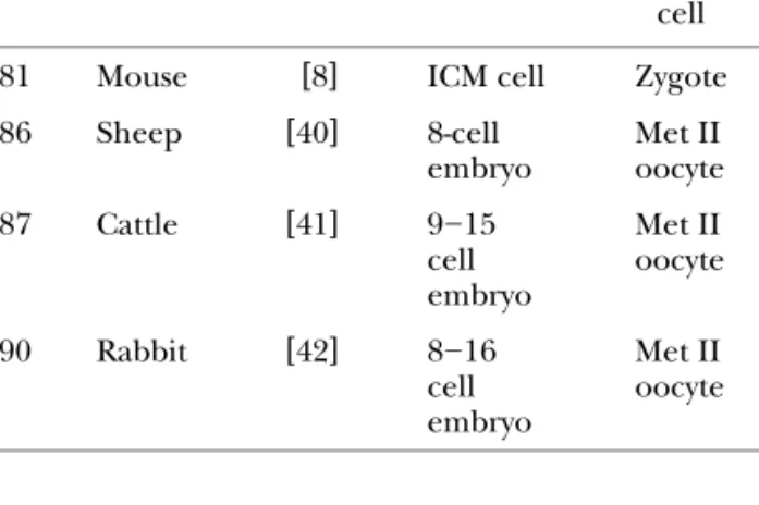
Table 1. The year when cloning was first reported for different species
Year Species Authors Donor Recipient cell enucleated
cell w x
1981 Mouse 8 ICM cell Zygote
w x
1986 Sheep 40 8-cell Met II
embryo oocyte w x 1987 Cattle 41 9]15 Met II cell oocyte embryo w x 1990 Rabbit 42 8]16 Met II cell oocyte embryo
blastomere of another two-cell embryo. Sendai virus was used to mediate cell fusion. The transfer of 160 reconstructed embryos to foster mothers resulted in the birth of a total of eight young. Since then, other investigators, including Tsunoda and his associates themselves, have obtained many cloned mice by transferring the nuclei of two- to eight-cell embryos
Ž .
into enucleated mature Met II oocytes, zygotes or
Ž .
the blastomeres of two-cell embryos Table 2 . According to McGrath and Solter,9 the nuclei of four- or eight-cell embryos introduced microsurgi-cally into enucleated zygotes or by using Sendai virus never supported the development of embryos to term. However, later investigators found that this was not the case when donor cell nuclei were introduced into oocytes by using electrofusion or Sendai virus.14]17 It
is interesting that nuclei of eight-cell mouse embryos introduced into enucleated zygotes could support only one or two cleavages18 or at best support
em-bryo development only up to the blastocyst stage.19 In
Table 2. Donor and recipient cells used for mouse cloning: only the studies resulting in the birth of live offspring are listed here
Nucleus donor Recipient Reference
2]4 cell embryo Met II oocyte 15,43]45 2]4 cell embryo 2-cell blastomere 46 2]8 cell embryo Met II oocyte 14]17
or zygote
4]8 cell embryo 2-cell blastomere 10 4]cell or morula Met II oocyte 16 embryo
contrast, the same nuclei introduced into enucleated oocytes or enucleated blastomeres of two-cell em-bryos supported development till mid-term17,20 or even to full-term.10,14 Apparently, the successrfailure of cloning experiments is largely dependent on the technical skill of investigators. Furthermore, the cell cycle phases of the donor and host cells at the time of fusion or nuclear transfer seem to contribute greatly to the outcome of the experiments.21,22 Even though
many investigators believe that enucleated zygotes and blastomeres of two-cell embryos are the best recipients of donor nuclei, we think that enucleated
Ž . 40
mature Met II oocytes, first used by Willadsen, are better suited for the production of live, cloned off
-Ž .
spring, at least for the mouse see below and per-haps for many other species. When enucleated oo-cytes are used as recipients, donor cells must be at either G0 or G1 phase of the cell cycle in order to obtain normal diploid embryos.21
Use of embryonic cells for production of mouse chimeras
ICM cells aggregated with eight- or 16-cell mouse embryos or those injected into blastocysts can con-tribute to the production of chimeric fetuses or young, suggesting totipotency of ICM cells.23]25
Ž .
Totipotency of embryonic stem ES cells was demon-strated in the same way.26,27According to Kato and Tsunoda,28 fetal germ cells of 15.5]16.5-day
post-Ž .
coitum dpc mouse embryos can contribute to the formation of chimeric embryos which, however, are apparently unable to develop to term.
Cloning using fetal somatic cells
Campbell et al29 cloned five sheep by fusing
enucle-ated oocytes with fetal epithelium cell line cultured for six to 13 passages. Cibelli et al30 reported the birth of three male calves following electrofusion of enucleated mature oocytes with fibroblasts collected from a 55-dpc fetus. They believed that fibroblasts were at the G1 stage of the cell cycle during fusion with oocytes. Cloning of the mouse using fetal so-matic cells has not been reported. Even primordial germ cells have not been used successfully for cloning.12
Cloning using adult somatic cells
The sheep ‘Dolly’ was the first animal ever cloned using adult somatic cells.1,31,32 Donor cells were
mammary gland cells from a 6-year-old ewe. The cells were electrofused with enucleated mature oocytes. Only one of 385 reconstructed couples developed to term. A key to this success, according to the authors, was to bring donor cells to the G0 phase of the cell cycle by ‘starving’ them before fusion with enucleated oocytes. Recently the mice2 and cattle3 were cloned
Ž
using adult somatic cells. Two calves one male and
.
one female were born after fusion of muscle cells with enucleated oocytes.3
Mouse clones were obtained using cumulus cells.2 According to Schultz et al,33 more than 90% of cu-mulus cells surrounding recently ovulated oocytes are in G0rG1 phases of the cell cycle. The first live clone mouse was born on 3 October, 1997 and was named ‘Cumulina’. She proved to be fertile. As of 30 Octo-ber 1998 we have over 80 mice cloned using cumulus cells, some of them being the 4th generation of the
Ž .
clone clone of clone of clone of clone . All cloned mice proved to be fertile.
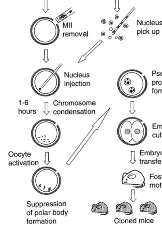
The cloning procedure we used is shown in Figure
Ž .
1. Donor oocytes ooplasm donors were collected
Ž .
from recently superovulated black B6D2F1 mice. Their Met II chromosome]spindle complexes were removed microsurgically. Meanwhile, cumulus cell]oocyte complexes were collected from recently
Ž .
superovulated agouti B6C3F1 females. Cumulus cells were dispersed with hyaluronidase, washed, the plasma membranes disrupted and their nuclei in-jected individually into enucleated oocytes. We used an injection pipette housed in a piezo-impact pipette drive unit. This unit, which drives the pipette a short
Ž .
distance e.g. 0.5mm very rapidly, allows the pipette to drill the zona pellucida easily and penetrate the oocyte’s plasma membrane without lysing the oocyte.34 We think that our success in cloning mice is
due, in part, to the use of this special pipette-driving unit.
Ž
We used a thin-walled, flush-ended pipette
ap-.
proximately 7 mm in diameter for injection. The
Ž .
plasma membrane of the cumulus cell 8]15mm was broken when the cell was drawn in and out of the pipette a few times. In most cases the nuclei we injected were devoid of visible cytoplasmic material. Each nucleus was injected within 5 min of its isola-tion.
The cumulus cell nucleus injected into an enucle-ated oocyte transformed into disarrayed
chromo-Figure 1. A diagram illustrating the cloning procedure we used2and described in the text.
somes. This disorder reflects an unusual situation in which single, condensed chromatids are each at-tached to a single pole of the spindle and are there-fore not aligned on a metaphase plate. After standing for 1]6 h in the medium, the oocytes were exposed to a medium containing both Sr2q and cytochalasin-B. The former activated the oocytes,35 while the
latter prevented subsequent polar body formation and therefore chromosome expulsion. We chose Sr2q
for mouse oocyte activation because, unlike an elec-tric shock, it induces repetitive rises in free
intracellu-2q Žw 2qx.
lar Ca concentration Ca i of the mouse
oo-36 w 2qx
cyte. Repetitive Ca i rises are known to be a salient feature of oocyte activation in a wide variety of animals.37,38Activated oocytes each contained two or
more pseudo-pronuclei in which DNA replication takes place. We transferred developing embryos into
Ž .
albino CD-1 foster females. Figure 2 shows the
orig-Ž .
inal cumulus cell nucleus donor and two genera-tions of cloned mice generated in this way. In the
Ž .
nucleus was first injected into an enucleated oocyte followed by oocyte activation. When we activated enucleated oocytes first, then injected them with the
Ž .
donor nuclei approx. 1 h later , all reconstituted oocytes fragmented without even developing into
Ž
normal two-cell embryos Wakayama and
Yanagi-.
machi, unpublished data .
It was thought that cloning mice would be difficult because the mouse embryonic genome begins to be expressed at the two-cell stage or even during the last zygotic stage, thus leaving too little time for the transferred nucleus to be reprogrammed.39 We left
Ž .
injected cumulus cell nuclei in the cytoplasm of unactivated oocytes for 1]6 h. Reprogramming, if it occurs, must be completed at least in the oocytes that successfully developed to live offspring. It is important to note that although large proportions of re -constructed oocytes could develop to blastocysts and
Ž
implant, most were unable to develop to term Figure
.
3 . The reasons for this are not at all clear at present. Genomic heterogeneous nature of the donor cells and unpredictable nature of the reprogramming processes39 could be the causes of this disappointing
and puzzling phenomenon.
We found that embryos developed from enucle-ated oocytes receiving Sertoli cell and neuron nuclei could develop and implant fairly well, but none
de-Figure 2. Two generations of cloned mice. The top row is the original cumulus cell donor. The second row is two clones from the above. The third row is four clones of the
Ž .
clones with permission of ProBio America, Inc. .
Figure 3. A diagram illustrating a sharp decline in the survival of cloned embryos during their
Ž . Ž .
developmentin vitro before implantation and in vivo after implantation . Only approximately 1% of cumulus nucleus-injected oocytes develop to term.
veloped to term.2 These cells were believed to be at the G0 phase of the cell cycle. Apparently G0 phase may be a preferable,1 but not necessarily the only,
condition necessary for successful cloning. In our experiments we used only three types of somatic cells. Thus far cumulus cells were better than Sertoli cells and neurons for cloning purposes, but there is no reason to believe that the latter two are inferior to cumulus cells. Technical improvement may make these cells as efficient as cumulus cells for cloning. It is very likely that there are still other cell types that are better suited for cloning than cumulus cells. Someday any types of cell could be used for cloning. Cloning experiments have just begun.
Acknowledgements
We thank ProBio America, for financial support of our study. We are grateful to Dr and Mrs Wesley Whitten and Mrs Charlotte Oser for their assistance in the preparation of the manuscript.
References
1. Wilmut I, Schnieke AE, McWhir J, Kind AJ, Campbell KHS
Ž1997 Viable offspring derived from fetal and adult mam-.
malian cells. Nature 385:810]813
2. Wakayama T, Perry AC, Zuccotti M, Johnson KR,
Yanagi-Ž .
machi R 1998 Full-term development of mice from enucle-ated oocytes injected with cumulus cell nuclei. Nature 394:369]374
3. Vignon X, Chesne P, LeBourhis P, Flechon D, Hyman Y,
Ž .
Renard JP 1998 Developmental potential of bovine embryos reconstructed from enucleated mature oocytes fused with cultured somatic cells. Compt Rend Acad Sci 321:735]745
Ž .
4. Lyon MF, Rastan S, Brown SDM, eds 1995 Genetic Varia -tions and Strains of the Laboratory Mouse, Vols. I and II. Oxford University Press, OxfordrNew York
Ž .
5. Silver LM 1995 Mouse Genetics: Concepts and Applications. Oxford University Press, New YorkrOxford
Ž .
6. Hogan B, Costantini F, Lacy E 1986 Manipulating the Mouse Embryo: A laboratory Manual. Cold Spring Harbor Labora-tory Press, Cold Spring Harbor
Ž .
7. Wassarman PM, DePamphilis ML, eds 1993 Guide to tech-niques in mouse development. Methods in Enzymology, Vol. 225. Academic Press, San Diego
Ž .
8. Illmensee K, Hoppe PC 1981 Nuclear transplantation in Mus musculus: developmental potential of nuclei from preimplantation embryos. Cell 23:9]18
Ž .
9. McGrath J, Solter D 1984 Inability of mouse blastomere nuclei transferred to enucleated zygotes to support
develop-mentin vitro. Science 226:1317]1319
Ž .
10. Tsunoda Y, Yasui T, Shioda Y, Nakamura K, Uchida T 1987 Full-term development of mouse blastomere nuclei trans-planted into enucleated 2-cell embryos. J Exp Zool 241:147]151
Ž .
11. Tsunoda Y, Kato Y 1993 Nuclear transplantation of embry-onic stem cells in mice. J Reprod Fert 98:537]540
Ž .
12. Tsunoda Y, Tokunaga T, Imai H, Uchida T 1989 Nuclear transplantation of male primordial germ cells in the mouse. Development 107:407]411
Ž .
13. Tsunoda Y, Kato Y 1998 Not only inner cell mass nuclei but also trophectoderm nuclei of mouse blastocysts have a devel-opmental totipotency. J Reprod Fert 113:181]184
Ž .
14. Cheong HT, Takahashi Y, Kanagawa H 1993 Birth of mice after transplantation of early cell-cycle stage embryonic nuclei into enucleated oocytes. Biol Reprod 48:958]963
Ž .
15. Kwon OY, Kato T 1996 Production of identical sextuplet mice by transferring metaphase nuclei from four-cell em-bryos. Proc Natl Acad Sci USA 93:13010]13013
Ž .
16. Tsunoda Y, Kato Y 1997 Full term development after trans-fer of nuclei from 4-cell and compact morula stage embryos to enucleated oocytes in the mouse. J Exp Zool 278:250]254
Ž .
17. Kwon OY, Kono T, Nakahara T 1997 Production of live young by serial nuclear transfer with mitotic stages of donor
Ž .
nuclei in mice. J Reprod Dev Japan 43:25]31
Ž .
18. Rabl JM, Gilligan B. Critser ES, First NL 1986 Nuclear mansplantation in mouse embryos: assessment of recipient cell stage. Biol Reprod 34:733]739
Ž .
19. Howlett SK, Barton SC, Surani MA 1987 Nuclear cyto-plasmic interactions following nuclear transplantation in mouse embryos. Development 101:915]923
Ž .
20. Barnes FL, Robl JM, First NL 1987 Nuclear transplantation in mouse embryos: assessment of nuclear function. Biol Re-prod 36:1267]1274
Ž .
21. Campbell KHS, Pasqualino L, Otaegui PJ, Wilmut I 1996 Cell cycle coordination in embryonic cloning by nuclear transfer. Rev Reprod 1:40]46
Ž .
22. Kono T 1997 Nuclear transfer and reprogramming. Rev Reprod 2:74]80
Ž .
23. Gardner R 1968 Mouse chimeras obtained by the injection of cells into the blastocyst. Nature 220:596]597
Ž .
24. Prather RS, Hageman L, First NL 1989 Preimplantation of mammalian aggregation and injection chimeras. Gamete Res 22:233]247
Ž .
25. Kato Y, Tsunoda Y 1995 Pluripotency of mouse embryonic cells in germ lines at 3.5]8.5 and 11.3 post-coitum after aggregation with precompact embryos. Dev Growth Diff 37:79]84
Ž .
26. Allen B, Evans M, Kaufmann MH, Robertson E 1984 Forma-tion of germ-line chimera from embryo-derived teratocarci-noma cell lines. Nature 309:255]256
Ž .
27. Wang ZQ, Kiefer F, Urbanek P, Wagner EF 1997 Genera-tion of completely embryonic stem cell-derived mutant mice using tetraploid blastocysts injection. Mech Dev 62:137]145
Ž .
28. Kato Y, Tsunoda Y 1995 Germ cell nuclei of male fetal mice can support development of chimeras to midgestation fol-lowing serial transplantation. Development 121:779]783
Ž .
29. Campbell McWhir J, Ritchie WA, Wilmut O 1996 Sheep cloned by nuclear transfer from a cultured cell line. Nature 380:64]66
30. Cibelli JB, Stice SL, Golueke PJ, Kane JT, Jerry J, Blackwell C,
Ž .
Ponce FA, De Leon PA, Robl JM 1998 Cloned transgenic calves produced from nonquiescent fetal fibroblasts. Science 280:1256]1258
31. Asworth D, Bishop M, Campbell K, Colman A, Kind A, Schnieke A, Blott S, Griffin H, Haley C, McWhir J, Wilmut I
Ž1998 DNA microsatellite analysis of Dolly. Nature 394:329.
32. Singer EN, Dubrova YE, Jeffrey AJ, Wilde C, Finch LME, Well
Ž .
M, Pealer M 1998 DNA fingerprinting Dolly. Nature 394:329]330
Ž .
33. Schultz AW, Whittingham DG, Snowden R 1996 Alterations in the cell cycle of mouse cumulus granulosa cells during expansion and mucificationin vivoandin vitro. Reprod Fert Dev 8:935]943
Ž .
34. Kimura Y, Yanagimachi R 1995 Intracytoplasmic sperm in-jection in the mouse. Biol Reprod 52:709]720
Ž .
35. Bos-Mikich A, Whittingham DG, Jones KT 1997 Meiotic and mitotic Ca2q oscillations affect cell composition in resulting blastocysts. Dev Biol 182:172]179
Ž .
36. Kline D, Kline JT 1992 Repetitive calcium transients and the role of calcium in exocytosis and cell cycle activation in the mouse egg. Dev Biol 149:80]89
Ž .
37. Miyazaki S, Shirakawa H, Nakada K, Honda Y 1993 Essential role of the inositol 1,4,5-triphosphate receptorrCa2qrelease
channel in Ca2q-waves and Ca2qoscillations at fertilization of
mammalian eggs. Dev Biol 158:62]78
Ž .
38. Kline D 1996 Activation of the mouse egg. Theriogenology 45:81]90
Ž .
39. Solter D 1998 Dolly is a clone } and no longer alone. Nature 394:315]316
Ž .
40. Willadsen SM 1986 Nuclear transplantation in sheep em-bryos. Nature 320:63]65
41. Prather RS, Barnes FL, Sims ME, Robl JM, Eyestone WH, First
Ž .
NL 1987 Nuclear transplantation in the bovine embryo:
assessment of donor nuclei and recipients oocyte. Biol Re-prod 37:859]866
Ž .
42. Collas P, Robl JM 1990 Factors affecting the efficiency of nuclear transplantation in the rabbit embryo. Biol Reprod 43:877]884
Ž .
43. Cheong HT, Takahashi Y, Kanagawa H 1994 Relationship between nuclear remodeling and subsequent development of mouse embryonic nuclei transferred to enucleated oocytes. Mol Reprod Dev 37:138]145
Ž .
44. Kono T, Tsunoda Y, Nakahara Y 1991 Production of identi-cal twin and triplet mice by nuclear transplantation. J Exp Zool 257:214]219
Ž .
45. Kono T, Kwon OY, Watanabe T, Nakahara T 1992 Develop-ment of mouse enucleated oocytes receiving a nucleus from different stages of the second cell cycle. J Reprod Fert 94:481]487
Ž .
46. Kono T, Kwon OY, Nakahara T 1991 Development of enu-cleated mouse oocytes reconstituted with embryonic nuclei. J Reprod Fert 93:165]172