O R I G I N A L A R T I C L E
Complement-dependent cytotoxicity and Luminex
technology for human leucocyte antigen antibody
detection in kidney transplant candidates
exposed to different sensitizing events
Nata
sa Katalini
c
1,2, Alma Star
cevi
c
1, Martina Mavrinac
3and Sanja Balen
1,41
Tissue Typing Laboratory, Clinical Institute for Transfusion Medicine, Clinical Hospital Centre Rijeka,
Croatia,
2Faculty of Medicine, Josip Juraj Strossmayer University of Osijek, Croatia,
3Department of Medical
Informatics, Faculty of Medicine, University of Rijeka, Croatia and
4Department of Clinical Laboratory
Diagnostics, Faculty of Medicine, University of Rijeka, Croatia
Correspondence and offprint requests to: Natasa Katalinic; E-mail: [email protected]
Abstract
Background:The aim of this study was to determine the frequency of exposure to different sensitizing events (SEs) and to assess their effects on human leucocyte antigen (HLA) alloimmunization in transplant candidates using two different HLA antibody screening techniques: complement-dependent cytotoxicity (CDC) and Luminex.
Methods: This retrospective study included HLA antibody screening results for 163 patients on the kidney transplant wait-ing list (WL) tested from March 2012 until the end of December 2015 at the Tissue Typwait-ing Laboratory, Rijeka, Croatia. All sera samples were tested using the CDC and Luminex techniques in parallel.
Results: Two-thirds of the patients [114 (70%)] on the WL were exposed to transfusions, pregnancies and/or kidney trans-plant. The pre-transplant sera of 104 (63.80%) patients were negative for antibodies. In the sera of 23 (14.11%) patients, HLA antibodies were detected by CDC and Luminex and in the sera of 36 (22.09%) patients by Luminex only.
Conclusion:In patients on kidney WL, previous organ transplantation represents the strongest immunogenic stimulus, followed by blood transfusions (the most frequent SE) and pregnancies. Although Luminex is more sensitive than CDC in HLA antibody detection, the decision on unacceptable HLA antigens in WL patients has to be based on the results of both assays and the patient’s immunization history.
Key words:HLA antibodies, HLA antibodies techniques, kidney transplantation, sensitization, waiting list
Received:January 30, 2017.Editorial decision:April 27, 2017
VCThe Author 2017. Published by Oxford University Press on behalf of ERA-EDTA.
This is an Open Access article distributed under the terms of the Creative Commons Attribution Non-Commercial License (http://creativecommons.org/ licenses/by-nc/4.0/), which permits non-commercial re-use, distribution, and reproduction in any medium, provided the original work is properly cited. For commercial re-use, please contact [email protected]
852
doi: 10.1093/ckj/sfx050
Advance Access Publication Date: 25 July 2017 Original Article
Introduction
Different sensitizing events (SEs), such as blood transfusions, preg-nancies and previous transplants, may induce the development of alloantibodies against human leukocyte antigen (HLA) [1,2]. In the organ recipient, preformed antibodies against graft antigens [donor-specific antibodies (DSAs)] may cause acute (hyper-acute) or chronic transplant rejection. The patients wait longer for trans-plantation and extended dialysis treatment results in lower graft survival than in non-sensitized patients [3–5]. Post-transplant development ofde novodonor-specific antibodies causes a higher incidence of graft rejections and increased risk of graft loss [6].
The HLA system is the most polymorphic system in humans. The complexity of antigenic epitopes represents a significant challenge for tissue typing laboratories (TTLs) in developing appropriate methods to detect and characterize the repertoire of HLA antibodies present in sensitized transplant candidates.
The conventional and most widely used method for determin-ing HLA antibodies is the cell-based complement-dependent cyto-toxicity (CDC) assay that was introduced by Terasaki and McClelland in the early 1960s [7]. The CDC technique has been in use at TTL Rijeka since 1971, when the laboratory was founded as the first one in Croatia. The technique is based on HLA mole-cules displayed in their natural configuration [2]. In HLA anti-body screening, the patient’s serum is incubated with a panel of HLA-typed T and/or B lymphocytes, whose HLA alleles provide a representative sample of the studied population. Results are expressed as the percentage of panel lymphocytes that react with a patient’s serum [panel reactive antibodies (PRA)]. As the same technique is used for crossmatching between the recipi-ent serum and the potrecipi-ential donor’s lymphocytes, PRA is useful in assessing the probability of a negative crossmatch result [8]. However, the CDC assay has a number of shortcomings related to the possibility of false negative (due to low antibody titres, non-cytotoxic antibodies or antibodies to HLA class II) or a false-positive result (the presence of autoantibodies, non-HLA antibodies or immune complexes) [9].
One of the main consequences of the CDC method’s lack of sensitivity is the considerable rate of graft failure in transplan-tation after a negative CDC crossmatch [10]. In order to improve graft survival, new antibody detection methods were intro-duced, including flow cytometry (FC) and solid phase assay (SPA)–enzyme-linked immunosorbent assay (ELISA) and Luminex technology. While FC is cell based, SPA methods use purified HLA molecules attached to plates (ELISA) or micro-spheres (Luminex). These methods can detect lower levels of HLA antibodies, allowing more precise determination of the HLA antibody specificity and differentiation of antibodies that activate complement from those that are non-complement fix-ing [11]. Currently the Luminex technology is the most sensitive and has been in use at TTL Rijeka since 2012 [12]. It is a semi-quantitative bead-based immunoassay for detection of immu-noglobulin G (IgG) and IgM antibodies to class I and class II HLA molecules that combines FC (xMAP technology) and fluorescent microparticles coated with HLA antigens at a high concentra-tion [9]. This technique enables identificaconcentra-tion of very low titres of class I and II HLA antibodies, accurate definition of acceptable and unacceptable HLA antigens in highly sensitized patients, and determination of epitope specificity, which is important for graft outcome. It is very useful in DSA monitoring after trans-plantation. This technology is also highly sensitive, leading to the detection of clinically irrelevant antibodies. While some specificities detected by SPA are considered to be relevant, they are not an absolute contraindication to transplantation [1,8].
The purpose of this study is to determine the frequency of exposure to SEs such as organ transplantation, blood trans-fusion and pregnancy and to assess their effects on HLA alloim-munization in patients on the kidney transplant waiting list (WL) using two different HLA antibody screening techniques in parallel: CDC and Luminex.
Materials and methods
We performed retrospective analysis of the HLA antibody screening results for 163 patients on the kidney transplant WL in Rijeka. A total of 664 sera samples were tested from March 2012 until the end of December 2015. Sera from all patients were tested by CDC, with or without di-thiotheitrol (DTT), and Luminex techniques in parallel. In order to compare the two techniques in terms of their detection of HLA IgG antibodies only, nine patients were excluded from the study, as their sera revealed the presence of non-HLA and/or IgM antibodies. Patients with a positive result in at least one serum sample were considered to be sensitized.
Information on SEs was obtained from potential recipients, their nephrologists and transfusion protocols for patients who underwent haemodialysis in Rijeka and from the questionnaire that accompanied each sample.
In our centre, patients on the WL are screened for HLA anti-body presence every 3 months, four times per year, using CDC and Luminex techniques in parallel.
In the CDC assay the patient’s serum is incubated with a panel of 50 HLA-typed unseparated T and B lymphocytes fol-lowing standard procedures. HLA antibody screening was per-formed with and without DTT addition. A PRA>5% was considered positive.
Serum screening by Luminex was performed in two stages according to the manufacturer’s instructions. The first stage was tested by LIFECODES LifeScreen Deluxe Beads, LMX (Immucor Transplant Diagnostics, Stamford, CT, USA). Serum with a posi-tive result in the LMX test was subjected to a second stage of test-ing by Luminex Stest-ingle Antigen (LSA) beads (LIFECODES LSA Class I and/or LSA Class II Single Antigens, Immucor Transplant Diagnostics). Analysis was performed using a fluorocytometer (LABScan 200 Flow Analyser, Luminex, Austin, TX, USA). The results were analysed using MatchIt software (Immucor Transplant Diagnostics). Currently, no standard median fluores-cence intensity (MFI) cut-off values exist, therefore the raw values of patient’s serum>1000 were considered positive, as has been previously indicated in the literature.
Continuous data were expressed as the arithmetic mean or median and categorical data asn(%). To determine statistically significant differences between the two techniques used in HLA antibody detection, data were compared with the use of the chi quadrat test and the Mann–WhitneyUtest. A two-sided P-value <0.05 was considered statistically significant. Statistical analy-sis was performed using MedCalc version 12.13. (MedCalc Software, Ostend, Belgium).
Results
Of the 163 patients included in the analysis, 65 (39.88%) patients were female and 98 (60.12%) were male. The average age was 55.85611.86 years.
One-third of the patients [49 (30%)] on the WL were not exposed to any SEs. Two-thirds of patients [114 (70%)] were exposed to one of the SEs (63 subjects) or to combinations of two (43 subjects) or three SEs (8 subjects) (Figure1).
The number of sensitized patients grew proportionately with the number of sensitizing factors. HLA antibodies were detected (by Luminex assay) in 10 (20.41%) patients who did not have any SEs, in 19 (30.16%) patients after one SE, and in 22 (51.16%) patients after two SEs. Exposure to three SEs caused HLA antibody generation in all patients. The relationship between HLA sensitization and the number of SEs is shown in Figure2.
Regardless of the combination, the most frequent SEs were blood transfusions, in 92 (56.44%) patients, while previous preg-nancies were reported in 57 (34.97%) patients and previous transplants were performed in 24 (14.72%) patients.
Considering the type of SE, 42 (25.77%) patients received a transfusion only, 20 (12.27%) women reported a history of pre-vious pregnancies only and 1 (0.61%) patient had a prepre-vious transplant. In terms of combinations of SEs, 28 (17.18%) women had a history of both transfusions and pregnancies, 14 (8.59%) recipients had previous transfusions and transplants, only 1 (0.61%) patient had both a previous transfusion and pregnancy, while all three SEs were reported in 8 (4.91%) patients.
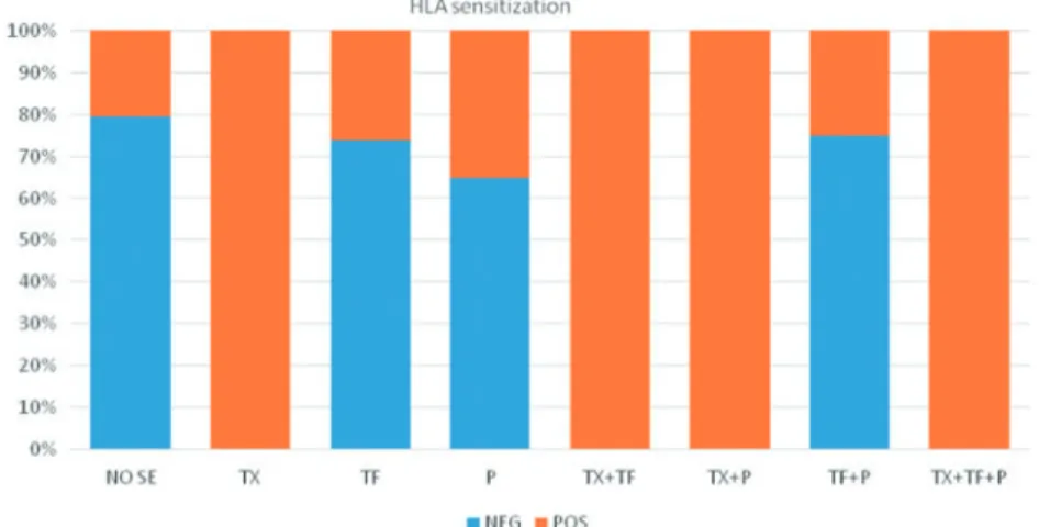
Figure3shows the proportion of the patients with detected HLA antibodies in pre-transplant sera according to the type of SE. Previous transplantation represents the strongest immuno-genic stimulus regardless of the combinations with other SEs, while pregnancy as an isolated event was the weakest sensitiz-ing factor.
HLA antibodies were not detected by either the CDC or Luminex techniques in the pre-transplant sera of 104 (63.80%) patients, while the sera of 59 (36.20%) patients were found to be positive for IgG HLA antibodies by the CDC and/or Luminex method.
In terms of the technique, HLA antibodies were detected in 23 (14.11%) patients by CDC and Luminex (CDCþLUMþ) and in an additional 36 (22.09%) by Luminex only (CDCLUMþ). The difference between the proportion of patients with HLA anti-bodies detected by CDC and/or Luminex was not statistically significant (v2¼0.18, P¼0.673).
Table1shows HLA sensitization detected by different tech-niques according to exposure to the SEs in patients on the kid-ney transplant WL. Considering different SEs, all patients with a history of previous transplantation only, or in combination with another SE, were sensitized.
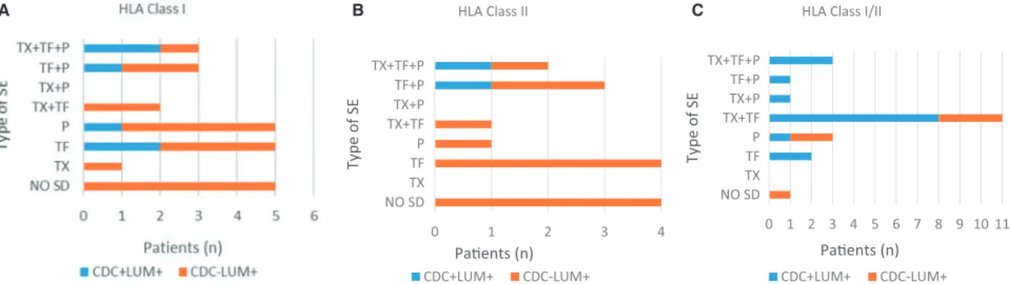
SEs induced the HLA sensitization in 49 patients; however, HLA antibodies were also detected in 10 patients without a his-tory of SEs. These antibodies were detected by Luminex only. Figure4A–C shows the prevalence of sensitized patients accord-ing to exposure to SEs and the class I and/or class II HLA antibod-ies detected by the two different techniques. Antibodantibod-ies against mixed HLA antigens class I/II were mostly detected by the CDC and Luminex in parallel. Luminex was more sensitive in the detection of HLA class I, and especially of class II, compared with the CDC assay. Thus the CDC technique finds it difficult to distin-guish class II antibodies using a panel of T and B lymphocytes.
The strength of the HLA antibody expressed as MFI was sig-nificantly higher in patients with antibodies detected by both techniques (CDCþLUMþ) compared with the group of patients with antibodies detected by Luminex only (CDCLUMþ) for HLA class I antibodies (MFI 4789 versus 3086; P<0.001) and class II Fig. 2.Distribution of patients with and without detected HLA antibodies
accord-ing to the number of SEs.
Fig. 3.Proportion of patients with and without HLA antibodies according to exposure to SEs. TX, transplantation; TF, blood transfusion; P, pregnancy. 30% 39% 26% 5%
Number of
sensizing events
0
1
2
3
Fig. 1.Distribution of patients on the kidney WL exposed to SEs (previous trans-plantation, pregnancy and/or blood transfusion).
antibodies (MFI 5715 versus 4606; P<0.001). The results are shown in Figure5A and B.
Ten patients who were not exposed to any senstizing event were HLA antibodies positive using Luminex technique and negative using CDC technique. Using Luminex technique Class I and Class II antibodies were detected. Among Class I antibodies 32 specificities were detected, and among Class II 21 specific-ities. Median values of peak MFI of HLA antibodies class I were significantly higher from median values of peak MFI of Class II specificities. (MFI 3324 versus 1779; P¼0.010) (Figure6).
Discussion
Previous transplants, blood transfusions and pregnancies are major SEs that can cause HLA alloimmunization [13]. Two-thirds of the patients (69.94%) on the WL at our centre were exposed to one or more sensitizing factor, which is consistent with the literature [14,15].
The risk of developing HLA antibodies is proportional to the number of sensitizing factors. The combination of SEs represents a stronger stimulus for HLA antibody generation than exposure to a single SE. In our study, most patients (38.65%) with a history of immunization had only one SE and HLA antibodies occurred in one-third (30.16%) of them, based on Luminex results. Fewer patients (26.38%) had been exposed to combinations of two SEs, and more than a half (51.16%) of these became sensitized. After exposure to three SEs (4.91%), HLA antibodies were detected in all patients (100%). Concerning the techniques used in antibody
detection, the difference in sensitivity between CDC and Luminex are interesting; the sensitivity mirrors the number of SEs. The higher sensitivity of Luminex is pronounced after exposure to a single SE (more patients had antibodies detected by Luminex only, while the CDC technique gave negative results). The difference between Luminex and CDC results disappears after two SEs, while exposure to three SEs caused sensitization detected by both tech-niques in almost all patients. The increased number of SEs causes antibody generation in higher concentrations that can be detected by less sensitive methods. The cumulative immunizing effect of different SEs has been recognized by several studies [13,16].
The most frequent SE was blood transfusion. More than half (56.44%) of the patients received one or more red blood concen-trates (RBCs), which is less than in previously published studies [14]. Most studies do not specify whether patients received trans-fusions after being placed on the WL or during the pre-transplant period. In this study, almost all transfused patients received RBCs (with or without leucocyte reduction) before registration on the WL. While being on the WL, only 11 (6.75%) patients were trans-fused. The US Renal Data System reports that 30% of transplant candidates on WLs received at least one blood transfusion, while
Yabuet al.[13] reported only 8.87% [13,17]. Other SEs were less
frequent; about one-third of transplant candidates had a history of pregnancy (34.97%) and the SE with least prevalence was pre-vious transplants (14.72%).
Evaluation of the impact of SEs on HLA sensitization varies according to the techniques used. Transfusions caused HLA anti-body generation in only 9.52% of transplant candidates when the serum screening was performed by CDC, but when tested by Luminex, the rate of sensitized patients increased to 25.77%. In our study the positivity rate detected by CDC is lower than in published data. Opelzet al.[18] reported that 28% of transfused patients developed HLA antibodies in the CDC assay. However, Luminex results in our study are in accordance with other reports. Hyunet al.[19] reported a 33% positive rate in transfused patients, while Leffellet al. [20] reported 20%. The reports on the positivity rate detected by CDC are mostly old, and lower results in our study can be explained by the differences in lymphocyte panels among centres and the various approaches to anaemia treatment by transfusions of different types of blood units.
HLA sensitization was detected in 10% (CDC) versus 12.27% (Luminex) of women with a history of pregnancies. The reported incidence is 18–30% tested by the CDC method and >50% when tested by Luminex [21–23]. The age of women in the tested groups may be the cause of the different results, as the titre of HLA antibodies declines over time [24]. In our study, only one patient had previous transplant as the sole SE.
HLA Class II 0 1 2 3 4 NO SD TX TF P TX+TF TX+P TF+P TX+TF+P Paents (n) Type of SE CDC+LUM+ CDC-LUM+ 0 1 2 3 4 5 6 7 8 9 10 11 NO SD TX TF P TX+TF TX+P TF+P TX+TF+P Paents (n) Type of SE
HLA Class I/II
CDC+LUM+ CDC-LUM+
A B C
Fig. 4.Total number of patients with HLA antibodies (A) class I, (B) class II and (C) class I/II detected by CDC and/or Luminex. TX, transplantation; TF, blood transfusion; P, pregnancy.
Table 1.Total number and proportion of patients exposed to differ-ent SEs according to the HLA antibody screening results by the CDC and Luminex techniques
Type of SE
HLA antibodies screening results
Neg,n(%) CDCLUMþ,n(%) CDCþLUMþ,n(%) No SE (n¼49) 39 (79.59) 10 (20.41) 0 TX (n¼1) 0 1 (100.00) 0 TF (n¼42) 31 (73.81) 7 (16.67) 4 (9.52) P (n¼20) 13 (65.00) 5 (25.00) 2 (10.00) TXþTF (n¼14) 0 6 (42.86) 8 (57.14) TXþP (n¼1) 0 0 1 (100.00) TFþP (n¼28) 21 (75.00) 5 (17.86) 2 (7.14) TXþTFþP (n¼8) 0 2 (25.00) 6 (75.00) Total (n¼163) 104 (63.80) 36 (22.09) 23 (14.11) Neg, negative; TX, transplantation; TF, blood transfusion; P, pregnancy.
Considering the difference between HLA class I and II anti-bodies detected by the two techniques, the higher sensitivity of Luminex is evident when a single class is present in the tested serum, especially class II antibodies. In our laboratory, the CDC technique is performed using a panel of unseparated (T and B) lymphocytes. HLA class I antigens are expressed on T lympho-cytes and class II antigens on B lympholympho-cytes. In the whole lym-phocyte population used in the CDC assay, the proportion of B lymphocytes is low, therefore the titre of HLA class II antibodies is below the sensitivity threshold of the CDC test. In patients with the presence of both class I and II antibodies, Luminex sen-sitivity is not so dominant when compared with the CDC
technique. A likely explanation may be that most patients posi-tive for both antibody classes had a history of previous kidney transplantation, confirming that solid organ transplantation represents the strongest immunizing event [19].
In our study, 10 male patients without reported SEs had anti-bodies that were detected by Luminex only. More specificities were found for class I antibodies. The MFI was low for both classes, but class I antibodies showed significantly higher MFIs compared with class II antibodies. The presence of HLA antibod-ies in non-immunized patients may be explained by the response to cross-reactive epitopes found in microorganisms, ingested proteins and allergens (natural alloantibodies), or as reactivity
Luminex, MFI 0 2000 4000 6000 8000 10000 12000 14000 16000 18000 CDC–LUM+ CDC+LUM+ Luminex, MFI 0 5000 10000 15000 20000 25000 CDC–LUM+ CDC+LUM+ A B
Fig. 5.Comparison of MFI values between HLA antibodies (A) class I and (B) class II detected by CDC and Luminex (CDCþLUMþ) and antibodies detected by Luminex only (CDCLUMþ) in sensitized patients on the kidney transplant WL exposed to SEs. The box-and-whisker plot shows median values of peak MFI for every specificity of HLA antibody that is considered positive (MFI1000) in each patient.
directed against denatured molecules (neo-epitopes) and exposed cryptic epitopes resulting from possible conformational changes of the HLA protein that may occur during the manufac-turing process [25–28].
Implementation of more sensitive techniques in HLA anti-body detection has enabled new insights into their occurrence, activity and clinical relevance in organ transplantation. The association between DSAs detected by CDC with hyperacute or acute graft rejection has been known for decades [10]. Reports on the clinical importance of DSAs detected in Luminex-based assays only are conflicting. Several studies have shown that the presence of DSAs detected exclusively by Luminex has no clini-cal relevance to graft survival [29,30]. Others, however, reported an association between DSAs and significantly increased graft loss even with a negative CDC and/or FC crossmatch [3,31,32]. The threshold of clinically relevant MFI values is also a matter of debate. Low levels of HLA DSAs detected by a Luminex assay before transplantation (MFI<1000 or<2000) are unlikely to have a deleterious effect on the graft [33,34]. Several authors have demonstrated significantly lower graft survival in patients with higher MFIs [3,35]. To assess the clinical impact of complement binding antibody on graft survival, several modifications of Luminex-based assays have been made. However, while some studies have shown that complement-fixing DSAs are more rel-evant in graft survival than non-complement-fixing DSAs [13, 36,37], some authors were unable to demonstrate such an asso-ciation [38,39]. Further investigations and methodology advan-ces are needed to bring us closer to these answers.
Conclusion
Exposure of patients on WLs to blood transfusions, pregnancies or previous transplants represents a risk factor of developing HLA antibodies that are considered a major immunologic bar-rier to successful transplantation. Blood transfusions are the most frequent and the most susceptible to our influence com-pared with other SEs. They should be minimized or avoided in transplant candidates whenever possible. HLA antibody testing
is critical in transplantation risk assessment, and CDC is widely used as a conventional cell-based method. It has low sensitivity and low resolution in determining antibody specificity, but high positive predictive value for antibody-mediated graft rejection. Luminex is a semi-quantitative SPA that is more sensitive than CDC, enabling better characterization of antibodies, but due to increased sensitivity, it may reveal the presence of antibodies with dubious clinical relevance. Based on the characteristics of both methods, the results have to be evaluated in combination with the clinical background and history of the patient’s expo-sure to SEs in order to determine unacceptable HLA antigen mismatches in the pre-transplant period.
Conflict of interest statement
None declared.References
1. Lobashevsky AL. Methodological aspects of anti-human leu-kocyte antigen antibody analysis in solid organ transplanta-tion.World J Transplant2014; 4: 153–167
2. Zeevi A, Girnita A, Duquesnoy R. HLA antibody analysis: sen-sitivity, specificity, and clinical significance in solid organ transplantation.Immunol Res2006; 36: 255–264
3. Lefaucheur C, Loupy A, Hill GS et al. Preexisting donor-specific HLA antibodies predict outcome in kidney trans-plantation.J Am Soc Nephrol2010; 21: 1398–1406
4. Rees L, Kim JJ. HLA sensitisation: can it be prevented?Pediatr
Nephrol2015; 30: 577–587
5. Meier-Kriesche HU, Kaplan B. Waiting time on dialysis as the strongest modifiable risk factor for renal transplant out-comes: a paired donor kidney analysis. Transplantation 2002; 74: 1377–1381
6. Willicombe M, Brookes P, Santos-Nunez Eet al. Outcome of patients with preformed donor-specific antibodies following alemtuzumab induction and tacrolimus monotherapy.Am J
Transplant2011; 11: 470–477 Luminex, MFI 1000 2000 3000 4000 5000 6000 7000 8000 9000 10000
HLA class I HLA class II
Fig. 6.Comparison of MFI values between HLA antibodies class I and II detected by CDC and Luminex in patients not exposed to SEs. The box-and-whisker plot shows median values of peak MFI for every specificity of HLA antibody class I and II that is considered positive (MFI1000) in each patient.
7. Terasaki PI, McClelland JD. Microdroplet assay of human serum cytotoxins.Nature1964; 998–1000
8. Tait BD, Su¨sal C, Gebel HMet al. Consensus guidelines on the testing and clinical management issues associated with HLA and non-HLA antibodies in transplantation.
Transplantation2013; 15: 19–47
9. Tait BD, Hudson F, Cantwell Let al. Review article: Luminex technology for HLA antibody detection in organ transplanta-tion.Nephrology (Carlton)2009; 14: 247–254
10. Patel R, Terasaki PI. Significance of the positive crossmatch test in kidney transplantation. N Engl J Med 1969; 280: 735–739
11. Minucci PB, Grimaldi V, Casamassimi Aet al. Methodologies for anti-HLA antibody screening in patients awaiting kidney transplant: a comparative study.Exp Clin Transplant2011; 9: 381–386
12. Katalinic N, Fucak M, Crnic Tet al. Pretransplantation moni-toring of HLA antibodies by complement dependent cytotox-icity and Luminex-based assays.Wien Klin Wochenschr2017; 129: 33–37
13. Yabu JM, Anderson MW, Kim D et al. Sensitization from transfusion in patients awaiting primary kidney transplant.
Nephrol Dial Transplant2013; 28: 2908–2918
14. Guichard-Romero A, Marino-Vazquez LA, Castelan N et al. Impact of pretransplant exposure to allosensitization fac-tors generating HLA antibodies in the Luminex era.Transpl
Immunol2016; 38: 33–39
15. Gombos P, Opelz G, Scherer Set al. Influence of test techni-que on sensitization status of patients on the kidney trans-plant waiting list.Am J Transplant2013; 13: 2075–2082 16. Triulzi DJ, Kleinman S, Kakaiya RMet al. The effect of
pre-vious pregnancy and transfusion on HLA alloimmunization in blood donors: implications for a transfusion-related acute lung injury risk reduction strategy. Transfusion 2009; 49: 1825–1835
17. US Renal Data System. USRDS 2010 Annual Data Report: Atlas of End-Stage Renal Disease in the United States. Bethesda, MD: National Institutes of Health, National Institute of Diabetes and Digestive and Kidney Diseases, 2010. https://www.usrds.org. (16 January 2017, date last accessed)
18. Opelz G, Graver B, Mickey MRet al. Lymphocytotoxic anti-body responses to transfusions in potential kidney trans-plant recipients.Transplantation1981; 32: 177–183
19. Hyun J, Park KD, Yoo Yvet al. Effects of different sensitiza-tion events on HLA alloimmunizasensitiza-tion in solid organ trans-plantation patients.Transplant Proc2012 Jan; 44: 222–225 20. Leffell MS, Kim D, Vega RMet al. Red blood cell transfusions
and the risk of allosensitization in patients awaiting primary kidney transplantation.Transplantation2014; 97: 525–533 21. Regan L, Braude PR, Hill DP. A prospective study of the
inci-dence, time of appearance and significance of anti-paternal lymphocytotoxic antibodies in human pregnancy. Hum
Reprod1991 6: 294–298
22. Vilches M, Nieto A. Analysis of pregnancy-induced anti-HLA antibodies using Luminex platform.Transplant Proc2015; 47: 2608–2610
23. Picascia A, Grimaldi V, Sabia C, et al. Comprehensive assessment of sensitizing events and anti-HLA antibody
development in women awaiting kidney transplantation.
Transpl Immunol2016; 36: 14–19
24. Scornik JC, Meier-Kriesche HU. Blood transfusions in organ transplant patients: mechanisms of sensitization and impli-cations for prevention.Am J Transplant2011; 11: 1785–1791 25. Morales-Buenrostro LE, Terasaki PI, Marino-Vazquez LA et al.
‘Natural’ human leukocyte antigen antibodies found in non-alloimmunized healthy males. Transplantation 2008; 86: 1111–1115
26. Pereira S, Perkins S, Lee JHet al. Donor specific antibody against denatured HLAA1: clinically nonsignificant? Hum
Immunol2011; 72: 492–498
27. El-Awar N, Terasaki PI, Nguyen Aet al. Epitopes of human leu-kocyte antigen class I antibodies found in sera of normal healthy males and cord blood.Hum Immunol2009; 70: 844–853 28. Cecka JM. Current methodologies for detecting sensitization
to HLA antigens.Curr Opin Organ Transplant2011; 16: 398–403 29. Su¨sal C, Ovens J, Mahmoud Ket al. No association of kidney
graft loss with human leukocyte antigen antibodies detected exclusively by sensitive Luminex single-antigen testing: a Collaborative Transplant Study report.Transplantation2011; 91: 883–887
30. Aubert V, Venetz JP, Pantaleo Get al. Are all donor-specific antibodies detected by solid-phase assay before transplan-tation clinically relevant?Transplantation2009; 87: 1897–1898 31. Amico P, Ho¨nger G, Mayr Met al. Clinical relevance of pre-transplant donor-specific HLA antibodies detected by single-antigen flow-beads.Transplantation2009; 87: 1681–1688 32. Patel AM, Pancoska C, Mulgaonkar Set al. Renal
transplanta-tion in patients with pre-transplant donor-specific antibod-ies and negative flow cytometry crossmatches. Am J
Transplant2007; 7: 2371–2377
33. Aubert V, Venetz JP, Pantaleo Get al. Low levels of human leukocyte antigen donor-specific antibodies detected by solid phase assay before transplantation are frequently clin-ically irrelevant.Hum Immunol2009; 70: 580–583
34. Szatmary P, Jones J, Hammad Aet al. Impact of sensitivity of human leucocyte antigen antibody detection by Luminex technology on graft loss at 1 year.Clin Kidney J2013; 6: 283–286 35. Salvade´ I, Aubert V, Venetz JP et al. Clinically-relevant
threshold of preformed donor-specific anti-HLA antibodies in kidney transplantation.Hum Immunol2016; 77: 483–489 36. Ramon DS, Huang Y, Zhao L, Rendulic T, Park JM, Sung RS,
Samaniego M. Use of complement binding assays to assess the efficacy of antibody mediated rejection therapy and pre-diction of graft survival in kidney transplantation. Hum
Immunol2017; 78: 57–63
37. Sicard A, Ducreux S, Rabeyrin M et al. Detection of C3d-binding donor-specific anti-HLA antibodies at diagnosis of humoral rejection predicts renal graft loss.J Am Soc Nephrol 2015; 26: 457–467
38. Thammanichanond D, Wiwattanathum P, Mongkolsuk T et al. Role of pretransplant complement-fixing donor-specific antibodies identified by C1q assay in kidney transplantation.
Transplant Proc2016; 48: 756–760
39. Ho¨nger G, Wahrmann M, Amico Pet al. C4d-fixing capability of low-level donor-specific HLA antibodies is not predictive for early antibody-mediated rejection.Transplantation2010; 89: 1471–1475