SALIVA AND SERUM LEVELS OF TNF-
α AND
IL-6 IN A SAMPLE OF ROMANIAN ADULT
SUBJECTS WITH TYPE 2 DIABETES MELLITUS
AND PERIODONTAL DISEASE
Adriana Monea, Lecturer, DMD, PhD
Department of Odontology and Periodontology,
Faculty of Dental Medicine, UMF Tîrgu-Mureş, Romania
Reinhard Gruber, Professor, PhD
Laboratory of Oral Cell Biology School of Dental Medicine, University of Bern, Switzerland
Nagy Elod, Associate Professor,MD PhD
Department of Pharmaceutical Biochemistry Faculty of Pharmacy,
UMF Tîrgu-Mureş, Romania
Gabriela Bereşescu, Lecturer, DMD, PhD
Department of Tooth Morphology Faculty of Medicine,
UMF Tîrgu-Mureş, Romania
Cosmin Moldovan, Lecturer, MD, PhD
Department of Histology Faculty of Medicine, UMF Tîrgu-Mureş, Romania
Monica Monea, Associate Prof., DMD, PhD
Department of Odontology and Periodontology,
Faculty of Dental Medicine, UMF Tîrgu-Mureş, Romania
Abstract
Introduction: Patients with diabetes mellitus can exert a stronger inflammatory response compared to systemically healthy individuals; however, if this accounts true patients with periodontal disease remains
unclear. We therefore determined salivary and serum TNF-α and IL-6 levels
in periodontal patients, with or without type 2 diabetes mellitus (T2D)
Material and methods: Twenty type 2 diabetes mellitus subjects with periodontal disease, 21 systemically healthy subjects with periodontal disease and 21 healthy controls were enrolled in the study. Periodontal
indices, HbA1c levels as well as saliva and serum TNF-α and IL-6 were
reported.
Results: TNF-α and IL-6 levels in the saliva were higher in type 2 diabetes mellitus subjects with periodontal disease compared to systemically healthy
IL-6 levels in the saliva than subjects with periodontal disease. The respective serum analysis, however, revealed only a minor impact of the type
2 diabetes mellitus and periodontal disease on TNF-α and IL-6 levels.
Conclusions: The data support the current hypothesis that the inflammation linked to periodontal disease is more severe in type 2 diabetic patients compared to the systemically healthy individuals.
Keywords: Diabetes mellitus, saliva, TNF-alpha, IL-6, periodontal disease Introduction
Diabetes mellitus is considered a risk factor for periodontal disease, although the underlying pathologic mechanisms that explain this association have not been completely understood (Mealey & Oates, 2006; Monea et al., 2012; Salvi et al., 2008). The hallmark of diabetes mellitus is hyperglycemia, which develops as a consequence of a lack of insulin (type 1 diabetes mellitus, T1D) or a resistance to insulin (type 2 diabetes mellitus, T2D). Hyperglycemia leads to advanced glycosylation end products (AGEs) (Brownlee, 1994), among which HbA1c is used for monitoring long term glycemic control (LeRoith & Olefsky, 2004). AGEs also affect the cells in many ways, one being the increase in their responsiveness to inflammatory signals such as those derived from gram-negative bacteria lipopolysaccharides (Araya et al., 2003). Thus, in diabetes mellitus, cells can exert a stronger inflammatory response compared to the respective controls in vitro.
In support of this hypothesis, some studies showed that IL-1 (Engebretson et al., 2004), IL-6 (Kurtis el al., 1999) and IL-8 (Engebretson et al., 2006) in the gingival crevicular fluid were higher in diabetic patients with periodontitis compared to healthy individuals with periodontitis. However, there is a controversy, as other studies reported similar levels of IL-1 (Kardesler et al., 2008) in patients with periodontitis with and without diabetes. This controversy remained when saliva has gained attention as a source for diagnostic tests (Miller et al., 2010; Yeh et al., 2010 ). For example, IL-6, MMP-8 and OPG levels were similar in diabetic and systemically healthy patients with periodontitis (Coast et al., 2010).
Moreover, T2D was associated with an increase in IL-1β and βTG
concentration in saliva, but independent of periodontal disease (Javed et al., 2012). Thus, saliva has gained attention as a source for diagnostic tests.
Neither of the studies, however, has determined the levels of TNF-α,
which is a key mediator of inflammatory reactions, and not necessarily follows the expression pattern of other inflammatory cytokines such as IL-6 (Kurtis et al., 2005). Thus there is a demand for saliva research evaluating the impact of diabetes on the cytokines levels in periodontal patients.
It was reasonable to determine inflammatory cytokines in saliva of periodontal patients, one cohort having T2D and the other cohort having normal blood glucose levels.
Material and method Study population
The first study group included 20 T2D subjects with periodontal disease, the second one 21 systemically healthy with periodontal disease subjects, and the third one 21 systemic and periodontal healthy controls. Diabetics were recruited from the Diabetes Clinic, County Hospital of
Tîrgu-Mureş, without knowing their glycemic control level, and were selected by
periodontal examination; subjects with periodontal disease but systemically healthy and controls were recruited from the Department of Odontology and
Periodontology, UMF Tîrgu-Mureş, after general and periodontal
examination. All subjects in the study groups were informed about the purpose of the study and signed an informed consent. The study was
approved by the Ethical Comity of UMF Tîrgu -Mureş.
Inclusion and exclusion criteria
Inclusion criteria for the study groups were as follows: age over 18; no major diabetic complications; presence of at least 20 teeth; moderate to severe periodontitis according to the criteria of the American Academy of Periodontology (Armitage, 1999); no periodontal treatment within the last six months; no antibiotic, corticoid or immunosuppressive administration within the last six months.
We excluded smokers and subjects that had any inflammatory disease within the last six months. Other additional exclusion criteria were: pregnancy or lactation, bleeding disorders, menstruation.
Periodontal status
Clinical examination was done using a specific algorithm, by registration of personal data, anamnesis information, extra- and intraoral examination. Periodontal status was assessed by measuring specific indices such as plaque index (PI) (Silness & Loe, 1964), gingival index (GI) (Loe & Silness, 1963), gingival bleeding index (GBI) of Ainamo et Bay (Ainamo & Bay, 1975), periodontal pocket depth (PD) in millimeters measured in six sites for each tooth and clinical attachment level (CAL) Christgau et al., 1998) .
Sample preparation
From each enrolled subject we collected 2 ml of un-stimulated saliva into a sterile tube, which was centrifuged for 3 min at 3.000 rpm and
clarified supernatant was filtered through a low protein binding membrane, separated in polyethylene recipients, then marked for identification and stored in the refrigerator until examination. Venous blood samples were obtained by venipuncture using an adequate closed system sample. Tubes
were then centrifuged at 3,000 rpm/10 min and stored at -20oC until
analyzed.
Immunoassay
Salivary and serum TNF-α and IL-6 levels were assessed with the
ELISA-sandwich method using a commercially available immunoassay kits
(OptEIA human TNF-α, Pharmingen, USA; DuoSet ELISA Development
System, R&D Systems, USA, respectively) according to the manufacturer’s guidelines. Results are reported in pg/ml.
We also determined blood glucose and glycated hemoglobin (HbA1c) levels for each diabetic subject, in order to evaluate the glycemic control level of diabetes mellitus.
Statistical analysis
The results were statistically analyzed using the Mann-Whitney U nonparametric test and Spearman’s correlations test. P values over 0.05 were considered statistically significant.
Results
Study population
Mean age was 56.73 ± 3.38 for diabetic subjects, characteristic for type 2 diabetes mellitus. The diabetic subjects enrolled in the study were all before treated with oral antidiabetic drugs, and periodontal disease was determined by the time of study inclusion. HbA1c levels (8-10.5%) and blood glucose levels (120-200 mg/dl) showed that all the enrolled diabetic subjects had medium to poor glycemic control of diabetes mellitus, and some of them had first degree obesity, having a medium BMI value of 28.75 (table 1). Biologic parameters of all subjects in the three study groups are described in table 1.
Table 1. Biologic parameters of subjects in study groups (T2DM= type 2 diabetes mellitus and periodontal disease, P= healthy with
periodontal disease, C=controls)
Saliva and serum levels of cytokines
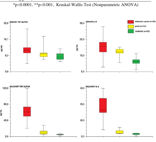
Salivary TNF-α and IL-6 levels were highly elevated in
parodontopathic diabetics compared with systemically healthy parodontopathic patients and controls, with a high statistic significance (p<0.0001) (table 2, figure 1).
Tabel 2. Salivary IL-6 and TNF-α variation in study groups (T2DM=
type 2 diabetes mellitus and periodontal disease, P= healthy with periodontal disease, C=controls)
Serum IL-6 levels in parodontopathic diabetics were significantly increased (p = 0.0144) compared to systemically healthy periodontopathic patients and extremely significant increased (p<0.0001) compared with healthy controls (table 3, figure 1).
Serum TNF-α in diabetics with periodontal disease was significantly
increased (p=0.0193) compared to systemically healthy perodontopathic patients and extremely significant increased (p=0.0003) compared with healthy controls (table 3, figure 1).
T2DP (n=20) P (n=21) C (n=21) Mean±Std.dev. Mean±Std.dev. Mean±Std.dev. Age (years) 56.37±13.89 52.37±11.12 53.09±14.10 BMI 28.90±5.38 22.01±3.19 25.06±4.74 Glycaemia (mg/dL) 168.46±21.63 84.20±.17 82.61±15.65 HbA1C (%) 9.02±0.84 - - PD (mm) 5.84±0.65 5.67±0.45 2.39±0.38 CAL (mm) 4.30±0.97 4.23±0.82 - PI 2.81±0.68 2.78±0.66 0.43±0.59 GI 2.32±0.40 2.24±0.31 0.22±0.42 GBI (%) 0.33±0.18 0.28±0.12 - T2DP (n=20) P (n=21) C (n=21) IL-6 (pg/mL)* 91.12±36.92 14.09±7.28 7.41±2.46 TNF (pg/mL)* 62.97±27.00 10.81±7.18 4.71±1.41 *p<0.0001, Mann-Whitney U test
Table 3. Serum IL-6 and TNF-α variation in study groups (T2DM= type 2 diabetes mellitus and periodontal disease, P= healthy with periodontal
disease, C=controls)
T2DM (n=20) P (n=21) C (n=21)
IL-6 (pg/mL)* 20.03 ± 6.81 16.98±2.57 11.41 ± 2.52 TNF (pg/mL)** 11.75 ± 4.56 9.88±3.27 7.95±1.81
*p<0.0001, **p<0.001, Kruskal-Wallis Test (Nonparametric ANOVA)
Figure 1. Box plots of the levels of serum and salivary TNF-α and IL-6 in diabetics with periodontitis, healthy with periodontitis and controls.
Correlation of saliva and serum levels of cytokines
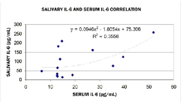
A marginally statistically significant correlation between IL-6 serum and salivary levels (R = 0.500, p = 0.057) resulted after applying Spearman’s nonparametric test. (Figure 2)
Figure 2. Serum and salivary IL-6 correlation in diabetics with periodontal disease
We found no correlations between serum IL-6 levels and age, BMI,
glycaemia, glycated hemoglobin and serum TNF-α .
Positive correlations with a tendency for statistical significance were found between salivary IL-6 levels in diabetics and the following parameters, after applying Spearman’s nonparametric test: age (R = 0.491, p = 0.063), body mass index (R = 0.456, p = 0.081), glycaemia (R = 0.465, p = 0.081) and glycated hemoglobin (R = 0.504, p = 0.056) (Figure 3).
Figure 3. Correlation of salivary IL-6 with HbA1c in diabetics with periodontal disease
Spearman’s nonparametric test revealed an average positive
significant correlation between serum and salivary TNF-α levels (R = 0.674,
p = 0.006) (Figure 4). No significant correlation was established between these values and the other parameters studied.
Discussions
Our results showed that salivary TNF-α and IL-6 levels in periodontal
patients were higher in diabetic patients with periodontitis compared to healthy individuals with periodontitis and controls. This might be one factor why in the presence of similar amounts of dental plaque and calculus, patients with T2D and periodontal disease had more severe periodontal problems than healthy subjects with periodontal disease.
In our study we measured elevated TNF-α and IL-6 levels in the
serum of diabetic subjects versus systemically healthy parodontopathic subjects and controls. These concentrations were increased not only when compared with own systemically healthy parodontopathic subjects and
controls, but were also higher than mean TNF-α and IL-6 values described in
literature in patients with periodontal disease but systemically healthy (Ng et al., 2007; Frodge et al., 2008; Coman, 2009). This confirms the hypothesis that diabetes mellitus is a co-factor in the onset and evolution of periodontal disease.
When we determined blood glucose and HbA1c levels, we noticed that all subjects had a medium to poor glycemic control, with mean values recorded for blood glucose level of 168.46±21.63 and 9.02±0.84 for HbA1c respectively. This might partially be due to the presence of periodontal disease, which may alter metabolic control of diabetes mellitus (Gurav, 2012).
Salivary IL-6 but not TNF-α tended to correlate with blood glucose
and HbA1c levels. The results have to be interpreted with care because of the small study group.
The clinical importance of our findings is that saliva IL-6 but also saliva TNF-α detection might serve as an indicator to predict the evolution of periodontal disease in subjects with type 2 diabetes mellitus. As a diagnostic fluid, saliva is yet insufficiently used in daily practice. It offers some advantages over serum, because of its non-invasive sampling method, which eliminates the need for clinicians’ special training. Furthermore, saliva analysis yielded values of the biochemical and immunological parameters comparable with those detected from blood samples (Gestenfeld et al. 2005).
Limitations of the study are that we included only non-smokers subjects, to minimize confounding variables. Further research should include smokers with T2D and periodontal disease, as smoking is a common habit in Romanian population.
One subject that remains to be explored is how initial periodontal therapy can influence salivary cytokines levels and glycemic control of T2D in parodontophatic patients.
Conclusion:
Our study showed elevated levels of TNF-α and Il-6 in the serum and
especially in the saliva of diabetics with periodontal disease, significantly higher than in systemically healthy subjects with periodontal disease and controls, confirming the hypothesis that the inflammation linked to periodontal disease is more severe in type 2 diabetic patients compared to the systemically healthy individuals.
The results of our study regarding the presence of TNF-α and IL-6 in
the serum and total saliva of diabetic patients allow us to conclude that saliva analysis is an efficient and safely enough tool for diagnosis and evaluation of periodontal disease progression in type 2 diabetic patients.
References:
Mealey, B.L. and T.W. Oates, Diabetes mellitus and periodontal diseases. J Periodontol, 2006. 77(8): p. 1289-303.
Mealey, B.L. and T.W. Oates, Diabetes mellitus and periodontal diseases. J Periodontol, 2006. 77(8): p. 1289-303.
Monea, A., T. Mezei, and M. Monea, The influence of diabetes mellitus on periodontal tissues: a histological study. Rom J Morphol Embryol, 2012. 53(3): p. 491-5.
Salvi, G.E., B. Carollo-Bittel, and N.P. Lang, Effects of diabetes mellitus on periodontal and peri-implant conditions: update on associations and risks. J Clin Periodontol, 2008. 35(8 Suppl): p. 398-409.
Brownlee, M., Lilly Lecture 1993. Glycation and diabetic complications. Diabetes, 1994. 43(6): p. 836-41.
Le Roith D., T.S., Olefsky J.M, ed. Diabetes mellitus: a fundamental and clinical text. 3rd edition ed. 2004, Lippincott Williams & Willkins.
Araya, A.V., et al., Ex vivo lipopolysaccharide (LPS)-induced TNF-alpha, IL-1beta, IL-6 and PGE2 secretion in whole blood from Type 1 diabetes mellitus patients with or without aggressive periodontitis. Eur Cytokine Netw, 2003. 14(3): p. 128-33.
Engebretson, S.P., et al., Gingival crevicular fluid levels of interleukin-1beta and glycemic control in patients with chronic periodontitis and type 2 diabetes. J Periodontol, 2004. 75(9): p. 1203-8.
Kurtis, B., et al., IL-6 levels in gingival crevicular fluid (GCF) from patients with non-insulin dependent diabetes mellitus (NIDDM), adult periodontitis and healthy subjects. J Oral Sci, 1999. 41(4): p. 163-7.
Engebretson, S.P., et al., The influence of diabetes on gingival crevicular fluid beta-glucuronidase and interleukin-8. J Clin Periodontol, 2006. 33(11): p. 784-90.
Kardesler, L., et al., Gingival crevicular fluid PGE2, IL-1beta, t-PA, PAI-2 levels in type 2 diabetes and relationship with periodontal disease. Clin Biochem, 2008. 41(10-11): p. 863-8.
Miller, C.S., et al., Current developments in salivary diagnostics. Biomark Med, 2010. 4(1): p. 171-89.
Yeh, C.K., et al., Current development of saliva/oral fluid-based diagnostics. Tex Dent J, 2010. 127(7): p. 651-61.
Costa, P.P., et al., Salivary interleukin-6, matrix metalloproteinase-8, and osteoprotegerin in patients with periodontitis and diabetes. J Periodontol, 2010. 81(3): p. 384-91.
Javed, F., M. Al-Askar, and K. Al-Hezaimi, Cytokine profile in the gingival crevicular fluid of periodontitis patients with and without type 2 diabetes: a literature review. J Periodontol, 2012. 83(2): p. 156-61.
Kurtis, B., et al., Gingival crevicular fluid levels of monocyte chemoattractant protein-1 and tumor necrosis factor-alpha in patients with chronic and aggressive periodontitis. J Periodontol, 2005. 76(11): p. 1849-55.
Armitage, G.C., Development of a classification system for periodontal diseases and conditions. Ann Periodontol, 1999. 4(1): p. 1-6.
Silness, J. and H. Loe, Periodontal disease in pregnancy. II. Correlation between oral hygiene and pereiodontal condition. Acta Odontol Scand, 1964. 22: p. 121-35.
Loe, H. and J. Silness, Periodontal disease in pregnancy I. Prevalence and severity. Acta Odontol Scand, 1963. 21: p. 533-51.
Ainamo, J. and I. Bay, Problems and proposals for recording gingivitis and plaque. Int Dent J, 1975. 25(4): p. 229-35.
Christgau, M., et al., Healing response to non-surgical periodontal therapy in patients with diabetes mellitus: clinical, microbiological, and immunologic results. J Clin Periodontol, 1998. 25(2): p. 112-24.
Ng, P.Y., et al., Candidate salivary biomarkers associated with alveolar bone loss: cross-sectional and in vitro studies. FEMS Immunol Med Microbiol, 2007. 49(2): p. 252-60.
Frodge, B.D., et al., Bone remodeling biomarkers of periodontal disease in saliva. J Periodontol, 2008. 79(10): p. 1913-9.
Coman M,L., Cercetări imunologice în boala parodontală, in Department of Odontology and Periodontology. 2009, University of Medicine and Pharmacy: Tg.Mures, Romania.
Gurav, A.N., Periodontal therapy - an adjuvant for glycemic control. Diabetes Metab Syndr, 2012. 6(4): p. 218-23.
Gerstenfeld, L.C., et al., Application of histomorphometric methods to the study of bone repair. J Bone Miner Res, 2005. 20(10): p. 1715-22.