Pathophysiological
mechanisms
of
absence
epilepsy:
A
computational
modelling
study
A thesis submitted to Cardiff University
for the degree of Doctor of Philosophy
by
Martynas Dervinis
School of Biosciences, Cardiff University, October 2016
Declaration
and
statements
DECLARATION This work has not been submitted in substance for any other degree or award at this or any other university or place of learning, nor is being submitted concurrently in candidature for any degree or other award. Signed ……… (candidate) Date ……… STATEMENT 1 This thesis is being submitted in partial fulfilment of the requirements for the degree of PhD. Signed ……… (candidate) Date ……… STATEMENT 2 This thesis is the result of my own independent work/investigation, except where otherwise stated, and the thesis has not been edited by a third party beyond what is permitted by Cardiff University’s Policy on the Use of Third Party Editors by Research Degree Students. Other sources are acknowledged by explicit references. The views expressed are my own. Signed ……… (candidate) Date ……… STATEMENT 3 I hereby give consent for my thesis, if accepted, to be available online in the University’s Open Access repository and for inter‐library loan, and for the title and summary to be made available to outside organisations. Signed ……… (candidate) Date ………
STATEMENT 4: PREVIOUSLY APPROVED BAR ON ACCESS
I hereby give consent for my thesis, if accepted, to be available for photocopying and for inter‐library loans after expiry of a bar on access previously approved by the Academic Standards & Quality
Committee.
Signed ……… (candidate) Date ………
Acknowledgements
I would like to thank my supervisor Vincenzo Crunelli for his support throughout my PhD, for all the discussions we had regarding the scientific matters and all the guidance while writing‐up my thesis. I am especially thankful for providing me with an opportunity to continue and complete my PhD after I could no longer continue working on my initial thesis project. I would like to thank my parents for their love and support that made this PhD possible. I am also very grateful to Sindy for all her love, dedication, and the most delicious food in the whole world, to Boris for all the countless Friday conversations we had that helped me to briefly forget the routine, and Andrius for being available to talk when I needed and all the small distractions.
Summary A typical absence is a non‐convulsive epileptic seizure that is a sole symptom of childhood absence epilepsy (CAE). It is characterised by a generalised hyper‐synchronous activity (2.5‐5 Hz) of neurons in the thalamocortical network that manifests as a spike and slow‐wave discharge (SWD) in the electroencephalogram. Although CAE is not a benign form of epilepsy, its physiological basis is not well understood. In an attempt to make progress regarding the mechanism of SWDs, I built a large‐scale computational model of the thalamocortical network that replicated key cellular and network electric oscillatory behaviours. Model simulation indicated that there are multiple pathological pathways leading to SWDs. They fell into three categories depending on their network‐level effects. Moreover, all SWDs had the same physiological mechanism of generation irrespective of their underlying pathology. They were initiated by an increase in NRT cell bursting prior to the SWD onset. SWDs critically depended on the T‐type Ca2+
current (IT) mediated firing in NRT and higher‐order thalamocortical relay cells (TCHO), as well as GABAB
synaptic receptor‐mediated IPSPs in TCHO cells. On the other hand, first‐order thalamocortical cells were inhibited during SWDs and did not actively participate in their generation. These cells, however, could promote or disrupt SWD generation if they were hyperpolarised or depolarised, respectively. Importantly, only a minority of active TC cells with a small proportion of them bursting were necessary to ensure the SWD generation. In terms of their relationship to other brain rhythms, simulated SWDs were a product of NRT sleep spindle (6.5‐14 Hz) and cortical δ (1‐4 Hz) pacemakers and had their oscillation frequency settle between the preferred oscillation frequencies of the two pacemakers with the actual value depending on the cortical bursting intensity. These modelling results are discussed in terms of their implications for understanding CAE and its future research and treatment.
Contents Declaration and statements ... 1 Acknowledgements ... 2 Summary ... 3 Contents ... 4 Abbreviations ... 7 Chapter 1 – Introduction: Absence seizures and childhood absence epilepsy... 12 1.1. Seizures ... 12 1.2. Epilepsy ... 14 1.3. Absence seizures ... 14 1.4. Childhood absence epilepsy... 15 1.4.1. Clinical symptoms ... 15 1.4.2. EEG features ... 18 1.4.3. Epidemiology ... 21 1.4.4. Aetiology ... 21 1.4.5. Treatment ... 27 1.4.6. Prognosis ... 29 1.4.7. Summary of human childhood absence epilepsy ... 30 Chapter 2 – Thalamocortical network properties ... 31 2.1. Physiological properties of thalamic cells ... 32 2.2. Physiological properties of neocortical cells ... 37 2.3. Connectivity of the thalamocortical network ... 44 2.3.1. Intra‐thalamic connectivity ... 44 2.3.2. Intra‐cortical connectivity ... 47 2.3.3. Thalamocortical connectivity ... 49 2.3.4. Corticothalamic connectivity ... 52 2.4. Oscillatory behaviours of the intact thalamocortical network ... 52
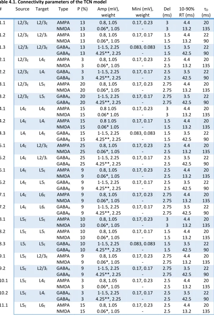
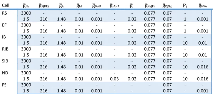
Chapter 3 – Mechanism of childhood absence epilepsy ... 63 3.1. Early experimental evidence ... 63 3.2. Modelling spike‐and‐wave oscillations based on GABAB receptor activation properties ... 69 3.3. Modelling failed to account for the recent experimental findings ... 73 3.4. A new computational model of spike‐and‐wave discharges is required ... 83 Chapter 4 – Methods ... 88 4.1. Organisation ... 88 4.2. Connectivity ... 89 4.3. Model neurons ... 98 4.4. Intrinsic currents ... 101 4.5. Synaptic currents ... 102 4.6. Intracellular ion concentration dynamics ... 103 4.7. Simulation techniques ... 103 4.8. Data recording and analysis ... 104 Chapter 5 – Results: Single cell models ... 105 5.1. Thalamocortical cells ... 105 5.2. Nucleus reticularis thalami cells ... 114 5.3. Neocortical cells ... 123 Chapter 6 – Results: Rhythms of the intact cortical network ... 128 Chapter 7 – Results: Rhythms of the intact thalamocortical network ... 141 Chapter 8 – Results: Paroxysmal oscillations ... 151 8.1. Cortical paroxysmal oscillation following GABAAR blockade ... 151 8.2. Spike‐and‐wave discharges elicited by increased tonic GABAA inhibition in the thalamus ... 153 8.3. Tests of changes in the thalamocortical network model to elicit spike‐and‐wave discharges .. 160 8.4. Processes initiating and terminating spike‐and‐wave discharges ... 168 8.5. The link between spike‐and‐wave discharges and sleep spindles ... 171 Chapter 9 – Discussion ... 175 9.1. Summary of results ... 175
9.2. Caveats and limitations ... 176 9.2.1. Physiological oscillations ... 176 9.2.2. Spike‐and‐wave discharge generation ... 181 9.3. Implications for understanding normal brain rhythms ... 181 9.4. Implications for understanding spike‐and‐wave discharges and absence epilepsy ... 183 9.5. Predictions ... 189 9.6. Future research directions ... 192 9.7. Strategy for devising more effective treatments for childhood absence epilepsy ... 194 Appendices ... 196 Appendix A: Intrinsic membrane currents in thalamocortical cell models... 196 Appendix B: Intrinsic membrane currents in nucleus reticularis thalami cell models ... 202 Appendix C: Intrinsic membrane currents in neocortical cell models ... 205 Appendix D: Synaptic membrane current models ... 210 References ... 213
Abbreviations 5‐HT 5‐hydroxytryptamine or serotonin ACh Acetylcholine AED Anti‐epileptic drug AHP Afterhyperpolarisation AMPA α‐amino‐3‐hydroxy‐5‐methyl‐4‐isoxazolepropionic acid AMPAR α‐amino‐3‐hydroxy‐5‐methyl‐4‐isoxazolepropionic acid receptor AP Action potential AS Absence seizure aTAS Atypical absence seizure BG Basal ganglia BOLD Blood‐oxygen‐level dependent CAE Childhood absence epilepsy [Ca2+] i Intracellular Ca2+ concentration [Ca2+] o Extracellular Ca2+ concentration CL Centrolateral thalamic nucleus CNS Central nervous system EEG Electroencephalogram EF Early firing regular spiking cell subtype eGABAAR Extrasynaptic γ‐aminobutyric acid A‐type receptor EPSP Excitatory postsynaptic potential FGPE Feline generalised penicillin epilepsy fMRI Functional magnetic resonance imaging FO First‐order FRB Fast repetitive bursting or chattering neocortical cell type FS Fast spiking neocortical cell type GABA γ‐aminobutyric acid GABAAR γ‐aminobutyric acid A‐type receptor GABABR γ‐aminobutyric acid B‐type receptor GAERS Genetic absence epilepsy rat from Strasbourg GAT‐1 γ‐aminobutyric acid transporter 1 GGE Genetic generalised epilepsy GLUT1 Glucose transporter type 1 protein
GPpe Globus pallidus pars externa GTCS Generalised tonic‐clonic seizure HCN Hyperpolarization‐activated cyclic nucleotide‐gated channel HO Higher‐order HT High‐threshold HVA High‐voltage‐activated IA A‐type K+ current IAHP Afterhyperpolarisation current IB Intrinsically bursting neocortical cell type ICAN Ca2+‐activated nonspecific cation current ID D‐type K+ current Ih Hyperpolarisation‐activated nonspecific cation current
IHVA High‐voltage‐activated Ca2+ current
IK[Ca] Ca2+‐activated K+ current
IK(DR) Delayed rectifier persistent K+ current
IK[Na] Na+ activated K+ current
IKir Inwardly rectifying K+ current
IKor Outwardly rectifying K+ current
ILAE International League Against Epilepsy IN Inhibitory cortical cell(s) (interneurons) INa Fast transient Na+ current
INa(P) Persistent Na+ current
IPSP Inhibitory postsynaptic potential IT Low‐voltage‐activated Ca2+ current
ITs Slow low threshold Ca2+ current
ITwindow Low‐voltage‐activated Ca2+ current (non‐inactivating) window component
JAE Juvenile absence epilepsy JME Juvenile myoclonic epilepsy Kir Inwardly rectifying K+ channels
LD Laterodorsal thalamic nucleus L1 Neocortical layer 1
L2/3 Neocortical layers 2 and 3 L2/3/4 Neocortical layers 2, 3, and 4 L4 Neocortical layer 4
L4/5/6 Neocortical layers 4, 5, and 6 L4py L4 regular pyramidal cell L4sp L4 star pyramidal cell L4ss L4 spiny stellate cell L5 Neocortical layer 5 L5/6 Neocortical layers 5 and 6 L5st Slender‐tufted L5 pyramidal cell L5tt Thick‐tufted L5 pyramidal cell L6 Neocortical layer 6 L6cc Cortically‐projecting L6 pyramidal cell L6ct Thalamus‐projecting L6 pyramidal cell LGN Lateral geniculate nucleus LSP Lower stable point LTCP Low threshold calcium potential LTS Low‐threshold spiking neocortical cell type LVA Low‐voltage‐activated M1 Primary motor cortex M2 Secondary motor cortex MD Mediodorsal thalamic nucleus mGluR Metabotropic glutamate receptor MRI Magnetic resonance imaging mRNA Messenger ribonucleic acid NA Noradrenaline or norepinephrine [Na+] i Intracellular Na+ concentration ND “Network driver” intrinsically bursting cell subtype NEC Non‐epileptic control NIPA2 Non imprinted in Prader‐Willi/Angelman syndrome 2 NREM Non‐rapid‐eye‐movement sleep NRT Nucleus reticularis thalami NRTFO Nucleus reticularis thalami sector associated with first‐order thalamic relays NRTHO Nucleus reticularis thalami sector associated with higher‐order thalamic relays NMDA N‐methyl‐D‐aspartate NMDAR N‐methyl‐D‐aspartate receptor Pc Paracentral thalamic nucleus
PoM Posterior medial thalamic nucleus PSC Postsynaptic current PSP Postsynaptic potential PSWD Poly‐spike‐and‐wave discharge PY Pyramidal cortical cell(s) REM Rapid‐eye‐movement sleep RI Neuronal membrane input resistance RIB repetitive intrinsically bursting neocortical cell type RS Regular spiking neocortical cell type S1 Primary somatosensory cortex S1po Perioral area of the primary somatosensory cortex S2 Secondary somatosensory cortex SC Superior colliculus
SK Small conductance Ca2+‐activated K+ channels
SNpr Substantia nigra pars reticulata STN Subthalamic nucleus SWC Spike‐and‐wave complex (a single SWD cycle) SWD Spike‐and‐wave discharge SWS Slow wave sleep or non‐rapid eye movement sleep TAS Typical absence seizure TC Thalamocortical relay nucleus/nuclei (dorsal thalamus) TCFO First‐order thalamocortical relay nucleus/nuclei TCHO Higher‐order thalamocortical relay nucleus/nuclei TCN Thalamocortical network TEA Tetrathylamonium THIP 4,5,6,7‐tetrahydroisoxazolo(5,4‐c)pyridin‐3‐ol or gaboxadol UP Unstable point USP Upper stable point VL Ventrolateral thalamic nucleus VB Ventrobasal thalamic complex VM Ventromedial thalamic nucleus VM Membrane potential VMb Basal ventromedial thalamic nucleus VPL Ventroposterolateral thalamic nucleus
VPM Ventroposteromedial thalamic nucleus WAG/Rij Wistar Albino Glaxo/Rijswijk rat
Chapter 1 – Introduction: Absence seizures and childhood absence epilepsy In this thesis I present a computational model of pathophysiological mechanisms underlying absence epilepsy in humans and animals. I begin by describing absence seizures (ASs) with their electrographic signature followed by the description of childhood absence epilepsy (CAE) – a pure form of genetic absence epilepsy in humans. I also summarise the current knowledge about the neural mechanisms underpinning this disease. I finish by providing a rationale for the modelling work carried out in this thesis and discuss its aims. 1.1.Seizures International League Against Epilepsy (ILAE) defines the epileptic seizure as an abnormal excessive or synchronous neural activity of the brain that is transient and manifests clinically (Fisher et al., 2005). Three essential elements are required for diagnosing an epileptic seizure: (1) the presence of a synchronous electrical brain activity leading to the seizure, (2) a clear onset and termination of this activity, and (3) accompanying clinical abnormalities. The presence of abnormal neural activity in humans is inferred using electrographic measures like the scalp surface electroencephalogram (EEG) because of its non‐invasive nature (Hrachovy and Frost, 2006, Verma and Radtke, 2006). Seizures are classified into types based on their pre‐, post‐, and inter‐ictal electric activity patterns (oscillations) and their origin and spread across the brain surface. Clinical utilisation of other techniques, like brain imaging, at present is restricted only to aiding the identification of seizure‐underlying structural abnormalities (Burch et al., 2012, Craven et al., 2012). The transient nature of a seizure is indicated by changes either in the EEG or clinical symptoms. Seizures could last from seconds to minutes and even to lengthy periods (>5 minutes) called status epilepticus (Panayiotopoulos, 2010). The onset and termination of seizures is not always abrupt and conspicuous. Pre‐ictal changes in the EEG pattern can reliably be detected for some seizures an hour in advance (Society, 2014) and clinical signs could be unfolding gradually (e.g., atypical absences) (Panayiotopoulos, 2010). The transition from a seizure to recovery could often be blurred for a lot of seizure types as clinical signs cede slowly and the EEG shows temporary post‐ictal alterations (e.g., post‐ictal suppression in generalised tonic‐clonic seizures – GTCSs) (Panayiotopoulos, 2010). These observations underscore the difficulty of reliable seizure detection and classification in epilepsy treatment and research. The presence of clinical signs forms a necessary part of a seizure diagnosis (Fisher et al., 2005). In the
absence of these symptoms an EEG seizure is deemed to be subclinical (Panayiotopoulos, 2010). A commonly associated sign of a seizure is the presence of involuntary jerky movements. Motor symptoms are classed into tonic, clonic, myoclonic, dystonic, atonic, and hypermotor (see Blume et al., 2001, for descriptions). Motor symptoms are salient, however other signs – autonomic, sensory, cognitive, mnemonic, and affective – are also present (see Panayiotopoulos et al., 2010, for examples of seizures involving various clinical symptoms). Clinical symptoms provide a basis for differentiating seizures into distinct types and give important clues about the neural network underlying a particular seizure type.
Table 1.1. Classification of seizures
Generalised seizures Tonic‐clonic (in any combination) Absence Typical Atypical with special features Myoclonic absence Eyelid myoclonia Myoclonic Myoclonic Myoclonic atonic Myoclonic tonic Clonic Tonic Atonic Focal seizures Unknown* Epileptic spasms *Seizures that cannot be clearly diagnosed into one of the preceding categories should be considered unclassified until further information allows their accurate diagnosis. This is not considered a classification category, however. Adapted from Berg et al. (2010).
Table 1.2 Descriptors of focal seizures according to degree of impairment during seizure Without impairment of consciousness or awareness With observable motor or autonomic components. This roughly corresponds to the concept of “simple partial seizure”. “Focal motor” and “autonomic” are terms that may adequately convey this concept depending on the seizure manifestations. Involving subjective sensory or psychic phenomena only, including an aura. With Impairment of consciousness or awareness. This roughly corresponds to the concept of complex partial seizure. “Dyscognitive” is a term that has been proposed for this concept (Blume et al., 2001). Evolving to a bilateral, convulsive seizure (involving tonic, clonic, or tonic and clonic components). This expression replaces the term “secondarily generalised seizure.” For more descriptors see Blume et al. (2001). Adapted from Berg et al. (2010). Based on their EEG features and clinical symptoms seizures are most broadly classified into generalised and focal (see Table 1.1) (Berg et al., 2010). Generalised seizures rapidly engage bilaterally distributed neural networks. Their origin in the brain is either diffuse or focused but inconsistent between seizures. In contrast, focal seizures always originate within the same unilateral ictogenic zone. They may become
bilateral (or secondarily generalised; see Table 1.2). 1.2.Epilepsy Epilepsy is defined as a brain disorder with a predisposition to generate unprovoked seizures and its diagnosis requires the occurrence of at least one seizure (Fisher et al., 2005). It is an umbrella term that involves a number of different diseases and syndromes. The new classification system groups epilepsies based on the age at the onset of seizures (Berg et al., 2010) and further divides them into genetic, structural, metabolic, immune, infectious, and of unknown origin based on underlying causes (Scheffer et al., 2014). Lifetime epilepsy affects 0.5‐1.5% of the world’s population – approximately 70 million people at this moment (Ngugi et al., 2010). It is one of the most common neurological disorders that is surpassed only by stroke and Alzheimer’s disease, yet its research financing is significantly smaller than any other major neurological disorder when adjusted for its prevalence (Meador et al., 2011, Young et al., 2012). Hence, increasing medical research could considerably reduce mortality, disability, social stigma, and associated physical, psychological and economic cost caused by epilepsy worldwide (Lozano et al., 2012, Young et al., 2012, WHO, 2015). 1.3.Absence seizures In my thesis I focus on ASs – a common type of seizures occurring in many epileptic disorders (Panayiotopoulos, 2010). It is the sole symptom of CAE and a primary symptom of juvenile absence epilepsy (JAE) and juvenile myoclonic epilepsy (JME). It is often present in many other epilepsy syndromes in combination with other seizure types: Epilepsy with myoclonic absences, eyelid myoclonia with absences or Jeavons syndrome, epilepsy with GTCSs, epilepsy with myoclonic‐atonic seizures, perioral myoclonia with absences, epilepsy with phantom absences, many of the epileptic syndromes starting in infancy or early childhood, as well as a few focal epilepsies including frontal lobe and occipital lobe epilepsies. ASs are classed as typical, atypical, or absences with special features – myoclonic absence and eyelid myoclonia (Table 1.1) (Berg et al., 2010). Typical absence seizures (TASs) are characterised by a mild to severe impairment of consciousness (absence) of a varying duration but mostly brief (Blumenfeld, 2005). The EEG signature of TAS is a bilateral oscillation pattern with each cycle containing a fast (approximately one third of the cycle) negative small amplitude spike component followed by a slower positive wave
component (spike‐/poly‐spike‐and‐wave discharge – SWD/PSWD) repeating with a 3‐4 Hz frequency (Weir, 1965, Gibbs et al., 1968, Hrachovy and Frost, 2006). If no other clinical symptoms are present, absences are called simple. In contrast, complex absences may involve clonic, myoclonic, tonic, and atonic motor components and automatisms, autonomic changes, and components of neocortical and limbic symptomatology (Panayiotopoulos, 2010). Expression of additional clinical symptoms is syndrome related. TASs differ from atypical ASs (aTASs) which have EEG field potentials oscillating with a lower frequency (1‐2.5 Hz) often irregular and invading limbic brain areas, mild to severe consciousness impairments, and changes in muscle tone and autonomic function (Panayiotopoulos, 2010). Atypical absences usually occur in children with severe genetic epilepsy syndromes and learning disabilities. The most recent classification (Berg et al., 2010) also distinguishes myoclonic absence – an absence seizure with clonic rather than myoclonic limb jerks and an increase in the muscle tone – and absence with eyelid myoclonia – jerking of eyelids and upward deviation of eyeballs and the head with a loss of consciousness (an absence) – occurring in Jeavons syndrome. All absence seizures are classed as generalised (Berg et al., 2010) meaning that they are expressed in the brain bilaterally from the very early stage of a seizure. Hence, the onset is diffuse or localised inconsistently and rapidly spreading. Although this may be the case for many ASs, TASs in CAE appear to defy this conjecture (see Section 1.4.2). 1.4.Childhood absence epilepsy The focus of the thesis is on typical rather than atypical ASs and on CAE. This type of epilepsy is of interest here because it is solely defined by the repeated occurrence of TASs (Panayiotopoulos, 2008). 1.4.1. Clinical symptoms The core aspect of the CAE diagnosis is the age at the onset of the first seizure. According to the ILAE classification (Commission, 1989), seizures associated with CAE occur in school age children with 6 to 7 years seeing the peak of the first incidences. The range appears to be 2‐10 years of age (Panayiotopoulos et al., 1989, Sadleir et al., 2006, 2008, Panayiotopoulos, 2008, Verrotti et al., 2011). A later onset indicates a different form of genetic generalised epilepsy (GGE; see Gallentine and Mikati, 2012, for a list of GGEs). CAE is a syndrome and as such requires the presence of other clinical signs for its diagnosis (Table 1.3). In short, TASs must be abrupt, rather severe, largely lacking motor components or coincident other forms of
a seizure, and brief but frequent. Abrupt here means a sudden onset and termination. No warning signs exist for TASs but it may take a few seconds to resume any pre‐ictal activity after the seizure termination (Hirsch and Panayiotopoulos, 2005, Panayiotopoulos, 2008). The impairment of consciousness is thought to be severe (Hirsch and Panayiotopoulos, 2005). However, recent studies found a varying degree of awareness impairment severity ranging from subclinical events indicated only by a characteristic EEG pattern through occasional clinical seizures with partial awareness to full‐blown absences (Sadleir et al., 2006, Sadleir et al., 2008). Evidence also indicates that sufficiently strong sensory stimulation – let it be a loud sound or a painful stimulus – would terminate an ongoing seizure (Rajna and Lona, 1989). Any ongoing directed activity usually ceases within the first 3 seconds of a seizure (Hirsch and Panayiotopoulos, 2005) but, again, occasionally may not be extinguished completely (Sadleir et al., 2006). The duration of a seizure in CAE is typically around 10 seconds (Panayiotopoulos et al., 1989, Sadleir et al., 2006). Although seizures shorter than 4 seconds were considered to signal CAE diagnosis exclusion in the past (Commission, 1989, Panayiotopoulos, 2008), recent studies observed approximately a quarter of all seizures to be shorter than 4 seconds (Sadleir et al., 2006, Ma et al., 2011).
Table 1.3. Inclusion and exclusion criteria for CAE Inclusion criteria: (1) Age at onset between 2 and 10 years. (2) Somewhat normal neurological state and development. (3) Brief (<1 minute, mostly 10 seconds) and frequent (dozens per day) ASs with abrupt and mostly severe impairment (loss) of consciousness. Automatisms are frequent but have no diagnostic significance. (4) EEG ictal discharges of generalised high‐amplitude spike and double (rarely three or more) spike‐ and slow‐wave complexes. They are rhythmic at around 3 Hz with a gradual and regular slowdown from the initial to the terminal phase of the discharge. Exclusion criteria: (1) Other than TASs such as GTCSs, or myoclonic jerks prior to or during the active stage of absences. (2) Eyelid myoclonia, perioral myoclonia, rhythmic massive limb jerking, and single or arrhythmic myoclonic jerks of the head, trunk, or limbs. However, mild myoclonic elements of the eyes, eyebrows, and eyelids may be featured – particularly in the first 3 seconds of the AS. (3) Mostly mild or no impairment of consciousness during seizures. (4) Visual (photic) and other sensory precipitation of clinical seizures. Adapted from Loiseau et al. (2002) with changes to reflect recent findings. Seizures in CAE are very frequent and vary from a few to perhaps hundreds a day (Horita et al., 1991, Ma et al., 2011). The frequency may depend on brain maturation as it declines with age and usually does not even occur every day in JAE (Engel, 2006, Panayiotopoulos, 2008). Hyperventilation is frequently used as a test for CAE as TASs could be induced by over‐breathing in more than 90% of all untreated CAE cases (Giannakodimos et al., 1995, Sadleir et al., 2006, Ma et al., 2011, Watemberg et al., 2015). In some patients ASs could be induced using intermittent photic stimulation (Sadleir et al., 2006). However, this is
deemed to be rare in CAE and rather suggestive of a syndrome that is likely to continue into the adulthood (Panayiotopoulos, 2008). Unprovoked absences are observed both during sleep and wakefulness (Kellaway et al., 1980, Horita et al., 1991, Halász et al., 2002, Sadleir et al., 2006, Zarowski et al., 2011) although the distinction between clinical and subclinical seizures is complicated by sleep (Horita, 2001). The link between sleep and ASs received increased attention and a few early studies seemed to indicate that SWDs were considerably more common during the slow wave sleep (SWS) compared to wakefulness (Kellaway et al., 1980, Kellaway, 1985, Nobili et al., 2001, Halasz et al., 2002). This interest coincided with the cortico‐reticular theory of ASs (see Section 3.1), which at its core had a claim that SWDs were abnormal sleep spindles, acquiring popularity (Kostopoulos, 2000). However, these studies did not include CAE patients (e.g., Halasz et al., 2002) or did not attempt to differentiate between different forms of GGEs (e.g., Kellaway et al., 1980), or if they did, they had small samples of medicated children (e.g., Nobili et al., 2001). It was reliably demonstrated that the first choice antiepileptic drugs (AEDs) for ASs – ethosuximide and sodium valproate – affect the sleep architecture (Roder and Wolf, 1981, Wolf et al., 1984, Zhang et al., 2014) and, therefore, could obscure the link between sleep‐related neural phenomena and SWDs. Attempts to establish a link between sleep spindles and SWDs were already hampered from the beginning since they failed to explain the early observations of ASs during wakefulness either in these very same studies or studies by other scientists (Panayiotopoulos et al., 1989, Horita et al., 1991). Distinct profiles of patients were observed that predominantly exhibited SWDs during wakefulness but not sleep and vice versa (Kellaway et al., 1980, Horita et al., 1991). Subsequently, studies employing large samples of patients with CAE or other epilepsies with ASs demonstrated that TASs were more common during wakefulness and when occurring they tended to befall in periods of inactivity, drowsiness, and superficial stages of sleep (Horita et al., 1991, Baldy‐Moulinier, 1992, Horita, 2001, Halász et al., 2002, Sadleir et al., 2006, Zarowski et al., 2011). These studies indicated a “sweet spot” for TASs corresponding to a transition period between sleep and wakefulness (Halász et al., 2002). Therefore rather than being abnormal misplaced sleep spindles, SWDs may well be a distinct phenomenon preferentially occurring at different brain arousal levels. Finally, the presence of any moderate to severe motor components during a seizure excludes the diagnosis of CAE with certainty (Panayiotopoulos, 2008). However, mild automatisms are frequent. They include a spontaneous eye opening, a slow upward drifting of the gaze, a twitching of the eyebrows, slow or fast irregular blinking, and a regular blinking with a 3 Hz frequency (Panayiotopoulos et al., 1989, Sadleir et al., 2006, Ma et al., 2011). Perioral myoclonic movements may also occur but are rare (Sadleir
et al., 2006). Aside these benign motor components, a patient remains motionless during a seizure but the posture is always maintained. 1.4.2. EEG features CAE is defined as exhibiting typical absences as its only seizure type and, therefore, having SWDs or PSWDs as its ictal EEG signature (Figure 1) (Hirsch and Panayiotopoulos, 2005). The oscillation frequency is around 3‐4 Hz with the initial phase usually being slightly faster (no more than 4 Hz and not less than 2.5 Hz) but slowing down at the end of a seizure. The intradischarge frequency does not fluctuate and there are no abrupt variations in a discharge amplitude (Panayiotopoulos et al., 1989). Fragmentation of the EEG discharge or presence of multiple SWDs per seizure and oscillation cycles containing more than 3 spikes are considered to be reasons for excluding CAE as a diagnosis (Panayiotopoulos, 2008). According to the ILAE classification, SWDs in CAE are regarded to be generalised (Panayiotopoulos, 2008). The current definition of generalised seizures makes an emphasis that seizures may have a focal onset but one that is inconsistent and rapidly transitioning into a generalised state (Berg et al., 2010). Finally, the interictal EEG in CAE is considered to be normal (Panayiotopoulos, 2008). Upon examination of a number of recent studies that recorded the EEG in CAE patients most of the described ictal EEG features with an exception of smooth slowing down of the ictal frequency appear to be problematic. It is evident that almost a half of all children having symptoms akin to CAE occasionally have seizures that start with a frequency between 4 and 5 Hz (Sadleir et al., 2006). Furthermore, half of all the seizures start as irregular or fragmented (Sadleir et al., 2006). Most of them become regular, whereas approximately 10% remain irregular before quickly terminating. Occasional regular seizures may also become disorganised briefly before terminating. Although rarely seizures with more than 3 spikes per cycle do appear (Sadleir et al., 2006, 2009). Another common observation in the majority of all diagnosed children is the appearance of focal or generalised SWD fragments shorter than 4 seconds during interictal periods that never develop into clinical seizures (Sadleir et al., 2006, 2009, Ma et al., 2009, Mariani et al., 2011). Hence, paroxysmal discharges occurring in CAE may not always fit into a clear‐ cut and robust prototypical SWD definition implied originally by Panayiotopoulos et al. (1989). Features of SWDs occurring in CAE were found to be less distinct from SWDs occurring in other types of GGE than previously suggested (Sadleir et al., 2009). The diagnosis of epilepsy syndrome alone was of limited use in predicting specific features of SWDs like fragmentation and occurrence of polyspikes and age, arousal levels, provocation, and unknown individual‐specific factors needed to be taken into account. However, there was a tendency for fragmentation and the polyspike frequency to increase with a more severe epilepsy diagnosis from CAE to JAE and JME but no clear‐cut boundaries between these syndromes were
observed in terms of their EEG signatures. Describing CAE as a generalised form of epilepsy (Gallentine and Mikati, 2012) and typical absences as generalised seizures (Berg et al., 2010) is perhaps the most controversial aspect of CAE diagnosis. It is true that TASs are expressed bilaterally across the brain surface as indicated by multiple scalp EEG electrodes and the widespread nature of the signal may appear somewhat instantaneous. However, its amplitude is largest when measured over the frontal lobe (Rodin et al., 1994, Holmes et al., 2004) and a careful examination of a large array of electrodes indicated that majority of seizures had a spatially contained onset focus (Holmes et al., 2004, Ma et al., 2011). Most of the time the onset was localised within the frontal cortex but occasionally it simultaneously involved parts of temporal or parietal lobes. Moreover, the initiation sites varied between patients and to a lesser degree even between seizures in the same patient. The latter was the reason why the classification of TASs as generalised was maintained with the definition of generalised seizures including those seizures that do not have a consistent onset focus and spread rapidly (Berg et al., 2010). More substance to the debate whether absences are truly generalised or truly focal were added recently
Figure 1. SWD of CAE. EEG recordings of a typical SWD occurring in CAE in an 8‐year‐old boy. The top part illustrates recordings made over various electrodes on the scalp. The bottom part shows zoomed
recordings over two of the electrodes. Abbreviations: Fp – fronto‐parietal; C – central; O – occipital; F – frontal; P – parietal. Adapted from Panayiotopoulos (2010).
by combined EEG‐functional magnetic resonance imaging (fMRI) studies. A consistent finding across studies that did not base imaging data analysis on Blood‐oxygen‐level dependent (BOLD) signal models alone found a preparatory activation starting from 15 to 2 seconds in advance of a seizure (Bai et al., 2010, Carney et al., 2010, Moeller et al., 2010, Szaflarski et al., 2010, Benuzzi et al., 2012). The first increase in activation was observed in the parietal brain regions with the posterior medial cortical areas (precuneus, middle, and posterior cingulate gyri) especially standing out. A few seconds later but still prior to the seizure onset the frontal areas would follow and their activity would peak already during the seizure. Again, medial areas (medial and orbital frontal cortices) were seen to particularly stand out. Thalamic activation was seen only during the seizure. The parietal‐to‐frontal spread of activation was also supported by an MEG study (Gupta et al., 2011). These findings suggest a few critical issues. First, they involve posterior cortical areas in the seizure preparatory activity. Unless the paroxysmal activity starts well in advance deep in the cortex before it could be detected in the scalp EEG, parietal areas should not be initiating seizures because of their activation timing relative to the SWD onset. Second, in that case they point to the frontal cortical areas alone or in collaboration with the thalamus as seizure initiators. Disentangling their distinct contributions to seizure initiation in human CAE remains an unresolved issue with two recent MEG studies showing that focal SWD sources were equally likely to be found in either or both areas (Tenney et al., 2013, Jacobs‐Brichford et al., 2014). Although the onsets were not fully consistent between seizures, MEG and fMRI studies strongly hint to focal initiation of TASs in CAE. The strongest evidence up to date, however, comes from experimental animal models of genetic absence epilepsy (see Section 3.3). The two most widely used ones – genetic absence epilepsy rat from Strasbourg (GAERS) and Wistar Albino Glaxo/Rijswijk rat (WAG/Rij) – were found to contain seizure initiation sites in the deep layers of the somatosensory cortex (Meeren et al., 2002, Manning et al., 2004, Sitnikova and van Luijtelaar, 2004, Gurbanova et al., 2006, Polack et al., 2007, Polack et al., 2009, van Raay et al., 2012, Zheng et al., 2012). This finding is hardly disputed but its significance to the human CAE has to be viewed with caution. Interestingly however, it may explain the so‐called seizure preparatory activity in the parietal cortical areas found in human fMRI studies. The interictal EEG features of CAE are considered to be mostly normal (Panayiotopoulos, 2010). There are a few exceptions however. One is the appearance of rhythmic posterior delta activity (~3 Hz) which, among other seizure types, has been associated with absence seizures and CAE (Gullapalli and Fountain, 2003, Guilhoto et al., 2006, Watemberg et al., 2007). This type of oscillation would wither in cases where seizures are successfully controlled by medication (Guilhoto et al., 2006). It is also aggravated by hyperventilation (Gullapalli and Fountain, 2003, Sadleir et al., 2006). Interestingly, EEG‐fMRI studies described in the previous paragraph localised the SWD preparatory activity within the parietal cortex.
This coincident overlap with the posterior delta may be important in terms of AS initiation. Other oft‐ reported interictal abnormalities are subclinical fragments of SWDs and focal spikes that show similar focal onset characteristics to generalised SWDs (Sadleir et al., 2006, Caraballo et al., 2008, Mariani et al., 2011, Kokkinos et al., 2013). 1.4.3. Epidemiology CAE prevalence in the general population is between 0.01% and 0.07%; among children it is in the range of 0.04‐0.07% (Jallon and Latour, 2005). The incidence rates range from 0.7 to 8 per 100,000 people and from 5.8 to 7.1 per 100,000 children (Jallon and Latour, 2005). CAE accounts for 1.5‐12.1% of all epilepsy cases and 12.8‐17.8% of all epilepsy cases under 15 years of age (Jallon and Latour, 2005). It is estimated that girls are being affected 2‐fold more often than boys (Jallon and Latour, 2005). 1.4.4. Aetiology CAE is classed as a form of GGE in the latest ILAE classification (Berg et al., 2010, Gallentine and Mikati, 2012, Scheffer et al., 2014) even though the genetic twin studies on this specific epilepsy syndrome have been scarce. A few studies that grouped CAE together with other forms of GGE found a concordance (the proportion of affected individuals with an affected twin) of 65‐95% among monozygotic twins and 10‐ 35% concordance among dizygotic twins (Zara et al., 1995, Berkovic et al., 1998, Kjeldsen et al., 2003, Corey et al., 2011, Vadlamudi et al., 2014). A few studies that singled out CAE for attention similarly found 55‐86% and 10‐22% incidence among monozygotic and dizygotic twins, respectively (Corey et al., 2011, Vadlamudi et al., 2014). These numbers indicate that CAE is highly genetic and that the whole concept of GGE group within the new classification is reasonable. GGEs are more common in close relatives of probands in genetic studies and simultaneous presence of several forms of GGEs is often observed in the same pedigree (Zara et al., 1995, Kjeldsen et al., 2003). However, GGE recurrence risks in first degree relatives are considerably lower than in monogenic disorders suggesting that a number of mutated genes combine to produce CAE. Moreover, the risk of developing a GGE declines even more rapidly with a second degree of separation between relatives indicating that genetic risk factors combine in a multiplicative rather than additive fashion (Zara et al., 1995). Combinations of different factors are expected to determine a seizure type and the age of onset among many defining characteristics of distinct forms of GGEs. Studies that had attempted to identify genetic mutations or polymorphisms conferring a risk of developing CAE found a few mutations that repeatedly implicated either an impairment in the γ‐
aminobutyric acid (GABA) type A receptor (GABAAR)‐mediated inhibition or an upregulation of low‐
voltage‐activated (LVA) T‐type Ca2+ channels. Five studies reported four different mutations of the
GABRG2 gene encoding the GABAA ion channel γ2 subunit (Wallace et al., 2001, Kananura et al., 2002,
Marini et al., 2003, Johnston et al., 2014, Lachance‐Touchette et al., 2014). The γ2 subunit contains a benzodiazepine modulatory site and at least one of these mutations was causing expression of ion channels with reduced or non‐functioning benzodiazepine sensitivity (Wallace et al., 2001, Bowser et al., 2002). In all three cases members of families with GABRG2 mutations had febrile seizures and other forms of more severe GGEs often in addition to ASs. Febrile seizures are convulsive whereas absences are not. They also differ in their age of onset. However, the two are alleviated by benzodiazepine and febrile seizures are exacerbated by an increased body temperature (Knudsen, 2000, Manning et al., 2003). A number of studies had focused on reduced function of the intra‐nucleus reticularis thalami (NRT) GABAAR‐mediated inhibition as a potential cause of SWDs (for a review see Beenhakker and Huguenard, 2009) which at the time made the authors to suggest that the mutation was potentially responsible for this effect (Wallace et al., 2001). Follow‐up studies showed, however, that the mutation was leading to reduced GABAAR‐mediated inhibition in the cortical layers 2 and 3 (L2/3), the layer 5 (L5), and the layer (L6) but not the thalamus (Tan et al., 2007, Hill et al., 2011, Witsch et al., 2015) via haploinsufficiency of the remaining non‐mutant allele and required additional susceptibility alleles (Tan et al., 2007, Reid et al., 2013). On the other hand, febrile seizures which was another major phenotype seen in families with GABRG2 mutation was shown to be the direct effect of the mutation itself rather than haploinsufficiency. This finding is compatible with another study that reported febrile seizure phenotype in a family without the AS phenotype. Febrile seizures in this family were mediated by a different GABRG2 mutation sparing the benzodiazepine sensitivity of the GABAA ion channel γ2 subunit (Baulac et al., 2001) but causing
translocation of GABAA channels away from synapses with increasing temperature (Bouthour et al., 2012). The two different genetic mechanisms for AS and febrile seizure phenotypes are also in line with the observation that the penetrance of the former was approximately three times lower than the latter indicative of the combined vs. direct influence of the mutation (Reid et al., 2013). Unfortunately, the genome screening in pure CAE samples of German (Kananura et al., 2002), Chinese (Lu et al., 2002), Japanese (Ito et al., 2005, Shi et al., 2010), Korean (Kim et al., 2012), and other European (Robinson et al., 2002) populations did not reveal any GABRG2 mutations indicating that this gene confers a rare rather than frequent susceptibility effect to both CAE and GGEs in general. Nevertheless, the discovery of the pathway leading to ASs via GABRG2 mutations firmly suggested that cortical overexcitability is part of a mechanism underlying CAE at least in some human patients. A variant GABRB3 of a gene encoding the GABAAR β3 subunit was also implicated in CAE. Initially one
study reported that a few alleles of this gene were not distributed equally in the genomes of affected individuals of 50 Austrian families with epilepsy compared to controls. Eighty percent of these individuals had CAE (Feucht et al., 1999). Subsequently, an allele with a common genetic variant in the exon 1a promoter region (C‐allele of rs4906902) that was over‐represented among CAE patients in the Austrian sample was shown to reduce the GABRB3 gene transcription rate (Urak et al., 2006). Three other mutations of this gene were identified among 8% of probands in a Latin American (Tanaka et al., 2008) and French‐Canadian (Lachance‐Touchette et al., 2010) CAE samples. The knockout of this gene in the mouse increases the synchrony of the neural activity of cells in a thalamic slice (Huntsman et al., 1999). However, the clinical phenotype in the knockout mouse exhibits myoclonic seizures and is similar to Angelman syndrome in humans rather than CAE (DeLorey et al., 1998). Moreover, the genome screening studies provided mixed results. The link between the C‐allele of rs4906902 and CAE was not replicated (Hempelmann et al., 2007) and there were also no GABRB3 mutations in an Austrian CAE sample (Feucht et al., 1999). Hence, GABRB3 mutations seem to confer a rare susceptibility effect and, if so, their exact neuropathological effect remains unknown. A single mutation was identified in a GABRA1 variant of the gene encoding the GABAA ion channel α1 subunit in a single patient with CAE. The mutation results in a truncated version of the subunit and impairs the functioning of the channel (Maljevic et al., 2006). Mice with the knockout of GABRA1 exhibit a phenotype similar to CAE with absence seizures and SWDs when bread into a background containing other epilepsy‐risk alleles (Arain et al., 2012). Hence similar to GABRG2 mutations, the GABRA1 mutation seem to produce epilepsy via haploinsufficiency of the remaining allele. The deletion of GABRA1 gene results in a modest reduction of GABAAR number in the cortex, as well as reduction in the GABAergic inhibition in the examined cortical L6 (Zhou et al., 2013). These same parameters were affected considerably more in the ventrobasal (VB) relay nucleus of the thalamus (Zhou et al., 2015). Whether one or both of these loci of impairment of the GABAergic inhibition are key factors contributing to the phenotype seen in the animal model of SWDs is not clear yet. And if GABRA1 mutations comprise a part of the genetic factors causing CAE, their contribution in the overall population must be small as indicated by a study that found none of these mutations in a Japanese CAE sample (Ito et al., 2005). The second major focus in genetic studies of CAE was on the role of LVA T‐type Ca2+ channels. However, the first few studies that implicated T‐type currents actually found two mutations in CACNA1A gene encoding the α1A subunit of the high‐voltage‐activated (HVA) P/Q‐type Ca2+ channel and not the LVA T‐
type channel (Jouvenceau et al., 2001, Imbrici et al., 2004). The first study reported a single patient with ASs, GTCSs and ataxia and the second one described a phenotype with absences and episodic ataxia in a
large family. None of the affected individuals had pure CAE. Furthermore, a number of inbred mutant animal models harbouring mutations in the CACNA1A gene – tottering, leaner, rolling Nagoya, rocker and wobbly mice (Fletcher et al., 1996, Doyle et al., 1997, Xie et al., 2007) – show a similar phenotype with absences, SWDs, and often ataxia. This is also true of animals with mutations in the CCHB4 gene encoding the β4 subunit of the P/Q‐type Ca2+ channel, lethargic mouse (Burgess et al., 1997), or in the CACNA2D2
gene encoding the α2δ2 subunit, ducky and entla mice (Barclay et al., 2001, Brill et al., 2004), or knockout mice (Song et al., 2004, Saito et al., 2009). A few of these animal models were found to have a reduced HVA Ca2+ current (Lorenzon et al., 1998, Wakamori et al., 1998, Barclay et al., 2001, Brill et al., 2004) but at the same time their LVA Ca2+ current was upregulated in thalamocortical (TC) cells possibly as a compensatory response (Zhang et al., 2002, Song et al., 2004). Bursts in TC cells mediated by T‐type Ca2+ current could possibly be initiating SWDs in these mutant animals or in human patients with CACNA1A gene mutations (see Section 2.1). However, the same role played by T‐type channels in cortical cells cannot be ruled out because none of these studies attempted to record T‐type Ca2+ current in these cells. Moreover, a recent study demonstrated that the cortex of the tottering mouse displays transient waves of increased excitation (Cramer et al., 2015). Hence, the mechanism mediating ASs in human patients likely relies on a secondary compensatory physiological process either in the thalamus or the cortex, or both structures. Unfortunately, no human genome screening studies looking for mutations in the genes encoding various subunits of HVA Ca2+ channels were ever carried out. CACNA1A gene mutations implicated the LVA T‐type Ca2+ channel in CAE indirectly. Studies that looked for mutations and polymorphic variations of genes CACNA1G, CACNA1H, and CACNA1I which code for the G, H, and I variants of the T‐type Ca2+ channel α 1 subunit, respectively, came up with varying conclusions. No mutations associated with CAE were identified in the CACNA1G gene in the Chinese, Japanese, and Hispanic CAE samples (Chen et al., 2003b, Singh et al., 2007), but one study reported a single patient with JME, atonic seizures, and a history of early CAE that had a rare variant of this gene (Singh et al., 2007). The T‐type Ca2+ channel with the α 1G subunit is expressed in the cortex and TC cells but not NRT cells (Talley et al., 1999). α1G‐knockout mice are largely resistant to pharmacologically induced SWDs via systemic administration of GABA type B receptor (GABABR) agonists (Kim et al., 2001). Previously mentioned murine SWD models with mutations or deletions of the CACNA1A gene are prevented from having seizures if the CACNA1G gene is also knocked out (Song et al., 2004). Finally, transgenic mice carrying a mutation that results in the overexpression of CACNA1G messenger ribonucleic acid (mRNA) in the cortex and the TC nuclei, among other parts of the central nervous system (CNS), have SWDs and increased T‐type currents in the thalamus (Ernst et al., 2009). Thus, alterations in the CACNA1G gene could be used either to induce or prevent SWDs in animals. Which, if not both, of the α1G subunits –
thalamic or cortical – are responsible is not clear. Evidence is also lacking for its involvement in CAE. As far as humans are concerned, two studies identified 12 different mutations and 2 common variations of the CACNA1H gene and a haplotype covering this gene that were associated with CAE in Chinese samples (Chen et al., 2003a, Liang et al., 2006). Although, none of these mutations were expressed in controls, they were inherited from parents none of whom reported a history of CAE. It may be that they were not diagnosed or that these mutations require a co‐expression of other risk alleles in order to produce CAE. When transferred to culture cells all of these mutations resulted in the membrane overexpression of T‐type Ca2+ channels (Vitko et al., 2007, Eckle et al., 2014) and most of them affected channel gating in ways that shifted channel voltage dependence towards more hyperpolarised potentials or channel opening was prolonged (Khosravani et al., 2004, Vitko et al., 2005). Interestingly, a recent study reported a CACNA1H mutation in a widely used genetic rat model of SWDs, GAERS (Powell et al., 2009). The mutation was found to cause a faster recovery from inactivation of the T‐type Ca2+ channel
with the α1H subunit and larger Ca2+ influxes during high‐frequency bursts in NRT cells of these animals. In
the thalamocortical network (TCN) the α1H subunit is almost exclusively expressed in the NRT and to a much smaller degree in the cortex (Talley et al., 1999). When GAERS were crossed with non‐epileptic control (NEC) animals, the incidence of SWDs and the time spent in a seizure correlated with the level of expression of the mutated gene. The homozygous mutants had more frequent and longer SWDs than heterozygous rats. The seizures were not completely absent in homozygous non‐mutant animals indicating that other risk alleles were present in these animals inherited from GAERS parents (Powell et al., 2009). CACNA1H gene mutations and the resultant increased bursting in NRT cells may partly account for the susceptibility to CAE. Yet mutations and polymorphisms of this kind are not common among European CAE patients (Chioza et al., 2006). So far CACNA1I gene was not associated with CAE and its mutations were absent in a single study of a Chinese CAE sample (Wang et al., 2006). A few polymorphisms of a gene CACNG3 coding the γ3 subunit of various Ca2+ channels were associated with CAE in a European sample (Everett et al., 2007b). A few variants of a gene CLCN2 encoding CL‐ channel were also associated with CAE (Everett et al., 2007a). However, no follow‐up studies have investigated effects of these polymorphisms in cell cultures or animal models. Ion channels have received most of attention in genetic studies of CAE. Yet the most important finding did not concern ion channels. Two studies reported that 12% of all early onset (before the age of 4) CAE cases had the SLC2A1 gene mutation (Suls et al., 2009, Arsov et al., 2012). The gene codes the glucose
transporter 1 (GLUT1) protein, which is responsible for transporting glucose across the blood–brain barrier. When tested in Danish CAE patients with a typical age of onset (4 years and later) the proportion was lower – 5% (Larsen et al., 2015). This is still a considerably large proportion when compared with mutations discussed previously and, as GLUT1 deficiency syndrome is rare in the general population (Larsen et al., 2015), the effect size associated with this finding is expected to be large. Another study reported that out of all tested patients with the SLC2A1 mutations more than half would have a GGE involving ASs of a variable age of onset (Mullen et al., 2010). However, examination of the symptoms in patients exhibiting ASs appears to suggest that some of the cases reported in the above studies may be part of a broader phenotype involving motor disorders and intellectual disability common among people having a GLUT1 deficiency syndrome as these patients were often found to exhibit mild ataxia, paroxysmal dyskinesia, and mild intellectual disability. It is typically assumed that physical and intellectual development of children with CAE is normal but this thinking does not seem to be entirely supported by recent studies showing that mental disorders (Caplan et al., 2008) and mild cognitive (Masur et al., 2013) and psychosocial (Wirrell et al., 1997, Wirrell, 2003) functioning impairments are more common among CAE patients in comparison to the rest of the population (see Section 1.4.6). Another recent study also showed that dysgraphia, a form of mild dystonia, was more common and more severe among children with absence epilepsy compared to controls (Guerrini et al., 2015) suggesting that motor symptoms in CAE could be largely overlooked. Therefore, motor and cognitive symptoms associated with many cases of absence epilepsy caused by the SLC2A1 gene mutation may not be unique to this form of absence epilepsy. Most of the reported SLC2A1 mutations were missense affecting amino acids comprising GLUT1 or less often non‐coding intron mutations resulting in mRNA splicing errors that, in turn, affect GLUT1 synthesis. An in vitro study inserting three of the missense mutations in the xenopus oocytes demonstrated that all of these mutations caused a reduction in the glucose flow through the glucose channel (Suls et al., 2009) suggesting that mutations of SLC2A1 result in glucose deficiency in the brain. Murine models of SWDs were found to exhibit an increased incidence of SWDs when blood levels of glucose fall (Reid et al., 2011). The mechanism by which low glucose could predispose for SWDs may involve astrocytes. In response to glucose astrocytic glia cells release adenosine which regulates neural excitability via intrinsic and synaptic membrane mechanisms (Scharbarg et al., 2016). Interestingly, the appearance of reactive astrocytes in the primary somatosensory cortex (S1) of GAERS was found to parallel the developmental onset of SWDs and the suppression of astrocytic reactivity also suppressed seizures (Akin et al., 2011). The dysfunction of thalamic astrocytes producing increased tonic GABAA inhibition of TC cells was also found in GAERS and certain other murine models (Cope et al., 2009). The effect was demonstrated to play a causal role in
SWD generation. Whether astrocytes are a part of the SWD mechanism or not, their involvement in the link between brain glucose deficiency and SWDs is speculative at this stage. Mutations of genes encoding other glucose transporters were not associated with CAE when tested (Hildebrand et al., 2014a). To finish, three mutations and three deletions of a gene coding the selective magnesium transporter non‐ imprinted in Prader‐Willi/Angelman syndrome 2 (NIPA2) located at 15q11.2 were identified among Chinese CAE patients (Tringham et al., 2012). The mutations impair protein trafficking to the membrane that results in a decrease in the intracellular magnesium concentration in cultured neurons transfected with mutant NIPA2. The outcome of this effect is cellular hyperexcitability and enhanced N‐methyl‐D‐ aspartate receptor (NMDAR) function due to reduced Mg2+ block (Xie et al., 2014). However, no variations in the NIPA2 gene specifically associated with CAE were identified in European and Australian CAE samples (Hildebrand et al., 2014b). In summary, 20 years of population genetic screening research has identified only a small fraction – perhaps less than 10% – of genetic basis of this syndrome. Three key findings have emerged from genetic studies. First is the implication of impaired GABAAR‐mediated inhibition in CAE. Mutations of GABRG2
gene encoding the γ2 subunit of the GABAA ion channel firmly came up in the number of studies. The
locus of the pathology appears to be in the cortex. The second important association involved mutations of the CACNA1H gene encoding the α1H subunit of the LVA T‐type Ca2+ channel expressed predominantly
in the NRT. Hyperexcitable NRT cells seem to contribute to SWD generation in combination with other ictogenic factors. Finally, mutations of SLC2A1 gene coding the GLUT1 protein were seen to affect the largest proportion of CAE patients with an identified genetic risk factor so far. At this stage the neural mechanism linking GLUT1 to ASs is unknown but may involve abnormal astrocytic function. 1.4.5. Treatment Treatment of epilepsy aims to control seizures with minimal adverse drug reactions (Panayiotopoulos, 2010). Seizure control takes a form of complete seizure freedom in the case of CAE. It is defined as an absence of seizures for a period three times longer than the inter‐seizure interval before the start of the treatment or an absence of seizures for 12 months, whichever is longer (Kwan et al., 2010). As seizures are extremely frequent in CAE, 12 months is the period required to be deemed free of seizures. A ketogenic diet is recommended for patients with GLUT1 deficiency syndrome (Klepper and Leiendecker, 2007) and, as discussed in the previous section, a considerable proportion of CAE patients appears to suffer from it (the SLC2A1 gene mutation). In a recent meta‐analysis 69% of patients with CAE or JAE
showed a half‐reduction in seizure frequency and 34% of patients became seizure free (Schoeler et al., 2015). Reviewed studies, unfortunately, were of small sample sizes only and a mixture of retrospective and prospective designs. AEDs are the only clinically accepted effective mean of achieving seizure freedom in CAE. Ethosuximide, sodium valproate, and lamotrigine are the first line monotherapies for ASs (Nunes et al., 2012). In case of a failure to control seizures, any two of the three can be combined or other AEDs tried. The latter include clobazam, clonazepam, levetiracetam, topiramate, and zonisamide (Nunes et al., 2012). Historically ethosuximide and valproate were regarded to achieve high levels of success – approximately 70% and 75%, respectively (Hirsch and Panayiotopoulos, 2005, Hwang et al., 2012). Lamotrigine was seen to have an efficacy of 50‐60%. However, none of these observations were based on well‐designed clinical trials that could make recommendations regarding clinical practice as indicated in a review of all studies published before 2010 (Posner Ewa et al., 2005, Glauser et al., 2006). The existing studies mostly used small samples, were short‐term with no follow‐ups and often retrospective. The most recent prospective cohort study found seizure control rates of 59% for ethosuximide and 56% for valproate at 12 months since the start of the treatment (Berg et al., 2014). The only ever large‐scale double‐blind and randomised clinical trial testing 453 CAE patients was completed in 2010. The results indicated 53%, 58%, and 29% complete seizure control efficacy within the first 16 weeks since the initial treatment for ethosuximide, valproate, and lamotrigine, respectively (Glauser et al., 2010). The efficacy in the same patients dropped to 45%, 44%, and 21% for the respective AEDs at 12 months since the initial treatment (Glauser et al., 2013). Only 37% of patients overall achieved seizure freedom by this time. 25% of patients in total withdrew due to intolerable side effects that were largest in the valproate group – 33%. Therefore, when potential selection biases are taken into account in prospective randomised study designs, one year seizure freedom is attained only in 44‐45% of all CAE patients receiving the most effective AEDs. It is worth noting that ASs are pharmacologically unique because they are aggravated by AEDs used to treat other types of, so‐called convulsive, seizures. Aggravation cases were documented for carbamazepine (Snead and Hosey 1985, Horn et al., 1986, Talwar et al., 1994, Parker et al., 1998, Parmeggiani et al., 1998, Yang et al., 2003), tiagabine (Knake et al., 1999, Vinton et al., 2005), vigabatrin (Parker et al., 1998, Yang et al., 2003), phenytoin (Osorio et al., 1989, Perucca et al., 1998, Genton, 2000) and phenobarbital (Perucca et al., 1998) in humans. De novo induced absences were observed even in a non‐epileptic patient treated with tiagabine (Zhu and Vaughn, 2002). All of these drugs upregulate GABAergic inhibition in the brain in addition to or as their main anticonvulsant effect (Macdonald and McLean, 1986, Brodie, 1995, Granger et al., 1995, Cunningham et al., 2000, Ben‐Menachem, 2011) and