Title page
Targeting B-cell receptor signalling in leukaemia and lymphoma-
how and why?
John C Allena, Fatima Talabb and Joseph R Slupskyc,d
a. Department of Molecular and Clinical Pharmacology, Institute of Translational Medicine, University of Liverpool.
b. Redx Oncology Plc
c. Department of Molecular and Clinical Cancer Medicine, Institute of Translational Medicine, University of Liverpool.
d. Corresponding author: Department of Molecular and Clinical Cancer Medicine, Institute of Translational Medicine, University of Liverpool, 1st Floor Sherrington Building, Ashton Street, Liverpool, United Kingdom, L69 3GE
Disclosures:
The authors have no conflict of interest with the contents of this review. FT is an employee of Redx Oncology Plc.
Targeting B-cell receptor signalling in leukaemia and lymphoma-
how and why?
1.
Introduction
B-lymphocytes are dependent on B-cell receptor (BCR) signalling for the constant maintenance of their physiological function, and in many B-cell malignancies this signalling pathway is prone to aberrant activation. This understanding has led to an ever increasing interest in the signalling networks activated following ligation of the BCR in both normal and malignant cells, and has been critical in establishing an array of small molecule inhibitors targeting BCR-induced signalling. By dissecting how different malignancies signal through BCR, researchers are contributing to the design of more customised therapeutics which have greater efficacy and lower toxicity than previous therapies. This allows clinicians access to an array of approaches to best treat patients whose malignancies have BCR signalling as a driver of pathogenesis. This review brings together old and new basic research on the pathways activated downstream of the BCR, and reports on how these pathways contribute to both B-cell homeostasis and neoplasia. We also report on how our understanding of BCR signalling has revealed drug targets both proximal and distal to the BCR that have been successfully exploited therapeutically, especially with respect to particular malignancies.
2.
BCR signalling in normal B-cells
The importance of BCR signalling
B-cell development and maturation is dependent on a diverse range of cellular responses relayed by antigen-independent (tonic) and antigen-dependent activation of the BCR1-4. This begins at the very earliest stages of B cell development where tonic signals through the pre-BCR combined with IL-7R activation function together to drive the proliferation and survival of pre-B cells in the bone marrow 5. Expression of functional BCR helps distinguish immature B-cells from pre-B cells during subsequent differentiation, and strong engagement of BCR at this stage enables negative selection (auto deletion) of self-reactive B-cell clones
6-8
. Immature (naïve) B-cells that survive this selection then egress from the bone marrow, and are able to receive environmental cues in the peripheral lymphoid tissues to differentiate into their various mature B-cell subsets9. During this period, and in the absence of antigen encounter, it is thought that tonic BCR signals maintain the survival of B-cells as they recirculate in the blood and follicular regions of the lymphoid tissues2,10 because experiments have shown that failure to express BCR on peripheral immature and mature B cells results in their death10. Differentiation of naïve B cells does not occur without engagement of the BCR by antigen and concomitant stimulation of cytokine and co-stimulatory receptors within lymphoid tissues. Here, it is the strength of BCR signalling that determines B cell fate, deciding whether B cells differentiate to follicular B cells, marginal zone B cells or are deleted through neglect11,12.
Proximal BCR signalling
The BCR is comprised of a membrane anchored ligand binding unit formed by immunoglobulin (Ig), and a signalling unit consisting of a heterodimer of Igα (CD79a) and Igβ
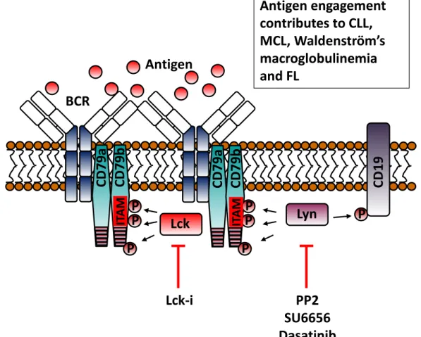
(CD79b) 13-15. Antigen binds to hypervariable regions present on the Ig component of the BCR causing receptor clustering and juxtapositioning into areas rich in signalling components16,17. Figure 1 illustrates the signalling networks activated following BCR stimulation. Upon antigen binding, two conserved tyrosine residues within the immuno-receptor tyrosine-based activation motifs (ITAMS) of CD79 become phosphorylated by Src-family kinases (SFK) such as Lyn (Figure 1A)18,19. Phosphorylated ITAMS act as docking sites for SH2 domains present in Syk, leading to its phosphorylation and subsequent activation (Figure 1B)20,21. SFK can also phosphorylate tyrosine residues outside the ITAM motifs of CD79 as well as tyrosine residues within the co-receptor protein CD19 (Figure 1A). Adaptor proteins such as BLNK, LAB and Nck are recruited to these extra-ITAM phosphotyrosine motifs in CD79 (Figure 1B)22-24. Active Nck recruits B-cell adapter for PI3K (BCAP) which becomes phosphorylated by Syk and attracts PI3Kδ via its p85α regulatory domain (Figure 1C)25,26. Additionally, the SFK-phosphorylated tyrosines within CD19 are additional binding sites for p85α and activation of PI3K (Figure 1C)27-29. However, it is suggested that PI3K can be activated independently of BCR engagement because the introduction of exogenous PI3K has been shown to rescue BCR negative cells from apoptosis30,31. A proposed mechanism for this rescue is the recruitment of PI3K to unphosphorylated motifs present on CD79a by the GTPase TC2132. Whatever the mechanism of activation, PI3Kδ converts PIP2 to PIP3 on
the plasma membrane, and, in doing so, generates a binding site for the pleckstrin homology (PH) domain of a collection of proteins normally residing in the cytosol of resting B-cells. Btk is one such protein, that additionally binds BLNK and becomes phosphorylated by Syk33, exposing an autophosphorylation site within Btk that culminates in its full activation (Figure 1C/D)34. PLCγ2 is also recruited to the plasma membrane via its PH domain and BLNK (Figure 1C/D), and is sequentially activated through phosphorylation by Btk and Syk35. Similarly, the Rac GTP-exchange factor Vav is also attracted to the BCR signalosome where it can be phosphorylated and activated by Syk (Figure 1C/E)36-38. Finally, PIP3 also attracts the kinases PDK1 and Akt to the plasma membrane (Figure 1C). Once
recruited, PDK1 phosphorylates Akt on T308 to increase the kinase activity of the latter. Stabilization of Akt kinase activity then occurs when it is phosphorylated on S473 by mTORC2 through a mechanism involving Akt-mediated phosphorylation of SIN139.
Distal BCR signalling
The proximal BCR signalling response quickly transcends to activate numerous signalling pathways summarised in Figure 1D. Active PLCγ2 is able to hydrolyse PIP2 to generate two
second messengers, IP3 and DAG. IP3 induces the release of Ca2+ from intracellular stores in
the endoplasmic reticulum (ER) and, together with DAG, this leads to the activation of PKCβ40. PKCβ is then able to mediate the stimulation of multiple downstream signalling pathways. For example, PKCβ together with DAG and Sos can generate active Ras (GTP-Ras), leading to activation of the cRaf-MEK-ERK pathway41-43. PKCβ can also phosphorylate CARMA1 (CARD11)44 to initiate the recruitment of MALT1 and Bcl-10, which forms the CBM complex45,46. This complex activates TAK1, a kinase that can catalyse the activation of both NFκB and JNK47. TAK1 activates NFκB by initially phosphorylating IKKβ47, which in turn phosphorylates IκBα, flagging it for ubiquitination and degradation by the proteasome48. This alleviates its inhibition of NFκB and allows it to translocate to the nucleus. Calmodulin (CaM) detects increased Ca2+ mobilisation from intracellular stores and activates the S/T phosphatase calcineurin (CN). CN dephosphorylates regulatory regions within NFAT causing
a conformational change that exposes its nuclear localisation sequence and results in its activation49.
The BCR signal can also be relayed to downstream signalling pathways independently of calcium release from the ER. As mentioned previously, generation of PIP3 by activation of
PI3Kδ causes the recruitment of Vav and generation of active GTP-loaded Rac1 and Rac2 (Figure 1E). These small G-proteins can facilitate F-actin reorganisation as well as activation of p38MAPK and JNK 24,50,51. Active Akt plays an important role in stimulating the mTORC1 pathway and in deactivating GSK3 and FoxO to facilitate increased cell growth and survival (Figure 1E)52-54. Collectively, these pathways are responsible for the control of a whole host of cellular functions such as; survival, proliferation, oxidative stress response, differentiation and DNA damage repair.
Negative regulation of BCR signalling
BCR signalling is highly regulated by a complex network of phosphatases, regulatory receptors and negative feedback signals in order to prevent its inappropriate activation. A primary example of this is how the SFK Lyn mediates the BCR signalling response. The protein tyrosine phosphatases (PTP) CD45 and CD148 dephosphorylate Y508 on the C-terminus of Lyn causing a conformational change that allows for its autophosphorylation and activation55,56. Then, depending on the context of the BCR signal, active Lyn can moderate the BCR signal by phosphorylating immuno-receptor tyrosine-based inhibitory motifs (ITIM) present on the regulatory receptors CD5, CD22, CD32 and CD7257-62. Phosphorylated ITIMs recruit phosphatases such as SHP-1, SHIP-1, SHIP-2, PTP-PEST, PTPN2, PTPROt, PTEN and PTPN22 which act on proximal targets to downregulate the BCR signalling response. Lyn controls its own inactivation by recruiting the phosphatase PTPN22, which forms a complex with the C-terminal Src kinase (Csk)63. Active Lyn binds and phosphorylates the Csk binding protein (Cbp) on Y314 and this serves to recruit Csk. PTPN22 then dephosphorylates the activating tyrosine residue on Lyn and allows Csk access to phosphorylate Lyn on Y508, resulting in the restoration of its closed inactive conformation64,65. Additionally, PTPN22 can regulate the stability of SFK and SYK by activating the E3-ubiquitin ligase c-Cbl, which ubiquitinylates these proteins and marks them for degradation66-68. Interestingly, SFK and Syk can recruit SOCS-1 which seems to augment their polyubiquitination and degradation by the proteasome65,69. However, the role of c-Cbl appears more complex than mere regulation of protein stability because it is reported to downregulate the BCR signal by binding to BLNK and preventing its association with PLCγ270. Other inhibitory receptors worth mentioning are those regulated by CD22 and CD32. Upon phosphorylation of their ITIMs by Lyn, PTEN and SHIP-1/2 respectively remove the 3’ and 5’ phosphate of PIP3 and attenuate signalling downstream of PI3K71-73. This regulatory
pathway is activated by prolonged exposure to autoantigens and forms the basis of auto-reactive B-cell elimination74. Lastly, the phosphatase SHP-1 has been reported to downregulate the BCR signal by dephosphorylating targets such as SFK, ITAMs, ZAP70, SYK, BLNK and LAB75.
Further regulation of the BCR signal is provided by negative feedback mechanisms that are independent of phosphatase activity. Following its activation, Btk can become phosphorylated on S180 by PKCβ activated by Btk-catalyzed stimulation of PLC2. This
disrupts the interaction of the PH domain of Btk with PIP3 and removes Btk from the plasma
membrane76. Furthermore, a recent report has demonstrated that Akt can also phosphorylate Btk on S51 and T495 which leads to its sequestration and degradation77. Another more distal example of negative feedback inhibition of BCR signalling is found in the regulation of NFκB by the CBM complex. It is reported that TAK1 can induce both kinase-independent and –dependent degradation of Bcl-10 by respectively coordinating the binding of E3 ubiquitin ligases and activating JNK78.
This meticulous regulation of the BCR allows it to constantly maintain its initiation, amplification, and termination under normal conditions. However, alteration in these pathways has the potential to transform B cells into an array of malignant diseases.
3.
BCR signalling in malignant B-cells
BCR signalling drives B-cell malignancies
It is widely accepted that signals emanating from the BCR play an important role in the initiation and progression of many B-cell malignancies. For example, gene expression profiling of chronic lymphocytic leukaemia cells (CLL) isolated from lymphatic tissues found that the BCR pathway was active in these cells to a greater degree than what is found in circulating CLL cells79. Furthermore, CLL cells use a restricted repertoire of IGHV genes, with many patients expressing virtually identical BCRs, known as sterotyped BCRs80-83. Similarly, mantle cell lymphoma (MCL) cells also express sterotyped BCR84,85, and display constitutive activation of BCR signalling components such as the PI3K pathway86,87. Other examples include diffuse large B-cell lymphoma (DLBCL)88, follicular lymphoma (FL)89, hairy cell leukaemia (HCL)90,91, Burkitts lymphoma (BL)92, Waldenström’s macroglobulinemia (WM)93, marginal zone lymphoma (MZL)94 and acute lymphoblastic leukaemia (ALL)95 cells which have all been shown to display some sort of defect in BCR signalling. However, the malignant cells in each of these diseases display distinct alterations in the BCR signalling pathway, reflecting defects originating from tonic/autonomous and/or chronic engagement of the BCR. What appears constant in many B-cell lymphomas is that IgM is the typical isotype of their BCR, probably due to the bias of this particular configuration of BCR towards stimulating survival and proliferation of B cells96. Below we will summarise some of the major aberrantly activated BCR signalling pathways found in different leukaemia and lymphomas, and highlight their importance in the initiation, survival and expansion of these malignant cells.
Chronic lymphocytic leukaemia (CLL)
CLL is a debilitating disease characterised by the gradual accumulation of mature B-cells that are resistant to apoptosis. The disease provides an excellent example of the prominent role BCR signalling plays in the pathogenesis of B-cell malignancies (Figure 1A). This role was determined from early studies of BCR structure in CLL cells which showed the genes coding for variable (antigen binding) regions of BCR heavy chain maintained germline sequences in approximately half of patients diagnosed with this disease. These patients with so-called unmutated CLL (UM-CLL) have disease which has less favourable prognosis than patients where these genes have been somatically hypermutated, so called mutated CLL (M-CLL)97. Other studies showed that BCRs on CLL cells from different patients could be virtually
identical with respect to IgVH genes and sequences, suggesting a common antigen or feature of the BCR that is involved in the pathology of this disease82. Common antigens targeted by BCR on CLL cells are reported to include epitopes associated with apoptosis and oxidation98, yeast/fungi cell wall components99, myosin100 and vimentin101, and BCR on CLL cells from UM-CLL patients are both polyreactive and responsive to BCR stimulation. In keeping with their ability to strongly respond to BCR engagement, UM-CLL cells generally have high expression and/or activation levels of many of the components of the BCR signalling pathway such as Syk102, Lyn, Btk, PLCγ2, PI3Kδ, GAB1, PTPN22103, PKCβ and NFκB. Furthermore, UM-CLL cells also generally express ZAP70104-106 and conflicting reports argue on one hand that this kinase mediates the phosphorylation of ITAM motifs and subsequent recruitment of Syk107, while others have shown that kinase dead ZAP70 can still enhance the BCR signalling response by acting as a scaffold protein108,109. Work from this Department has demonstrated that another kinase called Lck displays heterogeneous expression in CLL cells and is able to augment the BCR signalling response110. An important feature of BCR signalling in CLL cells that distinguishes it from other B cell malignancies is that it fails to activate the JNK pathway111, however, why this is the case requires further investigation. Direct engagement of the BCR is not the only way in which this receptor contributes to disease pathogenesis in CLL. Some BCR heavy chain structures are said to be represented stereotypically on CLL cells, and one study has demonstrated that particular regions, namely the FR2 and HCDR3, can interact to allow autonomous BCR signalling, particularly in CLL cells from UM-CLL patients, irrespective of antigen stimulation112.
In contrast to UM-CLL, CLL cells from patients with M-CLL express low surface IgM and show higher basal levels of Ca2+ and ERK activation consistent with constitutive low level stimulation of BCR113 resulting in induction of cell anergy114,115. Targeting either the ERK or the NF-AT pathway with specific inhibitors is reported to reduce the survival of anergic CLL cells, suggesting that BCR anergy contributes to clonal maintenance in M-CLL anergy114. Taken together, these findings in both UM- and M-CLL demonstrate that BCR signalling contributes to proliferation and survival of the malignant clone in CLL. However, the way in which BCR signalling is stimulated, be it through active engagement or through tonic/chronic/autonomous signalling may have impact on disease progression. CLL cells circulate between the peripheral blood (PB) and proliferation centres. Expansion of the malignant clone depends upon its ability to remain within proliferation centres such as the lymph node where there is a microenvironment rich in proliferation and survival signals113. BCR engagement on CLL cells induces increased adhesion116,117 and supressed expression of the receptor for sphingosine 1-phosphate118, both which lead to increased lymph node residency. This role of BCR in keeping CLL cells within lymph nodes has recently been exploited therapeutically where drugs targeting Btk (ibrutinib) and PI3K (idelalisib) induce lymphocytosis whilst reducing both lymphadenopathy and splenomegaly119,120.
Diffuse large B-cell lymphoma (DLBCL)
BCR signalling in a subset of DLBCL known as activated B-cell like (ABC)-DLBCL shows traits of active BCR signalling, and gene expression analysis revealed that a large amount of these cells display increased levels of BCR signalling components121. Furthermore, studies using internal reflection fluorescence (TIRF) microscopy found that the BCRs on ABC-DLBCL cells form clusters on the cell surface with proteins containing phosphotyrosine localising underneath88,122. Other studies investigating the mechanism of cell survival discovered that
these cells depend on anti-apoptotic signals provided by the NF-κB pathway123, found to be constitutively active in some cases of ABC-DLBCL owing to mutations in CARMA1124 (Figure 1D) and loss-of-function mutations in negative regulators of NF-κB125. However, malignant cells in the majority of ABC-DLBC contain wild type CARMA1 but still require this protein to maintain viability126. In these cases knockdown of BCR signalling units CD79a/b or any of its proximal signalling components (BLNK, Syk, Btk, PLCγ2, PI3K or PKCβ) induces apoptosis88. In particular, the malignant clones from approximately 20% of ABC-DLBCL cases bear mutations within the ITAM motifs of CD79a/b (Figure 1A) leading to reduced receptor endocytosis and increased surface expression of the BCR88. These cells have an enhanced BCR signalling response and are able to induce a greater and more prolonged activation of Akt. In particular, CD79b mutants are more resistant to moderation of the BCR signal by Lyn and consequently this may generate an oncogenic addiction to BCR-induced NF-κB activation88,127. Another protein that is frequently mutated in ABC-DLBCL is the adapter protein MYD88128 which is responsible for coupling toll-like receptors (TLR) to the NF-κB pathway. An L265P mutation in MYD88 was shown to augment autocrine secretion of the cytokines IL-6 and IL-10 resulting in increased ABC-DLBCL survival128. The relevance of such mutations to BCR-induced survival signals was highlighted when one group knocked down MYD88 in concert with CD79a/b, and found that this synergised to induce ABC-DLBCL cell killing128,129. Interestingly, this same mutation occurs in approximately 3% of CLL patients130, and MYD88 has recently been shown to work in concert with the BCR of ZAP70 positive CLL cells to stimulate cell survival and proliferation131.
Burkitt’s lymphoma (BL)
BL is an aggressive malignancy derived from germinal centre B-cells. A distinctive characteristic of this disease is a chromosomal translocation whereby the proto-oncogene MYC is linked to the heavy chain of BCR132. However, this alone is insufficient to cause BL, and studies in mice suggest that MYC works with active PI3K to generate the Burkett’s lymphoma phenotype133. In contrast to the chronically active BCR signal displayed in ABC-DLBCL, BL cells are resistant to BTK and CARMA1 knock down, but remain sensitive to a reduction in CD79a and Syk expression127,132. BCR ligation on BL cells induces activation of the PI3K pathway, and inhibition of this activation either by directly targeting PI3K or by targeting its downstream target mTORC1 kills these cells132. Tonic BCR signals are also present, and are the result of mutations within the transcription factor TCF3 (or E2A) or its negative regulator ID3132. TCF3 normally regulates the expression of the BCR and represses the levels of the phosphatase SHiP1. Thus, mutant forms found in BL lead to increased BCR expression and activation of the PI3K pathway.
Follicular lymphoma (FL)
FL cells are malignant germinal centre B-cells admixed with non-malignant cells such as T-cells, follicular dentritic cells (FDC), macrophages, fibroblasts and endothelial cells134,135. A hallmark of this disease is a t(14:18)(q32:q21) translocation resulting in an overexpression of Bcl-2 and resistance to apoptosis136. However, these cells also display high basal levels of active Syk, and resistance to apoptosis is at least partially mediated by PI3K activation137,138. It is clear that BCR plays a role in FL disease pathology139,140, autoreactivity is reported to be a feature of some FL clones141 and a more recent report has suggested BCR signals can be initiated within malignant cells by interaction of mannosyl residues on surface-expressed
BCR with lectins expressed on stromal cells in the tumour microenvironment (Figure 1A)142,143. Such antigen-independent signalling then drives expansion of the malignant clone. However, this is not the whole story because there also exist subclones that are unresponsive to BCR crosslinking due to elevated phosphatase activity142. Further investigation is required to understand the signalling mechanisms present in these latter clones, and how they contribute to disease.
Mantle cell lymphoma (MCL)
The malignant clone in MCL, like that in CLL, expresses a restricted repertoire of BCR genes, and it is thought that BCR signalling plays an important role in the survival of MCL cells and progression of disease (Figure 1A)144. A recent report found that BTK was constitutively activated in these cells and BCR crosslinking resulted in sustained activation of Syk. These kinases facilitated BCR-induced survival by mediating the secretion of autocrine factors and inducing adhesion to human bone marrow stromal cells (HMSCs) 145. Like in CLL, drugs targeting the BCR pathway such as ibrutinib and idelalisib have shown remarkable efficacy also in the treatment of MCL146.
4.
Targeting BCR signalling pathways
Proximal BCR signalling pathways Lyn inhibitors
Lyn is found overexpressed and constitutively active in many B cell malignancies where it contributes to high basal levels of tyrosine phosphorylated proteins. In CLL, this SFK is thought to contribute to malignant cell survival because experiments using PP2 and SU6656 [compounds which inhibit Lyn (Figure 1A)] induce apoptosis of CLL cells147. The prosurvival role of Lyn may be mediated by reported ability to deactivate procaspase-8 through phosphorylation-induced dimerization148, or by downregulating the tumour suppressor activity of PP2A149. Another study has suggested that Lyn plays an important role in CLL progression and lymphoid organ infiltration by malignant cells because of its ability to phosphorylate HS1 (hematopoietic cell-specific Lyn substrate-1) and regulate cytoskeletal functionality150. However, any conclusions drawn from these studies should be approached with caution because the drugs used to inhibit Lyn (PP2, SU6656 and dasatinib) also affect a broad range of SFKs when used at the concentrations reported147,151. Attempts to address the potential role of Lyn in CLL cells have used geldanamycin to disrupt Lyn’s association with HSP90. Although such treatment results in downregulation of Lyn expression and the induction of CLL cell apoptosis, mechanisms independent of Lyn may be inducing apoptosis because HSP90 regulates the stability of other proteins that may be important to the survival of CLL cells152. Nevertheless, a new study by Kim et al demonstrated that Lyn could be a promising target for treatment of bortezomib-resistant mantle cell lymphoma (BTZ-resistant MCL). In this study, treatment of these (BTZ-resistant cells with dasatinib caused inhibition of proliferation likely by preventing CD19 binding with Lyn and the p85 regulatory subunit of PI3K153. Similar observations were made when Lyn was depleted with siRNA, and therapeutic potential is strongly suggested by xenograft studies showing that tumour size in the mice injected with BTZ-resistant MCL cells can be significantly reduced by dasatinib treatment153. In humans there is at least one case report showing that a CLL patient with co-existing chronic myeloid leukaemia could be successfully treated with dasatinib154,
suggesting that general targeting of SFKs may be an effective approach. In this light, specific inhibition of Lyn may be a controversial target in the therapy of B cell malignancies155,156 because of its role as a positive and negative regulator of BCR signalling 157,158 through its ability to phosphorylate both ITAMs and ITIMs 155. Specific inhibition of Lyn may block negative feedback of BCR signalling and allow strong signals, perhaps mediated by other SFKs, to take precedence.
Lck inhibitors
An example of a SFK mediating BCR signalling in B cell malignancies is Lck. This SFK is classically expressed in T cells where it plays an important role in proximal antigen receptor (TCR) signalling. However, this kinase is also present in B1 B-lymphocytes and to participate in BCR signalling159. In CLL cells our work has shown that Lck is an important mediator of BCR signalling (Figure 1A), treatment of CLL cells with either a specific Lck inhibitor or Lck-specific siRNA resulted in inhibition of BCR-induced phosphorylation of IKK, ERK and Akt, and a reduction in BCR-mediated survival110. This same study also noted that constitutive activation of Lyn was not affected by the inhibitor compound used, indicating that Lck could be a potential target for treatment of CLL because of its proximal role in initiating BCR signalling. Work in this lab as well as in others has shown that CLL cells display heterogeneous Lck expression 160,161, and it is suggested that Lck induces CLL resistance to glucocorticoid-induced apoptosis 162. Although the level of Lck expression dictates the strength of BCR signalling, it is yet unclear how this relates to disease outcome.
Syk inhibitors:
Syk plays an important role in both autoimmune diseases and haematological malignancies
163
. Initial small molecule inhibitors targeting Syk were designed for the treatment of inflammatory diseases 164; however, pre-clinical studies suggested that Syk could also be a promising target for treatment of B cell malignancies 102,165-167. For example, Syk has been shown to be activated in 44% of DLBCL patient samples and responsible for regulating pathological BCR signalling 168, making Syk a valid potential target for the treatment of DLBCL. Syk was also shown to be constitutively active in peripheral B cells from a large number of CLL patients, and inhibition of this kinase in resulted in the induction of apoptosis through a mechanism involving downregulation of Mcl-1 protein levels102.
Fostamatinib is an orally available pro-drug of the active drug R406, and is a relatively selective Syk inhibitor 169. Treatment of CLL cells with fostamatinib resulted in a reduction in cell migration, adhesion and a moderate induction of apoptosis, achieved by an inhibition of both BCR and integrin signalling. 102,165,170. In-vivo studies in CLL and non-Hodgkin lymphoma mouse models supported the therapeutic potential of Syk inhibition in B cell malignancies
167,171
. In the clinic, 55% of CLL patients treated with fostamatinib achieved partial response compared to 22% of DLBCL, 11% of MCL and 10% in FL patients 172.
Cerdulatinib is a dual Syk/JAK kinase inhibitor that showed activity in both ABC- and GCB- types of DLBCL. This compound induced apoptosis in DLBCL as well as G1/S cell cycle arrest
173
. There are more selective Syk inhibitors currently under devolvement, namely entospletinib, PRT318 and P505-15. These compounds have shown encouraging results in pre-clinical studies in CLL 174and in DLBCL 168.
BTK inhibitors
BTK is a non-receptor protein tyrosine kinase that belongs to TEC family kinases and plays a crucial role in BCR signalling 175. Loss of function mutations within BTK result in X-linked agammaglobulinemia, a disorder characterised by the absence of mature B lymphocytes and immunodeficiency through lack of antibodies176. BTK is an important mediator of BCR-induced calcium flux, NF-B activation and B-cell proliferation, making it a central player in B cell physiology175. With respect to B cell malignancies, BTK knockdown studies in ABC-DLBCL cell lines have demonstrated this kinase is essential for tumour growth and cell survival88. Signalling through BTK is also essential for the survival and proliferation of the malignant B cells in Waldenström Macroglobulinemia (WM), a lymphoplasmacytic lymphoma, where mutation of MYD88 leads to induction of these signals177. Furthermore, BTK plays a key role in the malignant behaviour of CLL and MCL cells where chronic antigen-stimulation of the BCR facilitates disease progression144,178,179. Taken collectively, these studies have been used to justify targeting BTK as a valid therapeutic option to treat B-cell malignancies180.
Ibrutinib, a first-in-class inhibitor of BTK, is an orally available small molecule that covalently binds to Cys-481 and irreversibly blocks the kinase activity of BTK (Figure 1D). The effect of this blockade is observable also in CLL cells isolated from patients being administered this compound, these cells exhibit reduced levels of PLCγ and ERK phosphorylation, of expression of genes induced by BCR and NF-B pathways181. Studies of in-vitro models that resemble the tumour microenvironment and of in-vivo xenografts have shown that ibrutinib inhibits cell proliferation, survival and migration of CLL cells182,183. Ibrutinib has pleiotropic inhibitory effects allowing it to attenuate signalling induced by the BCR, CD40, BAFF, TLR and chemokine receptors. This indicates that the therapeutic effect of ibrutinib is likely due to regulation of microenvironmental influences rather than to direct induction of apoptosis. Ibrutinib is also shown to inhibit secretion of the chemokines CCL3 and CCL4 from CLL cells, and reduced serum levels of these chemokines is observed in CLL patients treated with this agent116,182,183. An interesting feature of most CLL and MCL patients treated with ibrutinib is that they show transient lymphocytosis due to egress of malignant cells from the protective microenvironment in the lymph nodes119,184-186, and this is likely due to inhibition of chemokine- and BCR-induced integrin 41-mediated adhesion116,117,187,188.
Clinical trials in relapse or refractory CLL patients have demonstrated high frequency of durable remission even in patients with high risk CLL (e.g. mutated TP53, del17p and del11q)
189
. Ibrutinib has been approved by the United States Food and Drug Administration (FDA) for the treatment of relapsed or refractory MCL and CLL184 and for WM190. However, resistance to ibrutinib in MCL and CLL has been described. One of the mechanisms of ibrutinib resistance is cysteine-to-serine mutation at C481 in the binding site of BTK191,192 as well as gain-of-function mutations within PLC2193. Many other reversible and irreversible BTK inhibitors are still under development to overcome such resistance to improve selectivity and tolerability180. Moreover, combination studies are ongoing in order to achieve better and more durable responses to ibrutinib190.
PI3K inhibitors
PI3K plays an important role in mediating signalling through the BCR, adhesion molecules and chemokine receptors and is therefore key to B cell survival, migration and proliferation194. There are four catalytic isoforms of PI3K: p110α, p110β, p110γ and p110δ.
With respect to B cells, the δ isoform is particularly important because PI3Kδ deletion or mutation in mice results in lack of B1 lymphocytes, lower numbers of mature B cells and impaired antibody production195. Importantly, PI3K expression is limited to lymphocytes and particular subsets of myeloid lineage cells such as mast cells and neutrophils, and this makes compounds that specifically target this isoform particularly attractive because the profile of toxicities will be less than what could be expected from pan PI3K inhibitors or inhibitors which target the more ubiquitously epressed and isoforms of this protein. In CLL, PI3Kδ has been shown to be constitutively active in the malignant cells196, and its inhibition reduces malignant cell viability making targeting of this PI3K isoform using small molecular inhibitors a potential treatment approach197. Idelalisib (formerly GS-1101 or CAL-101) is a highly selective inhibitor of PI3Kδ197 which has an efficacy that has allowed fast-track approval for the treatment of relapsed and refractory CLL in the U.S.A. (Figure 1C).198 Idelalisib is reported to inhibit the prosurvival effect of BCR stimulation in CLL cells199, and its cytotoxic effect is dose- and time-dependent being mediated by induction of the caspase pathway regardless of p53 or IgHV mutation status. Idelalisib treatment also leads to blocking several microenvironmental signals such as CD40L, TNFα, BAFF, ET1, fibronectin adhesion and nurse-like and stromal cell contact199-201, likely by also affecting cells such as NK and T cells which produce cytokines known to promote CLL cells survival and proliferation such as TNF-α, CD40L, IL-6 and interferon-γ200,202,203. Clinically, CLL patients treated with Idelalisib exhibit lymphocytosis due to redistribution of CLL cells from lymphoid tissues to peripheral blood204,205. This redistribution may be caused by reduction in PI3K-mediated cell adhesion and migration120,199, or by re-expression of the receptor for sphingosine-1 phosphate and induction of lymphocyte egress from lymph nodes/proliferation centres as has been recently suggested118. In terms of adverse drug reactions idelalisib has been linked to a characteristic set including, most notably, diarrhoea/colitis, increased liver transaminases, skin rash and pneumonitis. Very little is currently known about the biological basis of these adverse drug reactions because they affect only a subset of patients. Thus, more research is needed so that patients at risk of developing these adverse drug reactions can be identified, and methodologies to prevent them introduced.
Future perspectives
BCR pathway inhibitors have had remarkable success in the treatment of B-cell malignancies, particularly for patients displaying resistance to conventional chemotherapeutic agents. What is more, these small molecule inhibitors offer a less toxic alternative to conventional chemotherapy and only minor side effects are reported with their use. However, long term treatment with these molecules may prove more toxic than previously thought. For example, United States prescribing information for idelalisib contains a black box risk for serious/fatal diarrhoea198,206. Moreover, drug resistance of the type observed with the usage of ibrutinib is also a problem191-193. To combat the problems associated with toxicity, new generation drugs against PI3K, such as duvalisib, and BTK, such as acalabrutinib, are either in development or have been introduced into the clinic207,208. Furthermore, new targets can be developed within this pathway, and our work has suggested Lck as one target for the treatment of CLL110. E3 ubiquitin ligases are also potential targets, recent research has begun to reveal roles for these proteins in B lymphomagenesis209-211. Clearly, investigation into BCR signalling and compounds which
inhibit this pathway requires ongoing investigation, and it is likely that any inhibitors used will be employed within combinatorial therapeutic approaches in order to increase their efficacy.
References
1. Monroe JG. ITAM-mediated tonic signalling through pre-BCR and BCR complexes. Nat Rev Immunol. 2006;6(4):283-294. 10.1038/nri1808.
2. Kraus M, Alimzhanov MB, Rajewsky N, Rajewsky K. Survival of resting mature B lymphocytes depends on BCR signaling via the Igalpha/beta heterodimer. Cell. 2004;117(6):787-800. 10.1016/j.cell.2004.05.014.
3. Cariappa A, Pillai S. Antigen-dependent B-cell development. Curr Opin Immunol. 2002;14(2):241-249.
4. Pierce SK, Liu W. The tipping points in the initiation of B cell signalling: how small changes make big differences. Nat Rev Immunol. 2010;10(11):767-777. 10.1038/nri2853. 5. Corfe SA, Paige CJ. The many roles of IL-7 in B cell development; mediator of survival, proliferation and differentiation. Semin Immunol. 2012;24(3):198-208. 10.1016/j.smim.2012.02.001.
6. Goodnow CC, Crosbie J, Adelstein S, et al. Altered immunoglobulin expression and functional silencing of self-reactive B lymphocytes in transgenic mice. J Immunol. 2009;183(9):5442-5448.
7. Goodnow CC, Crosbie J, Jorgensen H, Brink RA, Basten A. Induction of self-tolerance in mature peripheral B lymphocytes. Nature. 1989;342(6248):385-391. 10.1038/342385a0. 8. Hartley SB, Crosbie J, Brink R, Kantor AB, Basten A, Goodnow CC. Elimination from peripheral lymphoid tissues of self-reactive B lymphocytes recognizing membrane-bound antigens. Nature. 1991;353(6346):765-769. 10.1038/353765a0.
9. Rickert RC. New insights into pre-BCR and BCR signalling with relevance to B cell malignancies. Nat Rev Immunol. 2013;13(8):578-591. 10.1038/nri3487.
10. Lam KP, Kuhn R, Rajewsky K. In vivo ablation of surface immunoglobulin on mature B cells by inducible gene targeting results in rapid cell death. Cell. 1997;90(6):1073-1083. 11. Niiro H, Clark EA. Regulation of B-cell fate by antigen-receptor signals. Nat Rev Immunol. 2002;2(12):945-956. 10.1038/nri955.
12. Bojarczuk K, Bobrowicz M, Dwojak M, et al. B-cell receptor signaling in the pathogenesis of lymphoid malignancies. Blood Cells Mol Dis. 2015;55(3):255-265. 10.1016/j.bcmd.2015.06.016.
13. Shaw AC, Mitchell RN, Weaver YK, Campos-Torres J, Abbas AK, Leder P. Mutations of immunoglobulin transmembrane and cytoplasmic domains: effects on intracellular signaling and antigen presentation. Cell. 1990;63(2):381-392.
14. Sanchez M, Misulovin Z, Burkhardt AL, et al. Signal transduction by immunoglobulin is mediated through Ig alpha and Ig beta. J Exp Med. 1993;178(3):1049-1055.
15. Williams GT, Peaker CJ, Patel KJ, Neuberger MS. The alpha/beta sheath and its cytoplasmic tyrosines are required for signaling by the B-cell antigen receptor but not for capping or for serine/threonine-kinase recruitment. Proc Natl Acad Sci U S A. 1994;91(2):474-478.
16. Ketchum C, Miller H, Song W, Upadhyaya A. Ligand mobility regulates B cell receptor clustering and signaling activation. Biophys J. 2014;106(1):26-36. 10.1016/j.bpj.2013.10.043. 17. Cheng PC, Cherukuri A, Dykstra M, et al. Floating the raft hypothesis: the roles of lipid rafts in B cell antigen receptor function. Semin Immunol. 2001;13(2):107-114. 10.1006/smim.2000.0302.
18. Stepanek O, Draber P, Drobek A, Horejsi V, Brdicka T. Nonredundant roles of Src-family kinases and Syk in the initiation of B-cell antigen receptor signaling. J Immunol. 2013;190(4):1807-1818. 10.4049/jimmunol.1202401.
19. Yao XR, Flaswinkel H, Reth M, Scott DW. Immunoreceptor tyrosine-based activation motif is required to signal pathways of receptor-mediated growth arrest and apoptosis in murine B lymphoma cells. J Immunol. 1995;155(2):652-661.
20. Kurosaki T, Johnson SA, Pao L, Sada K, Yamamura H, Cambier JC. Role of the Syk autophosphorylation site and SH2 domains in B cell antigen receptor signaling. J Exp Med. 1995;182(6):1815-1823.
21. Rowley RB, Burkhardt AL, Chao HG, Matsueda GR, Bolen JB. Syk protein-tyrosine kinase is regulated by tyrosine-phosphorylated Ig alpha/Ig beta immunoreceptor tyrosine activation motif binding and autophosphorylation. J Biol Chem. 1995;270(19):11590-11594. 22. Kabak S, Skaggs BJ, Gold MR, et al. The direct recruitment of BLNK to immunoglobulin alpha couples the B-cell antigen receptor to distal signaling pathways. Mol Cell Biol. 2002;22(8):2524-2535.
23. Castello A, Gaya M, Tucholski J, et al. Nck-mediated recruitment of BCAP to the BCR regulates the PI(3)K-Akt pathway in B cells. Nat Immunol. 2013;14(9):966-975. 10.1038/ni.2685.
24. Malhotra S, Kovats S, Zhang W, Coggeshall KM. Vav and Rac activation in B cell antigen receptor endocytosis involves Vav recruitment to the adapter protein LAB. J Biol Chem. 2009;284(52):36202-36212. 10.1074/jbc.M109.040089.
25. Leu CM. Nck, a missing adaptor between the B-cell receptor complex and the BCAP/PI3K/Akt pathway. Cell Mol Immunol. 2014;11(2):120-122. 10.1038/cmi.2013.53. 26. Okada T, Maeda A, Iwamatsu A, Gotoh K, Kurosaki T. BCAP: the tyrosine kinase substrate that connects B cell receptor to phosphoinositide 3-kinase activation. Immunity. 2000;13(6):817-827.
27. Carter RH, Doody GM, Bolen JB, Fearon DT. Membrane IgM-induced tyrosine phosphorylation of CD19 requires a CD19 domain that mediates association with components of the B cell antigen receptor complex. J Immunol. 1997;158(7):3062-3069. 28. Tuveson DA, Carter RH, Soltoff SP, Fearon DT. CD19 of B cells as a surrogate kinase insert region to bind phosphatidylinositol 3-kinase. Science. 1993;260(5110):986-989.
29. Buhl AM, Pleiman CM, Rickert RC, Cambier JC. Qualitative regulation of B cell antigen receptor signaling by CD19: selective requirement for PI3-kinase activation, inositol-1,4,5-trisphosphate production and Ca2+ mobilization. J Exp Med. 1997;186(11):1897-1910. 30. Srinivasan L, Sasaki Y, Calado DP, et al. PI3 kinase signals BCR-dependent mature B cell survival. Cell. 2009;139(3):573-586. 10.1016/j.cell.2009.08.041.
31. Cambier JC, Johnson SA. Differential binding activity of ARH1/TAM motifs. Immunol Lett. 1995;44(2-3):77-80.
32. Delgado P, Cubelos B, Calleja E, et al. Essential function for the GTPase TC21 in homeostatic antigen receptor signaling. Nat Immunol. 2009;10(8):880-888. 10.1038/ni.1749.
33. Baba Y, Hashimoto S, Matsushita M, et al. BLNK mediates Syk-dependent Btk activation. Proc Natl Acad Sci U S A. 2001;98(5):2582-2586. 10.1073/pnas.051626198. 34. Park H, Wahl MI, Afar DE, et al. Regulation of Btk function by a major autophosphorylation site within the SH3 domain. Immunity. 1996;4(5):515-525.
35. Kim YJ, Sekiya F, Poulin B, Bae YS, Rhee SG. Mechanism of B-cell receptor-induced phosphorylation and activation of phospholipase C-gamma2. Mol Cell Biol. 2004;24(22):9986-9999. 10.1128/MCB.24.22.9986-9999.2004.
36. Johmura S, Oh-hora M, Inabe K, et al. Regulation of Vav localization in membrane rafts by adaptor molecules Grb2 and BLNK. Immunity. 2003;18(6):777-787.
37. Fu C, Turck CW, Kurosaki T, Chan AC. BLNK: a central linker protein in B cell activation. Immunity. 1998;9(1):93-103.
38. Deckert M, Tartare-Deckert S, Couture C, Mustelin T, Altman A. Functional and physical interactions of Syk family kinases with the Vav proto-oncogene product. Immunity. 1996;5(6):591-604.
39. Yang G, Murashige DS, Humphrey SJ, James DE. A Positive Feedback Loop between Akt and mTORC2 via SIN1 Phosphorylation. Cell Rep. 2015;12(6):937-943. 10.1016/j.celrep.2015.07.016.
40. Newton AC. Protein kinase C: poised to signal. Am J Physiol Endocrinol Metab. 2010;298(3):E395-402. 10.1152/ajpendo.00477.2009.
41. Coughlin JJ, Stang SL, Dower NA, Stone JC. RasGRP1 and RasGRP3 regulate B cell proliferation by facilitating B cell receptor-Ras signaling. J Immunol. 2005;175(11):7179-7184.
42. Stone JC. Regulation and Function of the RasGRP Family of Ras Activators in Blood Cells. Genes Cancer. 2011;2(3):320-334. 10.1177/1947601911408082.
43. Roose JP, Mollenauer M, Ho M, Kurosaki T, Weiss A. Unusual interplay of two types of Ras activators, RasGRP and SOS, establishes sensitive and robust Ras activation in lymphocytes. Mol Cell Biol. 2007;27(7):2732-2745. 10.1128/MCB.01882-06.
44. Sommer K, Guo B, Pomerantz JL, et al. Phosphorylation of the CARMA1 linker controls NF-kappaB activation. Immunity. 2005;23(6):561-574. 10.1016/j.immuni.2005.09.014.
45. Shinohara H, Yasuda T, Aiba Y, et al. PKC beta regulates BCR-mediated IKK activation by facilitating the interaction between TAK1 and CARMA1. J Exp Med. 2005;202(10):1423-1431. 10.1084/jem.20051591.
46. Rosebeck S, Rehman AO, Lucas PC, McAllister-Lucas LM. From MALT lymphoma to the CBM signalosome: three decades of discovery. Cell Cycle. 2011;10(15):2485-2496.
47. Wang C, Deng L, Hong M, Akkaraju GR, Inoue J, Chen ZJ. TAK1 is a ubiquitin-dependent kinase of MKK and IKK. Nature. 2001;412(6844):346-351. 10.1038/35085597. 48. Suzuki H, Chiba T, Kobayashi M, Takeuchi M, Furuichi K, Tanaka K. In vivo and in vitro recruitment of an IkappaBalpha-ubiquitin ligase to IkappaBalpha phosphorylated by IKK, leading to ubiquitination. Biochem Biophys Res Commun. 1999;256(1):121-126. 10.1006/bbrc.1999.0296.
49. Crabtree GR, Olson EN. NFAT signaling: choreographing the social lives of cells. Cell. 2002;109 Suppl:S67-79.
50. Wang X, Zhang F, Chen F, et al. MEKK3 regulates IFN-gamma production in T cells through the Rac1/2-dependent MAPK cascades. J Immunol. 2011;186(10):5791-5800. 10.4049/jimmunol.1002127.
51. Minden A, Lin A, Claret FX, Abo A, Karin M. Selective activation of the JNK signaling cascade and c-Jun transcriptional activity by the small GTPases Rac and Cdc42Hs. Cell. 1995;81(7):1147-1157.
52. Gold MR, Scheid MP, Santos L, et al. The B cell antigen receptor activates the Akt (protein kinase B)/glycogen synthase kinase-3 signaling pathway via phosphatidylinositol 3-kinase. J Immunol. 1999;163(4):1894-1905.
53. Limon JJ, Fruman DA. Akt and mTOR in B Cell Activation and Differentiation. Front Immunol. 2012;3:228. 10.3389/fimmu.2012.00228.
54. Zhang X, Tang N, Hadden TJ, Rishi AK. Akt, FoxO and regulation of apoptosis. Biochim Biophys Acta. 2011;1813(11):1978-1986. 10.1016/j.bbamcr.2011.03.010.
55. Zhu JW, Brdicka T, Katsumoto TR, Lin J, Weiss A. Structurally distinct phosphatases CD45 and CD148 both regulate B cell and macrophage immunoreceptor signaling. Immunity. 2008;28(2):183-196. 10.1016/j.immuni.2007.11.024.
56. Yamaguchi H, Hendrickson WA. Structural basis for activation of human lymphocyte kinase Lck upon tyrosine phosphorylation. Nature. 1996;384(6608):484-489. 10.1038/384484a0.
57. Ravetch JV, Lanier LL. Immune inhibitory receptors. Science. 2000;290(5489):84-89. 58. Ono M, Bolland S, Tempst P, Ravetch JV. Role of the inositol phosphatase SHIP in negative regulation of the immune system by the receptor Fc(gamma)RIIB. Nature. 1996;383(6597):263-266. 10.1038/383263a0.
59. Wang J, Koizumi T, Watanabe T. Altered antigen receptor signaling and impaired Fas-mediated apoptosis of B cells in Lyn-deficient mice. J Exp Med. 1996;184(3):831-838.
60. Smith KG, Tarlinton DM, Doody GM, Hibbs ML, Fearon DT. Inhibition of the B cell by CD22: a requirement for Lyn. J Exp Med. 1998;187(5):807-811.
61. Maeda A, Kurosaki M, Ono M, Takai T, Kurosaki T. Requirement of SH2-containing protein tyrosine phosphatases SHP-1 and SHP-2 for paired immunoglobulin-like receptor B (PIR-B)-mediated inhibitory signal. J Exp Med. 1998;187(8):1355-1360.
62. Adachi T, Flaswinkel H, Yakura H, Reth M, Tsubata T. The B cell surface protein CD72 recruits the tyrosine phosphatase SHP-1 upon tyrosine phosphorylation. J Immunol. 1998;160(10):4662-4665.
63. Cloutier JF, Veillette A. Association of inhibitory tyrosine protein kinase p50csk with protein tyrosine phosphatase PEP in T cells and other hemopoietic cells. EMBO J. 1996;15(18):4909-4918.
64. Cloutier JF, Veillette A. Cooperative inhibition of T-cell antigen receptor signaling by a complex between a kinase and a phosphatase. J Exp Med. 1999;189(1):111-121.
65. Ingley E. Src family kinases: regulation of their activities, levels and identification of new pathways. Biochim Biophys Acta. 2008;1784(1):56-65. 10.1016/j.bbapap.2007.08.012. 66. Cohen S, Dadi H, Shaoul E, Sharfe N, Roifman CM. Cloning and characterization of a lymphoid-specific, inducible human protein tyrosine phosphatase, Lyp. Blood. 1999;93(6):2013-2024.
67. Sohn HW, Gu H, Pierce SK. Cbl-b negatively regulates B cell antigen receptor signaling in mature B cells through ubiquitination of the tyrosine kinase Syk. J Exp Med. 2003;197(11):1511-1524. 10.1084/jem.20021686.
68. Shao Y, Yang C, Elly C, Liu YC. Differential regulation of the B cell receptor-mediated signaling by the E3 ubiquitin ligase Cbl. J Biol Chem. 2004;279(42):43646-43653. 10.1074/jbc.M404082200.
69. Katkere B, Rosa S, Drake JR. The Syk-binding ubiquitin ligase c-Cbl mediates signaling-dependent B cell receptor ubiquitination and B cell receptor-mediated antigen processing and presentation. J Biol Chem. 2012;287(20):16636-16644. 10.1074/jbc.M112.357640.
70. Yasuda T, Maeda A, Kurosaki M, et al. Cbl suppresses B cell receptor-mediated phospholipase C (PLC)-gamma2 activation by regulating B cell linker protein-PLC-gamma2 binding. J Exp Med. 2000;191(4):641-650.
71. Mayo LD, Donner DB. The PTEN, Mdm2, p53 tumor suppressor-oncoprotein network. Trends Biochem Sci. 2002;27(9):462-467.
72. Brauweiler A, Tamir I, Dal Porto J, et al. Differential regulation of B cell development, activation, and death by the src homology 2 domain-containing 5' inositol phosphatase (SHIP). J Exp Med. 2000;191(9):1545-1554.
73. Vivier E, Daeron M. Immunoreceptor tyrosine-based inhibition motifs. Immunol Today. 1997;18(6):286-291.
74. Seda V, Mraz M. B-cell receptor signalling and its crosstalk with other pathways in normal and malignant cells. Eur J Haematol. 2015;94(3):193-205. 10.1111/ejh.12427.
75. Pao LI, Badour K, Siminovitch KA, Neel BG. Nonreceptor protein-tyrosine phosphatases in immune cell signaling. Annu Rev Immunol. 2007;25:473-523. 10.1146/annurev.immunol.23.021704.115647.
76. Kang SW, Wahl MI, Chu J, et al. PKCbeta modulates antigen receptor signaling via regulation of Btk membrane localization. EMBO J. 2001;20(20):5692-5702. 10.1093/emboj/20.20.5692.
77. Mohammad DK, Nore BF, Hussain A, Gustafsson MO, Mohamed AJ, Smith CI. Dual phosphorylation of Btk by Akt/protein kinase b provides docking for 14-3-3zeta, regulates shuttling, and attenuates both tonic and induced signaling in B cells. Mol Cell Biol. 2013;33(16):3214-3226. 10.1128/MCB.00247-13.
78. Moreno-Garcia ME, Sommer K, Rincon-Arano H, et al. Kinase-independent feedback of the TAK1/TAB1 complex on BCL10 turnover and NF-kappaB activation. Mol Cell Biol. 2013;33(6):1149-1163. 10.1128/MCB.06407-11.
79. Herishanu Y, Perez-Galan P, Liu D, et al. The lymph node microenvironment promotes B-cell receptor signaling, NF-kappaB activation, and tumor proliferation in chronic lymphocytic leukemia. Blood. 2011;117(2):563-574. 10.1182/blood-2010-05-284984.
80. Fais F, Ghiotto F, Hashimoto S, et al. Chronic lymphocytic leukemia B cells express restricted sets of mutated and unmutated antigen receptors. J Clin Invest. 1998;102(8):1515-1525. 10.1172/jci3009.
81. Tobin G, Thunberg U, Karlsson K, et al. Subsets with restricted immunoglobulin gene rearrangement features indicate a role for antigen selection in the development of chronic lymphocytic leukemia. Blood. 2004;104(9):2879-2885. 10.1182/blood-2004-01-0132.
82. Agathangelidis A, Darzentas N, Hadzidimitriou A, et al. Stereotyped B-cell receptors in one-third of chronic lymphocytic leukemia: a molecular classification with implications for targeted therapies. Blood. 2012;119(19):4467-4475. 10.1182/blood-2011-11-393694.
83. Messmer BT, Albesiano E, Efremov DG, et al. Multiple distinct sets of stereotyped antigen receptors indicate a role for antigen in promoting chronic lymphocytic leukemia. J Exp Med. 2004;200(4):519-525. 10.1084/jem.20040544.
84. Hadzidimitriou A, Agathangelidis A, Darzentas N, et al. Is there a role for antigen selection in mantle cell lymphoma? Immunogenetic support from a series of 807 cases. Blood. 2011;118(11):3088-3095. 10.1182/blood-2011-03-343434.
85. Thelander EF, Rosenquist R. Molecular genetic characterization reveals new subsets of mantle cell lymphoma. Leuk Lymphoma. 2008;49(6):1042-1049. 10.1080/10428190801947559.
86. Martinez N, Camacho FI, Algara P, et al. The molecular signature of mantle cell lymphoma reveals multiple signals favoring cell survival. Cancer Res. 2003;63(23):8226-8232.
87. Rizzatti EG, Falcao RP, Panepucci RA, et al. Gene expression profiling of mantle cell lymphoma cells reveals aberrant expression of genes from the PI3K-AKT, WNT and TGFbeta signalling pathways. Br J Haematol. 2005;130(4):516-526. 10.1111/j.1365-2141.2005.05630.x.
88. Davis RE, Ngo VN, Lenz G, et al. Chronic active B-cell-receptor signalling in diffuse large B-cell lymphoma. Nature. 2010;463(7277):88-92. 10.1038/nature08638.
89. Sachen KL, Strohman MJ, Singletary J, et al. Self-antigen recognition by follicular lymphoma B-cell receptors. Blood. 2012;120(20):4182-4190. 10.1182/blood-2012-05-427534.
90. Arons E, Roth L, Sapolsky J, Suntum T, Stetler-Stevenson M, Kreitman RJ. Evidence of canonical somatic hypermutation in hairy cell leukemia. Blood. 2011;117(18):4844-4851. 10.1182/blood-2010-11-316737.
91. Forconi F, Cencini E, Sicuranza A, Sozzi E, Lauria F. Molecular insight into the biology and clinical course of hairy cell leukemia utilizing immunoglobulin gene analysis. Leuk Lymphoma. 2011;52(1):15-23. 10.3109/10428194.2010.530362.
92. Schmitz R, Ceribelli M, Pittaluga S, Wright G, Staudt LM. Oncogenic mechanisms in Burkitt lymphoma. Cold Spring Harb Perspect Med. 2014;4(2). 10.1101/cshperspect.a014282.
93. Varettoni M, Zibellini S, Capello D, et al. Clues to pathogenesis of Waldenstrom macroglobulinemia and immunoglobulin M monoclonal gammopathy of undetermined significance provided by analysis of immunoglobulin heavy chain gene rearrangement and clustering of B-cell receptors. Leuk Lymphoma. 2013;54(11):2485-2489. 10.3109/10428194.2013.779689.
94. Yan Q, Huang Y, Watkins AJ, et al. BCR and TLR signaling pathways are recurrently targeted by genetic changes in splenic marginal zone lymphomas. Haematologica. 2012;97(4):595-598. 10.3324/haematol.2011.054080.
95. Melo JV. The diversity of BCR-ABL fusion proteins and their relationship to leukemia phenotype. Blood. 1996;88(7):2375-2384.
96. Dogan I, Bertocci B, Vilmont V, et al. Multiple layers of B cell memory with different effector functions. Nat Immunol. 2009;10(12):1292-1299. 10.1038/ni.1814.
97. Damle RN, Wasil T, Fais F, et al. Ig V gene mutation status and CD38 expression as novel prognostic indicators in chronic lymphocytic leukemia. Blood. 1999;94( 6):1840-1847. 98. Catera R, Silverman GJ, Hatzi K, et al. Chronic lymphocytic leukemia cells recognize conserved epitopes associated with apoptosis and oxidation. Mol Med. 2008;14(11-12):665-674. 10.2119/2008-00102.Catera.
99. Hoogeboom R, van Kessel KP, Hochstenbach F, et al. A mutated B cell chronic lymphocytic leukemia subset that recognizes and responds to fungi. J Exp Med. 2013;210(1):59-70. 10.1084/jem.20121801.
100. Chu CC, Catera R, Hatzi K, et al. Chronic lymphocytic leukemia antibodies with a common stereotypic rearrangement recognize nonmuscle myosin heavy chain IIA. Blood. 2008;112(13):5122-5129. 10.1182/blood-2008-06-162024.
101. Binder M, Lechenne B, Ummanni R, et al. Stereotypical chronic lymphocytic leukemia B-cell receptors recognize survival promoting antigens on stromal cells. PLoS One. 2010;5(12):e15992. 10.1371/journal.pone.0015992.
102. Gobessi S, Laurenti L, Longo PG, et al. Inhibition of constitutive and BCR-induced Syk activation downregulates Mcl-1 and induces apoptosis in chronic lymphocytic leukemia B cells. Leukemia. 2009;23(4):686-697. 10.1038/leu.2008.346.
103. Negro R, Gobessi S, Longo PG, et al. Overexpression of the autoimmunity-associated phosphatase PTPN22 promotes survival of antigen-stimulated CLL cells by selectively activating AKT. Blood. 2012;119(26):6278-6287. 10.1182/blood-2012-01-403162.
104. Lanham S, Hamblin T, Oscier D, Ibbotson R, Stevenson F, Packham G. Differential signaling via surface IgM is associated with VH gene mutational status and CD38 expression in chronic lymphocytic leukemia. Blood. 2003;101(3):1087-1093. 10.1182/blood-2002-06-1822.
105. Allsup DJ, Kamiguti AS, Lin K, et al. B-cell receptor translocation to lipid rafts and associated signaling differ between prognostically important subgroups of chronic lymphocytic leukemia. Cancer Res. 2005;65(16):7328-7337. 10.1158/0008-5472.CAN-03-1563.
106. Wiestner A, Rosenwald A, Barry TS, et al. ZAP-70 expression identifies a chronic lymphocytic leukemia subtype with unmutated immunoglobulin genes, inferior clinical outcome, and distinct gene expression profile. Blood. 2003;101(12):4944-4951. 10.1182/blood-2002-10-3306.
107. Chen L, Widhopf G, Huynh L, et al. Expression of ZAP-70 is associated with increased B-cell receptor signaling in chronic lymphocytic leukemia. Blood. 2002;100(13):4609-4614. 10.1182/blood-2002-06-1683.
108. Chen L, Huynh L, Apgar J, et al. ZAP-70 enhances IgM signaling independent of its kinase activity in chronic lymphocytic leukemia. Blood. 2008;111(5):2685-2692. 10.1182/blood-2006-12-062265.
109. Gobessi S, Laurenti L, Longo PG, Sica S, Leone G, Efremov DG. ZAP-70 enhances B-cell-receptor signaling despite absent or inefficient tyrosine kinase activation in chronic lymphocytic leukemia and lymphoma B cells. Blood. 2007;109(5):2032-2039. 10.1182/blood-2006-03-011759.
110. Talab F, Allen JC, Thompson V, Lin K, Slupsky JR. LCK is an important mediator of B-cell receptor signaling in chronic lymphocytic leukemia B-cells. Mol Cancer Res. 2013;11(5):541-554. 10.1158/1541-7786.MCR-12-0415-T.
111. Petlickovski A, Laurenti L, Li X, et al. Sustained signaling through the B-cell receptor induces Mcl-1 and promotes survival of chronic lymphocytic leukemia B cells. Blood. 2005;105(12):4820-4827. 10.1182/blood-2004-07-2669.
112. Duhren-von Minden M, Ubelhart R, Schneider D, et al. Chronic lymphocytic leukaemia is driven by antigen-independent cell-autonomous signalling. Nature. 2012;489(7415):309-312. 10.1038/nature11309.
113. Packham G, Krysov S, Allen A, et al. The outcome of B-cell receptor signaling in chronic lymphocytic leukemia: proliferation or anergy. Haematologica. 2014;99(7):1138-1148. 10.3324/haematol.2013.098384.
114. Apollonio B, Scielzo C, Bertilaccio MT, et al. Targeting B-cell anergy in chronic lymphocytic leukemia. Blood. 2013;121(19):3879-3888, S3871-3878. 10.1182/blood-2012-12-474718.
115. Muzio M, Apollonio B, Scielzo C, et al. Constitutive activation of distinct BCR-signaling pathways in a subset of CLL patients: a molecular signature of anergy. Blood. 2008;112(1):188-195. 10.1182/blood-2007-09-111344.
116. de Rooij MFM, Kuil A, Geest CR, et al. The clinically active BTK inhibitor PCI-32765 targets B-cell receptor- and chemokine-controlled adhesion and migration in chronic lymphocytic leukemia. Blood. 2012;119(11):2590-2594. 10.1182/blood-2011-11-390989. 117. Spaargaren M, Beuling EA, Rurup ML, et al. The B cell antigen receptor controls integrin activity through Btk and PLCγ2. The Journal of Experimental Medicine. 2003;198(10):1539-1550. 10.1084/jem.20011866.
118. Till KJ, Pettitt AR, Slupsky JR. Expression of functional sphingosine-1-phosphate receptor 1 is increased by idelalisib but not fostamatinib or ibrutinib in chronic lymphocytic leukaemia cells. J Immunol. 2015;194(5):2439 - 2446. Prepublished on Epub 2015 Jan 28 as DOI doi: 10.4049/jimmunol.1402304.
119. Woyach JA, Smucker K, Smith LL, et al. Prolonged lymphocytosis during ibrutinib therapy is associated with distinct molecular characteristics and does not indicate a suboptimal response to therapy. Blood. 2014;123(12):1810-1817. 10.1182/blood-2013-09-527853.
120. Fiorcari S, Brown WS, McIntyre BW, et al. The PI3-kinase delta inhibitor idelalisib (GS-1101) targets integrin-mediated adhesion of chronic lymphocytic leukemia (CLL) cell to endothelial and marrow stromal cells. PLoS One. 2013;8(12):e83830. 10.1371/journal.pone.0083830.
121. Monti S, Savage KJ, Kutok JL, et al. Molecular profiling of diffuse large B-cell lymphoma identifies robust subtypes including one characterized by host inflammatory response. Blood. 2005;105(5):1851-1861. 10.1182/blood-2004-07-2947.
122. Tolar P, Sohn HW, Liu W, Pierce SK. The molecular assembly and organization of signaling active B-cell receptor oligomers. Immunol Rev. 2009;232(1):34-41. 10.1111/j.1600-065X.2009.00833.x.
123. Davis RE, Brown KD, Siebenlist U, Staudt LM. Constitutive nuclear factor kappaB activity is required for survival of activated B cell-like diffuse large B cell lymphoma cells. J Exp Med. 2001;194(12):1861-1874.
124. Lenz G, Davis RE, Ngo VN, et al. Oncogenic CARD11 mutations in human diffuse large B cell lymphoma. Science. 2008;319(5870):1676-1679. 10.1126/science.1153629.
125. Thomas RK, Wickenhauser C, Tawadros S, et al. Mutational analysis of the IkappaBalpha gene in activated B cell-like diffuse large B-cell lymphoma. Br J Haematol. 2004;126(1):50-54. 10.1111/j.1365-2141.2004.05000.x.
126. Ngo VN, Davis RE, Lamy L, et al. A loss-of-function RNA interference screen for molecular targets in cancer. Nature. 2006;441(7089):106-110. 10.1038/nature04687.
127. Young RM, Staudt LM. Targeting pathological B cell receptor signalling in lymphoid malignancies. Nat Rev Drug Discov. 2013;12(3):229-243. 10.1038/nrd3937.
128. Ngo VN, Young RM, Schmitz R, et al. Oncogenically active MYD88 mutations in human lymphoma. Nature. 2011;470(7332):115-119. 10.1038/nature09671.
129. Lim KH, Yang Y, Staudt LM. Pathogenetic importance and therapeutic implications of NF-kappaB in lymphoid malignancies. Immunol Rev. 2012;246(1):359-378. 10.1111/j.1600-065X.2012.01105.x.
130. Puente XS, Pinyol M, Quesada V, et al. Whole-genome sequencing identifies recurrent mutations in chronic lymphocytic leukaemia. Nature. 2011;475(7354):101-105. 10.1038/nature10113.
131. Wagner M, Oelsner M, Moore A, et al. Integration of innate- into adaptive- immune responses in ZAP-70 positive chronic lymphocytic leukaemia. Blood. 2015. 10.1182/blood-2015-05-646935.
132. Schmitz R, Young RM, Ceribelli M, et al. Burkitt lymphoma pathogenesis and therapeutic targets from structural and functional genomics. Nature. 2012;490(7418):116-120. 10.1038/nature11378.
133. Sander S, Calado DP, Srinivasan L, et al. Synergy between PI3K signaling and MYC in Burkitt lymphomagenesis. Cancer Cell. 2012;22(2):167-179. 10.1016/j.ccr.2012.06.012. 134. Kridel R, Sehn LH, Gascoyne RD. Pathogenesis of follicular lymphoma. J Clin Invest. 2012;122(10):3424-3431. 10.1172/JCI63186.
135. Stevenson FK, Stevenson GT. Follicular lymphoma and the immune system: from pathogenesis to antibody therapy. Blood. 2012;119(16):3659-3667. 10.1182/blood-2011-11-367730.
136. Hiddemann W, Cheson BD. How we manage follicular lymphoma. Leukemia. 2014;28(7):1388-1395. 10.1038/leu.2014.91.
137. Leseux L, Hamdi SM, Al Saati T, et al. Syk-dependent mTOR activation in follicular lymphoma cells. Blood. 2006;108(13):4156-4162. 10.1182/blood-2006-05-026203.
138. Gulmann C, Espina V, Petricoin E, 3rd, et al. Proteomic analysis of apoptotic pathways reveals prognostic factors in follicular lymphoma. Clin Cancer Res. 2005;11(16):5847-5855. 10.1158/1078-0432.CCR-05-0637.
139. Zuckerman NS, McCann KJ, Ottensmeier CH, et al. Ig gene diversification and selection in follicular lymphoma, diffuse large B cell lymphoma and primary central nervous system lymphoma revealed by lineage tree and mutation analyses. Int Immunol. 2010;22(11):875-887. 10.1093/intimm/dxq441.
140. Bahler DW, Levy R. Clonal evolution of a follicular lymphoma: evidence for antigen selection. Proc Natl Acad Sci U S A. 1992;89(15):6770-6774.
141. Kuppers R. Mechanisms of B-cell lymphoma pathogenesis. Nat Rev Cancer. 2005;5(4):251-262. 10.1038/nrc1589.
142. Amin R, Mourcin F, Uhel F, et al. DC-SIGN-expressing macrophages trigger activation of mannosylated IgM B-cell receptor in follicular lymphoma. Blood. 2015;126(16):1911-1920. 10.1182/blood-2015-04-640912.
143. Linley A, Krysov S, Ponzoni M, Johnson PW, Packham G, Stevenson FK. Lectin binding to surface Ig variable regions provides a universal persistent activating signal for follicular lymphoma cells. Blood. 2015;126(16):1902-1910. 10.1182/blood-2015-04-640805.
144. Jares P, Colomer D, Campo E. Molecular pathogenesis of mantle cell lymphoma. J Clin Invest. 2012;122(10):3416-3423. 10.1172/JCI61272.
145. Bernard S, Danglade D, Gardano L, et al. Inhibitors of BCR signalling interrupt the survival signal mediated by the micro-environment in mantle cell lymphoma. Int J Cancer. 2015;136(12):2761-2774. 10.1002/ijc.29326.
146. Campo E, Rule S. Mantle cell lymphoma: evolving management strategies. Blood. 2015;125(1):48-55. 10.1182/blood-2014-05-521898.
147. Contri A, Brunati AM, Trentin L, et al. Chronic lymphocytic leukemia B cells contain anomalous Lyn tyrosine kinase, a putative contribution to defective apoptosis. J Clin Invest. 2005;115(2):369-378. 10.1172/JCI22094.
148. Zonta F, Pagano MA, Trentin L, et al. Lyn-mediated procaspase 8 dimerization blocks apoptotic signaling in B-cell chronic lymphocytic leukemia. Blood. 2014;123(6):875-883. 10.1182/blood-2013-02-485540.
149. Zonta F, Pagano MA, Trentin L, et al. Lyn sustains oncogenic signaling in chronic lymphocytic leukemia by strengthening SET-mediated inhibition of PP2A. Blood. 2015;125(24):3747-3755. 10.1182/blood-2014-12-619155.