the better approach
Demineralized Bone Matrix:
Development of a Process for Optimal Performance
the better approach
Introduction
MTF is a non-profit organization founded in 1987 by academic orthopaedic surgeons dedicated to providing tissue of the highest quality and safety for transplantation. Everything we do at MTF begins with safety. MTF has distributed almost 4.2 million allografts since our inception, and we have never experienced a case of viral disease transmission. MTF’s exemplary safety record is directly attributed to our commitment to the donor families and to the tissue recipients we serve. This tremendous commitment provides our customers with the assurance that this gift of human tissue is safe and that it comes from a trustworthy source.
We also think beyond safety. While safety governs every decision we make, we know that quality and efficacy also matter. Current techniques used by some tissue banks to clean, process and sterilize demineralized allografts have been shown to be detrimental to the quality of the tissue. These methods vary widely from bank to bank, because the industry donor criteria and processing standards are open to interpretation. Demineralized allograft tissue of less-than-optimal quality may yield a graft that does not perform its intended function, leading to a less-than-optimal clinical outcome.
Principles of Bone Healing and Graft
Incorporation
1Four components are necessary for bone healing and/or bone graft incorporation: the presence of host cells, a signal to trigger differentiation of the host cells to bone forming cells, a scaffold or matrix on which the new bone can form, and an adequate blood supply.
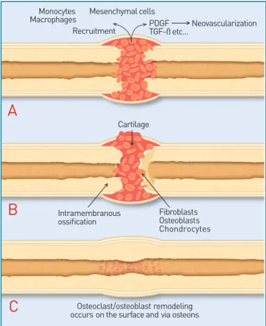
When bone fracture or injury occurs, there is loss of mechanical integrity of the bone and disruption to the blood supply. The healing cascade begins immediately. The three phases are: inflammation, repair and remodeling. Inflammation is the process by which host cells remove debris from the injured site, prepare the local matrix into a site which can support cell growth, and enable new bone to be formed. Revascularization, which is required for new bone to grow, begins in the inflammation phase. Repair includes the recruitment and differentiation of host cells into osteoblasts, which in turn produce new bone at the injured site. Lastly, remodeling is the resorption of immature or extraneous bone coupled with reorientation of bone along the direction of mechanical loading to provide adequate
structural support. These phases (Figure 1) are regulated
by the release of local cytokines.
Monocytes Mesenchymal cells Macrophages
Recruitment PDGFTGF-ß etc... Neovascularization
Fibroblasts Osteoblasts Chondrocytes Cartilage
A
B
C
Intramembranous ossification Osteoclast/osteoblast remodeling occurs on the surface and via osteonsFigure 1: Example of the healing cascade in fracture repair. The three phases of fracture repair include A) the inflammatory phase, B) the reparative phase, and C) the remodeling phase.1
New bone formation and bone healing are influenced by several factors in the bone graft material, some of which can be controlled, such as:
•
Bone forming potential•
Porosity•
pHHost factors are not as easily controlled:
•
Age•
Systemic disease•
Vascularity•
Presence of infection•
Quantity and quality of host cells•
Use of anti-inflammatory drugsBone grafts are often used as bone void fillers to assist with bone healing. Grafts can provide support or cell signals. During the healing process, the bone grafts are incorporated into the host bone by remodeling and/or creeping
substitution. Bone graft materials used as bone void fillers can be described as osteogenic, osteoinductive and/or osteoconductive.
Osteogenic tissues are capable of forming new bone from living cells. Osteoprogenitor cells proliferate and differentiate into osteoblasts (bone-building cells) and eventually into osteocytes (mature bone cells). These cells
represent the osteogenic potential of the graft.1
Osteoinductive tissues are ones which promote chemotaxis, mitogenesis and formation of osteoprogenitor cells
that have osteogenic capacity (as described above).2
Osteoinductive materials will form bone when implanted
into tissues which would not otherwise form new bone.3
Osteoconductive tissues allow for fibrovascular tissue development and osteoprogenitor cell invasion of a porous structure. This material then acts as a temporary scaffold
which will be replaced with newly formed bone.2
Rationale for Clinical Use of DBM
Autograft bone provides all three components necessary for bone healing. It has been widely used during bone grafting procedures due to availability of donor graft sites and good incorporation upon transplantation. However, the use of autograft bone requires a second surgical site (to procure bone) which has associated morbidity risks. Allogeneic demineralized bone matrix (DBM) eliminates the need for a second surgical site, and its demineralized state results in bone that is osteoconductive and has osteoinductive potential.
How DBM Works
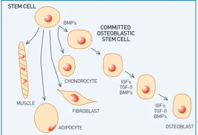
The exact mechanism of the osteoinductive potential of DBM has not been very well defined. However, it is thought that the removal of the mineral component of bone exposes the active bone morphogenetic proteins (BMPs) present in the DBM while retaining the inherent osteoconductive properties of the bone. When implanted, the active BMPs are thought to signal the host mesenchymal cells thus causing the cells to proliferate and differentiate into chondroblasts, which in turn will form a cartilage matrix. Ultimately, the cartilage matrix is converted into a calcified extracellular matrix. This calcified matrix will become vascularized, and osteoprogenitor cells will form new bone on this matrix. This will be followed by the formation of bone marrow and
marrow elements.3 STEM CELL COMMITTED OSTEOBLASTIC STEM CELL BMP’s chondrocyte adiPocyte fiBroBlast osteoBlast igf’s tgf-ß BMP’s igf’s tgf-ß BMP’s Muscle
Figure 2: Schematic showing differentiation of a stem cell into an active osteoblast, and demonstrating the influence of BMP-2.1
What Constitutes Good DBM
MTF has developed and validated a demineralization procedure for processing bone into DBM. This procedure provides safe, high-quality allograft DBM and was developed through rigorous testing to ensure that the osteoinductive potential of the tissue was not compromised.
Many factors contribute to the high quality of MTF DBM, including: • Quality tissue • Careful processing • A good carrier • Quality control • Scientific evidence
Quality Tissue
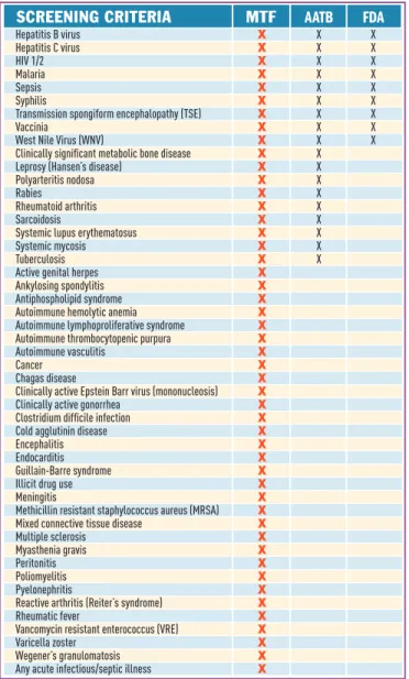
MTF’s quality and safety standards consistently meet or exceed the requirements of the American Association of Tissue Banks (AATB) as well as the guidelines for screening and testing of tissue donors set forth by the Food and Drug Administration (FDA). The AATB and FDA set only minimal guidelines to ensure safety of tissue. Potential MTF donors must pass through an extensive quality assurance process. Screening begins at the site of recovery with a comprehensive medical and social history that includes the cause of death. Tissue and blood samples are tested for infectious diseases, including hepatitis, HIV and syphilis. A team of medical/ technical specialists from the infectious disease and tissue banking fields evaluates all information, including test results, before the donor is released for processing. Figure 3 lists those areas in which MTF voluntarily defers donors for safety or quality reasons even when not required by the FDA or AATB.
the better approach
Screening criteria MTF AATB FDA
Hepatitis B virus X X X Hepatitis C virus X X X HIV 1/2 X X X Malaria X X X Sepsis X X X Syphilis X X X Transmission spongiform encephalopathy (TSE) X X X Vaccinia X X X West Nile Virus (WNV) X X X Clinically significant metabolic bone disease X X Leprosy (Hansen’s disease) X X Polyarteritis nodosa X X Rabies X X Rheumatoid arthritis X X Sarcoidosis X X Systemic lupus erythematosus X X Systemic mycosis X X Tuberculosis X X Active genital herpes X Ankylosing spondylitis X Antiphospholipid syndrome X Autoimmune hemolytic anemia X Autoimmune lymphoproliferative syndrome X Autoimmune thrombocytopenic purpura X Autoimmune vasculitis X Cancer X Chagas disease X Clinically active Epstein Barr virus (mononucleosis) X Clinically active gonorrhea X Clostridium difficile infection X Cold agglutinin disease X Encephalitis X Endocarditis X Guillain-Barre syndrome X Illicit drug use X Meningitis X Methicillin resistant staphylococcus aureus (MRSA) X Mixed connective tissue disease X Multiple sclerosis X Myasthenia gravis X Peritonitis X Poliomyelitis X Pyelonephritis X Reactive arthritis (Reiter’s syndrome) X Rheumatic fever X Vancomycin resistant enterococcus (VRE) X Varicella zoster X Wegener’s granulomatosis X Any acute infectious/septic illness X
Figure 3: Tissue bank screening criteria. Note: MTF voluntarily exceeds both AATB and FDA screening criteria.
Careful Processing
To maintain biological integrity, MTF processes all tissue using aseptic techniques in ISO Class 4 (certified) clean rooms. MTF’s use of these clean rooms is designed to prevent any environmental contamination of the tissue and thus eliminates the need for terminal sterilization by high-dose gamma radiation, which has been shown to compromise the biological and biomechanical integrity of allograft tissue (Figure 4).4,5
160 ———––––––––—---——————————————————— 140 ———––––––––—---——————————————————— 120 ———––––––––—---——————————————————— 100 ———––––––––—---——————————————————— 80 ———––––––––—---——————————————————— 60 ———––––––––—---——————————————————— 40 ———––––––––—---——————————————————— 20 ———––––––––—---——————————————————— 0 ———––––––––————————---———————————— 1 2 3 dBM lot n 0 kGy n 12 kGy n18 kGY n 25 kGY al P mmol/mg pr ot ein/min
Figure 4: Gamma radiation decreases osteoinductive potential of DBM powder in a dose-dependent manner, as measured using an in vitro alkaline phosphatase (ALP) assay. 5
For example, in an experimental study where DBX®
Putty was exposed to a terminal gamma radiation dose of 17.2 kGy, the osteoinductive potential was reduced by
approximately 50% (Figure 5).6 2.5 ———––––––––—---——————————————————— 2 ———––––––––—---——————————————————— 1.5 ———––––––––—---——————————————————— 1 ———––––––––—---——————————————————— 0.5 ———––––––––—---——————————————————— 0 ———––––––––————————---———————————— donor 1 donor 2 donor 3
n 0 kGy n 17 kGy os teoinductivity sc or e
Figure 5: Gamma radiation decreases the osteoinductive potential of DBX Putty using the in vivo athymic mouse assay. 6
A Good Carrier
DBM in its native form is a fine powder and as such is difficult to deliver to an operative site. DBM formulations should be designed to resist movement under irrigation while maintaining the physical integrity of the tissue form. Also, ideal DBM formulations should be provided ready
for use and not require any mixing or thawing prior to use. Therefore, MTF adds a carrier material to the demineralized bone to improve cohesion and provide an implant with good handling characteristics.
The carrier used in DBX is sodium hyaluronate which has been proven to be extremely safe. MTF uses high-quality, medical grade sodium hyaluronate, ISO 13485 certified, produced through fermentation processes using Good Manufacturing Practice (GMP) guidelines. It is designed to be isotonic and non-hemolytic. The sodium hyaluronate used in DBX tissue is not of animal origin.
Sodium hyaluronate was chosen because it is a polysaccharide formed by plasma membrane proteins. It is naturally
occurring in the human body in the joints, eyes, extracellular matrix of skin and musculoskeletal tissue. Sodium
hyaluronate plays an essential role in cell proliferation, migration and adhesion and has been correlated to
angiogenesis.7,8 It also confers positional stability of the tissue.9
Formulations
DBX is a demineralized bone matrix that has osteoinductive potential and is osteoconductive. It is composed of
demineralized bone from human donors in a biocompatible carrier. The various DBX forms have been designed to meet surgical needs while maximizing the amount of bone delivered to the surgical site.
The demineralized bone powder is produced by first milling
cortical bone to the appropriate size (212 – 850µm), followed
by removal of the minerals from the cortical bone via an acid extraction procedure. The demineralized milled cortical bone is then rinsed and buffered to ensure neutral pH. Lastly, the demineralized bone is lyophilized (residual moisture content < 6%) to ensure stability during staging prior to mixing with appropriate carriers.
DBX Demineralized Bone Matrix is non-hemolytic, ensuring compatibility with the surrounding autogenous blood cells. The DBX formulations have been specifically designed to model the pH of human blood.
DBX Paste
DBX Paste provides a flowable consistency of granulated cortical bone in sodium hyaluronate. The bone content of DBX Paste is 26% bone (weight/weight).
DBX Putty
DBX Putty provides a moldable consistency of granulated cortical bone in sodium hyaluronate. The bone content of DBX Putty is 31% (weight/weight).
DBX Mix
DBX Mix provides a morselized cortical-cancellous bone texture in sodium hyaluronate, which eliminates the need to combine bone chips with a DBM. The bone content of DBX Mix is 35% (weight/weight).
DBX Pre-Formed Shapes
Certain carriers allow MTF to pre-shape the DBM tissue form into predetermined sizes and shapes. For example, DBX Strip provides a cohesive and flexible preformed DBM combined with sodium hyaluronate and gelatin. The bone content of DBX Strip is 45% (weight/weight).
DBX Strip differs slightly from other DBX forms since, in addition to sodium hyaluronate, its carrier includes porcine gelatin to give it unique handling characteristics. The combination of demineralized bone, gelatin, and sodium hyaluronate results in a pre-formed strip formulation.
Quality Control
Every lot of DBX undergoes strict release criteria testing. Sterility is tested per USP <71>. Each lot is also tested via an in vitro or in vivo assay for osteoinductive potential.
DBM Processing
MTF’s demineralized bone matrices are produced from both cancellous and cortical bone which are subjected to controlled cleaning processes (including hydrogen peroxide and ethanol), followed by demineralization with hydrochloric acid. MTF’s aseptic processes have been designed to preserve the biological integrity of the tissue. Each donor lot of DBM is verified for osteoinductive potential prior to distribution.
Osteoinductivity Testing
Osteoinductive materials have the ability to induce new bone formation even when implanted into non-bony tissue such as muscle. When bone is formed in this manner, the material is said to have osteoinductive potential. Osteoinductive potential can be measured using either in vivo or in vitro test methods. MTF utilizes three methods to measure DBM osteoinductive potential prior to distribution: 1) athymic mouse muscle pouch assay, 2) alkaline phosphatase assay, or 3) BMP-2 content assay.
the better approach
Athymic Mouse Assay
The athymic mouse assay for in vivo osteoinductive
potential is based upon the Urist10 model and is designed to
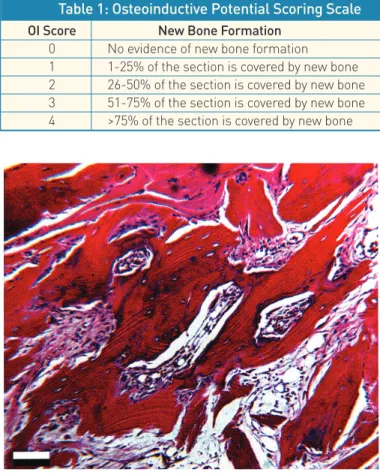
histologically confirm that DBX tissues have osteoinductive potential prior to distribution. In this model, implantation of demineralized bone in the hamstring muscle pouch of an athymic mouse will result in ectopic bone formation if the bone has osteoinductive potential. Athymic mice are used as they lack a thymus gland, and therefore are unable to mount an immune response against the human tissue. New bone formation is measured histologically after 28 days implantation (minimum) and scored using the scale in Table 1. This score is expressed as percent of new bone
formation in the visualized area. Figure 6 displays a
representative histological section used for scoring osteoinductive potential.
Table 1: Osteoinductive Potential Scoring Scale
OI Score New Bone Formation
0 No evidence of new bone formation
1 1-25% of the section is covered by new bone
2 26-50% of the section is covered by new bone
3 51-75% of the section is covered by new bone
4 >75% of the section is covered by new bone
Figure 6: Example of histological section used to grade osteoinductivity in the athymic mouse Bar = 100 micron.
Alkaline Phosphatase Assay
Alkaline phosphatase (ALP) is an enzyme which is
expressed by bone growing cells during new bone formation. Demineralized bone will activate these bone growing cells, thus resulting in increased levels of the ALP enzyme. In vitro test methods have been developed whereby bone growing cells are exposed to DBM and the resulting level of in vitro ALP enzyme is then measured. Correlations between the ALP levels and in vivo osteoinductivity scores from the athymic mouse model demonstrate that the ALP assay can be used as another means of measuring the osteoinductive potential of
DBM tissue forms.11
BMP-2 Content Assay
In vitro measurement via Enzyme-Linked ImmunoSorbent Assay (ELISA) of BMP-2 levels in DBM can be used as a surrogate measurement for the in vivo osteoinductive potential of DBX. Bone induction is a sequential, multi-step cascade which involves various growth factors. Bone morphogenetic proteins (and other intrinsic growth factors) in bone are exposed by the demineralization process. However, BMP-2 has been shown to be the best single predictor of osteoinductive potential based on statistical analysis of the correlation between in vitro levels of various growth factors and in vivo osteoinductivity.12,13
ELISA is a quick and quantitative screening technique that can be used in place of methods with animal variability or a subjective, semi-quantitative scoring system. Therefore, measurement of BMP-2 level in DBM using ELISA techniques can be used as a surrogate in vitro test for evaluating
osteoinductive potential of DBM.
There is a strong correlation (R2 = 0.72) between in vitro ELISA results on DBM and in vivo osteoinductive potential of DBX Putty in the athymic mouse model.
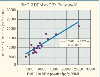
BMP-2 quantified in the final formulation of DBX Putty was found to be of equivalent levels as in the respective DBM powder (from the same donor lots). The good correlation
(R2 = 0.82) between BMP-2 levels in DBX Putty and DBM
powder (n=18, Figure 7), demonstrates that the addition of sodium hyaluronate to DBM powder does not alter intrinsic growth factor levels.14
Additionally, the strong correlation between BMP-2 levels and in vivo osteoinductive potential for DBM in sodium hyaluronate (R2 = 0.72) and DBM in saline (R2 = 0.78) demonstrates that there is no significant change in BMP-2 levels or in vivo osteoinductive potential of DBM after mixing DBM and the
sodium hyaluronate carrier into the final DBX tissue form.14
Taken in its entirety, this data supports the validity of the BMP-2 ELISA assay as a batch release test method for OI.
BMP-2 DBM vs DBX Putty (n=18) 30000 ––––––––——————————––– —--——---——————— 25000 ––––––––—————————–––— —--——---——————— 20000 ––––––––————---—–––––—---—————————————— 15000 ––––––––————---—–––––—---—————————————— 10000 ––––––––————---—–––––—---—————————————— 5000 ––––– –––————---—–––––—---—————————————— 0 –––––––————---—————--————————— 0 5000 10000 15000 20000 25000 30000 BMP-2 in dBM powder (pg/g dBM) y=-0.7909 x + 2391.6 r2=0.8201 B M P -2 in d B M P ut ty (p g/ g d B M )
Figure 7: Correlation of BMP-2 levels in DBM powder and DBX Putty (R2 = 0.82) from the same lots (n=18 donor lots).
In summary, each lot of DBX is validated using one or more of the following: the Urist athymic mouse model, the in vitro BMP-2 test, or the in vitro ALP assay.
Scientific Evidence
IndicationsDBX is intended for use in voids or gaps that are not intrinsic to the stability of the bony structure. It is intended for treatment of surgically or traumatically created osseous defects of the posterolateral spine, pelvis, and extremities. Additionally, DBX Putty and Paste are intended for the augmentation of deficient maxillary and mandibular alveolar ridges and the treatment of oral/maxillofacial and dental intra-osseous defects.
DBX is for single patient use only.
Clinical Studies
Numerous studies have demonstrated the clinical effectiveness of MTF’s DBX Demineralized Bone Matrix Tissue forms.
Study 1: DBX Putty plus autograft has been shown to be as effective as autograft alone.15
A mix of DBX Putty and autograft was shown to be as effective as autograft alone in a multicenter, randomized, controlled clinical study of patients undergoing
posterolateral spine fusion for the treatment for degenerative disc disease (DDD).
•
Equivalent fusion rates were achieved between thetwo groups at 24 months.
• DBX and autograft = 100% at 24 mo.
• Autograft alone = 96% at 24 mo.
•
Both groups exhibited significant decrease in VASpain scores at 6 months relative to baseline that is sustained through 24 months.
•
The authors conclude that DBX Putty is at least aseffective as autograft alone when used as a bone graft extender in the treatment of DDD via posterolateral spine fusion.
Study 2: DBX Putty plus local bone and bone marrow aspirate has been shown to be as effective as
autograft alone.16
Results indicate that DBX is as effective as autograft alone when used as a bone graft extender when combined with bone marrow aspirate from the iliac crest and local bone in the treatment of degenerative disc disease.
•
Equivalent fusion rates were achieved at 12-monthspost-surgery:
• Iliac crest autograft = 97.6%
• DBX Putty + local bone + iliac crest bone marrow
aspirate = 93.9%
•
At 12 months, postoperative VAS pain scores improved41.7% for the autograft control group and 53.1% in the DBX Putty + local bone and bone marrow aspirate group.
•
Oswestry disability index rates improved 51.5% for theautograft control group and 53.5% in the DBX Putty + local bone and bone marrow aspirate group.
•
Representative 12-month radiographs are shown inFigure 8.
Figure 8: A-P and Lateral radiographs at 12-months
demonstrated DBX Putty + local bone and bone marrow aspirate is as effective as autograft alone.16
the better approach
Study 3: DBX Putty effectively treats periodontalintraosseous defects.17
Intraosseous periodontal defects in systemically healthy patients were treated with either DBX Putty or with demineralized freeze-dried bone allograft (DFDBA). Only
sites with defects ≥ 3 mm in depth were considered.
A statistical analysis was conducted at 6 months
postoperative. Both groups of patients yielded significant improvements in percent bone fill, with 37% ± 18.5% bone fill achieved in patients treated with DFDBA and 50% ± 25% in cases of patients treated with DBX Putty.
Summary
At MTF, we are driven by our strong commitment to safety. It is because of this commitment that we continue to maintain an exemplary safety record, providing our customers with allograft tissue from a source they can trust. As part of our philosophy, we believe that providing safe tissue is not enough—we also must not compromise the biological properties of the demineralized bone. MTF’s processes for bone demineralization and creation of DBM tissue forms have been designed and validated to ensure the safety of the allografts without adversely affecting their biological performance. The processes and studies described here demonstrate that MTF’s demineralization processes and use of sodium hyaluronate in DBX have no harmful effects on the natural biological properties or in vivo performance of the allografts.
The demineralization process and additionally the use of sodium hyaluronate in DBX by MTF yields a safe, effective allograft designed and validated to maintain the natural healing function of allograft bone.
References
1. Mehta S, Collings C. Orthobiologics: Improving Fracture Care Through Science: Lippincott Williams & Wilkins; 2007.
2. Mouch CS, Einhorn TA. Bone Morphogenetic Proteins
and Other Growth Factors to Enhance Fracture Healing and Treatment of Nonunions. In: Lieberman JR,
Friedlander GE, editors. Bone Regeneration and Repair: Biology and Clinical Applications: Humana Press; 2005. p 169-194.
3. Boyan BB, McMillan J, Lohman CH, Ranly DM,
Schwartz Z. Bone Graft Substitutes: Basic Information for Successful Clinical Use with Special Focus on Synthetic Graft Substitutes. In: Laurencin CT, editor. Bone Graft Substitutes: ASTM; 2003. p 231-259.
4. Vangsness CT, Jr., Wagner PP, Moore TM, Roberts MR.
Overview of Safety Issues Concerning the Preparation and Processing of Soft-Tissue Allografts. Arthroscopy 2006; 22(12):1351-8.
5. Han B, Yang Z, Nimni M. Effects of Gamma Irradiation
on Osteoinduction Associated with Demineralized Bone Matrix. J Orthop Res 2008; 26(1):75-82.
6. Gertzman AA, Sunwoo M, Raushi D, Dunn M. The Effect
of Cold Gamma Radiation Sterilisation on the Properties of Demineralised Bone Matrix. In: Kennedy JF, Philips GO, Williams PA, editors. Sterilisation of tissues using ionising radiations: CRC Press; 2005. p 151-156.
7. Fraser JR, Laurent TC. Turnover and Metabolism in
Hyaluronan. In: Evered D, Whelan J, editors. Biology of Hyaluronan: Wiley; 1989. p S41-S59.
8. Orlidge A, D’Amore P. Cell Specific Effects of
Glycosaminoglycans on the Attachment and Proliferation of Vascular Wall Components. Microvasc Res
1986;31:S41-S43.
9. Gertzman A, Sunwoo M. A Pilot Study Evaluating Sodium Hyaluronate as a Carrier for Freeze-Dried Demineralized
Bone Powder.Cell Tissue Bank 2001;2:S87-S94.
10. Urist MR. Bone: Formation by Autoinduction. Science
1965; 150(698):893-9.
11. Han B, Tang B, Nimni ME. Quantitative and Sensitive In
Vitro Assay for Osteoinductive Activity of Demineralized Bone Matrix. J Orthop Res 2003; 21(4):648-54.
12. Blum B, Moseley J, Miller L, Richelsoph K, Haggard W. Measurement of Bone Morphogenetic Proteins and Other Growth Factors in Demineralized Bone Matrix. Orthopedics 2004;27(1 Suppl):s161-5.
13. Murray SS, Brochmann EJ, Harker JO, King E, Lollis RJ, Khaliq SA. A Statistical Model to Allow the Phasing out of the Animal Testing of Demineralised Bone Matrix Products. Altern Lab Anim 2007; 35(4):405-9.
14. Chnari E, Javoroncov M, Gertzman AA, Sunwoo MH,
Dunn MG. Bone Morphogenetic Protein 2 (BMP-2) Levels are Predictive of the Osteoinductive Potential of
Demineralized Bone Matrix.ORS, 2010.35: 485.
15. Becker VV, Nardone E, Robbins S, Anderson DG, Maiman D, Fry M, McDonnell D, Bethel A, Jones D. Comparison of Autograft to DBX Demineralized Bone Matrix Putty Combined with Autograft using
Posterolateral Lumbar Fusion.Spine 2005;5(4):S56-57.
16. Dube M, Luckett M. Comparison of Iliac Crest Autograft to DBX Demineralized Bone Matrix Putty Combined with Local Autograft and Iliac Aspirate used in Posterolateral
Spinal Fusion.Spine 2007;6(5):117S.
17. Bender SA, Rogalski JB, Mills MP, Arnold RM, Cochran
DL, Mellonig JT. Evaluation of Demineralized Bone Matrix Paste and Putty in Periodontal Intraosseous Defects. J Periodontol 2005; 76(5):768-77.