Submitted21 June 2018 Accepted 7 October 2018 Published14 November 2018 Corresponding author Jevrosima Stevanovic, rocky@vet.bg.ac.rs Academic editor Joseph Gillespie
Additional Information and Declarations can be found on page 13
DOI10.7717/peerj.5887
Copyright 2018 Cirkovic et al. Distributed under
Creative Commons CC-BY 4.0
OPEN ACCESS
Honey bee viruses in Serbian colonies of
different strength
Dragan Cirkovic1,*, Jevrosima Stevanovic2, Uros Glavinic2, Nevenka Aleksic3,
Spomenka Djuric4, Jelena Aleksic5and Zoran Stanimirovic2,*
1Department of Chemical and Technological Sciences, State University of Novi Pazar, Novi Pazar, Serbia 2Department of Biology, Faculty of Veterinary Medicine, University of Belgrade, Belgrade, Serbia 3Department of Parasitology, Faculty of Veterinary Medicine, University of Belgrade, Belgrade, Serbia 4Department of Economics and Statistics, Faculty of Veterinary Medicine, University of Belgrade, Belgrade,
Serbia
5Institute of Molecular Genetics and Genetic Engineering (IMGGE), University of Belgrade, Beograd, Serbia
*These authors contributed equally to this work.
ABSTRACT
Protection of honey bees is of great economic importance because of their role in pollination. Crucial steps towards this goal are epidemiological surveys of pathogens connected with honey bee losses. In this study deformed wing virus (DWV), chronic bee paralysis virus (CBPV), acute bee paralysis virus (ABPV) and sacbrood virus (SBV) were investigated in colonies of different strength located in five regions of Serbia. The relationship between colony strength and virus occurrence/infection intensity were assessed as well as the genetic relationship between virus sequences from Serbia and worldwide. Real-time RT-PCR analyses detected at least one virus in 87.33% of colonies. Single infection was found in 28.67% colonies (21.33%, 4.00%, 2.67% and 0.67% in cases of DWV, ABPV, SBV and CBPV, respectively). In the majority of colonies (58.66%) more than one virus was found. The most prevalent was DWV (74%), followed by ABPV, SBV and CBPV (49.30%, 24.00% and 6.70%, respectively). Except for DWV, the prevalence of the remaining three viruses significantly varied between the regions. No significant differences were found between colony strength and either (i) the prevalence of DWV, ABPV, SBV, CBPV and their combinations, or (ii) DWV infection levels. The sequences of honey bee viruses obtained from bees in Serbia were 93–99% identical with those deposited in GenBank.
SubjectsBiodiversity, Entomology, Veterinary Medicine, Virology
Keywords Apis mellifera, Viruses, Colony strength, Phylogeny, Beekeeping, Bee pathology
INTRODUCTION
Honey bees (Apis mellifera) are well-known beneficial insects for their popular products, and much more for their important role in pollination (Venturini et al., 2017). Unfortunately, huge losses of managed honey bee colonies were reported worldwide (Van Engelsdorp et al., 2008;Van Engelsdorp et al., 2009;Bacandritsos et al., 2010;Van Engelsdorp et al., 2012;Lee et al., 2015;Antúnez et al., 2016;Kulhanek et al., 2017;Brodschneider et al., 2018), but no single factor was confirmed to be a certain cause of colony mortality (Van
most often been cited (Francis, Nielsen & Kryger, 2013;McMenamin & Genersch, 2015;
Steinhauer et al., 2018).
More than 22 honey bee viruses have been identified and described so far (Genersch, 2010) which exist or co-exist in individual bees or colonies, but may remain unnoticed (Chen & Siede, 2007;Brutscher, McMenamin & Flenniken, 2016). However, several viruses transferred byV. destructor considered to pose increasing risk to colonies’ health (Martin
et al., 2012) including deformed wing virus (DWV), chronic bee paralysis virus (CBPV),
acute bee paralysis virus (ABPV) and the sacbrood virus (SBV), all of them seeming to have worldwide occurrence and distribution (Genersch, 2010;Simeunović et al., 2014a;Brutscher, McMenamin & Flenniken, 2016).
In Southeastern Europe, the presence and prevalence of bee viruses have been investigated in Hungary, Slovenia and Croatia (Bakonyi et al., 2002;Forgách et al., 2008;
Toplak et al., 2012;Tlak Gajger et al., 2014;Tlak Gajger, Bičak & Belužić, 2014). In Serbia, four honey bee viruses were reported: ABPV, Egypt bee virus J strain (EBV), cloudy wing virus (CWV) and the black queen cell virus (BQCV) byKulinčević, Ball & Mladjan (1990), and DWV and ABPV bySimeunović et al. (2014a). However, due to the lack of information on the prevalence of SBV and CBPV, as well as the long time which passed since the previous investigations necessity demands newer research.
There is limited information about the relation between colony strength and presence of bee viruses.
The present study was aimed at: (1) surveying the prevalence of DWV, SBV, ABPV and CBPV in honey bee colonies of different strength in Serbia; (2) exploring the differences between virus prevalence/intensity of infection and colony strength; and (3) phylogenetic analyses to reveal the relationship between viruses found in Serbia and those deposited in GenBank.
MATERIAL AND METHODS
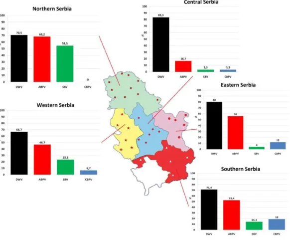
One hundred and fifty colonies were sampled from 32 apiaries (approximately five colonies per apiary) located in five administrative regions of Serbia (Fig. 1) in autumn (in period from September 25 to October 5) 2017.
On each apiary two strong, one medium and two weak colonies were chosen. Colony strength assessment and classification were done as inCavigli et al. (2016). The selected colonies were without visible signs of any disease.
About a hundred workers, both foragers and house bees were chosen for each sample, placed in sterile test tubes on dry ice, and stored at−20◦C until being processed.
Thirty randomly selected specimens taken from each bee sample were pulverized and homogenized in 5 mL of PBS solution. After centrifugation, from 140µL of the supernatant the RNA was extracted with ZR Viral RNA KitTM(Zymo Research, Orange, CA, USA).
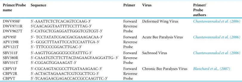
The obtained sequences were amplified in Rotor-Gene Q 5plex (Qiagen, Hilden, Germany) and the target viruses detected with the Rotor-Gene Probe RT-PCR Kit (Qiagen, Germany), in separate single-step reactions. The primer pairs and probes for DWV, ABPV and SBV (Table 1) were the same as used byChantawannakul et al. (2006)and for CBPV
Figure 1 Prevalence patterns of investigated viruses in honey bee colonies in five regions of Serbia.
Full-size DOI: 10.7717/peerj.5887/fig-1
those deployed by Blanchard et al. (2007). The final primer concentration 800 nM and probe concentration of 400 nM proved optimum. The analyses were done in conditions defined in the work ofSimeunović et al. (2014a). With each set of sample reactions, standard dilutions of the control sample were run and a threshold level set according to the standard curve obtained.
Selected RNA isolates were subjected to endpoint RT-PCRs using primer pairs and following the recommendations fromANSES (2011). The sequencing of each amplicon was done in both orientations in ABI 3130 Genetic Analyzer (Applied Biosystems, Foster City, CA, USA).
The obtained partial nucleotide (nt) sequences of honeybee viruses were identified by the BLAST search (http://blast.ncbi.nlm.nih.gov/Blast.cgi) against the GenBank database. Sequences encoding a partial coding sequence (cds) of polyprotein gene of DWV, a capsid protein gene of ABPV, a partial cds of RNA-dependent RNA polymerase (RdRp) gene of CBPV, and a partial cds of polyprotein gene of SBV were recovered. They were used for phylogenetic analyses along with related sequences deposited in GenBank. The best models of sequence evolution according to the Bayesian Information Criterion assessed MEGA version 6 (Tamura et al., 2007) were as follows: T92 + G for DWV and SBV, T92 + I for
Table 1 Primers and probes (TaqMan ProbeR
) used for RNA molecular identification of investigated viruses in real-time RT-PCR. Primer/Probe
name
Sequence Primer Virus Primer/
Probe authors DWV958F DWV9711R DWV9627T 50 -AAATTCTCTCACAGTCCAAG-30 50 CAACAGGTAATTTTCCTTTAG-30 50 -CATGCTCGAGGATTGGGTCGTCGT-30 Forward Reverese Probe
Deformed Wing Virus Chantawannakul et al. (2006)
APV95F APV159R APV121T 50 - TCCTATATCGACGACGAAAGACAA-30 50 - GCGCTTTAATTCCATCCAATTGA-30 50 - TTTCCCCGGACTTGAC-30 Forward Reverese Probe
Acute Bee Paralysis Virus Chantawannakul et al. (2006)
SBV311F SBV380R SBV331T 50 -AAGTTGGAGGCGCGYATTTG-30 50 -CAAATGTCTTCTTACDAGAAGYAAGGATTG- 30 50 -CGGAGTGGAAAGAT-30 Forward Reverese Probe
Sacbrood Virus Chantawannakul et al. (2006)
CBPV1F CBPV2R CBPVT 50 -CGCAAGTACGCCTTGATAAAGAAC-30 50-ACTACTAGAAACTCGTCGCTTCG-30 50 -TCAAGAACGAGACCACCGCCAAGTTC-30 Forward Reverese Probe
Chronic Bee Paralyses Virus Blanchard et al., (2007)
ABPV, and K2 + G + I for CBPV. Evolutionary relations assessed using these models of sequence evolution and the Neighbor-Joining (NJ) algorithm were shown as phylograms. Statistical support was tested with 1,000 nonparametric bootstrap (BS) replicates, with 50%≥BS≤74% considered moderate support, and BS≥75% considered good support. Statistical analysis
Depending on data characteristics (testing for normality), the results were presented through the mean and standard deviation, or the median and interquartile range were used. Differences were tested using ANOVA,t-test, or, where appropriate, non-parametric Mann–Whitney U test and Kruskal–Wallis test. Pearson chi-square analysis (or Fisher’s exact test) were applied where necessary. Data analysis was performed using IBM SPSS Statistics ver. 21.0 software (IBM, Armonk, NY, USA).
RESULTS
All of the four viruses in examined samples of adult bees were detected. Their prevalence differed depending on the region of sampling (Fig. 1). In 150 honey bee samples (colonies), the prevalence of these four viruses was as follows: 74% of DWV, 49.30% of ABPV, 24.00% of SBV and 6.70% of CBPV. Samples negative for all four viruses comprised 12.67% of the colonies investigated (Fig. 2).
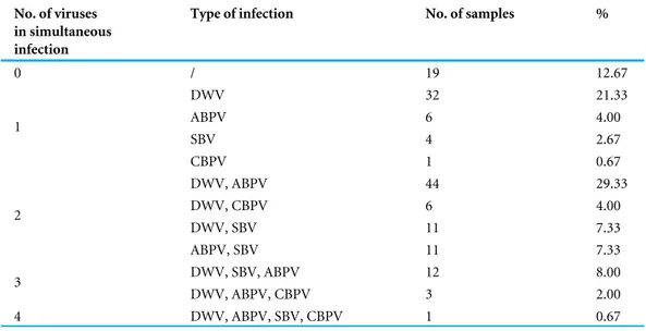
In 87.33% of samples analysed at least one virus was detected. Single infection was found in 28.67% of colonies (DWV, ABPV, SBV and CBPV in 21.33%, 4.00%, 2.67% and 0.67% colonies, respectively,Table 2). The majority of colonies (58.66%) were found to be infected with more than one virus. DWV had the highest prevalence in all regions (66.70–83.30%), while the least prevalent virus was CBPV (0–19%). Except for DWV, the prevalence of the remaining three viruses was significantly different between different regions (χ2 test: DWVP=0.554, ABPVP=0.001, SBVP< 0.001, CBPVP=0.030).
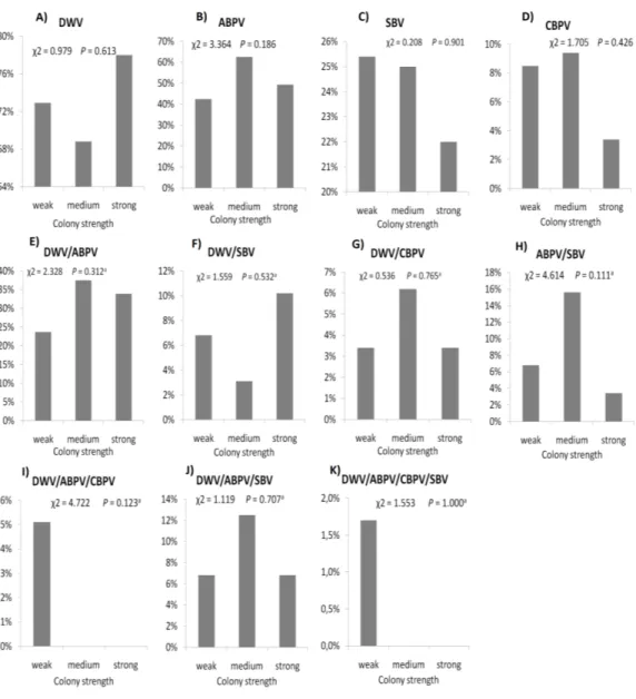
The prevalence of each virus in weak, medium and strong colonies is shown inFig. 3. DWV was most prevalent in strong colonies (78%), followed by weak (72.90%) and
Figure 2 Overall prevalence of CBPV, SBV, ABPV and DWV in Serbian bees (analyzed in 150 sam-ples).
Full-size DOI: 10.7717/peerj.5887/fig-2
Table 2 Prevalences of single and simultaneous virus infections in honey bee samples from Serbia. No. of viruses
in simultaneous infection
Type of infection No. of samples %
0 / 19 12.67 DWV 32 21.33 ABPV 6 4.00 SBV 4 2.67 1 CBPV 1 0.67 DWV, ABPV 44 29.33 DWV, CBPV 6 4.00 DWV, SBV 11 7.33 2 ABPV, SBV 11 7.33 DWV, SBV, ABPV 12 8.00 3 DWV, ABPV, CBPV 3 2.00 4 DWV, ABPV, SBV, CBPV 1 0.67 Notes.
DWV, deformed wing virus; CBPV, chronic bee paralysis virus; ABPV, acute bee paralysis virus; SBV, sacbrood virus. medium colonies (68.80%). The highest number of ABPV-positive samples was recorded in medium colonies (62.50%), followed by strong (49.20%) and weak colonies (42.40%). SBV was found in 25.40% of weak colonies, 25.00% of medium ones and in 22.00% of strong colonies. CBPV were found in 9.40% of medium colonies, 8.50% weak and 3.40% strong colonies. No significant differences were recorded in the prevalence of DWV, ABPV, SBV and CBPV infections (and their combinations) between weak, medium and strong
Figure 3 Prevalence of DWV, ABPV, SBV, CBPV and their combinations (A–K) in weak, medium and strong colonies.
Full-size DOI: 10.7717/peerj.5887/fig-3
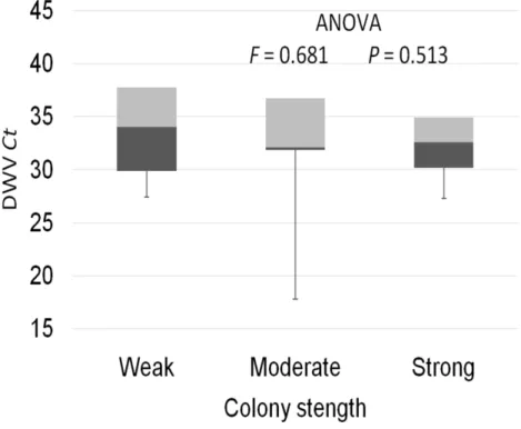
colonies were recorded with theχ2 test (Fig. 3). The significance of differences in virus infection levels (expressed through Ct values) between colonies of different strength were also tested. In order to avoid any confounding factor originated from the presence of other viruses in multiple infections, only single infections were taken into consideration in the data analysis. The numbers of samples with single ABPV, SBV and CBPV infections were not statistically valid for the comparison of their Ct values in strong, medium and weak colonies; therefore, only DWV infection intensity was eligible for testing in respect to colony strength. No significant differences were found between DWV infection levels in colonies of different strength (Fig. 4; ANOVA,F=0.681,P=0.513). In addition, the
Figure 4 Intensity of DWV infection (Ct values) in weak, medium and strong colonies.
Full-size DOI: 10.7717/peerj.5887/fig-4
Figure 5 Intensity of virus infections (Ct values) in single DWV infection and in double infections (DWV+ABPV, DWV+CBPV and DVW+SBV).
Full-size DOI: 10.7717/peerj.5887/fig-5
DWV infection and double infections caused by DWV and ABPV, SBV or CBPV (ANOVA, F=7.510,P< 0.001).
Phylogenetic analyses
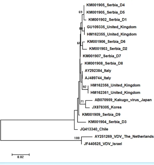
Phylogenetic trees showing evolutionary relations between Serbian and worldwide honeybee viruses DWV, ABPV, SBV and CBPV are shown inFigs. 6–9, respectively.
Figure 6 Neighbour-Joining tree of studied DWV sequences.The tree was constructed using a 420 nt long aligned matrix of 18 sequences encoding a partial coding sequence (cds) of polyprotein gene of DWVs. VDVs (AY251269andJF440525) were used as outgroups to root the tree. Viruses are indicated with GenBank Access. Nos. and the country of origin. Numbers at nodes represent bootstrap support. Bar on the left shows the number of nucleotide substitutions per site.
Full-size DOI: 10.7717/peerj.5887/fig-6
Nine DWV sequences detected in Serbian honeybees were deposited in GenBank: Serbia D1 (KM001902); Serbia D2 (KM001903); Serbia D3 (KM001904); Serbia D4 and D5 (KM001905, these two sequences were identical and thus they were deposited in the GenBank under the same accession number); Serbia D6 (KM001906); Serbia D7 (KM001907); Serbia D8 (KM001908); and Serbia D9 (KM001909). BLAST search found 99 to 98% nucleotide identities with DWV sequences in the database. Eighteen additional DWVs sequences from the GenBank were used for phylogenetic analysis, and VDVs (AY251269andJF440525) were used as outgroups to root the tree. The length of the aligned matrix was 420 nt. Evolutionary relations of studied DWVs are shown inFig. 6. Five Serbian DWVs organized into two moderately supported clusters, comprising three
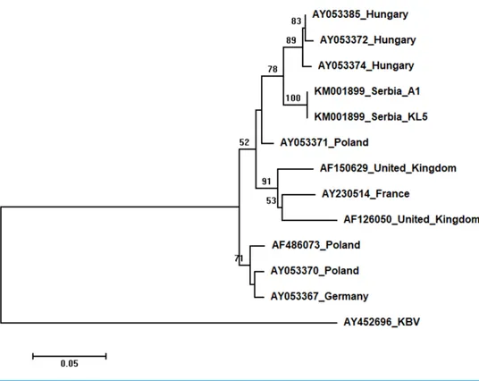
Figure 7 Neighbour-Joining tree of studied ABPV sequences.The tree was constructed using a 398 nt long aligned matrix of 12 sequences encoding a capsid protein of ABPVs. KBV (AY452696) was used as outgroups to root the tree. Viruses are indicated with GenBank Access. Nos. and the country of origin. Numbers at nodes represent bootstrap support. Bar on the left shows the number of nucleotide substitu-tions per site.
Full-size DOI: 10.7717/peerj.5887/fig-7
and two sequences, respectively, were closely related to DWVs from the United Kingdom, while others were dispersed throughout the tree.
Two Serbian ABPV sequences, KL4 and KL5, were identical, and thus they were deposited in the GenBank under the same accession number (KM001899). They showed 97 to 93% nucleotide identities to ABPV sequences in the database. Ten additional European ABPV sequences from the GenBank were used for phylogenetic analysis, and KBV (AY452696) was used as outgroup to root the tree shown inFig. 7. The length of the aligned matrix was 398 nt. Serbian ABPVs were closely related to the Hungarian ones while Western and Northern European viruses formed separate clusters.
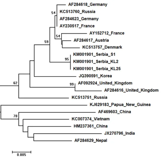
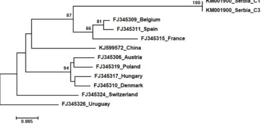
Three identical Serbian SBV sequences, S1, KL2 and KL25, deposited in the GenBank under the same accession number,KM001901, showed 99 to 94% sequence identity rates with other SBVs in the database. Seventeen additional SBVs from the GenBank were used for phylogenetic analysis. The length of the aligned matrix was 570 nt, and the recovered tree is shown inFig. 8. Three Serbian SBVs cluster together with SBVs from the continental Europe. Two identical Serbian CBPV sequences, CBPV-1 and CBPV-3, deposited in the GenBank under the same accession number (KM001900) show 96 to 93% sequence identity with other CBPVs in the GenBank. The length of the aligned matrix comprising Serbian and ten additional CBPVs was 429 nt. The relations of studied CBPVs are shown inFig. 9.
Figure 8 Neighbour-Joining tree of studied SBV sequences.The tree was constructed using a 429 nt long aligned matrix of 20 sequences encoding a partial coding sequence (cds) of polyprotein gene of SBVs. Viruses are indicated with GenBank Access. Nos. and the country of origin. Numbers at nodes represent bootstrap support. Bar on the left shows the number of nucleotide substitutions per site
Full-size DOI: 10.7717/peerj.5887/fig-8
DISCUSSION
In the era of intensive agriculture and serious decline in pollinator populations worldwide, primarily honey bees (Goulson et al., 2015), it is of great importance to gain an insight into the distribution and prevalence of factors most often connected with bee losses in any geographic region (Van Engelsdorp et al., 2008;Van Engelsdorp et al., 2009;Van Engelsdorp et al., 2012;Cavigli et al., 2016). In this study, samples from clinically healthy colonies in Serbian apiaries were analysed by real-time RT-PCR in order to detect honey bee viruses (DWV, ABPV, SBV and CBPV) and determine their prevalence patterns and prevalence. The results revealed DWV to be the most prevalent virus in Serbian apiaries, not unlike in many other countries: Hungary (Bakonyi et al., 2002), France (Tentcheva et al., 2004;Mouret et al., 2013), Austria (Berenyi et al., 2006), Slovenia (Toplak
et al., 2012) and Uruguay (Giacobino et al., 2016). High prevalence of DWV (74%) and
Figure 9 Neighbour-Joining tree of studied CBPV sequences.The tree was constructed using a 570 nt long aligned matrix of 12 sequences encoding a partial coding sequence (cds) of RNA-dependent RNA polymerase (RdRp) gene of CBPV.FJ345326was used as outgroup to root the tree. Viruses are indicated with GenBank accession numbers and the country of origin. Numbers at nodes represent bootstrap sup-port. Bar on the left shows the number of nucleotide substitutions per site.
Full-size DOI: 10.7717/peerj.5887/fig-9
relation toV. destructor mite infestation and their persistence as subclinical infection in apparently healthy colonies (Gauthier et al., 2007;Mouret et al., 2013;Wells et al., 2016). The average prevalence of SBV in Serbian samples was 24%, and none of the investigated colonies exhibited signs of sacbrood disease. The absence of disease signs in all recorded SBV-positive colonies may be the result of prominent hygienic behaviour (Swanson et
al., 2009), previously confirmed for honey bees throughout Serbia (Stanimirović et al.,
2002;Stanimirović, Stevanović & Ćirković, 2005;Stanimirović et al., 2008;Stanimirović et
al., 2011). The frequency of SBV in Serbia is similar with 40.24% recorded in Croatia
(Tlak Gajger et al., 2014), considerably lower than 86% from France (Tentcheva et al.,
2004) and 100% from Uruguay (Antúnez et al., 2006), but several times higher than 1.1%, 1.4% and 2% reported in Spain (Antúnez et al., 2012), England (Baker & Schroeder, 2008) and Hungary (Forgách et al., 2008), respectively. Low prevalence of CBPV in the samples is typical for asymptomatic colonies (Tentcheva et al., 2004). The rate of 0–19% CBPV-positive samples affirmed in Serbia is in accordance with the results obtained in the majority of Austrian federal states (Berenyi et al., 2006), Chinese provinces (Ai, Yan & Han, 2012), Korea (Choe et al., 2012), Slovenia (Toplak et al., 2012), and the apiaries from Denmark (Nielsen, Nicolaisen & Kryger, 2008) and France (Tentcheva et al., 2004).
Among monitored honey bee viruses in Serbia, the highest incidence was recorded for DWV (66.7–83.3%). No significant differences in its prevalence among Serbian regions is not surprising knowing its global occurrence (Wilfert et al., 2016) and its dominance over other viruses invariable environmental conditions(Giacobino et al., 2016).
The second most common virus in Serbian apiaries was ABPV, but its incidence (16.7– 68.2%) significantly varied between the regions. The prevalence of SBV and CBPV also displayed dissimilar patterns in environmentally different regions. Additional investigations
are necessary to explain the observed significant differences. It can be assumed that these results may reflect the beekeepers’ negligence of apicultural measures (Stanimirović et al.,
2007a), but also may have risen from different means ofV. destructor control (Nielsen,
Nicolaisen & Kryger, 2008), which may be the reason only in ABPV infection, since not all
viruses are transmitted by varroa mites (Glenny et al., 2017).
Nevertheless, differences in orographic factors and forage quality between regions should be also considered as the environment was suggested as a key factor interacting with local bee populations and ecogenotypes (Stanimirović, Stevanović & Ćirković, 2005;Giacobino
et al., 2016). Our results concerning 87.33% samples with at least one virus and 58.66%
with two or more are similar to those observed in Austria (Berenyi et al., 2006), France (Tentcheva et al., 2004;Gauthier et al., 2007) and Slovenia (Toplak et al., 2012).
Interestingly, no significant differences were found in the presence of DWV, ABPV, SBV and CBPV infections (and their combinations) in colonies of various strength. In addition, no significant differences were affirmed between single DWV infection levels (expressed through a Ct value) in colonies of different strength. These results may speak in favour of crucial influence of predisposing factors—pathogens, parasites, poor-quality nutrition, pesticides, and unfavourable climate conditions—on bee vitality (Stanimirović et al., 2007a;Simeunovic et al., 2014b;Abbo et al., 2017;Annoscia et al., 2018;Glavinic et al.,
2017;Stevanovic et al., 2016). Special emphasis should be put on the negative influence of
infestation withV. destructor, a biological and mechanical vector of at least two viruses, DWV and APBV, (Ryabov et al., 2014;Abbo et al., 2017) and a possible factor that could contributeNosema ceranae spreading (Glavinić et al., 2014). In addition, we may assume that bees highly infected with viruses do not return from the field committing ‘‘altruistic suicide" to regulate colony virus load as in cases ofV. destructor and/orN. ceranaeinfected bees (Kralj & Fuchs, 2006;Higes et al., 2008). In our study, the presence of another virus(es), ABPV, SBV or CBPV, in co-infections significantly influenced the intensity of DWV infection. The observed differences in DWV Ct values between co-infections and single DWV infections could be explained with the influence of simultaneous replication of the another present virus, wherein the influence may be stimulatory or suppressive. However, we should have in mind recent characterization of DWV master variants (DWV-A, DWV-B, and DWV-C) and their impact on bee health (McMenamin & Flenniken, 2018).
Very small percentages of multiple infections in comparison with single infections found in this study point out the possibility that the former are related with severe V. destructor infestations commonly observed in Serbian apiaries (Stanimirović et al., 2007a;
Stanimirović et al., 2017).
High identity rates among relatively short studied nucleotide sequences of DWVs account for the poorly supported and unresolved phylogenetic tree (Fig. 6). However, the observed close genetic distance between all DWVs is concordant with the hypothesis of their relatively recent evolutionary diversification and worldwide spread, potentially connected to the geographic expansion of their main vector,V. destructor(Berenyi et al., 2007;Wilfert
et al., 2016). On the other hand, Serbian and Hungarian ABPVs are closely related, and
this may be explained by the geographical vicinity and trade between beekeepers of the two countries. Both Serbian and Hungarian ABPVs are relatively distant from those from
the Western and Northern Europe, and this finding is in accordance with the report of
Bakonyi et al. (2002) that Hungarian ABPVs are not closely related with Western and
Northern European ABPVs. Although Serbian SBVs are closely related with SBVs from the continental Europe, further analysis, involving sequences from neighbouring countries, are required for determining whether similar separation exists with SBVs, as it has been affirmed in case of Serbian ABPV. CBPVs from Serbia, France, Belgium and Spain are monophyletic but Serbian CBPVs occupy a rather long branch indicating a non-negligible genetic distance between Serbian and mentioned CBPVs. These findings may indicate that CBPV (which is taxonomically and genetically very different from the other three honey bee viruses analysed in this study), may have different epizootiological character, and hence, is less intensively involved in the geographical spread of honey bee virus strains (Ribière, Olivier & Blanchard, 2010). Alternatively, unique genetic properties (higher mutation rate or segment rearrangements) may explain the genetic seclusion of the Serbian CBPVs. However, for better understanding of viral diversity in honey bee colonies, additional analyses are needed. This is in accordance with the opinion ofGalbraith et al. (2018), who also emphasized the importance of virus development dynamics and its possible impact on honey bees. Studies on bee pathogens causing colony decline in Serbia were mainly focused onNosema sp. (Stanimirovic et al., 2007b;Stevanovic et al., 2011;Stevanovic et al., 2013;Glavinić et al., 2014;Simeunovic et al., 2014b) andV. destructor (Stanimirović et al., 2002;Stanimirović, Stevanović & Ćirković, 2005;Stanimirovic et al., 2005;Stevanovic et al., 2008;Stanimirović et al., 2011;Radakovic et al., 2013;Gajic et al., 2013;Glavinić et al.,
2014;Stanimirović et al., 2017) with only one study dealing with bee viruses (Simeunović
et al., 2014a). Therefore, our work represents an important contribution towards better
understanding of bee pathogens in Serbia.
CONCLUSIONS
This work represents the first thorough investigation aimed at the constitution of the epidemiological baseline regarding molecular identification, prevalence patterns and prevalence of honey bee viruses in Serbia. The geographic origin and strength of honey bee colonies in Serbia proved to be insufficient to induce significant differences in the prevalence of the investigated viruses. Infection intensity of DWV presented through Ct value greatly depends on the presence of co-infection with other viruses. However, single ABPV, SBV and CBPV infections were not frequent enough to allow the comparison of their Ct values. In addition, the sequence analyses of Serbian honey bee viruses confirmed their identity and enabled an insight into their phylogenetic relationship with those found worldwide.
ADDITIONAL INFORMATION AND DECLARATIONS
Funding
This work was supported by the Ministry of Education, Science and Technological Development of the Republic of Serbia (grant number III 46002, led by Prof. dr Zoran
Stanimirović). The funders had no role in study design, data collection and analysis, decision to publish, or preparation of the manuscript.
Grant Disclosures
The following grant information was disclosed by the authors:
Ministry of Education, Science and Technological Development of the Republic of Serbia: III 46002.
Competing Interests
The authors declare there are no competing interests.
Author Contributions
• Dragan Cirkovic conceived and designed the experiments, analyzed the data, authored
or reviewed drafts of the paper, approved the final draft.
• Jevrosima Stevanovic analyzed the data, authored or reviewed drafts of the paper,
approved the final draft.
• Uros Glavinic performed the experiments, prepared figures and/or tables.
• Nevenka Aleksic authored or reviewed drafts of the paper, approved the final draft. • Spomenka Djuric analyzed the data, prepared figures and/or tables.
• Jelena Aleksic analyzed the data, prepared figures and/or tables, for second version
of the paper, she made new phylogenetic trees, made new Figures 6, 7, 8 and 9 in better resolution and corrected text and Figure legends in connection with Phylogenetic analyses.
• Zoran Stanimirovic conceived and designed the experiments, analyzed the data,
contributed reagents/materials/analysis tools, authored or reviewed drafts of the paper, approved the final draft.
DNA Deposition
The following information was supplied regarding the deposition of DNA sequences: The Deformed wing virus isolate Serbia D1 polyprotein gene, partial cds sequence described here are accessible via GenBank accession numbersKM001902.1.
The Deformed wing virus isolate Serbia D2 polyprotein gene, partial cds sequence described here are accessible via GenBank accession numberKM001903.1.
The Deformed wing virus isolate Serbia D3 polyprotein gene, partial cds sequence described here are accessible via GenBank accession numberKM001904.1.
The Deformed wing virus isolate Serbia D4 polyprotein gene, partial cds sequence described here are accessible via GenBank accession numberKM001905.1.
The Deformed wing virus isolate Serbia D6 polyprotein gene, partial cds sequence described here are accessible via GenBank accession numberKM001906.1.
The Deformed wing virus isolate Serbia D7 polyprotein gene, partial cds sequence described here are accessible via GenBank accession numberKM001907.1.
The Deformed wing virus isolate Serbia D8 polyprotein gene, partial cds sequence described here are accessible via GenBank accession numberKM001908.1.
The Deformed wing virus isolate Serbia D9 polyprotein gene, partial cds sequence described here are accessible via GenBank accession numberKM001909.1.
Data Availability
The following information was supplied regarding data availability: NCBI:KM001899.1–KM001909.1
University of Belgrade, Biology Department: https://biologija.vet.bg.ac.rs/uzgoj-i-nega-pcela/.
Supplemental Information
Supplemental information for this article can be found online athttp://dx.doi.org/10.7717/ peerj.5887#supplemental-information.
REFERENCES
Abbo PM, Kawasaki JK, Hamilton M, Cook SC, DeGrandi-Hoffman G, Li WF, Liu J, Chen YP. 2017.Effects of Imidacloprid andVarroa destructoron survival and health of European honey bees,Apis mellifera.Insect Science24:467–477
DOI 10.1111/1744-7917.12335.
Ai H, Yan X, Han R. 2012.Occurrence and prevalence of seven bee viruses inApis mellif-eraandApis ceranaapiaries in China.Journal of Invertebrate Pathology109:160–164
DOI 10.1016/j.jip.2011.10.006.
Annoscia D, Brown S, Di Prisco G, De Paoli E, Del Fabbro S, Zanni V, Galbraith D, Caprio E, Grozinger CM, Pennacchio F, Nazzi F. 2018.Haemolymph removal by the parasiteVarroa destructorcan trigger the proliferation of the Deformed Wing Virus in mite infested bees (Apis mellifera), contributing to enhanced pathogen virulence.bioRxiv DOI 10.1101/257667.
Antúnez K, Anido M, Garrido-Bailon E, Botias C, Zunino P, Martínez-Salvador A, Higes M. 2012.Low prevalence of viruses in Spain during 2006 and 2007.Research in Veterinary Science93:1441–1445DOI 10.1016/j.rvsc.2012.03.006.
Antúnez K, D’Alessandro B, Corbella E, Ramallo G, Zunino P. 2006.Honeybee viruses in Uruguay.Journal of Invertebrate Pathology93:67–70
DOI 10.1016/j.jip.2006.05.009.
Antúnez K, Invernizzi C, Mendoza Y, Zunino P. 2016.Honeybee colony losses in Uruguay during 2013–2014.Apidologie48:364–370
DOI 10.1007/s13592-016-0482-2.
Bacandritsos N, Granato A, Budge G, Papanastasiou I, Roinioti E, Caldon M, Falcaro C, Gallina A, Mutinelli F. 2010.Sudden deaths and colony population decline in Greek honey bee colonies.Journal of Invertebrate Pathology105:335–340
DOI 10.1016/j.jip.2010.08.004.
Baker A, Schroeder DD. 2008.Occurrence and genetic analysis of picorna-like viruses infecting worker bees ofApis melliferaL. populations in Devon, South West England. Journal of Invertebrate Pathology98:239–242DOI 10.1016/j.jip.2008.02.010.
Bakonyi T, Farkas R, Szendroi A, Dobos-Kovacs M, Rusvai M. 2002.Detection of acute bee paralysis virus by RT-PCR in honey bee andVarroa destructorfield samples: rapid screening of representative Hungarian apiaries.Apidologie33:63–74
Berenyi O, Bakonyi T, Derakhshifar I, Köglberger H, Nowotny N. 2006.Occurrence of six honeybee viruses in diseased Austrian apiaries.Applied and Environmental Microbiology72:2414–2420DOI 10.1128/AEM.72.4.2414-2420.2006.
Berenyi O, Bakonyi T, Derakhshifar I, Köglberger H, Topolska G, Ritter W, Pechhacker H, Nowotny N. 2007.Phylogenetic analysis of deformed wing virus genotypes from diverse geographic origins indicates recent global distri-bution of the virus.Applied and Environmental Microbiology73:3605–3611
DOI 10.1128/AEM.00696-07.
Blanchard P, Ribiere M, Celle O, Lallemand P, Schurr F, Olivier V, Faucon JP. 2007. Evaluation of a real-time two-step RT-PCR assay for quantitation of Chronic bee paralysis virus (CBPV) genome in experimentally-infected bee tissues and in life stages of a symptomatic colony.Journal of Virological Methods141:7–13
DOI 10.1016/j.jviromet.2006.11.021.
Brodschneider R, Gray A, Adjlane N, Ballis A, Brusbardis V, Charrière JD, Chlebo R, Coffey MF, Dahle B, De Graaf DC, Dražić MM, Evans G, Fedoriak M, Forsythe I, Gregorc A, Grzęda U, Hetzroni A, Kauko L, Kristiansen P, Martikkala M, Martín-Hernández R, Medina-Flores CA, Mutinelli F, Raudmets A, Ryzhikov VA, Simon-Delso N, Stevanovic J, Uzunov A, Vejsnæs F, Wöhl S, Zammit-Mangion M, Danihlík J. 2018.Multi-country loss rates of honey bee colonies during winter 2016/2017 from the COLOSS survey.Journal of Apicultural Research57:452–457
DOI 10.1080/00218839.2018.1460911.
Brutscher LM, McMenamin AJ, Flenniken ML. 2016.The buzz about honey bee viruses. PLOS Pathogens12:e1005757DOI 10.1371/journal.ppat.1005757.
Cavigli I, Daughenbaugh KF, Martin M, Lerch M, Banner K, Garcia E, Brutscher LM, Flenniken ML. 2016.Pathogen prevalence and abundance in honey bee colonies in-volved in almond pollination.Apidologie47:251–266
DOI 10.1007/s13592-015-0395-5.
Chantawannakul P, Ward L, Boonham N, Brown M. 2006.A scientific note on the detection of honeybee viruses using real-time PCR (TaqMan) inVarroamites col-lected from a Tai honeybee (Apis mellifera) apiary.Journal of Invertebrate Pathology 91:69–73DOI 10.1016/j.jip.2005.11.001.
Chen YP, Siede R. 2007.Honey bee viruses.Advances in Virus Research70:33–80
DOI 10.1016/S0065-3527(07)70002-7.
Choe SE, Nguyen LTK, Noh JH, Koh HB, Jean YH, Kweon CH, Kang SW. 2012. Prevalence and distribution of six bee viruses in KoreanApis ceranapopulations. Journal of Invertebrate Pathology109:330–333DOI 10.1016/j.jip.2012.01.003. Forgách P, Bakonyi T, Tapaszti Z, Nowotny N, Rusvai M. 2008.Prevalence of
pathogenic bee viruses in Hungarian apiaries: situation before joining the European Union.Journal of Invertebrate Pathology98:235–238DOI 10.1016/j.jip.2007.11.002. Francis RM, Nielsen SL, Kryger P. 2013.Varroa-virus interaction in collapsing honey
bee colonies.PLOS ONE8:e57540DOI 10.1371/journal.pone.0057540.
French Agency for Food, Environmental and Occupational Health and Safety (ANSES). 2011.Screening for several honeybee viruses using PCR (in-house method) (ABPV,
IAPV, KBV, DWV, SBV, BQCV, CBPV).Available athttp:// www.anses.fr/ en
(accessed on 5 July 2017).
Gajic B, Radulovic Z, Stevanovic J, Kulisic Z, Vucicevic M, Simeunovic P, Stan-imirovic Z. 2013.Variability of the honey bee miteVarroa destructorin Ser-bia, based on mtDNA analysis.Experimental and Applied Acarology61:97–105
DOI 10.1007/s10493-013-9683-9.
Galbraith DA, Fuller ZL, Ray AR, Brockmann A, Frazier M, Gikungu MW, Martinez FI, Kapheim KM, Kerby JT, Kocher SD, Losyev O, Muli E, Patch HM, Rosa C, Sakamoto JM, Stanley A, Vaudo AD, Grozinger CM. 2018.Investigating the viral ecology of global bee communities with high-throughput metagenomics.Scientific Reports8:8879 DOI 10.1038/s41598-018-27164-z.
Gauthier L, Tentcheva D, Tournaire M, Dainat B, Cousserans F, Colin ME, Bergoin M. 2007.Viral load estimation in asymptomatic honey bee colonies using the
quantitative RT-PCR technique.Apidologie38:426–435DOI 10.1051/apido:2007026. Genersch E. 2010.Honey bee pathology: current threats to honey bees and beekeeping.
Applied Microbiology and Biotechnology87:87–97DOI 10.1007/s00253-010-2573-8. Giacobino A, Molineri AI, Pacini A, Fondevila N, Pietronave H, Rodríguez G,
Palacio A, Bulacio Cagnolo N, Orellano E, Salto CE, Signorini ML, Merke J. 2016.Varroa destructorand viruses association in honey bee colonies under different climatic conditions.Environmental Microbiology Reports8:407–412
DOI 10.1111/1758-2229.12410.
Glavinic U, Stankovic B, Draskovic V, Stevanovic J, Petrovic T, Lakic N, Stan-imirovic Z. 2017.Dietary amino acid and vitamin complex protects honey bee from immunosuppression caused byNosema ceranae.PLOS ONE12:e0187726
DOI 10.1371/journal.pone.0187726.
Glavinić U, Stevanović J, Gajić B, Simeunović P, Ðurić S, Vejnović B, Stanimirović Z. 2014.Nosema ceranaeDNA in honey bee haemolymph and honey bee miteVarroa destructor.Acta Veterinaria-Beograd64:349–357DOI 10.2478/acve-2014-0033. Glenny W, Cavigli I, Daughenbaugh KF, Radford R, Kegley SE, Flenniken ML. 2017.
Honey bee (Apis mellifera) colony health and pathogen composition in migratory beekeeping operations involved in California almond pollination.PLOS ONE 12:e0182814DOI 10.1371/journal.pone.0182814.
Goulson D, Nicholls E, Botias C, Rotheray EL. 2015.Bee declines driven by com-bined stress from parasites, pesticides, and lack of flowers.Science347:1255957
DOI 10.1126/science.1255957.
Higes M, Martín-Hernández R, Botías C, Bailón EG, González-Porto AV, Barrios L, Del Nozal MJ, Bernal JL, Jiménez JJ, Palencia PG, Meana A. 2008.How natural infection byNosema ceranaecauses honeybee colony collapse.Environmental Microbiology10:2659–2669DOI 10.1111/j.1462-2920.2008.01687.x.
Kralj J, Fuchs S. 2006.Parasitic mites influence flight duration and homing ability of infestedApis melliferaforagers.Apidologie37:577–587DOI 10.1051/apido:2006040. Kulhanek K, Steinhauer N, Rennich K, Caron DM, Sagili RR, Pettis JS, Ellis JD, Wilson
national survey of managed honey bee 2015–2016 annual colony losses in the USA. Journal of Apicultural Research56:328–340DOI 10.1080/00218839.2017.1344496. Kulinčević J, Ball B, Mladjan V. 1990.Viruses in honey bee colonies infested withVarroa
jacobsoni: first findings in Yugoslavia.Acta VeterinariaBeograd40:37–42.
Lee K, Steinhauer N, Rennich K, Wilson ME, Tarpy DR, Caron DM, Rose R, Delaplane KS, Baylis K, Lengerich EJ, Skinner JA, Wilkes JT, Sagili R, VanEngelsdorp D. 2015.A national survey of managed honey bee 2013–2014 annual colony losses in the USA.Apidologie46:292–305DOI 10.1007/s13592-015-0356-z.
Martin SJ, Highfield AC, Brettell L, Villalobos EM, Budge GE, Powell M, Schroeder DC. 2012.Global honey bee viral landscape altered by a parasitic mite.Science 336:1304–1306DOI 10.1126/science.1220941.
McMenamin AJ, Flenniken ML. 2018.Recently identified bee viruses and their impact on bee pollinators.Current Opinion in Insect Science26:120–129
DOI 10.1016/j.cois.2018.02.009.
McMenamin AJ, Genersch E. 2015.Honey bee colony losses and associated viruses. Current Opinion in Insect Science8:121–129DOI 10.1016/j.cois.2015.01.015. Mouret C, Lambert O, Piroux M, Beaudeau F, Provost B, Benet P, Colin ME, L’Hostis
M. 2013.Prevalence of 12 infectious agents in field colonies of 18 apiaries in Western France.Revue de Medecine Veterinaire164:577–582.
Nielsen SL, Nicolaisen M, Kryger P. 2008.Incidence of acute bee paralysis virus, black queen cell virus, chronic bee paralysis virus, deformed wing virus, Kashmir bee virus and sacbrood virus in honey bees (Apis mellifera) in Denmark.Apidologie 39:310–314DOI 10.1051/apido:2008007.
Radakovic M, Stevanovic J, Djelic N, Lakic N, Knezevic-Vukcevic J, Vukovic-Gacic B, Stanimirovic Z. 2013.Evaluation of the DNA damaging effects of amitraz on human lymphocytes in the Comet assay.Journal of Biosciences38:53–62
DOI 10.1007/s12038-012-9287-2.
Ribière M1, Olivier V, Blanchard P. 2010.Chronic bee paralysis: a disease and a virus like no other?Journal of Invertebrate Pathology103:S120–S131
DOI 10.1016/j.jip.2009.06.013.
Ryabov EV, Wood GR, Fannon JM, Moore JD, Bull JC, Chandler D, Mead A, Bur-roughs N, Evans JD. 2014.A virulent strain of deformed wing virus (DWV) of honeybees(Apis mellifera) prevails afterVarroa destructor-mediated, orin vitro, transmission.PLOS Pathogens10:e1004230DOI 10.1371/journal.ppat.1004230. Simeunovic P, Stevanovic J, Cirkovic D, Radojicic S, Lakic N, Stanisic LJ, Stanimirovic
Z. 2014b.Nosema ceranaeand queen age influence the reproduction and pro-ductivity of the honey bee colony.Journal of Apicultural Research53:545–554
DOI 10.3896/IBRA.1.53.5.09.
Simeunović P, Stevanović J, Vidanović D, Nišavić J, Radović D, Stanišić LJ, Stan-imirović Z. 2014a.A survey of deformed wing virus and acute bee paralysis virus in honey bee colonies from Serbia using real-time RT-PCR.Acta Veterinaria-Beograd 64:81–92DOI 10.2478/acve-2014-0009.
Stanimirović Z, Aleksić N, Stevanović J, Ćirković D, Mirilović M, Djelić N, Stojić V. 2011.The influence of pulverised sugar dusting on the degree of infestation of honey bee colonies withVarroa destructor.Acta Veterinaria-Beograd61:309–325
DOI 10.2298/AVB1103309S.
Stanimirović Z, Ćirković D, Pejin II, Pejović D. 2007a.Strategy for ecologic con-trol in fightingVarroa destructor.Veterinarski Glasnik61:11–35 (in Serbian)
DOI 10.2298/VETGL0702011S.
Stanimirović Z, Glavinić U, Lakić N, Radović D, Ristanić M, Tarić E, Stevanović J. 2017. Efficacy of plant-derived formulation Argus Ras inVarroa destructorcontrol.Acta Veterinaria-Beograd67:191–200 DOI 10.1515/acve-2017-0017.
Stanimirović Z, Pejović D, Stevanović J, Vučinić M, Mirilović M. 2002.Investigations of hygienic behaviour and disease resistance in organic beekeeping of two hon-eybee ecogeographic varieties from Serbia.Acta Veterinaria-Beograd52:169–180
DOI 10.2298/AVB0203169S.
Stanimirovic Z, Stevanovic J, Bajic V, Radovic I. 2007b.Evaluation of genotoxic effects of fumagillin by cytogenetic testsin vivo.Mutation Research/Genetic Toxicology and Environmental Mutagenesis628:1–10DOI 10.1016/j.mrgentox.2006.09.014. Stanimirović Z, Stevanović J, Ćirković D. 2005.Behavioural defenses of the honey bee
ecotype from Sjenica—Pester againstVarroa destructor.Acta Veterinaria-Beograd 55:69–82DOI 10.2298/AVB0501069S.
Stanimirovic Z, Stevanovic J, Jovanovic S, Andjelkovic M. 2005.Evaluation of genotoxic effects of ApitolR
(cymiazole hydrochloride)in vitroby measurement of sister chromatid exchange.Mutation Research/Genetic Toxicology and Environmental Mutagenesis588:152–157DOI 10.1016/j.mrgentox.2005.10.003.
Stanimirović Z, Stevanović J, Mirilović M, Stojić V. 2008.Heritability of hygienic behaviour in grey honey bees (Apis mellifera carnica).Acta Veterinaria-Beograd 58:593–601DOI 10.2298/AVB0806593S.
Steinhauer N, Kulhanek K, Antúnez K, Human H, Chantawannakul P, Chauzat MP, VanEngelsdorp D. 2018.Drivers of colony losses.Current Opinion in Insect Science 26:142–148DOI 10.1016/j.cois.2018.02.004.
Stevanovic J, Schwarz RS, Vejnovic B, Evans J, Irwin RE, Glavinic U, Stanimirovic Z. 2016.Species-specific diagnostics ofApis melliferatrypanosomatids: a nine-year survey (2007–2015) for trypanosomatids and microsporidians in Serbian honey bees. Journal of Invertebrate Pathology139:6–11DOI 10.1016/j.jip.2016.07.001.
Stevanovic J, Simeunovic P, Gajic B, Lakic N, Radovic D, Fries I, Stanimirovic Z. 2013. Characteristics ofNosema ceranaeinfection in Serbian honey bee colonies.Apidologie 44:522–536DOI 10.1007/s13592-013-0203-z.
Stevanovic J, Stanimirovic Z, Genersch E, Kovacevic SR, Ljubenkovic J, Radakovic M, Aleksic N. 2011.Dominance ofNosema ceranaein honey bees in the Balkan countries in the absence of symptoms of colony collapse disorder.Apidologie 42:49–58DOI 10.1051/apido/2010034.
Stevanovic J, Stanimirovic Z, Radakovic M, Stojic V. 2008.In vitro evaluation of the clastogenicity of fumagillin.Environmental and Molecular Mutagenesis49:594–601
DOI 10.1002/em.20409.
Swanson JA, Torto B, Kells SA, Mesce KA, Tumlinson JH, Spivak M. 2009.Odorants that induce hygienic behavior in honeybees: identification of volatile compounds in chalkbrood-infected honeybee larvae.Journal of Chemical Ecology35:1108–1116
DOI 10.1007/s10886-009-9683-8.
Tamura K, Dudley J, Nei M, Kumar S. 2007.MEGA4: molecular evolutionary ge-netics analysis (MEGA) software version 4.0.Molecular Biology and Evolution 24:1596–1599DOI 10.1093/molbev/msm092.
Tentcheva D, Gauthier L, Zappulla N, Dainat B, Cousserans F, Colin ME, Bergoin M. 2004.Prevalence and seasonal variations of six bee viruses inApis mellifera L. andVarroa destructormite populations in France.Applied and Environmental Microbiology70:7185–7191DOI 10.1128/AEM.70.12.7185-7191.2004.
Tlak Gajger I, Bičak J, Belužić R. 2014.The occurence of honeybee viruses in apiaries in the Koprivnica-Križevci district in Croatia.Veterinarski Arhiv84:421–428.
Tlak Gajger I, Kolodziejek J, Bakonyi T, Nowotny N. 2014.Prevalence and distribution patterns of seven different honeybee viruses in diseased colonies: a case study from Croatia.Apidologie45:701–706DOI 10.1007/s13592-014-0287-0.
Toplak I, Rihtarič D, Ciglenečki UJ, Hostnik P, Jenčič V, Barlič-Maganja D. 2012. Detection of six honeybee viruses in clinically affected colonies of Carniolan gray bee (Apis mellifera carnica).Slovenian Veterinary Research49:89–96.
Van Engelsdorp D, Caron D, Hayes J, Underwood R, Henson M. 2012.A national survey of managed honey bee 2010–11 winter colony losses in the USA: results from the Bee Informed Partnership.Journal of Apicultural Research51:115–124
DOI 10.3896/IBRA.1.51.1.14.
Van Engelsdorp D, Evans JD, Saegerman C, Mullin C, Haubruge E, Nguyen BK, Frazier M, Frazier J, Cox-Foster D, Chen Y, Underwood R, Tarpy DR, Pettis JS. 2009.Colony collapse disorder: a descriptive study.PLOS ONE4:e6481
DOI 10.1371/journal.pone.0006481.
Van Engelsdorp D, Hayes J, Underwood RM, Pettis J. 2008.A survey of honey bee colony losses in the US, fall 2007 to spring 2008.PLOS ONE3:e4071
DOI 10.1371/journal.pone.0004071.
Venturini E, Drummond FA, Hoshide AK, Dibble AC, Stack LB. 2017.Pollination reservoirs in lowbush blueberry (Ericales: Ericaceae).Journal of Economic Entomology 110:333–346DOI 10.1093/jee/tow285.
Wells T, Wolf S, Nicholls E, Groll H, Lim KS, Clark SJ, Swain J, Osborne JL, Haughton AJ. 2016.Flight performance of actively foraging honey bees is re-duced by a common pathogen.Environmental Microbiology Reports8:728–737
DOI 10.1111/1758-2229.12434.
Wilfert L, Long G, Leggett HC, Schmid-Hempel P, Butlin R, Martin SJM, Boots M. 2016.Deformed wing virus is a recent global epidemic in honeybees driven by Varroamites.Science351:594–597 DOI 10.1126/science.aac9976.