Translation initiation: structures,
mechanisms and evolution
Assen Marintchev and Gerhard Wagner*
Department of Biological Chemistry and Molecular Pharmacology, Harvard Medical School, Boston, USA
Abstract. Translation, the process of mRNA-encoded protein synthesis, requires a complex apparatus, composed of the ribosome, tRNAs and additional protein factors, including aminoacyl tRNA synthetases. The ribosome provides the platform for proper assembly of mRNA, tRNAs and protein factors and carries the peptidyl-transferase activity. It consists of small and large subunits. The ribosomes are ribonucleoprotein particles with a ribosomal RNA core, to which multiple ribosomal proteins are bound. The sequence and structure of ribosomal RNAs, tRNAs, some of the ribosomal proteins and some of the additional protein factors are conserved in all kingdoms, underlying the common origin of the translation apparatus. Translation can be subdivided into several steps : initiation, elongation, termination and recycling. Of these, initiation is the most complex and the most divergent among the different kingdoms of life. A great amount of new structural, biochemical and genetic information on translation initiation has been accumulated in recent years, which led to the realization that initiation also shows a great degree of conservation throughout evolution. In this review, we summarize the available structural and functional data on translation initiation in the context of evolution, drawing parallels between eubacteria, archaea, and eukaryotes. We will start with an overview of the ribosome structure and of translation in general, placing emphasis on factors and processes with relevance to initiation. The major steps in initiation and the factors involved will be described, followed by discussion of the structure and function of the individual initiation factors throughout evolution. We will conclude with a summary of the available information on the kinetic and thermodynamic aspects of translation initiation.
1. Introduction 198
2. Ribosome structure and organization of the translation apparatus 200
2.1 Nomenclature 200 2.2 Ribosome structure 201 3. Overview of translation 203 3.1 Translation initiation 203 3.2 Translation elongation 203 3.2.1 Mechanism 203
3.2.2 Higher order organization of the eukaryotic translation apparatus and channeling of tRNA 208
3.3 Translation termination and recycling 210
* Author for correspondence : G. Wagner, Department of Biological Chemistry and Molecular Pharmacology, 240 Longwood Avenue, Harvard Medical School, Boston, MA 02115, USA.
4. Translation initiation 210
4.1 General translation initiation factors 211 4.2 Subunit dissociation/anti-association 212 4.3 Initiator aa-tRNA recognition 213 4.4 Start site recognition 214
4.4.1 Overview 214
4.4.2 Roles of individual factors 215 4.5 Subunit joining and factor release 217
4.6 Processes specific for eukaryotic translation initiation 219 4.6.1 Cap binding and scanning 219
4.6.2 Polyadenylation 222
4.7 Leaderless mRNAs – a minimal ‘ universal ’ system 224 4.8 Reinitiation and leaky scanning 225
4.9 Initiation at non-AUG codons and the stringency of start codon selection 227
5. Structure/function of initiation factors 231
5.1 Universally conserved factors 232 5.1.1 IF1/eIF1A 232
5.1.2 IF2/eIF5B 234 5.2 IF3 and eIF1 237
5.2.1 IF3 237 5.2.2 eIF1 238 5.3 eIF2 240
5.4 Eukaryotic factors required for eIF2 function 247 5.4.1 eIF5 247
5.4.2 eIF2B 248 5.5 eIF3 254
5.6 Factors involved in cap binding and scanning 257 5.6.1 eIF4E 257
5.6.2 eIF4G 259 5.6.3 eIF4A 262
5.6.4 eIF4B and eIF4H 264 5.7 PABP 265
6. Kinetic aspects of translation initiation and its regulation 266
6.1 General considerations 267 6.2 Translation initiation in bacteria 269 6.3 Translation initiation in eukaryotes 270
7. Concluding remarks 273
8. Acknowledgments 274
9. References 274
1. Introduction
The field of translation and translation initiation in particular has experienced an unprecedented growth in recent years, both in terms of accumulation of new data and of much deeper under-standing of the underlying processes. We now have insights into the structure and location of most translation initiation factors (IFs) and can discuss their roles on a structural and mechanistic level.
Here, we have attempted to summarize at least a fraction of the landslide of new information and present the emerging picture of various aspects of translation initiation. The main focus of this review is on the mechanism of translation initiation, the structure and function of the IFs and the organization of the initiation complexes (ICs). A look on translation initiation from an evolutionary perspective emphasizes both the common origins and organization of trans-lation and the great diversity among species. We give special attention to the organization of the translational apparatus in the cell, the concept of channeling of factors and intermediates, and their implications. The endless variety of mechanisms of translation regulation and alterna-tive initiation are beyond the scope of this review and are only included where they have a direct relation to our understanding of the general mechanisms of initiation.
Most of our knowledge about translation comes from eubacteria and eukaryotes. In recent years, the archaeal system has started to attract more attention, in part because it is remarkably similar to that in eukaryotes, but much simpler and involves fewer translation factors. As archaeal translation is related to eukaryotic translation, statements about eukaryotes will be assumed throughout this review to apply to archaea as well and vice versa, unless otherwise specified. The organellar translational apparatus is evolutionarily related to its eubacterial counterpart, but has undergone long independent evolution in its specific environment and will not be discussed here. Section 2 contains a brief overview of the structure of the ribosome. In Section 3 we present an overview of translation, with emphasis on factors and processes with relevance for our understanding of translation initiation. The mechanism of translation initiation is discussed in Section 4. Section 5 contains a summary of our knowledge about the structures of individual IFs and the organization of the ICs. Finally, in Section 6 we try to look at translation initiation and its regulation from a kinetic perspective.
It was not humanly possible to discuss all individual reports on any subject (or even only the ones we are aware of ). Therefore, we have tried to present what we see as the prevailing views and refer the reader to recent specialized reviews for details. While browsing through the sea of sometimes contradictory publications, we tried to follow some general ‘ guidelines ’ :
(1) With the risk of ignoring groundbreaking discoveries, we rarely mention isolated reports, contradicting the consensus from the rest of the field, unless the results appear sound and unambiguous. On some occasions, we have discussed controversies, mainly to emphasize that a ‘ mainstream ’ concept has been seriously challenged and promote the broader acceptance of the alternative.
(2) Detection of relatively weak interactions depends on the limitations of the method used, concentrations and experimental conditions. Even relatively strong interactions in the sub-micromolar range can be lost during centrifugation, a method routinely used in translation studies. Therefore, we have tried to be cautious with negative binding results, and especially Abbreviations: IC, initiation complex ; IF, translation initiation factor ; eIF, translation eukaryotic initiation factor ; EF, elongation factor ; eEF, eukaryotic elongation factor ; aa-tRNA, aminoacyl-tRNA ; aaRS, tRNA synthetase ; cryo-EM, cryo-electron microscopy ; A site (of the ribosome), aminoacyl-tRNA site ; P site, peptidyl-aminoacyl-tRNA site ; E site, exit site ; ASL, anticodon stem-loop (of the aminoacyl-tRNA) ; PTC, peptidyl transferase center ; GAC, GTPase-associated center ; SRL, sarcin/ricin-binding loop (in the large ribosomal subunit) ; GEF, guanine nucleotide exchange factor ; GAP, GTPase activating protein ; SD, Shine–Dalgarno sequence ; RBS, ribosome-binding site ; ORF, open reading frame ; UTR, untranslated region ; IRES, internal ribosome entry site ; Gcnx, general (translational) control non-derepressible ; Gcd–, general control derepressed ; Sui, suppresor of initiator codon mutation ; NTD, N-terminal domain ; CTD, C-terminal domain ; ZBD, Zn-binding domain ; RRM, RNA recognition motif ; WT, wild type.
with reports that a mutation completely abolishes binding. On the other hand, interactions observed only in vitro at non-physiological concentrations (especially if they were mainly electrostatic) were only considered reliable if it was known that the factors involved are brought in proximity via other interactions.
(3) A modification or mutation often has an effect in some systems, and some mRNAs, but not others, and the effect may depend largely on the experimental conditions. Therefore, if effects were seen in some studies but not in others, we have generally favored the presence of an effect, unless there were clear contradictions.
(4) Data obtained under non-physiological conditions were considered with extreme caution. (5) If a factor is reported to bind to other factors both individually and in combination, it is
hard to know if the interactions are cooperative, anti-cooperative or independent, without quantitative binding data. If a factor is found to bind to two other factors simultaneously, but not individually, more often than not, it also binds to each individual factor, even if the binding was too weak to detect by the method used.
(6) Similarly to point (2) above, reports of mRNA binding by RNA-binding proteins were also subject to scrutiny, because any sequence- or structure-specific RNA-binding protein usually has a fairly high non-specific RNA-binding affinity, which cannot be taken as proof that it actually binds to mRNAin vivo.
(7) On the other hand, there are numerous reports of high-affinity sequence/structure-specific mRNA binding by ‘ non-specific ’ factors, such as eukaryotic initiation factor 4A (eIF4A) and eIF4G, for example. Although such high-affinity binding sites may be absent from most mRNAs, it cannot be assumed that a ‘ non-specific ’ mRNA-binding eIF binds all mRNAs with the same affinity. Furthermore, as it appears that any imaginable regulation mechanism is in fact used somewhere, it is likely to find more and more viral or cellular mRNAs using high-affinity ‘ non-specific ’ factor-binding sites for translation initiation.
We apologize for any omissions of important work and views due to space limitations or ignorance on our part.
2. Ribosome structure and organization of the translation apparatus 2.1 Nomenclature
The ribosomes, ribosomal subunits and ribosomal RNAs (rRNAs) are identified by their sedimentation coefficients : the intact ribosomes are 70S in eubacteria and archaea, and 80S in eukaryotes ; the small subunits are 30S and 40S respectively ; and the large subunits are 50S and 60S respectively. The rRNA in the eubacterial and archaeal small subunit is 16S (18S in eukaryotes), and the rRNAs in the large subunit are 23S (26S and 58S inS. cerevisiae, and 28S and 58S in human respectively) and 5S. The ribosomal proteins are given designation ‘ S ’ and a number for proteins in the small ribosomal subunit and ‘ L ’ and a number for those belonging to the large ribosomal subunit. For example, S1 is small ribosomal subunit protein 1. As men-tioned above, part of the bacterial and eukaryotic ribosomal proteins are homologous to each other and it is accepted that homologous ribosomal proteins have similar location on the ribosome and probably perform similar functions in ribosome biogenesis and/or translation. Unfortunately, for historic reasons, ribosomal proteins conserved between bacteria and eukaryotes do not have the same names in both nomenclatures, and alternative nomenclatures exist for the eukaryotic ribosomes (for a comparison of the nomenclatures of yeast ribosomal
proteins and their relationships to mammalian, archaeal and eubacterial ribosomal proteins see Planta & Mager, 1998).
Additional designations exist in eukaryotic translation for the pre-IC of the small ribosomal subunit with IFs, before it is bound to mRNA (43S), and for the IC on the mRNA (48S), also based on their respective sedimentation coefficients.
2.2 Ribosome structure
In recent years, several X-ray structures of ribosomal subunits and of the 70S bacterial ribosome were determined, some at a resolution as high as 24 A˚ . Some of these structures contained tRNAs and translation factors. In addition, cryo-electron microscopy (cryo-EM) reconstructions are also available for structures and complexes corresponding to various steps along the translation pathway (reviewed in Ramakrishnan, 2002). Whereas X-ray structures of the small ribosomal subunit and of the intact ribosome are only available from eubacteria, a high-resolution X-ray structure of an archaeal 50S subunit has also been determined (Banet al. 2000) and the high degree of similarity to its eubacterial counterpart reinforces the universal con-servation of ribosome structure throughout evolution. Only cryo-EM information is available for the eukaryotic ribosome. The resolution of the cryo-EM reconstruction of the yeast 80S ribosome (y15 A˚ ) was sufficient to fit common structural elements from the X-ray structures of the bacterial 30S and archaeal 50S subunits and model homologous proteins. Regions of unassigned electron density provided indications where ribosomal proteins without bacterial homologs could be located, although the identity of the proteins could not be inferred (Spahn et al. 2001).
The structure of the small subunit can be subdivided into head, neck, platform, and body, which have obvious relationships to the structural domains of the 16S rRNA : 5k-domain, central domain, 3k-major domain, and 3k-minor domain (Fig. 1). The 5k-domain corresponds to the body ; the central domain – to most of the platform ; and the 3k-major – to the head. The neck provides a relatively flexible connection between the head and the rest of the small subunit. The 3k-minor domain consists of the last two helices (44 and 45) and the 3k-end of the rRNA. It was noted that the structural domains are ‘ nearly structurally autonomous ’ and that ‘ this organization immediately suggests that the domains are designed to move relative to one another during protein synthesis ’. The long helix 44 lies across the body and ends into the platform and neck. Therefore, helix 44 is connected to all major domains and could relay conformational changes and movement along the entire small subunit. In contrast, the large subunit consists of a rigid core and mobility is restricted primarily to segments on the periphery (Yusupovet al. 2001).
The mRNA binding site of the ribosome is on the small subunit, along the neck region between the head and the body, whereas the peptidyl transferase center (PTC) is on the large subunit (Fig. 1c). The ribosome has three binding sites for tRNA, shared between the two subunits. The aminoacyl (A) site has high affinity for aminoacyl-tRNA (aa-tRNA) ; the peptidyl (P) site has high affinity for peptidyl-tRNA ; and the exit (E) site has high affinity for deacylated tRNA. The anticodon stem-loop (ASL) of the tRNA is oriented toward the mRNA on the small subunit, whereas the acceptor end of the tRNA, to which the amino acid is attached, binds to the large subunit, and the acceptor ends of the tRNAs in the A and P sites are in the PTC. The GTPase-associated center (GAC) and the sarcin/ricin-binding loop (SRL) on the large ribosomal subunit are important for stimulation of GTP hydrolysis by several translation factors
(Fig. 1c). They are located at the base of the L7/L12 stalk near the aa-tRNA entry site (the nomenclatures concerning the GAC vary and the term is often used with a broader meaning to include both the L7/L12 stalk and the SRL). The nascent peptide exits through a channel in the large subunit (reviewed in Ramakrishnan, 2002 ; Tenson & Ehrenberg, 2002).
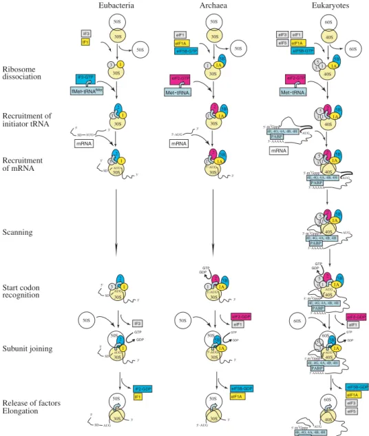
mRNA 3' 5' L7 SRL PTC L7 SRL PTC (a) (b) (c)
Fig. 1.Ribosome structure. (a) Domain organization of the 16S rRNA (from Fig. 3a, Yusupovaet al. 2001, with permission). The 5k-domain (blue) corresponds to the body, the central domain (purple) to the plat-form, the 3k-major domain (red) to the head, and the 3k-minor domain (yellow) – to the last two helices (44 and 45) and the 3k-end of the rRNA. (b) The structure of the 30S ribosomal subunit from the structure of the 70S ribosome, PDB code 1JGP (Yusupovaet al. 2001). 16S rRNA is shown as a ribbon and colored as in (a). Ribosomal proteins are shown in grey. The mRNA is in brown and its 3k-end (in the ‘ Entry ’ channel) and 5k-end (in the ‘ Exit ’ channel) are shown. (c) Stereo view of the structure of the 70S ribosome, PDB codes 1JGP and 1GIY (Yusupova et al. 2001). The small ribosomal subunit is in semitransparent surface representation and colored in grey. The mRNA is in brown, the A-, P-, and E-site tRNAs are in blue, coral and green respectively. The 23S and 5S rRNAs from the large subunit are shown as ribbon and colored in violet, and the large ribosomal proteins are in beige. The peptidyl-transferase centre (PTC), the sarcin/ricin-binding loop (SRL), and the L7 ribosomal protein are labeled.
3. Overview of translation
The process of protein synthesis can be subdivided into several major stages : initiation, elongation, termination and recycling. Translation initiation will be discussed in depth in the following sections and, therefore, only a brief description is presented in this section. In dis-cussing elongation, termination and recycling, specific attention will be paid to processes and factors with relevance to initiation. Some highlights from this section, which we would like to bring to the reader’s attention are : (1) The elongation factors (EFs) delivering aa-tRNA to the ribosome are homologous to thec subunit of the eukaryotic initiation factor 2 (eIF2). (2) The large domain rearrangement in elongation factor EF1A (formerly EF-Tu) upon GTP hydrolysis is an exception rather than the rule for this family of G proteins. Accordingly, the nearly 1000-fold lower affinity of EF1A . GDP for aa-tRNA, compared to EF1A . GTP, may also be an exception. (3) In eukaryotes, some events within translation are organized at a higher level, which is termed channeling ; tRNA, factors and intermediates are predominantly channeled along the translation pathway and rarely able to diffuse freely. This is in part also true for yeast, especially with respect to channeling of tRNAs between aminoacyl-tRNA synthetases (aaRS), eukaryotic elongation factor 1A (eEF1A), and ribosomes. As part of closing the tRNA channeling cycle, eEF1B, the exchange factor (GEF) for eEF1A, forms a stable complex with eEF1A . GTP and is only released upon aa-tRNA binding to eEF1A . GTP, whereas free eEF1A . GDP has nanomolar affinity for unacylated tRNA.
3.1 Translation initiation
Translation initiation covers all the steps between subunit dissociation upon termination in the previous translation cycle, and the assembly at an mRNA start codon of a ribosome ready for elongation. During translation initiation, the ribosome, with an initiator aa-tRNA in the P-site, is assembled on mRNA, with the help of a set of IFs. The main tasks that are performed by the translation apparatus during initiation (not necessarily in this order) are : (1) subunit dis-sociation and anti-asdis-sociation, (2) selection of the initiator aa-tRNA, (3) selection of the correct translation start site, and (4) subunit joining at the start codon. At the end of initiation, the ribosome is ready to accept the first elongator tRNA and form the first peptide bond, which marks the beginning of the next stage, elongation (Fig. 2).
3.2 Translation elongation
Translation elongation is the process of synthesis of the polypeptide chain, by the ribosome assembled at the start codon, until a stop codon is reached.
3.2.1 Mechanism
During elongation, an aa-tRNA is first bound to the A-site and if proper base-pairing between the mRNA codon in the A-site and the tRNA anticodon is established, a peptide bond is formed with the peptide attached to the tRNA in the P-site, accompanied by transfer of the peptide (now 1 amino acid longer) to the A-site tRNA. Then, the peptidyl tRNA is moved from the A- to the P-site, and the deacylated tRNA from the P-site is moved to the E-site, displacing from there the tRNA deacylated in the previous cycle. The mRNA is coordinately translocated by one codon. Thus, tRNA-mRNA base-pairing and the correct reading frame are retained (reviewed in Merrick & Nyborg, 2000 ; Ramakrishnan, 2002).
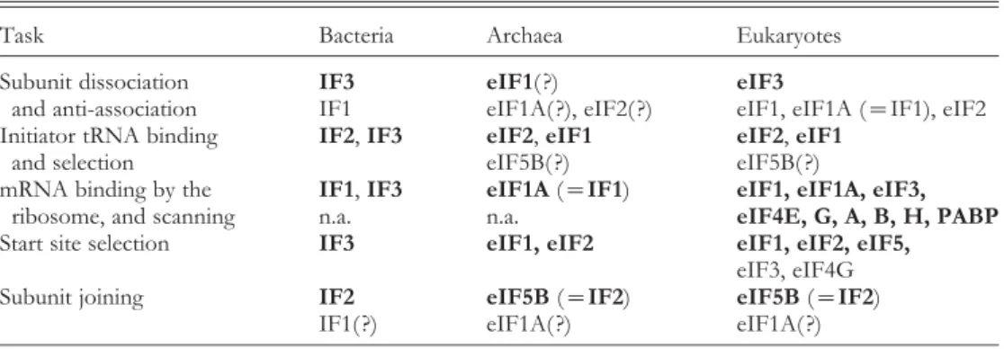
eIF2-GTP 3 5 3 3 5 3 5 3 5 3 5 40S 40S 40S 40S 40S 2 5B Eukaryotes Ribosome dissociation Recruitment of initiator tRNA eIF1 Recruitment of mRNA Scanning 1A 1 eIF2-GTP Release of factors Elongation Met-tRNAi mRNA eIF1A eIF5B-GTP 5B 1A 1 2 5B 1A 1 GTP GDP 5B 1A GDP GTP 2 5B 1A 1 eIF2-GDP 40S eIF1 eIF1A eIF5B-GDP 60S 60S 40S 60S 60S Start codon recognition 40S 2 5B 1A 1 4E, 4G, 4A, 4B, 4H m7Gppp AUG AAAAA PABP PABP 4E, 4G, 4A, 4B, 4H m7Gppp AAAAA PABP PABP 4E, 4G, 4A, 4B, 4H m7Gppp AAAAA PABP PABP AUG AUG 4E, 4G, 4A, 4B, 4H m7Gppp AAAAA PABP PABP AUG 4E, 4G, 4A, 4B, 4H m7Gppp AAAAA PABP PABP AUG 4E, 4G, 4A, 4B, 4H m7Gppp AAAAA PABP PABP AUG Subunit joining 30S 30S 30S 30S 30S 2 5B 1A 1 mRNA AUG 5B 1A 1 2 5B 1A 1 AUG GTP GDP 5B 1A AUG GDP GTP 2 5B 1A 1 AUG eIF2-GDP 30S eIF1 eIF1A eIF5B-GDP 50S 50S 30S 50S 50S eIF1 Met-tRNAi eIF1A eIF5B-GTP eIF3 eIF5 30S 30S 30S 30S 30S 1 3 mRNA 2 1 3 2 1 3 2 1 GDP GTP 2 1 3 30S IF3 IF1 IF2-GDP 50S 50S 30S 50S 50S IF3
fMet-tRNAfMet IF1 IF2-GTP AUG SD AUG SD AUG SD AUG SD AUG 5 60S 50S 50S AUG SD eIF3 eIF5 3' 3' 5' 5' 3' 3' Archaea Eubacteria 5'- 3'- 5'-5' 5' 5' 3' 3' 3' 3' 3' 3' 3'-
5'-Fig. 2.Translation initiation. Schematic representation of translation initiation in eubacteria, archaea and eukaryotes. The universally conserved pairs of proteins IF1/eIF1A and IF2/eIF5B are in yellow and blue respectively. eIF2 (present only in archaea and eukaryotes) is in red. The cap/poly-A-binding complex (present only in eukaryotes) is in light blue. The rest of the initiation factors (IFs) are in grey. The 5k- and 3k-ends of mRNA are labeled. SD, Shine–Dalgarno sequence. Every effort has been made to provide a correct temporal and spatial representation of the events ; however, the exact timing of recruitment and release of factors is not always known. Furthermore, the recruitment of IFs and RNAs need not follow a precise order, but may be a stochastic process. Note that GTP hydrolysis by IF2/eIF5B occurs after subunit joining and is required for release of IF2/eIF5B from the ribosome. Only the 5k-end-dependent initiation mechanism is shown for archaea, but internal SD-dependent initiation is also used in these organisms on polycistronic mRNAs. The scheme for eukaryotic initiation presumes that the scanning 43S ribosomal complex remains associated with the 5k-cap (see text for details). eIF1 and eIF2 and bacterial IF3 need to be displaced from their original positions for subunit joining to occur (and are shown as ‘ leaving ’ before subunit joining), but could remain associated with the ribosome. The other IFs remain associated with the ribosome during subunit joining and some even early in elongation (see text for details).
In bacteria, the aa-tRNA is brought to the ribosome as part of an EF1A . GTP . aa-tRNA ternary complex. Elongation factor EF1A (formerly EF-Tu) is universal and binds to most combinations of tRNAs and the amino acids attached to their 3k-end. The binding affinity of EF1A is determined by its affinities for the tRNA portion and for the aminoacyl portion of the aa-tRNA and the lack of apparent specificity is achieved through combinations of high affinity for the tRNA and low – for the amino acid, and vice versa. The affinity of EF1A is lower for certain aa-tRNAs, such as the initiator tRNA fMet-tRNAfMet(recognized by IF2), the seleno-cysteine-tRNA (Sec-tRNASec, recognized by a specialized EF SelB), and some aa-tRNA com-binations that are intermediates for further modification of the attached amino acid, like conversion of Asp-tRNAAsn into Asn-tRNAAsn in some species. The latter group gains high affinity for EF1A after modification. The same recognition mechanism of aa-tRNA is used by the eukaryotic EF1A homolog, eEF1A (reviewed in Francklynet al. 2002).
Initial binding of the EF1A . GTP . aa-tRNA ternary complex to the ribosome near the GAC places the aa-tRNA in a hybrid A/T site, where the ASL of the tRNA is near the A-site mRNA codon in the decoding center of the small subunit, but the rest of the tRNA is not yet positioned in the A-site. EF1A . GTP . aa-tRNA ternary complexes containing non-cognate tRNA have equal chance to bind to the ribosome as complexes containing the correct tRNA complementary to the codon in the A-site. After initial binding of the EF1A . GTP . aa-tRNA ternary complex to the ribosome, selection against the incorrect tRNAs is performed at two stages. First, coordinated conformational changes in the ternary complex and the ribosome allow the anticodon of the aa-tRNA to contact the mRNA codon in the A site. The codon-anticodon pairing has dual roles : (1) It stabilizes the complex between EF1A . GTP . aa-tRNA and the ribosome. The affinity of ternary complexes containing non-cognate tRNA is low and they quickly dissociate without GTP hydrolysis by EF1A. (2) Discrimination between cognate tRNAs and near-cognate tRNAs (forming non-canonical base pairs) is performed by ‘ inspec-tion ’ of the geometry of the minor groove in the first two base pairs of the codon in the A-site. Non-canonical base pairs (e.g. G . U) are tolerated in the third, ‘ wobble ’ position. The discrimi-nation in the first two positions is mediated by the universally conserved nucleotides 530, 1492 and 1493, whose bases become inserted in the minor groove of the codon-anticodon base pairs (Ogleet al. 2001). The binding of a cognate tRNA to the codon in the A-site promotes a ‘ closed ’ conformation of the small subunit required for ribosome-stimulated GTP hydrolysis by EF1A.
Upon GTP hydrolysis, EF1A is released and the aa-tRNA is accommodated in the A-site, with the acceptor end being inserted into the PTC of the large subunit. A second round of selection occurs at this stage : the rates of accommodation of the near-cognate aa-tRNAs in the PTC (01 sx1
) are much slower than their rates of dissociation (rejection) :y6 sx1
, leading toy100-fold discrimination. In contrast, cognate aa-tRNAs bind more tightly with negligible (<03 sx1
) dissociation rates, compared to their higher rates of accommodation (y7 sx1
). In summary, the discrimination against non-cognate aa-tRNAs is achieved predominantly at the first selection step, before GTP hydrolysis, whereas discrimination against near-cognate aa-tRNAs is achieved both at the first step before GTP hydrolysis (10- to 100-fold), and at the second step, after GTP hydrolysis (y100-fold), yielding overall misincorporation rates for near-cognate aa-tRNAs in the order of 10x3
to 10x4
. Such high fidelity could not have been achieved based only on differences in binding affinities of the cognateversusnear-cognate aa-tRNAs, but instead rely on induced fit, where only the binding of a cognate aa-tRNA leads to acceleration of rate-limiting structural rearrangement steps (reviewed in Rodnina &
Wintermeyer, 2001). The steric restrictions based on the geometry of canonical versus non-canonical base pairs are very important in discrimination between cognate and near-cognate tRNAs. This becomes especially clear in cases where a near-cognate codon-anticodon pair has the same or even higher binding energy than the cognate pair, but is still efficiently eliminated during translation (Ogleet al. 2001).
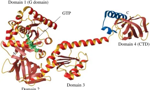
The structures of bacterial EF1A (EF-Tu) have been determined in a GTP- and GDP-bound form and with GTP and bound aa-tRNA (Fig. 3). EF1A is composed of three domains. The G-domain (domain I) binds the nucleotide and is packed against domains II and III. Two segments, called ‘ Switch 1 ’ and ‘ Switch 2 ’ are important for regulation of GTP binding and hydrolysis. The aa-tRNA binds across domains II and III with the amino-acid moiety binding to domain II, at the interface with domain I. Domains I and II are the most conserved, whereas
(a) (b)
EF1A from the EF1A.EF1B complex
EF1A.GDP
I
II
EF1A.GTP
(=EF1A.GTP.fMet-tRNAfMet)
III
(c)
(d) (e) (f)
eEF1A from the eEF1A.eEF1Bα complex
Archaeal eEF1A.GDP eIF2γ.GTP (=eIF2γ.GDP) I II III I II III I II III I II III I II III
Fig. 3.Structures of proteins from the EF1A (EF-Tu) family in different conformations. Domain I (the G domain) is in dark blue, domain II is in blue, and domain III is in light blue. Domains II and III from all proteins were structurally aligned, in order to illustrate the different orientations of domain I with respect to the rest of the protein. (a) Structure ofT. aquaticusEF1A (EF-Tu) in complex with the non-hydrolyzable GTP analog GDPNP, PDB code 1EFT (labeled as EF1A . GTP for simplicity). The structure is super-imposable with the structure of EF1A from the EF1A . GTP . Cys-tRNACysternary complex, PDB code 1B23 (not shown). (b) Structure ofT. thermophilusEF1A from the complex with the nucleotide exchange factor EF1B (EF-Ts), PDB code 1AIP. (c) Structure ofT. aquaticusEF1A . GDP, PDB code 1TUI. (d) Structure ofP. abyssieIF2c. GTP, PDB code 1KK1. The domain orientation is identical in the structures of P. abyssieIF2c. GDP, PDB code 1KK2, and apo-eIF2c, PDB code 1KK0, and very similar (y14xrotation of domain I) inM. jannaschiiapo-eIF2c, PDB code 1S0U (not shown). (e) Structure of yeast eEF1A from the complex with the nucleotide exchange factor eEF1Ba-CTD, PDB code 1G7C. (f) Structure ofS. solfa-taricuseEF1A . GDP, PDB code 1JNY. Note that the structure of EF1A in (b) is similar to the EF1A . GDP and eEF1A . GDP structures in (c) and (f), whereas the structure of eEF1A in (e) is closer to that of EF1A . GTP in (a). eEF1Bais not related to bacterial EF1B and the two proteins bind to different surfaces of eEF1A and EF1A respectively (not shown).
domain III is not conserved in some members of the EF1A family. Cryo-EM reconstructions of EF1A . GTP . aa-tRNA binding to the ribosome indicate that the sarcin/ricin-binding loop (SRL) interacts with the G-domain near the nucleotide-binding site, whereas the L7/L12 stalk is on the opposite side of the G-domain (reviewed in Merrick & Nyborg, 2000 ; Ramakrishnan, 2002).
After aa-tRNA binding in the A-site and peptide bond formation, a second factor, EF2 . GTP (formerly EF-G) binds to the same site on the ribosome as EF1A, and triggers translocation of the A-site tRNA to the P-site with concomitant translocation of the mRNA by one codon. EF2 hydrolyzes GTP in the process and the resulting EF2 . GDP dissociates from the ribosome, leaving the A-site open for binding another EF1A . GTP . aa-tRNA ternary complex. The trans-location is thought to go through hybrid, ‘ A-P ’ and ‘ P-E ’ states of the tRNAs, in which the acceptor ends of the tRNAs move first, followed by simultaneous translocation of the mRNA and the anticodon ends of the tRNAs. The translocation involves a ‘ ratchet-like ’ rotation of the small subunit with respect to the large subunit. The structure of EF2 . GDP bears a re-markable resemblance with the EF1A . GTP . aa-tRNA complex and has sparked a long-lasting search for other cases of molecular mimicry in translation. It appears, however, that most claims for mimicry are not as obvious and the important characteristics are interactions with common sites and fitting into the same cavities on the ribosome, which not always require extensive mimicry in shape and structure (reviewed in Lancasteret al. 2002 ; Ramakrishnan, 2002 ; Valle et al. 2003).
EF1A . GDP is recycled to its active GTP-bound form by a guanine nucleotide exchange factor (GEF), EF1B (formerly EF-Ts). No GEF has been reported for EF2 (EF-G) and it has been assumed that spontaneous dissociation of GDP from EF2 is fast enough to allow equili-bration between the GTP- and the GDP-bound forms. Under optimal conditions, elongation in bacteria proceeds at a rate of 10–15 amino acids per second and with fairly low error rate of 10x3
to 10x4
(reviewed in Merrick & Nyborg, 2000 ; Rodnina & Wintermeyer, 2001). Elongation is well conserved among all kingdoms of life and the eukaryotic factors eEF1A (formerly eEF1a) and eEF2 are homologous to bacterial EF1A (EF-Tu) and EF2 (EF-G) respectively. The structures of GTP- and GDP-bound EF1A demonstrate that a large re-arrangement occurs upon GTP hydrolysis, involving almost 90xrotation between the G-domain (domain I) and domains II and III (Fig. 3). This rearrangement has been ascribed to the entire family of EF1A-like G proteins. It is likely that the same is true for eEF1A : the structure of archaeal eEF1A . GDP (Vitagliano et al. 2001) resembles that of EF1A . GDP, whereas the structures of the complex of the eukaryotic EF1A homolog, eEF1A with its exchange factor eEF1B, with or without GDP or GDPNP, are closer to the ‘ active ’ GTP-bound structure of EF1A (y25xrotation), than to the GDP- and EF1B-bound conformations of EF1A (y60x rotation) (Andersenet al. 2000, 2001). The structures of archaeal eEF1A in complex with eEF1B, or of eukaryotic eEF1A . GDP are not known, but the sequence identities between archaeal and eukaryotic eEF1A and eEF1B arey50 andy20 % respectively, indicating, that the corre-sponding structures are likely to be similar in both kingdoms. It has been suggested, however, that the structure of eukaryotic eEF1A . GDP could be similar to that of the eEF1A . eEF1B complex (Andersen et al. 2000, 2001). The truth may be somewhere in the middle, because one group has found indications for a flexible, extended conformation of free eEF1A (Budkevichet al. 2002).
Structural data from other members of the family, however, suggest that this large-scale rearrangement could be the exception, rather than the rule (Fig. 3). No significant domain
rearrangements are seen in the structures of another EF1A homolog, eIF2c in apo-form (Schmittet al. 2002 ; Roll-Mecaket al. 2004), GDP-bound, and GTP-bound forms (Schmittet al. 2002), all of which were found to be close (y15x) to the ‘ active ’ EF1A . GTP conformation. A more distant member of the same G protein family, IF2/eIF5B (which will be discussed in detail in Section 5) displays only 8xof rotation between the GTP- and GDP-bound forms (Roll-Mecaket al. 2000). A direct implication of these findings is that the relative affinities of the GTP- and GDP-bound forms of the above factors for their ligands need not necessarily be drastically different and, thus, the release of the GDP-bound proteins may not be instantaneous.
Despite the homology between the bacterial and eukaryotic EFs, bacterial EFs cannot work with eukaryotic ribosomes and vice versa. It was found, however, that if the proteins of the L7/L12 stalk (the GTPase-associated center) of the bacterial ribosome were removed in vitro and replaced with their eukaryotic counterparts, then these modified bacterial ribosomes could use rat eEF1A and eEF2, but not the bacterial EFs (Uchiumiet al. 2002).
In fungi, there is an additional translation elongation factor eEF3, not found in other eukaryotes, which is essentialin vivoand required for each cycle of elongationin vitro(reviewed in Belfield et al. 1995). The structure of eEF3 (residues 1–980) from S. cerevisiae consists of four domains, including an N-terminal HEAT domain and two ABC domains (Andersenet al. 2004).
GTP hydrolysis by EF1A (EF-Tu) is accompanied with y1000-fold reduction in its affinity for aa-tRNA, whereas eEF1A . GDP retains significant affinity for aa-tRNA (Crechet & Parmeggiani, 1986). Another interesting difference between EF1A and eEF1A is that their GEFs are unrelated and even bind to different regions of the proteins. The GEF for bacterial EF1A is EF1B (EF-Ts), which binds to domains I (the G domain) and III of EF1A (reviewed in Merrick & Nyborg, 2000). eEF1B (formerly eEF1b), the GEF for eEF1A, binds pre-dominantly to domain II of eEF1A, as well as to domain I – from almost the opposite side compared to the binding site of bacterial EF1B (EF-Ts) to EF1A (EF-Tu) (Andersenet al. 2000).
3.2.2 Higher order organization of the eukaryotic translational apparatus and channeling of tRNA
Channeling is a phenomenon, characteristic for multi-step enzymic processes, where reaction intermediates are not allowed to diffuse freely in the medium, but are passed on from one active center to the next. Among the benefits of channeling for the overall efficiency of the process are higher effective concentration of the intermediates at the enzyme active site and protection of unstable, highly reactive or insoluble intermediates from contact with the environment (Fersht, 1998).
It has been found, that the translational apparatus in eukaryotes is highly organized, to the extent that most components are not able to diffuse freely out of permeabilized cells. Such permeabilized cells were able to sustain high rates of translation over long periods of time, if supplied with only amino acids and energy sources. The aa-tRNAs are highly sensitive to deacylation and it appears that they are never free in the cell, but instead are transferred from the aaRS directly to eEF1A. What was even more remarkable is that even the relatively stable deacylated tRNAs were not able to diffuse freely in cells from higher eukaryotes (Negrutskii et al. 1994 ; Stapulionis & Deutscher, 1995). Both eEF1A and eEF1B have been found to bind to F-actin. It is not clear to what extent this phenomenon applies to yeast, but there is strong
evidence at least for the channeling of aa-tRNAs from the aaRS directly to eEF1A. As explained above, eEF1B binds tightly to eEF1A . GTP and is only released upon aa-tRNA binding. Furthermore, eEF1A . GTP stimulates the activity of the aaRS (reviewed in Negrutskii & El’skaya, 1998).
The kinetic aspects of eIF2B-catalyzed nucleotide exchange on eIF2 is discussed in more detail in Section 5.4.2. Here, we will discuss nucleotide exchange by eEF1B and EF1B (EF-Ts) in the context of channeling of aa-tRNA. The stable binding of eEF1B to its product, eEF1A . GTP slows down significantly the rate of exchange in the absence of aa-tRNA, indicating that eEF1B is in fact ‘ optimized ’ for channeling, and not for working as a stand-alone enzyme. In addition to ensuring protection of aa-tRNA from deacylation, such behavior of eEF1B has other potential advantages : an enzyme cannot change the equilibrium in a reaction, unless it does not dissociate from the product. The higher affinity of eEF1B for the product eEF1A . GTP, than for the substrate eEF1A . GDP, combined with the high concentrations of eEF1B relative to its substrate, indicates that the equilibrium between eEF1A . GTP and eEF1A . GDP is shifted toward the GTP-bound form in the complex eEF1B–eEF1A. Of course, the original equilibrium would be restored if eEF1B dissociated from eEF1A . GTP. This increases the concentration of eEF1A . GTP (in the form of eEF1B–eEF1A . GTP) available for binding to aa-tRNA. Furthermore, eEF1A and all three subunits of eEF1B have been reported to interact with individual aaRSs and it has been proposed that eEF1B has an additional role in facilitating the transfer of aa-tRNA (Bec et al. 1994 ; Sang Lee et al. 2002). If eEF1A and the tRNA are brought together before they are even converted into their ‘ active ’ forms, eEF1A . GTP and aa-tRNA respectively, then their binding to each other is transformed into a first-order, concentration-independent reaction. To complete the cycle, eEF1A . GDP binds to both deacylated tRNA and to aaRS with nanomolar affinity (Petrushenkoet al. 2002). Thus, eEF1A . GDP can re-bind the tRNA and assist in its delivery to the aaRS.
It is clear, that channeling in translation cannot be absolute, because some steps require a certain degree of diffusion. One such example is the delivery of aa-tRNA to the ribosome by the universal eEF1A, where the ‘ correct ’ and ‘ incorrect ’ eEF1A . GTP . aa-tRNA complexes bind randomly to the ribosome and can be distinguished only after binding to the ribosome. One can only speculate what additional benefits can be obtained from the organization of the translational apparatus in higher order structures. One such possibility is that, if a tRNA exiting the ribosomal E-site is picked by the corresponding aaRS and aminoacylated, the resulting aa-tRNA will be transferred back to eEF1A . GTP in the vicinity of the same codon of mRNA, to which it was basepaired in the previous cycle. Then, the probability of that same aa-tRNA binding to the same codon in the context of the next ribosome is increased. The result of this purely hypothetical scenario is that the frequency of futile cycles of binding and release of ‘ incorrect ’ aa-tRNAs may be decreased and the overall efficiency of translation increased.
The situation is quite different with bacterial EF1A (EF-Tu) and EF1B (EF-Ts). EF1B appears ‘ optimized ’ for stand-alone operation, because its dissociation from the product EF1A . GTP is not much slower than that from the substrate EF1A . GDP, although it still is
y5-fold slower (Gromadski et al. 2002). There are no reports, to our knowledge, of binding of EF1B to any aaRS (of course, the presence of weak transient interactions cannot be excluded). aa-tRNAs in bacteria are obviously as sensitive to deacylation as they are in eukaryotes and it is ensured that they are at least predominantly protein-bound. It appears, however,
that this is done ‘ kinetically ’ – through quick binding to EF1A . GTP, rather than via physical channeling. It would be interesting to know (but maybe hard to test) whether EF1A . GTP and the aa-tRNA could bind to each other before being released from EF1B and aaRS respectively.
The structures of the eEF1A . eEF1B complex (Andersenet al. 2000) and the EF1A . EF1B (EF-Tu . EF-Ts) complex (Kawashima et al. 1996) provide an explanation for the different properties of the two GEFs. eEF1B stabilizes an active-like conformation, similar to that of EF1A . GTP (Andersen et al. 2000), thus ‘ preparing ’ it for GTP binding (Fig. 3). Accordingly, eEF1B binds more tightly to the ‘ product ’ eEF1A . GTP, than to the ‘ substrate ’ eEF1A . GDP (Crechet & Parmeggiani, 1986 ; Janssen & Moller, 1988). This is not the case in bacteria, where EF1A in the EF1A . EF1B resembles free EF1A (Kawashimaet al. 1996), and EF1B binds almost equally well to EF1A . GTP and EF1A . GDP (Gromadskiet al. 2002).
3.3 Translation termination and recycling
Translation termination is the process of recognition of an in-frame stop codon in the mRNA, release of the nascent polypeptide and dissociation of the ribosomal complexes.
Recognition of a stop codon in the A-site is performed by two ‘ class I ’ release factors in bacteria, RF1 and RF2. RF1 recognizes the UAA and UAG stop codons, whereas RF2 recognizes UAA and UGA. Eukaryotes have only one class I termination factor eRF1, which recognizes all three stop codons. A class II release factor, RF3 . GDP in bacteria, binds to the ribosome in the presence of a class I factor. Upon release of the nascent peptide, the GDP bound to RF3 is exchanged to GTP, accompanied by conformational changes and dissociation of the class I factor. GTP hydrolysis, in turn, causes dissociation of RF3. A ribosome-recycling factor, RRF, together with EF2 (formerly EF-G) then completes the process by dissociating the two subunits. Eukaryotes do not have an RRF, but unlike the non-essential bacterial RF3, eRF3 is essential and could also be fulfilling the role of an RRF (reviewed in Merrick & Nyborg, 2000 ; Ramakrishnan, 2002 ; Kisselevet al. 2003).
4. Translation initiation
In this section, we discuss the process of translation initiation : its mechanism and the roles of individual IFs (Fig. 2, Tables 1 and 2). The next section is dedicated to the structural aspects of initiation, whereas the last section contains a view at translation initiation from an enzyme kinetics perspective. Although we have tried to discuss every topic only once, in the most appropriate context, a certain degree of redundancy was inevitable. Some of the highlights of this section are as follows : (1) eIF4F remains associated with the IC during scanning and even transiently during elongation. In more general terms, IFs appear to be recruited earlier than previously thought and to be released much later than previously thought : factors are often displaced from their original locations, but remain associated with the IC, although more weakly. (2) The affinity of eIF2 . GDP for the initiator tRNA (Met-tRNAi) is not much lower than that of eIF2 . GTP and the difference appears to involve mainly recognition of the Met moiety by eIF2 . GTP. (3) The IFs do not directly ‘ inspect ’ the identity of the start codon and recognition is mediated by the complementarity and geometry of the interaction of the initiator tRNA and mRNA in the context of the ribosome.
4.1 General translation initiation factors
The relationships between bacterial, archaeal and eukaryotic IFs can be found in Table 1 and Fig. 2, and will be discussed in depth in Section 5. All archaeal initiation factors (aIFs) have eukaryotic homologs and are designated in the literature as either aIFs or eIFs. In this review, we will call them eIFs, both for simplicity and to underline the relationship with their eukaryotic homologs.
Table 1. Functions in translation initiation and the factors involved
Task Bacteria Archaea Eukaryotes
Subunit dissociation IF3 eIF1( ?) eIF3
and anti-association IF1 eIF1A( ?), eIF2( ?) eIF1, eIF1A (=IF1), eIF2
Initiator tRNA binding IF2,IF3 eIF2,eIF1 eIF2,eIF1
and selection eIF5B( ?) eIF5B( ?)
mRNA binding by the IF1,IF3 eIF1A(=IF1) eIF1, eIF1A, eIF3,
ribosome, and scanning n.a. n.a. eIF4E, G, A, B, H, PABP
Start site selection IF3 eIF1, eIF2 eIF1, eIF2, eIF5,
eIF3, eIF4G
Subunit joining IF2 eIF5B(=IF2) eIF5B(=IF2)
IF1( ?) eIF1A( ?) eIF1A( ?)
Factors in bold have a principal role. ( ?) indicates function not proven.
Table 2. Interactions involving translation initiation factors in eukaryotesa
Rib. mRNA tRNA 1a 1A 2 2B 3 4A 4B 4E 4G 4H 5 5B PABP Rib. ++b ++ + +± ++ ± ++ + ++ mRNA ++ + ± ++ + + + ++ + + tRNA ++ + ± ++ ± 1 + ± + ++c + + 1A +± + + ± ++ 2 ++ ± ++ + + ++ + +± 2B ± ++ 3 ++ ++ ++ + + + + ++ 4A + ± ++ ± 4B + + ± + 4E + + 4G ++ + + ++ + + + 4H + ± 5 + + ± +± ++ + + 5B ++ ± ++ + PABP + + + Otherd ++ ++ + ++ ++ ++ ++ ++ ++ + ++ a
‘ eIF ’ is omitted from the names of the factors. b
‘+’, One reported interaction ; ‘++’, more than one interactions ;¡, interaction not proven. c
Interactions reported in at least one species are listed, whether present in all eukaryotes or not. d
Interactions with other proteins, whether with or without a direct role in translation. The list is likely incomplete and some of the interactions may not be referenced in the text.
Briefly, bacterial IF1 is homologous to eIF1A, IF2 is homologous to eIF5B, and IF3 has no archaeal or eukaryotic homolog. IF3 was previously thought to correspond to eIF3, but recent data clearly indicate that it is functionally similar to eIF1, present in both archaea and eukaryotes. Some bacteria have an eIF1 homolog (YciH inE. coli), but its function is unclear and its absence from a number of bacterial species makes it an unlikely candidate for a general IF.
Archaea and eukaryotes also have eIF2 (unrelated to IF2). There is an eIF4A homolog in all kingdoms, named W2 (not related to the W2 domain found in some eIFs), or more recently IF4A in bacteria. However, the role of IF4A/eIF4A in bacteria and archaea has not been completely understood. A number of IFs have only been found in eukaryotes : eIF2B, eIF3, eIFs 4B, 4E, 4G, 4H, eIF5 and the poly-A-binding protein (PABP).
The distinction between general IFs and proteins with a regulatory role in translation is not always clear-cut. The function of a protein called eIF2A is unclear, but its deletion in yeast has no obvious phenotype and it is thus unlikely to act as a general IF (Zollet al. 2002). On the other hand, PABP, which is not always considered a general IF, has an important role on polyadenylated mRNAs, which are the majority of the mRNAs in the cell, and is considered here a general IF.
Eukaryotic initiation factor 5A (eIF5A) and its bacterial homolog, elongation factor P (EF-P) are essential factors with a number of roles in translation. One role shared between the bacterial and eukaryotic homologs is stimulation of peptide bond formation, especially the first peptide bond, in an amino acid-dependent manner (reviewed in Ganoza et al. 2002). eIF6 is found in archaea and eukaryotes and appears to be involved in ribosome biogenesis and (in eukaryotes) nuclear export and signaling (Ceciet al. 2003).
4.2 Subunit dissociation/anti-association
Dissociation of ribosomes at the end of termination is an active process involving a combination of termination, elongation and initiation factors. Anti-association activity involves binding to already dissociated subunits and prevention of subunit association, and is mainly mediated by IFs. The role of anti-association is not only to provide a pool of ribosomal subunits for initiation, but also to prevent premature assembly of translationally inactive ribosomes during initiation as well as ribosome assembly at an incorrect site.
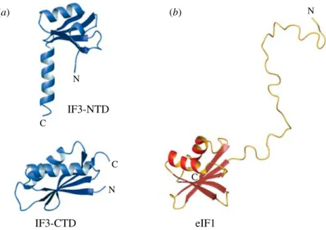
In bacteria, IF3 is responsible for subunit dissociation and anti-association. Its C-terminal domain (IF3-CTD) binds to the subunit interface surface of the platform of the small subunit, thus directly competing with the large subunit for binding. Binding of IF3-CTD to the small subunit is stabilized by the N-terminal domain (IF3-NTD) and/or the inter-domain linker (Dallas & Noller, 2001). The binding of IFs 1, 2, 3 and the initiator tRNA fMet-tRNAfMet to the small ribosomal subunit is strongly cooperative (Weiel & Hershey, 1982 ; Zucker & Hershey, 1986) and, therefore, all other factors also contribute to IF3’s anti-association activity. In eukaryotes, eIF3 (unrelated to IF3, see above and Table 2) carries the main subunit dis-sociation and anti-asdis-sociation function, and similarly to bacteria, its activity (and affinity for the small subunit) is enhanced in the presence of other factors : eIF1, eIF2 . GTP . Met-tRNAi, mRNA and small U-rich RNAs (Kolupaeva et al. 2005). eIF1 (unrelated to IF1 or IF3) binds to the same surface of the small subunit as IF3-CTD in bacteria, and its binding is cooperative with eIF3 binding (Pestova & Kolupaeva, 2002 ; Lomakin et al. 2003). Binding of eIF1 and eIF1A (homologous to IF1) to the small ribosomal subunit is cooperative (Maag & Lorsch, 2003)
and, thus, eIF1A can also indirectly promote anti-association. The ternary complex of eIF2 (unrelated to IF2) with GTP and Met-tRNAistabilizes eIF3 binding and also provides a steric block against subunit joining. As archaea do not have eIF3, subunit anti-association would have to rely on binding of eIF1, eIF1A and the ternary complex eIF2 . GTP . Met-tRNAi, with eIF1 and the ternary complex providing a steric block against subunit association.
4.3 Initiator aa-tRNA recognition
The selection of the initiator tRNA involves several tasks : (1) recognition of the initiator tRNA (proper charging) by the aaRS ; (2) discrimination against the initiator tRNA by EFs ; (3) discrimination against uncharged or mischarged initiator tRNA by IFs ; and (4) discrimination against elongator tRNAs by IFs.
The first task involves interaction of the aaRS with not only the acceptor end of the tRNA, but also directly with the anticodon. No elongation or IF directly binds the anticodon of the tRNA. In bacteria, there is a second enzymic step – formylation of Met-tRNAfMet, which has multiple roles : it increases the stability of the resulting fMet-tRNAfMetto deacylation, reduces the affinity for EF1A (task 2) and increases the affinity for IF2 (task 3). In eukaryotes, task 2 is accomplished by post-transcriptional modification of tRNAi. Recognition of properly charged initiator tRNA (tasks 3 and 4) is described below.
In bacteria, the initiator tRNA is tRNAfMet, and in eukaryotes it is tRNAi (or tRNAiMet) – specific for methionine, but distinct from the methionine-specific elongator tRNA (tRNAMet). The initiator tRNA must be charged with the correct amino acid, formylmethionine (fMet-tRNAfMet) in bacteria, and methionine (Met-tRNA
i) in eukaryotes. In bacteria, formylation increases the stability of fMet-tRNAfMetto deacylation, whereas the free eukaryotic Met-tRNA
i and the elongator aa-tRNAs are fairly unstable.
In bacteria, selection for fMet-tRNAfMet occurs at two levels. The G protein IF2 binds specifically to the fMet moiety and the acceptor end of the tRNA with moderate affinity (KD of y05mM) and has negligibly low affinity (KD >1 mM) for the deacylated tRNA and free fMet (Guenneugueset al. 2000). IF2 and fMet-tRNAfMetbind cooperatively to the small ribo-somal subunit. Whereas GTP or GDP binding and fMet-tRNAfMet binding to free IF2 are independent in solution, off the ribosome, it was reported that IF2 . GTP stabilizes fMet-tRNAfMetbinding to the small subunit more than IF2 . GDP does (Antounet al. 2003).
IF3 is also involved in tRNAfMetselection, but does not distinguish between acylated and deacylated tRNAfMet. IF3 does, however discriminate against elongator tRNAs and has been proposed to act indirectly – through the ribosome. The initiator tRNAfMethas three conserved GC base pairs (nucleotides 29–31 and 39–41) in the anticodon stem, which, upon IF3-induced conformational changes in the small subunit, could be directly inspected by the conserved nucleotides G1338 and A1339 in the head (Dallas & Noller, 2001).
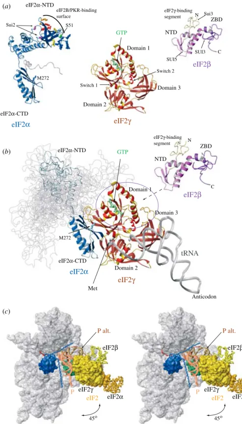
In archaea and eukaryotes, eIF2 (unrelated to IF2) is responsible for selection and recruitment of Met-tRNAito the ribosome. eIF2 is a heterotrimer and its biggest,csubunit is homologous to EF1A, eEF1A and SelB/eEFSec (Leibundgut et al. 2005). Like the EFs, eIF2 binds to Met-tRNAimore tightly in its GTP-bound form, recognizing determinants from both the tRNA and the Met moiety. As discussed in the previous section, the distinction between ‘ specific ’ and ‘ non-specific ’ binding is only quantitative and aa-tRNA recognition by the universal factors, EF1A/eEF1A and specific factors such as SelB/eEFSec is similar. The affinity of eIF2 for Met-tRNAi is y10 nM (Kapp & Lorsch, 2004), much higher than the affinity of bacterial
(structurally unrelated) IF2 for fMet-tRNAfMetand the release of eIF2 from Met-tRNA
irequires GTP hydrolysis by eIF2. The affinity of eIF2 for Met-tRNAidrops onlyy20-fold upon GTP hydrolysis, with y100-fold increase in dissociation rate ; therefore, eIF2 . GDP dissociation from Met-tRNAi may not be instantaneous (Kapp & Lorsch, 2004). After eIF2 hydrolyzes GTP and is released from the IC, the acceptor end of Met-tRNAiis free and could potentially interact with eIF5B (the eukaryotic homolog of IF2), which at that stage needs to be properly oriented to promote subunit joining (see Section 4.5). Such an interaction could stabilize Met-tRNAi binding and provide an additional level of selection for Met-tRNA for the time interval between eIF2 release and subunit joining. Unfortunately, direct binding between eIF5B and Met-tRNAi has not been reported and there is only indirect data in support of such an interaction (Choiet al. 1998, 2000 ; Marintchev et al. 2003). The putative interaction of eIF5B with Met-tRNAiwould be expected to be weak, in order for eIF5B not to compete with eIF2 off the ribosome. An additional difficulty is that Met-tRNAi is both unstable and difficult to prepare and label in large amounts.
There is no indication that eIF1, which is a functional analog of IF3, is involved in direct or indirect selection for tRNAi. However, the GC base pairs in the initiator tRNA and the corresponding nucleotides in the head of the small subunit, proposed to be involved in initiator tRNA selection (see above) are conserved between bacteria and eukaryotes, raising the possibility that at some stage, or under specific conditions, the eukaryotic small subunit could be involved in discrimination against elongator tRNAs.
4.4 Start site recognition 4.4.1 Overview
This point in translation initiation is the most divergent among kingdoms and one of the reasons why for a long time it was thought that eukaryotic and bacterial translation initiation are unrelated. Bacteria have a conserved sequence motif, called Shine–Dalgarno (SD) or ribosome-binding site (RBS), several nucleotides upstream of the start codon. The SD is complementary to the 3k-end of the 16S rRNA and the 5–7 nt spacer allows the start codon to be positioned in the P-site of the decoding center of the small subunit. The mRNAs in eubacteria usually contain more than one open reading frame (ORF), i.e. encode more than one protein, and the ribosomes assemble directly at the translation start sites. Regulation of translation initiation in bacteria is usually mediated by mRNA secondary structures and proteins binding at or near the SD element or the start codon (reviewed in Hershey & Merrick, 2000 ; Jackson, 2000). An interesting ‘ riboswitch control ’ mechanism was discovered recently, where small molecules bind directly to the 5k-leader sequence of mRNAs encoding enzymes involved in their metabolism, causing rearrangement of secondary structures. Depending on the position and nature of the affected secondary structure elements, the metabolites stimulate or inhibit the expression of the enzymes at the level of transcription or translation initiation (reviewed in Nudler & Mironov, 2004). No structures are available yet for riboswitches regulating translation initiation, but the first structure of a riboswitch that regulates transcription was recently published : the purine-binding domain of the guanine riboswitch in complex with hypoxanthine (Bateyet al. 2004).
In eukaryotes, the majority of mRNAs contain only one ORF ; their 5k-end is ‘ capped ’ with an m7
G-cap through a reverse, 5kx5kbond ; and they have a poly-A tail at their 3k-end. Both the 5k-cap and the 3k-poly-A tail are important for efficient translation. The 43S IC is first recruited to the 5k-cap through interactions with a set of eIFs, called eIF4F or cap-binding complex. The
IC then scans along the mRNA until the start codon. This is usually, but not always, the first AUG, as the nucleotide context around the AUG significantly influences initiation efficiency. The optimal sequence context for the AUG start codon in higher eukaryotes is GCCA/ GCCAUGG, (Kozak, 1986, 1987). The most important nucleotides are the A or G at position
x3 (where the A of the AUG codon is+1) and the G at+4. The consensus sequence context in plants and other eukaryotes is similar to that in vertebrates, although it may be quite different in some organisms, such asS. cerevisiae(reviewed in Kozak, 1991). The length and the presence or absence of secondary structure in the 5k-untranslated region (5k-UTR) are major determinants of translation efficiency. In addition to the 3k-poly-A tail, sequences in the 3k-untranslated region (3k-UTR) often regulate translation, usually serving as binding sites for translation regulators. Regulatory proteins binding to the 5k-UTR have also been found. A number of eukaryotic mRNAs, including many viral mRNAs, have an alternative mode of translation : the ribosome is recruited directly to an internal site at or near the start codon, called internal ribosome entry site (IRES). The IRESs can be fairly long and structured RNA segments. There are several types of IRES, differing in both length and structure of the RNA. Translation initiation at an IRES is typically independent of the presence of a 5k-cap and requires only a subset of the eIFs, involved in canonical cap-dependent translation initiation. The factor requirements vary among different types of IRESs (reviewed in Jackson, 2000 ; Pestovaet al. 2001). On the extreme is the case of the cricket paralysis virus IRES, which does not even require Met-tRNAi(Pestova & Hellen, 2003). In addition to the canonical and IRES-dependent translation initiation, the scanning IC has been found to skip RNA segments and resume scanning on certain mRNAs. Since the 3k-end of the eukaryotic 18S rRNA is not complementary to the bacterial SD sequence (or to any sequence near the start site of the eukaryotic mRNA), the eukaryotic apparatus cannot recognize a bacterial translation start site. Similarly, bacteria cannot recognize the start signals in eukaryotic mRNAs. For an in-depth review of start site recognition mechanisms in bacteria and eukaryotes, and their variations see Jackson (2000).
Archaeal translation is not as well studied as translation in eubacteria and eukaryotes. Like eubacteria, archaea have SD-like sequences and polycistronic mRNAs, which are not capped or polyadenylated. However, it appears that in most mRNAs, the first start codon is at or near the 5k-end and is not preceded by an SD sequence. SD elements are used mainly in polycistronic mRNAs for initiation at internal start sites. mRNAs, where the start site is at or within a few nucleotides from the 5k-end, are called ‘ leaderless ’ mRNAs. They have also been found in eubacteria and eukaryotes, and, unlike other types of mRNA, leaderless mRNAs can be trans-lated in cell extracts derived from all kingdoms (reviewed in Mollet al. 2002). The properties of leaderless mRNAs make them particularly interesting from evolutionary and mechanistic perspectives and will be discussed in more detail in Section 4.7 below.
4.4.2 Roles of individual factors
In addition to the small ribosomal subunit, which recognizes the SD element, bacterial IF3 has a central role in start site selection : it can dissociate ICs assembled on non-canonical codons or on canonical codons located at or near the 5k-end. Although AUG is the predominant start codon in bacteria (y90 %), GUG and UUG are also considered ‘ canonical ’ and representy8 % andy1 % of the start codons respectively. IF3 cannot dissociate ICs preformed on canonical start sites and is released upon start site recognition (reviewed in Hershey & Merrick, 2000). This indicates that the small subunit can spontaneously undergo the conformational changes
associated with start site recognition, leading to a state with lower affinity for IF3. The discrimination between ‘ good ’ and ‘ bad ’ start sites is indirect and based on the balance between the stabilities of the two alternative conformations : IF3 is able to reverse the changes in the IC, unless they are stabilized by proper interactions of the small subunit, the initiator tRNA, and the mRNA (Dallas & Noller, 2001). As no IF in either bacteria or eukaryotes directly ‘ inspects ’ the start codon, the identity of AUG as the start codon is defined by the anticodon of the initiator tRNA (reviewed in Hershey & Merrick, 2000 ; Hinnebusch, 2000).
In eukaryotes, the dynamic discrimination between ‘ good ’ and ‘ bad ’ start sites is taken to a new level of complexity and multiple factors are involved. As translation initiation on most mRNAs involves scanning from the 5k-cap to the start codon, selection needs to be achieved in the context of an IC sliding along mRNA and efficient discrimination against incorrect initiation sites must be sustained over longer periods of time. The processes of 5k-cap and 3k-poly-A recognition and scanning do not have bacterial or archaeal counterparts and will be discussed separately in Section 4.6 below.
eIF1 discriminates against non-AUG codons and its mechanism of action is probably similar to that proposed for IF3 (see above), as is its binding site on the small ribosomal subunit (Lomakinet al. 2003). In eukaryotes, only AUG is a ‘ canonical ’ start codon, but UUG and GUG appear to be the preferred ‘ non-canonical ’ start codons (see Section 49 below). eIF1 dis-criminates also between ‘ good ’ and ‘ bad ’ nucleotide context of the start codon (the ‘ Kozak ’ consensus element). This function of eIF1 is performed indirectly – via conformational changes in the small ribosomal subunit and may not be based on sequence-specific recognition by the ribosome, but rather on the propensity of the mRNA segment for the conformation required to fit in the mRNA-binding groove of the small subunit. eIF1, similarly to bacterial IF3, also prevents formation of ICs on AUG codons located at or near the 5k-end (reviewed in Hershey & Merrick, 2000 ; Pestovaet al. 2001).
Other factors directly involved in start codon selection are eIF2 and eIF5. eIF2 in its GTP-bound form brings Met-tRNAi to the IC. Upon start codon recognition, eIF2 hydrolyzes GTP and is subsequently released from the IC. In eukaryotes, GTP hydrolysis requires the presence of eIF5, which serves as a GTPase-activating protein (GAP) for eIF2. GTP hydrolysis and the subsequent release of eIF2 are required for progression of the ICs toward the next stage – subunit joining. Accordingly, an increase in the rate of GTP hydrolysis by eIF2 or decrease of its affinity for Met-tRNAi leads to higher error rates of initiation (reviewed in Donahue, 2000 ; Hershey & Merrick, 2000 ; Pestova et al. 2001). Conversely, any factor that stabilizes binding of eIF1 to the IC, like eIF3, for example, would be expected to promote higher fidelity of initiation.
As mentioned above, archaea have SD-like elements, but also many leaderless mRNAs. Furthermore, although archaea have eIF1 and eIF2, they do not have eIF3 and eIF5. The absence of eIF3 suggests that eIF1 binding to the IC may be weaker than in eukaryotes. Consequently, eIF1-mediated start site discrimination could be less efficient.
A parallel between archaea and eukaryotes suggests that eIF2 GTP hydrolysis and nucleotide exchange are more stringently controlled in eukaryotes, hence the need for a GAP (eIF5) and a GEF (eIF2B). Given the high degree of homology between eukaryotic and archaeal eIF2, the eukaryotic factor probably has retained the intrinsic ability to hydrolyze GTP, but this activity is efficiently repressed. According to this scenario, an alternative role of eIF5 could be to help derepress the GTPase activity of eIF2, rather than (or in addition to) acting as a classical GAP factor, stabilizing a transition state in GTP hydrolysis. In support of such an interpretation,
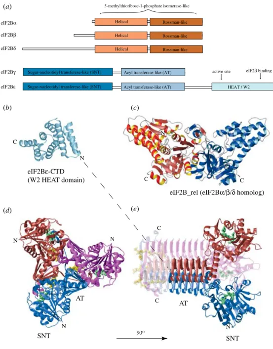
eIF5 is homologous to the two core domains of eIF2b, covering almost the entire length of archaeal eIF2b, except for a short eIF2c-binding segment that is absent in eIF5 (eukaryotic eIF2band eIF5 also have additional, non-homologous segments, responsible for mutual binding, see Section 5). Furthermore, mutations in, or deletion of the second of the eIF2b domains shared with eIF5 cause increased rate of spontaneous GTP hydrolysis by eIF2 (Donahue, 2000 ; Hashimotoet al. 2002).
4.5 Subunit joining and factor release
Subunit joining depends on the proper orientation of the initiator aa-tRNA, achieved upon start codon recognition. A small subunit with an initiator aa-tRNA in the P-site base-paired to the start AUG codon of mRNA can bind to the large subunit and form a translationally active ribosome, in the absence of additional factors. In all kingdoms subunit joining is promoted by a universally conserved G-protein, called IF2 in bacteria and eIF5B in eukaryotes, which, like the initiator tRNA, needs to be properly positioned. The other universally conserved factor, IF1/eIF1A (in bacteria and eukaryotes respectively), when bound alone to the small subunit, stimulates the rates of both subunit joining and dissociation, and also stabilizes binding of IF2/eIF5B to the small subunit. The two factors appear to be coordinately released after subunit joining in all kingdoms (Benneet al. 1973 ; Choi et al. 2000 ; Olsen et al. 2003) and therefore IF1/eIF1A is likely also involved in subunit joining at the end of translation initiation.
In addition to proper positioning of the initiator tRNA, subunit joining requires certain IFs to be released or at least displaced from their original location. The event that triggers factor release in all kingdoms is start site recognition. As explained above, both bacterial IF3 and eukaryotic eIF1 bind to the interface surface of the small subunit and need to be displaced from there before subunit joining. In eukaryotes, start site recognition also induces GTP hydrolysis by eIF2 and release of eIF2 . GDP. There is no obvious need for the IFs to physically dissociate from the 40S subunit in order for subunit joining to occur, as long as they do not block the interaction with the 60S subunit either directly or indirectly. After subunit joining, eIFs 1, 2, 3 and 5 do not co-sediment with the ICs in sucrose gradient centrifugation and could already be released at that stage. Alternatively, the remaining interactions are too weak to withstand centrifugation. For example, eIF3 is involved in several interactions with the 40S subunit, some of which involve solvent-exposed surfaces and can be retained even after subunit joining. Therefore, eIF3 and other eIFs could still be associated with the ICs during and even after subunit joining ( Jackson, 2000). eIF3 also interacts with mRNA, with affinity dependent on RNA structure and/or sequence. It was found that eIF3 stays associated with the 40S subunit after the release of eIF2, and eIF1 could also be associated with such complexes through its interaction with eIF3 (Unbehaunet al. 2004). Recently, direct evidence for transient presence of IFs after the onset of elongation was obtained in mammals (Poyryet al. 2004), as well as more indirect indication in yeast (Rajkowitschet al. 2004). Both reports demonstrated time-dependence of the ability of ribosomes to reinitiate after translating a short ORF. The former group dem-onstrated direct requirement for the presence of eIF4F or at least the central domain of eIF4G and eIF4A in the first IC (for the short ORF) and that these eIFs could not bind after the short ORF was already translated. As eIF4G binding to the 48S IC is mediated by eIF3, the above results implied that eIF3, and possibly most other factors associated with it, can remain on the 80S for a short period of time. Clearly, eIF2 . GTP . Met-tRNAi needs to be
regenerated from eIF2 . GDP and tRNAi, before reinitiation can occur. There was no apparent need for either eIF4E or PABP (Poyryet al. 2004). As discussed in Section 4.6 below, it is not clear if and with what rates eIF4E dissociates from the cap and/or eIF4G. There is also no indication whether binding to eIF4E or PABP affects the association of eIF4G with the IC during scanning, at the start codon or after subunit joining. There are somewhat con-troversial reports that eIF2 stays on the 40S subunit even after being released from Met-tRNAi, and is later transferred to the 60S subunit (Ramaiah et al. 1992), but other authors found the association to be unstable under physiological conditions (Chakrabarti & Maitra, 1992). In view of the above results, transient association of eIF2 . GDP with the ribosomes does not seem surprising.
The dependence of reinitiation on the presence of a set of eIFs on the terminating ribosome provides an explanation for the rather unexpected finding that deletion or mutations of eIF5B that slow down subunit joining or release of eIF5B after subunit joining, inhibit reinitiation (Lee et al. 2002 ; Shinet al. 2002). Both slow subunit joining and slow eIF5B release delay the onset of elongation and extend the time interval between GTP hydrolysis by eIF2 and ter-mination, thus allowing the eIFs to dissociate. This interpretation relies on the assumption that the association of at least part of the eIFs required for reinitiation with the 48S complex is destabilized upon GTP hydrolysis by eIF2, which is supported by the different stability and composition of 48S complexes subjected to centrifugation before and after GTP hydrolysis by eIF2.
IF2/eIF5B and IF1/eIF1A are coordinately released after subunit joining (Benneet al. 1973 ; Choiet al. 2000 ; Olsenet al. 2003). The release is triggered by GTP hydrolysis by IF2/eIF5B. The GTPase activity of IF2/eIF5B (like the GTPase activities of the elongation factors EF1A/ eEF1A and EF2/eEF2) is stimulated by interaction with the GAC on the large ribosomal subunit. IF2/eIF5B . GDP has lower affinity for the ribosome than IF2/eIF5B . GTP and dis-sociates quickly (Benneet al. 1973 ; Pestovaet al. 2000 ; Leeet al. 2002 ; Shinet al. 2002 ; Antoun et al. 2003).
One kinetic study usingE. coliribosomes and factors andB. stearothermophilusIF2 found no difference in the activity of IF2 . GTPversusIF2 . GDP, and no role of GTP hydrolysis in release of IF2 upon subunit joining, contradicting previous data and casting doubts over the similarity of subunit joining between bacteria and eukaryotes (Tomsicet al. 2000). A recent report from the Ehrenberg group reconfirmed the importance of GTP binding and hydrolysis by IF2 for subunit joining (Antounet al. 2003). It was proposed that the conflicting results in (Tomsicet al. 2000) may have been due to possible contamination with GTP during the preparation of IF2 (Antounet al. 2003). IF2 from the thermophilicB. stearothermophilus, used in (Tomsicet al. 2000), is known to bind more tightly to the E. coli ribosome than the endogenous IF2 from E. coli (Brombachet al. 1986 ; Severiniet al. 1990). The use of a foreign protein with high affinity for the E. coliribosome is convenient for biochemical assays (Brombachet al. 1986 ; Severiniet al. 1990), but hardly appropriate for kinetic analysis of translation initiation in E. coli, which affects the validity of the obtained results. A detailed explanation of the expected effects of such an IF2 variant/mutant with high affinity for the ribosome in its GDP-bound state can be found in Antoun et al. (2003). Briefly, if a mutant (or exogenous) IF2 . GDP binds too tightly to the ribosome, it will have a slower rate of dissociation after subunit joining, which can, depending on the 50S subunit concentration, make IF2 . GDP dissociation the rate-limiting step. The end result is that no difference in the activity of the mutant IF2 . GTP and IF2 . GDP will be observed, because the processes will be equally slow.