Eosinophilic esophagitis: Updated consensus
recommendations for children and adults
Chris A. Liacouras, MD, Glenn T. Furuta, MD, Ikuo Hirano, MD, Dan Atkins, MD, Stephen E. Attwood, MD, FRCS, FRCSI, MCh, Peter A. Bonis, MD, A. Wesley Burks, MD, Mirna Chehade, MD, Margaret H. Collins, MD, Evan S. Dellon, MD, MPH,
Ranjan Dohil, MD, Gary W. Falk, MD, MS, Nirmala Gonsalves, MD, Sandeep K. Gupta, MD, David A. Katzka, MD, Alfredo J. Lucendo, MD, PhD, Jonathan E. Markowitz, MD, MSCE, Richard J. Noel, MD, Robert D. Odze, MD, FRCP,
Philip E. Putnam, MD, FAAP, Joel E. Richter, MD, FACP, MACG, Yvonne Romero, MD, Eduardo Ruchelli, MD, Hugh A. Sampson, MD, Alain Schoepfer, MD, Nicholas J. Shaheen, MD, MPH, Scott H. Sicherer, MD, Stuart Spechler, MD, Jonathan M. Spergel, MD, PhD, Alex Straumann, MD, Barry K. Wershil, MD, Marc E. Rothenberg, MD, PhD,* and Seema S. Aceves, MD, PhD* Aurora and Denver, Colo, Milwaukee, Wis, Cincinnati, Ohio, Rochester, Minn, Philadelphia, Pa, Basel and Lausanne, Switzerland, Chapel Hill and Durham, NC, Boston, Mass, Chicago, Ill, San Diego, Calif, New York, NY, Indianapolis, Ind, Tomelloso, Spain, Greenville, SC, and North Shields, United Kingdom
INFORMATION FOR CATEGORY 1 CME CREDIT
Credit can now be obtained, free for a limited time, by reading the review articles in this issue. Please note the following instructions.
Method of Physician Participation in Learning Process:The core material for these activities can be read in this issue of the Journal or online at the JACI Web site:www.jacionline.org. The accompanying tests may only be submitted online at
www.jacionline.org. Fax or other copies will not be accepted.
Date of Original Release: July 2011. Credit may be obtained for these courses until June 30, 2013.
Copyright Statement:CopyrightÓ2011-2013. All rights reserved. Overall Purpose/Goal:To provide excellent reviews on key aspects of aller-gic disease to those who research, treat, or manage alleraller-gic disease.
Target Audience:Physicians and researchers within the field of allergic disease.
Accreditation/Provider Statements and Credit Designation:The Ameri-can Academy of Allergy, Asthma & Immunology (AAAAI) is accredited by the Accreditation Council for Continuing Medical Education (ACCME) to pro-vide continuing medical education for physicians. The AAAAI designates these educational activities for a maximum of 1AMA PRA Category 1 Creditä. Phy-sicians should only claim credit commensurate with the extent of their participa-tion in the activity.
Activity Objectives
1. To define the diagnostic guidelines for eosinophilic esophagitis (EoE). 2. To list and define allergic manifestations and allergy tests related to
an-tigenic causes of the disease.
3. To list and define dietary and medical treatments of EoE.
Recognition of Commercial Support:This CME activity has not received external commercial support.
Disclosure of Significant Relationships with Relevant Commercial Com-panies/Organizations:C. A. Liacouras is a consultant for Cephalon, a speaker for Nutricia and Abbott, and a physician member of the American Partnership for Eosinophilic Disorders. G. T. Furuta is a consultant for Meritage and Sunovion and has received research support from the National Institutes of Health and the American Partnership for Eosinophilic Disorders. I. Hirano is a consultant for Meritage Pharma. D. Atkins is a consultant for Sunovion and has received re-search support from AstraZeneca. P. A. Bonis has received rere-search support from TIGER. A. W. Burks is a consultant for ActoGeniX NV, Intelliject, McNeil Nutritionals, Novartis, Pfizer, and Schering-Plough; is a minority stockholder for Allertein and MastCell, Inc; is on the Dannon Co Probiotics Advisory Board; is on the Nutricia Expert Panel; has received research support from the National
Institutes of Health, the Food Allergy and Anaphylaxis Network (FAAN), and the Wallace Research Foundation; has provided legal consultation/expert witness testimony on the topic of food allergy; is on the FAAN medical Board of Directors; is an American College of Allergy, Asthma & Immunology (ACAAI) Dermato-logical Allergy Committee Member; is a National Institutes of Health (NIH) HAI Study Section Member; is on the US Food and Drug Administration (FDA) Reviewer Board; and is on the Journal of Allergy and Clinical Immunology (JACI) Editorial Board. M. H. Collins is a consultant for Sunovion, Meritage Pharma, and Cephalon and is a member of the American Partnership for Eosino-philic Disorders (APFED) medical advisory panel. E. S. Dellon has received re-search support from AstraZeneca, the American College of Gastroenterology, and the NIH/UNC. R. Dohil is a stockholder in Meritage Pharmaceuticals. G. W. Falk is a consultant for Eurand. N. Gonsalves has received research support from the Campaign Urging Research for Eosinophilic Disease (CURED) and Da-vid & Denise Bunning. S. K. Gupta is a speaker for Abbott and a consultant for Baxter and Meritage. H. A. Sampson is a consultant for Allertein Therapeutics, LLC; has received research support from the Food Allergy Initiative and the Na-tional Institute for Allergy and Infectious Diseases (NIAID)/NIH; is a consultant and scientific advisor for the Food Allergy Initiative; is a medical advisor for the FAAN; is a scientific advisor for the University of Nebraska–Food Allergy Re-search and Resource Program (FARRP); and is 45% owner of Herbal Springs, LLC. A. Schoepfer has received research support from AstraZeneca, GlaxoSmith-Kline, and Dr Falk Ah. N. J. Shaheen is a consultant for AstraZeneca. S. H. Sicherer is a consultant for the Food Allergy Initiative and has received research support from the NIAID/NIH. S. Spechler is a consultant for Torax Medical, Xenoport, and Ironwood Pharmaceuticals and has received research support from Takeda Pharmaceuticals. J. M. Spergel is on the DBV Scientific board of directors; has re-ceived research support from Cephalon, APFED, and the NIH/DOD; and is on the APFED Medial Advisory Board and the American Academy of Allergy, Asthma & Immunology (AAAAI)’s Annual Meeting Program Committee. B. K. Wershil is a speaker for Prometheus Pharmaceutical, Inc; has received research support from Meritage; and is Secretary/Treasurer for Children’s Digestive Health Foundation. M. E. Rothenberg has equity interest to reslizumab in Cephalon; is a consultant for Array Biopharma, Biocrystal Pharmaceuticals, and Endo Pharmaceuticals; has received research support from the National Institutes of Health, the FAAN, and the Hana Foundation; and is on the American Partnership for Eosinophilic Disor-ders Medical Advisory Board and the International Eosinophilic Society Executive Council. S. S. Aceves is coinventor of the Meritage Pharma Oral Viscous Budeso-nide and is chair of the medical advisory panel of APFED. The rest of the authors have declared that they have no conflict of interest.
Eosinophilic esophagitis (EoE) is a clinicopathologic condition of increasing recognition and prevalence. In 2007, a consensus recommendation provided clinical and histopathologic
guidance for the diagnosis and treatment of EoE; however, only a minority of physicians use the 2007 guidelines, which require fulfillment of both histologic and clinical features. Since 2007, the number of EoE publications has doubled, providing new disease insight. Accordingly, a panel of 33 physicians with expertise in pediatric and adult allergy/immunology,
gastroenterology, and pathology conducted a systematic review of the EoE literature (since September 2006) using electronic databases. Based on the literature review and expertise of the panel, information and recommendations were provided in each of the following areas of EoE: diagnostics, genetics, allergy testing, therapeutics, and disease complications. Because accumulating animal and human data have provided evidence that EoE appears to be an antigen-driven immunologic process that involves multiple pathogenic pathways, a new conceptual definition is proposed highlighting that EoE represents a chronic, immune/antigen-mediated disease characterized clinically by symptoms related to esophageal dysfunction and histologically by eosinophil-predominant inflammation. The diagnostic guidelines continue to define EoE as an isolated chronic disorder of the esophagus diagnosed by the need of both clinical and pathologic features. Patients commonly have high rates of concurrent allergic diatheses, especially food
sensitization, compared with the general population. Proved therapeutic options include chronic dietary elimination, topical corticosteroids, and esophageal dilation. Important additions since 2007 include genetic underpinnings that implicate EoE susceptibility caused by polymorphisms in the thymic stromal lymphopoietin protein gene and the description of a new potential disease phenotype, proton pump inhibitor-responsive esophageal eosinophila. Further advances and controversies regarding diagnostic methods, surrogate disease markers, allergy testing, and treatment approaches are discussed. (J Allergy Clin Immunol 2011;128:3-20.)
Key words: Eosinophils, eosinophilic, esophagitis, esophageal, food allergy
Discuss this article on the JACI Journal Club blog: www. jaci-online.blogspot.com.
Since the publication of the eosinophilic esophagitis (EoE) consensus recommendation (CR) in 2007,1scientific publications focusing on EoE have nearly doubled, and the recognition of patients who have eosinophil-predominant esophagitis has in-creased dramatically. Early studies described aspects of the condi-tion in children, but it has become clear that adults have a similar
disorder. Practice patterns in multiple subspecialties (adult and pe-diatric gastroenterology, allergy/immunology, pulmonary medi-cine, and otolaryngology) have begun to include EoE in the differential diagnosis of various clinical presentations. Increased recognition, along with the chronic nature of EoE, has led to a steady increase in prevalence.
A salient aspect of the 2007 CR was that EoE was defined as a clinicopathologic entity in which esophageal eosinophilia was a necessary but not sufficient criterion for diagnosis. Most recent publications pertaining to EoE, however, have included cohorts of patients in whom the diagnosis was based solely on the histologic finding of esophageal eosinophilia. In fact, a recent study and a 2010 survey of 1836 physician members of the American College of Gastroenterology, the American Academy of Allergy, Asthma & Immunology, and the North American Society of Pediatric Gastroenterology, Hepatology, and Nutrition identified that only one third of respondents used the CR definition.2,3 Furthermore, the 2007 CR statements regarding the clinical presentation, pathogenesis, treatment, and complica-tions have elucidated ambiguities and controversy in the context of expanding clinical experience and increased scientific investigation.
To address these concerns, an interdisciplinary expert panel was convened with the following goals: (1) to provide clarity to the definition, nomenclature, clinical presentation, histology, and diagnostic testing; (2) to report on various disease phenotypes that might exist; (3) to evaluate allergic manifestations and allergy tests related to antigenic causes of the disease; (4) to review, reassess, and provide recommendations on dietary and medical treatments; and (5) to review the use of esophageal dilation and complications associated with EoE. The interdisciplinary panel subsequently generated this report with the hope that the follow-ing information will provide a framework to improve care and develop future studies on EoE.
METHODOLOGY
A task force of 33 physicians with recognized expertise in the clinical evaluation, endoscopy, histopathology, genetics, allergy, and treatment of EoE was gathered to address specific clinically relevant topics. The expert panel consisted of pediatric and adult gastroenterologists, allergists, and pathologists. A systematic review of the English-language medical literature between September 2006 and August 2010 was performed by using electronic databases (MEDLINE, PubMed, and Ovid). Relevant data were discussed among committee members in a series of conference calls. Critical evaluations included study design, numbers of patients, definitions used, outcomes reported, and potential biases. The chair of each committee synthesized the Complete information for the authors, including affiliations and study responsibilities, is
shown inAppendix E1in this article’s Online Repository atwww.jacionline.org. *These authors contributed equally to this work.
Received for publication February 12, 2011; accepted for publication February 17, 2011. Available online April 7, 2011.
Reprint requests: Chris A. Liacouras, MD, Exton Specialty Center, The Children’s Hos-pital of Philadelphia, 481 John Young Way, Exton, PA 19341. E-mail:liacouras@ email.chop.edu.
0091-6749/$36.00
Ó2011 American Academy of Allergy, Asthma & Immunology doi:10.1016/j.jaci.2011.02.040
Abbreviations used
APT: Atopy patch test
CR: Consensus recommendation EoE: Eosinophilic esophagitis GERD: Gastroesophageal reflux disease
hpf: High-power field PPI: Proton pump inhibitor SPT: Skin prick test
data, and inconsistencies were resolved by means of discussion until a consensus was achieved. The recommendations of each committee included a review and update of the 2007 Consensus Report, clinical recommendations, and proposals for future research. The manuscript was reviewed and approved by all 33 participants.
REVIEW OF 2007 CR AND GUIDELINES
The original 2007 consensus definition of EoE, based on both a threshold number of eosinophils and clinical parameters of upper intestinal symptoms for which gastroesophageal reflux disease (GERD) was not the underlying cause, might have been problematic. First, the histologic finding of 15 or more eosino-phils per high-power field (hpf) in an esophageal biopsy spec-imen carries no proved biological significance or power to discriminate among various esophageal diseases. Second, the exhortation to eliminate GERD as a potential cause of esopha-geal eosinophilia (as determined by best clinical practice, which can include either failure of proton pump inhibitors [PPIs] to resolve symptoms and ongoing eosinophilia or a normal pH impedance monitoring study) has not been rigorously applied or validated.
The reasons for this are multiple and relate, in part, to the identification of at least 2 groups of patients with eosinophil-predominant esophageal inflammation. One group, best described as having GERD with more eosinophils than usual, has abnormal pH monitoring results and a clinicopathologic response to PPIs.4,5 Another group, best described as having PPI-responsive esopha-geal eosinophilia, has normal pH study results but nevertheless shows a clinicopathologic response to proton pump inhibition.6 Whether this latter group represents GERD that was not identified by means of pH impedance–monitoring studies*or a clinical re-sponse to the potential anti-inflammatory properties of PPIs is not yet certain. In neither of these groups has an association with an antigenic or immunologic cause of esophageal eosino-philia been thoroughly studied.
Another inherent problem was the use of the abbreviation ‘‘EE.’’ Although easily understood by allergists and pathologists, among gastroenterologists EE classically defines ‘‘erosive esoph-agitis.’’ The use of the abbreviation EoE rather than EE for EoE should eliminate the potential for this confusion.
No studies have been published since the CR that would clearly permit diagnosis or phenotype discrimination based on pathogno-monic clinical/histologic features or biomarkers. Thus although many studies performed since 2007 have used the 2007 CR as proposed, the majority do not, leaving diagnostic uncertainty both for patients and within the published literature.7Because of the in-creasing recognition of patients with esophageal eosinophilia and the clinical demand for a more relevant diagnostic guideline, an ur-gent need has developed to revise the previously published CR.8-10
CONCEPTUAL DEFINITION AND DIAGNOSTIC GUIDELINES FOR EoE
Proposed conceptual definition of EoE
Recognition of the differences between esophageal eosino-philia as a histologic descriptor and EoE as a disease, in itself, is critical (Table I). As clinical experience has developed and as more patients are being identified, varying phenotypes based on symptoms or anatomic abnormalities (eg, stricturing) might de-fine a ‘‘spectrum’’ of EoE. As supported by a number of past and recent basic/translational studies and clinical experience demonstrating that the underlying cause of EoE is likely an aber-rant ‘‘immune’’ or ‘‘antigenic’’ response associated with consis-tent endoscopic, histologic, and genetic abnormalities, a conceptual definition for EoE is proposed.11-45Use of this con-ceptual definition not only will provide a framework to refine our perceptions and hypotheses but also will guide future diagnos-tic tests, therapeudiagnos-tic modalities, and pathogenediagnos-tic studies on EoE.
Conceptual definition
Eosinophilic esophagitis represents a chronic, immune/ antigen-mediated esophageal disease characterized clinically by symptoms related to esophageal dysfunction and histologically by eosinophil-predominant inflammation.
Proposed diagnostic guideline for EoE
In conjunction with this conceptual definition of EoE, recent clinical experience and research supports revisions in the original diagnostic guidelines for EoE. Rationales for statements in the current guidelines compared with the 2007 CR are listed inTable II.
Diagnostic guideline
EoE is a clinicopathologic disease. Clinically, EoE is characterized by symptoms related to esophageal dysfunction. Pathologically, 1 or more biopsy specimens must show eosinophil-predominant inflammation. With few exceptions, 15 eosinophils/hpf (peak value) is considered a minimum threshold for a diagnosis of EoE. The disease is isolated to the esophagus, and other causes of esophageal eosinophilia should be excluded, specifically PPI-responsive esophageal eosinophilia. The disease should remit with treatments of dietary exclusion, topical corti-costeroids, or both. EoE should be diagnosed by clinicians, taking into consideration all clinical and pathologic information; neither of these parameters should be interpreted in isolation.
TABLE I.Diseases associated with esophageal eosinophilia
GERD EoE
Eosinophilic gastrointestinal diseases Celiac disease
Crohn disease Infection
Hypereosinophilic syndrome Achalasia
Drug hypersensitivity Vasculitis
Pemphigoid vegetans Connective tissue disease Graft-versus-host disease
For optimal pathologic evaluation, multiple biopsy specimens from the proximal and distal esophagus should be obtained and evaluated for a variety of pathologic features. Pathologists should report all abnormalities associated with EoE, such as the peak eosinophil value (obtained from the area with the highest density of eosinophils), eosinophilic microabscesses, surface layering of eosinophils, extracellular eosinophil granules, basal cell hyper-plasia, dilated intercellular spaces, and lamina propria fibrosis. In a few circumstances patients might have strong clinical evidence for EoE and have less than 15 eosinophils/hpf, with other histologic features indicative of eosinophilic inflammation.
An emerging body of literature and clinical experience de-scribes a subset of patients whose symptoms and histopathologic findings are responsive to PPI treatment and who might or might not have well-documented GERD. Until more is known regarding this subgroup of patients, these patients should be given diagnoses of PPI-responsive esophageal eosinophilia. Future studies should be performed to determine whether PPIs help to diminish an immune/antigen-driven response, as is known to occur in patients with EoE.
SUMMARY OF LITERATURE SINCE THE 2007 CR History and physical examination
Update of 2007 recommendations.Several studies have confirmed previously described clinical features of EoE, but no pathognomonic features have been identified. Clinical manifes-tations of EoE in children are nonspecific and vary by age such that diagnosis based on symptoms alone is not feasible. Infants and toddlers often present with feeding difficulties, whereas school-aged children are more likely to present with vomiting or pain.46,47Dysphagia is a predominant symptom in adolescents. EoE in children is most often present in association with other manifestations of atopic diathesis (food allergy, asthma, eczema, chronic rhinitis, and environmental allergies) and is responsive to elimination of specific dietary antigens in that population.
The typical patient with EoE is an atopic male (male/female ratio, 3:1) who presents in childhood or during the third or fourth
decades of life; however, EoE can occur at any age.48,49EoE oc-curs in most racial and ethnic groups, although many studies have reported predominance in non-Hispanic whites; the reason for this requires further investigation.50-54 Physical examinations are useful in children to identify normal growth patterns and in both children and adults to identify comorbid allergic diseases; however, no features on physical examination are specific in mak-ing the diagnosis of EoE. In addition, no oral or pharyngeal man-ifestations of EoE have been identified, although some children who have EoE might present with laryngeal symptoms.55
Symptoms in adult patients with EoE are somewhat stereotyp-ical and include dysphagia, chest pain, food impaction, and upper abdominal pain. Solid-food dysphagia continues to be the most common presenting symptom.48,49,56,57When examining all pa-tients presenting with dysphagia in endoscopy units, EoE has a prevalence of up to 15%.56,57In some series chest pain is the second leading symptom in adults with EoE.6,58Whether chest pain from EoE can be differentiated from GERD or is due to esophageal hy-persensitivity to acid remains to be determined.6,58,59,60Food im-paction necessitating endoscopic bolus removal occurs in 33% to 54% of adults with EoE.61 Upper abdominal pain, symptoms of GERD, and nonspecific throat symptoms, including globus, have also been reported in some adults with EoE. Recent clinical obser-vations suggest that chest discomfort associated with EoE might have different features than those reported in patients with GERD. A subgroup of patients has been increasingly recognized who have (1) a typical EoE symptom presentation, (2) have had GERD diagnostically excluded, and (3) demonstrated a clinicopathologic response to PPIs.6,58,59,62,63Terms used to describe these patients in-cludePPI-responsive esophageal eosinophiliaandPPI-responsive EoE. The latest term is controversial because limited evidence to support the effect of PPIs in an ‘‘immune/antigen-driven’’ inflam-matory response exists. Potential explanations include healing of a disrupted epithelial barrier to prevent further immune activation, decreased eosinophil longevity, inherent anti-inflammatory proper-ties of PPIs, or unreliable diagnostic testing.64-67
A validated symptom-assessment tool is not available, such that recent studies attempting to correlate symptoms with histology TABLE II.Rationale for definition of and diagnostic guidelines for EoE
1.Change in EE abbreviation.EE often has been used as an abbreviation for erosive esophagitis. Use of the abbreviation EoE rather than EE for eosinophilic esophagitis should eliminate the potential for confusion.
2.Inclusion of the word chronic.Clinical experience supports that EoE is a chronic disease that will require long-term follow-up and treatment. 3.Inclusion of the term immune/antigen driven.An increasing body of clinical, translational, and basic evidence supports a role of an aberrant immune
response (potentially reversible with treatment) as an underlying pathogenetic feature of EoE.
4.Continued use of the word clinicopathologic. No biomarker or pathognomonic element has been identified that would eliminate the need for both symptoms and an abnormal histology to make the diagnosis.
5.No change in threshold number of 15 eosinophils/hpf.Since the 2007 CR, no studies have identified a clear ‘‘lower limit of esophageal eosinophilia’’ or threshold number that would define EoE or have identified other histologic features or pattern of disease distribution that are pathognomonic of EoE. 6.No change in the use of hpf as the unit of measurement for eosinophilia.No studies have yet determined a standardized size of an hpf, and this might be
practically unachievable. This issue is problematic because the size of an hpf can alter the reported number of eosinophils per hpf.
7.Inclusion of topical steroids/diet exclusions as a treatment. Current clinical evidence exists to include this paradigm to differentiate EoE from other diseases. Other potential therapies might exist but have not yet been supported in the literature.
8.Exclusion of GERD reference.A number of other causes of esophageal eosinophilia have been identified, and a broader statement has been included that allows for clinical discretion to be used.
9.Inclusion of patients with less than 15 eosinophils/hpf.A small number of patients with EoE (and who are treated with a PPI) might have less than the threshold number of eosinophils on their mucosal biopsy specimens associated with other features of eosinophilic inflammation, including microabscess formation, superficial layering, or extracellular eosinophil granules. Potential reasons for this finding include but are not limited to inadequate biopsy specimens, sampling error, chronic disease, or partial treatment response.
have too little objective basis and have yielded conflicting results. Some studies found such a correlation,40,49,62,68,69and others did not.57,70-72The correlation of symptom severity with the density of esophageal eosinophilia is therefore still controversial and is currently insufficient to permit either diagnosis at presentation or a critical assessment of the efficacy of therapy. The absence of symptoms in the face of active inflammation is particularly problematic because it is uncertain whether persistent inflamma-tion will result in complicainflamma-tions such as stricture formainflamma-tion.
Committee clinical recommendations.Any patient with symptoms suggestive of EoE should undergo a careful history, with a particular focus on eating and swallowing habits. Both children and adults with EoE often rapidly adapt eating habits to manage their impaired esophageal function; a number of these compensatory behaviors will escape detection unless the clinician maintains a high index of suspicion (seeTable E1in this article’s Online Repository atwww.jacionline.org).
In children physical examination is essential to assess param-eters of growth and nutrition that might be affected by the effect of the disease itself (eg, feeding difficulties that limit intake) or by attempts at therapy that involve severe dietary restrictions. Appropriate evaluations should be undertaken when signs indic-ative of other conditions that might involve the esophagus (eg, Crohn disease and eosinophilic gastroenteritis) or that might mimic the condition (eg, GERD and achalasia) are present.
Because an emerging group of patients with PPI-responsive esophageal eosinophilia has been identified, clinical judgment, as well as information derived from therapeutic response to PPI, pH monitoring, or both, should be taken into careful consideration to distinguish esophagitis related to GERD from that caused by EoE. PPI responsiveness or diagnostic testing (pH monitoring) might not adequately distinguish GERD and EoE.
Committee future recommendations. Validated symptom-assessment tools that can be used to discriminate EoE from other causes of esophageal eosinophilia and to monitor the effect of treatments in therapeutic trials are urgently needed. Studies to identify a reliable biomarker of inflammation will be required to limit the number of endoscopies needed to confirm control over the inflammatory process. Additional mechanistic studies clarifying PPI-responsive esophageal eosinophilia will aid in our understanding of the pathogenesis of EoE. In the future these studies could also use translational methods (eg DNA microarrays or specific gene and protein levels through immuno-chemistry, ELISA, or both) to incorporate biologic measures to further refine the clinical definition of EoE.
Endoscopic and radiologic features
Update of 2007 recommendations.A number of studies have confirmed the presence of esophageal abnormalities iden-tifiable by means of endoscopy in patients with EoE, including fixed esophageal rings (sometimes called corrugated rings or trachealization), transient esophageal rings (sometimes called feline folds or felinization), whitish exudates, longitudinal fur-rows, edema, diffuse esophageal narrowing, narrow-caliber esophagus, and esophageal lacerations induced by passage of the endoscope (a manifestation of mucosal fragility that, when severe, gives the esophagus the appearance of crepe paper). However, because all of these endoscopic features have been described in other esophageal disorders, none can be considered pathognomonic for EoE.
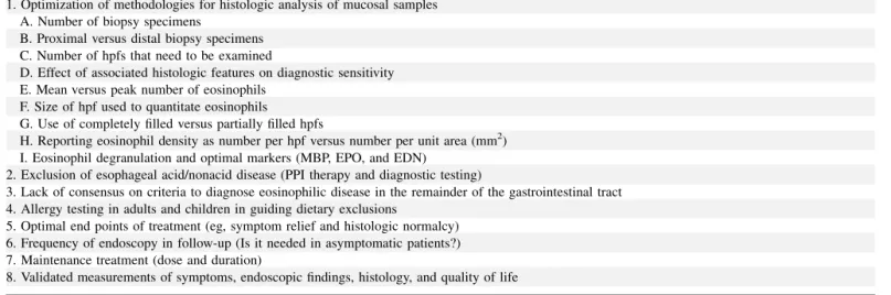
Two studies have provided information on the diagnostic utility of these endoscopic findings. In one study of 222 patients with dysphagia who had endoscopy with esophageal biopsy, 33 (15%) had histologic evidence of EoE.57Among 21 patients who had en-doscopic features suggestive of EoE, the diagnosis was confirmed by means of biopsy in only 8 (38%). Ten (9.8%) of 102 patients with a normal endoscopic examination had histologic evidence of EoE. Esophageal eosinophilia was frequently found in patients who had other causes of dysphagia (eg, reflux esophagitis and peptic stricture). Another study described similar findings.56 Among 261 patients with dysphagia who had endoscopy with esophageal biopsy, 31 (12%) had histologic evidence of EoE. However, only 12 (34%) of 35 patients with esophageal rings seen on endoscopy were confirmed to have esophageal eosino-phils on biopsy. The optimal number of mucosal biopsy speci-mens that should be obtained to maximize the diagnostic yield of EoE has begun to be addressed.73,74By using 15 eosinophils/ hpf as a threshold for diagnosis, one study identified a diagnostic sensitivity of 84%, 97%, and 100% for obtaining 2, 3, and 6 bi-opsy specimens, respectively.73
Barium contrast radiography can identify a number of the anatomic and mucosal abnormalities of EoE, but the sensitivity of radiography as a diagnostic test for this condition appears to be low. One study found that barium swallow results were normal in 12 of 17 children with EoE, including 4 who had endoscopy for food impaction.75
Committee clinical recommendations.Endoscopy with esophageal biopsy remains the only reliable diagnostic test for EoE. However, the finding of isolated esophageal eosinophilia without determining corroborating symptoms and ruling out other causes of esophageal eosinophilia is inadequate to make the diagnosis of EoE. In the appropriate clinical setting the finding of any of the endoscopic features described above supports but does not establish the diagnosis of EoE (seeTable E2andFig E1in this article’s Online Repository atwww.jacionline.org). Esophageal biopsy specimens should be taken to seek histologic evidence of EoE in patients with unexplained dysphagia, even if results of endoscopy appear normal or identify a potential cause of dys-phagia other than EoE.
Two to 4 mucosal biopsy specimens of the proximal and distal esophagus should be obtained. In children and, when indicated, in adults biopsy specimens of the gastric antrum and duodenum should be obtained once to exclude other potential causes of esophageal eosinophilia. There are limited data to support routine gastric or duodenal biopsies in adults in the absence of symptoms or endoscopic abnormalities suggesting other gastrointestinal disorders, although it is reasonable for these biopsies to be performed.
Radiography is not a recommended routine diagnostic test for EoE but can be helpful in selected cases not only to characterize anatomic abnormalities that can be difficult to define endoscop-ically but also to provide information on the length and diameter of complicated esophageal strictures. Findings of a narrow-caliber esophagus (see definition in the ‘‘Disease complications’’ section) or proximal cervical esophageal stricture might be overlooked. Communication with the radiologist regarding indi-cations for the esophagram is important so that the entire esophagus, including the caliber and distensibility of the esoph-ageal lumen, will be fully assessed.
identifying the endoscopic features of EoE and to define the diagnostic utility of the individual endoscopic features of EoE.73,74
Histologic findings
Update of 2007 recommendations. Eosinophils and extracellular eosinophil granules. No prospective studies have determined a threshold number of esophageal eosinophils that can establish a diagnosis of EoE with high specificity and sensitivity and consistently allow differentiation of EoE from other causes of esophageal eosinophilia. One study that related peak eosinophil counts in esophageal biopsy specimens from patients with EoE to symptom frequency or severity reported a lack of correlation between eosinophil density and symptoms in untreated patients with new diagnoses.70However, another study that correlated a composite score found some correlation between symptom subcomponents (dysphagia and anorexia/early satiety) and inflammation.46 Pediatric patients who had esophageal biopsy specimens obtained between 1982 and 1999 with 15 or more eosinophils/hpf and as few as 5 or more eosinophils/hpf were significantly more likely to have increased eosinophil numbers in subsequent esophageal biopsy specimens. Surface layering and microabscesses were found only in biopsy specimens that had 15 or more eosinophils/hpf.76These data are supported by a case-control study that found that the odds ratios for the findings of basal zone hyperplasia and extracellular eosinophil granules were 44 and greater than 100 for patients with EoE (defined as >20 eosinophils/hpf) versus those without EoE, respectively. In that study epithelial desquamation and microabscesses were pre-sent only in patients with greater than 20 eosinophils/hpf.77
Some studies have shown that significant eosinophilic inflam-mation occurs in the proximal esophagi of adults with EoE but not GERD78; others have not confirmed this finding.6Previous stud-ies reported patients with EoE with increased eosinophil numbers in the distal esophagus.79Some have found that a significant pro-portion of adult patients with greater than 15 eosinophils/hpf had GERD/PPI-responsive esophageal eosinophilia.6Currently, nei-ther histopathology nor distribution of inflammatory changes in esophageal biopsy specimens predicts response to PPI therapy. However, eosinophilic microabscesses and surface layering of eosinophils are more typical of findings associated with EoE than GERD. In a limited number of patients, the presence of extracellular eosinophil granules (depicted by extracellular depo-sition of granule proteins, including eosinophil peroxidase, major basic protein, and eosinophil-derived neurotoxin) was found to be a useful feature for histologic distinction of EoE from GERD.80-82 One pediatric study showed that basal cell hyperplasia and extra-cellular eosinophil granules correlated with symptoms.77
From a technical standpoint, one study identified a benefit of evaluating the peak number of eosinophils per hpf, as opposed to the average number, by using the number of eosinophils, extracellular eosinophil granules, epithelial changes, and eosinophils per hpf in patients with EoE.51Unfortunately, the actual size of the hpf de-scribed in many studies is quite variable and frequently not reported. These limitations continue to create significant problems in compar-ing data across institutions and between different studies.83
Associated histologic features and other cell types observed in patients with EoE. Lamina propria fibrosis is found in most biopsy specimens from children and adults with EoE and has been shown to be less prevalent in biopsy specimens from patients with GERD and healthy subjects.78,84,85In some studies subepithelial fibrosis was one of the histologic features that
improved after treatment with topical steroids or anti–IL-5 (mepoli-zumab).40,69Other histologic findings, such as basal zone hyperpla-sia, elongation of rete pegs, and dilated intercellular spaces, are also consistently associated with EoE, but their diagnostic specificity is less certain.86,87Some studies have also identified that mast cells are increased in biopsy specimens from patients with EoE compared with those from patients with GERD.24,41,81,88IgE-bearing cells are more common in biopsy specimens from patients with EoE com-pared with those from patients with GERD and are also not detected in control specimens.24,41The number of intraepithelial regulatory T cells are increased in esophageal biopsy specimens from patients with EoE and those with GERD compared with normal mucosa but are not significantly different when comparing EoE with GERD.29B-cell numbers are increased in biopsy specimens from patients with EoE compared with those seen in control subjects as well.41
In both murine and translational studies, the cytokine IL-5 remains a focal point. IL-5 has been identified in human biopsy specimens and has been shown to drive eosinophil-mediated esophageal remodeling in murine models.11,26,89Periostin, an ex-tracellular matrix protein associated with heart and lung repair and remodeling, has been shown to be increased in the esophagi of patients with EoE. The presence of periostin correlates with in-creased eosinophil levels in patients with EoE but not in patients whose eosinophil levels are less than the threshold criteria that were used to define EoE.90 Confirming and expanding prior genetic studies, expression of eotaxin-1/CCL11 and eotaxin-3/ CCL26genes have been reported to be increased in biopsy spec-imens from patients with EoE compared with those seen in con-trol specimens.91,92 Fibroblast growth factor 9, IL-13, IL-15, and TGF-b1 levels are also increased in biopsy specimens from patients with EoE and patients with GERD compared with those seen in normal biopsy specimens.93
Committee clinical recommendations.Histopathologic features of esophageal mucosal biopsy specimens must be interpreted in conjunction with the patient’s clinical information (see Table E3 in this article’s Online Repository at www. jacionline.org and Fig 1) Until more specific studies are per-formed, it is important that all histologic features, including peak eosinophil counts obtained from the most densely populated hpf, eosinophil microabscess formation, superficial layering of eosinophils, extracellular eosinophil granules, basal cell hyper-plasia, dilated intercellular spaces, rete peg elongation, subepi-thelial lamina propria fibrosis, and increases in numbers of other cell types, such as lymphocytes, be evaluated and noted in pathology reports. Inflammatory changes in patients with EoE might be focal and might not be present in all biopsy spec-imens from a single patient. Because of the nonspecific nature of symptoms in children, assessment of gastric and duodenal mu-cosa is recommended.
of esophageal eosinophilia. The diagnostic significance and specificity of other features of eosinophil-predominant inflam-mation, such as eosinophilic microabscess forinflam-mation, superficial layering of eosinophils, and the presence of extracellular eosin-ophil granules, are also still unknown.
Prospective studies are needed to help identify histologic features that can differentiate EoE from other causes of esopha-geal eosinophilia, in particular GERD. Standardization of the unit for eosinophil enumeration (hpf vs square millimeters) might facilitate comparison of patients between different studies. Studies that validate associated features of inflammation (seeTable E3) might allow discrimination between EoE and GERD and might also potentially allow differentiation of EoE phenotypes. Studies should address practical issues, such as the optimal number of tis-sue samples to survey, the anatomic location with the highest yield of diagnostic mucosal biopsy specimens (proximal/middle/distal), and the best method of quantitating eosinophils.7,94,95 Transcrip-tome analysis of mucosal samples might potentially determine novel pathogenetic mechanisms and allow for molecular diagnos-tics. Expression of eotaxin-3, periostin, COX-2, IL-5, IgE, and a large panel of EoE-specific transcripts (eg, EoE transcriptome) should be investigated in larger studies to determine whether they are helpful in distinguishing EoE from GERD.
Other diagnostic modalities
Update of 2007 recommendations.Studies before 2007 showed that in patients with reflux esophagitis, standard trans-nasal and wireless capsule pH-recording systems demonstrate variability in acid pH monitoring.66,67One recent study detected significantly greater symptom association with the addition of im-pedance to pH testing compared with pH testing alone; however, the clinical implication of this association has not been deter-mined.96The performance characteristics of transnasal and wire-less pH and combination pH impedance testing in patients with EoE are uncertain.
In a cohort study adults with esophageal eosinophilia in their mid and proximal esophagi without endoscopic evidence of
erosive reflux disease underwent pH monitoring while off PPI therapy.6At baseline, pH testing revealed that 15 (71%) of 21 pa-tients demonstrated pathologic acid reflux disease, whereas 6 did not. Both groups were then treated with twice-daily PPI therapy for 2 months and underwent follow-up endoscopy. Among subjects with pathologic acid reflux, 12 (80%) had resolution of esophageal eosinophilia, suggesting the eosinophils might have been present in response to acid reflux. Three subjects had persistent eosino-philia despite PPI therapy because they might have had 2 diseases (GERD and EoE) or because they were nonadherent in taking their PPIs. Among the 6 subjects with baseline esophageal eosinophilia with normal pH test results, 2 (33%) demonstrated resolution of their eosinophilia with PPI therapy, despite the lack of objective evidence for acid reflux disease, suggesting either a lack of diag-nostic accuracy of the pH-monitoring study to identify pathologic acid reflux at baseline or that PPIs have an independent mechanism for improvement of esophageal eosinophilia. Four (66%) subjects demonstrated persistent esophageal eosinophilia, reflecting either classic EoE or lack of subject adherence to PPI therapy.
When studied systematically in pediatric patients, neither acid nor nonacid reflux occurs in patients with EoE in a manner that differs from that seen in age-matched control subjects.97,98Other tests have assisted in understanding more about the pathophysiology of EoE but have not added effect in making the diagnosis of EoE. One study demonstrated that patients with EoE sensed 0.1 N hydro-chloric acid earlier than comparison control groups.59The associa-tion of motility disturbances with EoE remain controversial, with some studies demonstrating abnormalities, such as high amplitude contractions, increased esophageal pressurization, and disordered wave patterns, whereas others reveal normal motility.48,97,99-101 Prolonged esophageal manometry and pH testing in children with EoE found that ineffective peristalsis correlated with dysphagia in children with EoE compared with control subjects.97,100
Endoscopic ultrasonography has been shown to detect a greater mucosal and muscular thickness in patients with EoE compared with that seen in control subjects.97,99Impedance planimetry (En-doFLIP; Crospon, Inc, Carlsbad, Calif), a technology that uses a
FIG 1. Histology of the esophagus (mucosal biopsy specimens). A, Normal esophagus. B, EoE.
bag filled with a conductive solution and multiple impedance electrodes to simultaneously measure pressure and volume, detected significant pathologic changes in esophageal wall com-pliance and distensibility, which could be an important measure of esophageal dysfunction in patients with EoE.102 High-resolution esophageal manometry with pressure topography dem-onstrated abnormal esophageal pressurization patterns in adults with EoE compared with that seen in patients with GERD or healthy control subjects and might reflect reduced esophageal compliance.101 Endoscopic confocal laser microscopy might detect structural changes not apparent on visible light (routine) endoscopy specific to EoE.94
Committee clinical recommendations. Esophageal pH monitoring (and pH impedance, where available) is a useful diagnostic test to evaluate for GERD in patients with esophageal eosinophilia. Other testing modalities do not yet offer clear clinical benefit in diagnostic testing.
Committee future recommendations. Well-designed studies to investigate the role of pH and pH impedance monitoring in patients with GERD and EoE are needed, particularly in adults with EoE, in whom differentiation between EoE and GERD appears to be more problematic than in children and adolescents. Analysis of esophageal dysfunction might provide vital informa-tion for monitoring disease progression and therapeutic response. Prolonged ambulatory motility monitoring, as opposed to sta-tionary monitoring, can provide optimal insights because abnormalities might only be intermittently present in patients with EoE. Other testing modalities require further investigation.
GENOTYPIC FEATURES OF ESOPHAGEAL EOSINOPHILIA
A growing body of literature supports the immunologic basis and genotypic features of EoE. During the past 4 years, a number of studies have helped shape the new conceptual definition of EoE as an immune/antigen-mediated disease.
Update of 2007 recommendations
In the 2007 CR the basic genetic features of EoE were limited to one study identifying the esophageal transcriptome of patients with EoE and its distinction from the transcriptome of healthy patients, as well patients with nonspecific chronic esophagitis (likely reflux associated with esophageal peak eosinophils counts <
_6 eosinophils/hpf).103Studies have validated the expression of a
unique EoE transcriptome and validated that it differentiates EoE from GERD, with eotaxin-3 being abundantly overexpressed in patients with EoE.91IL-13 has been found to be specifically upre-gulated in the esophagi of patients with EoE and might function as a master regulator of the EoE transcriptome.22By using prior re-sults that focused on the correlation of the EoE transcriptome with eosinophil levels, recent studies identified specific genome-wide transcripts that correlate with IL-13 and mast cells.104,105These transcripts do not fully overlap with the originally described eosinophil-associated transcriptome, suggesting the existence of unique mechanisms involved in esophageal inflammation. Nota-bly, abnormal gene expression is primarily reversible with disease remission, although a set of epithelial differentiation genes has been shown to remain abnormal and might be important in predis-posing to disease relapse.26,106
Genetic susceptibility loci discussed in the 2007 CR were limited to genetic variants identified in the eotaxin-3 gene based
on a candidate gene approach. Now, a genome-wide analysis, probing 550,000 common genetic variants in both a discovery and replication cohort from multiple institutions, has identified the first genome-wide EoE susceptibility locus at 5q22.38Two genes located in the susceptibility haploblock include thymic stromal lymphopoietin(TSLP), a cytokine involved in TH2 cell
determi-nation. In a broad analysis of genetic variants within 53 candidate genes involved in allergic responses, epithelial responses, or both, theTSLPgene was also identified as a strong susceptibility locus for EoE, particularly when atopy was controlled, providing strong collective evidence for the role of this pathway in EoE pathogen-esis.39A genetic variant in theTSLPreceptor gene, located on a pseudoautosomal region of the X-chromosome, was also linked with EoE susceptibility in male patients, providing early insight into a potential mechanistic contribution for this pathway into disease pathogenesis and the known clustering of EoE in male pa-tients.39Additionally, a common deletion variant in the filaggrin gene (2282del4), originally identified as a major contributing gene to atopic dermatitis, was identified to be markedly overrep-resented in patients with EoE compared with control subjects without EoE.32Interestingly, the filaggrin association appeared to be independent of the presence of atopic dermatitis in patients with EoE. Finally, a pilot study suggested that particular genetic variants in theTGFBgene might correlate with the presence of esophageal TGF-blevels and response to therapy.69
Committee clinical recommendations.The clinical use of specific genotypes to predict EoE diagnosis, prognosis, or both is not yet ready for clinical application. However, esophageal gene expression is likely to emerge as a key molecular analysis that helps differentiate EoE from other states, including GERD, to determine glucocorticoid-responding and nonresponding patients and to distinguish treated EoE (particularly useful when patients are medically treated with topical steroids before a PPI-confirmed diagnosis of EoE has been made). Clearly, genetic factors contribute to EoE susceptibility, and a combination of common variants likely play a major role in specifying the particular patient phenotype.
Committee future recommendations. The committee values the need for larger and well-characterized cohorts for genome-wide and candidate gene analysis, particularly examin-ing more rare genetics variants than included in traditional genome-wide association studies. Identifying specific genotypes of patients with esophageal eosinophilia who are resistant to PPI therapy (classic EoE) and those with PPI-responsive esophageal eosinophilia is warranted. Further analysis of the genetic variants that contribute to atopic features of EoE (including the apparent nonatopic EoE group) and determining the relationship of these variants to other atopy and GERD susceptibility loci are areas of research need. Multisite studies aimed at validating the value of the EoE transcriptome for molecular diagnosis are also needed. In addition, adequately powered studies using well-defined patient populations designed to distinguish EoE from other esophageal eosinophilic conditions are needed.
GENERAL ALLERGIC EVALUATION Update of 2007 recommendations
multiple concurrent allergic diatheses, with 28% to 86% of adults and 42% to 93% of pediatric patients having another allergic disease.53,107-112Several studies have reported that 50% to 60% of patients with EoE have a prior history of atopy.53,107,113Since 2007, the rates of allergic diatheses have been better described, especially among adults with EoE. The majority of patients with EoE have sensitization to food allergens, aeroallergens, or both based on positive skin prick test (SPT) responses or serum specific IgE test results. There is a subset of patients with EoE who are not sensitized to food allergens, aeroallergens, or both, as determined by using specific IgE testing. Current studies dem-onstrate local IgE production and increase FceRI-positive cell numbers in patients with EoE.41,42
IgE-mediated food allergy. The testing method and def-initions of food allergy vary among studies, but estimates of IgE-mediated immediate food hypersensitivity in patients with EoE range from 15% to 43%.53,107Higher rates of food-induced ana-phylaxis can occur in patients with EoE based on current data us-ing established guidelines for the diagnosis of food-induced anaphylaxis.114The presence of documented IgE-mediated food allergy can be a predictive factor for EoE in adult and pediatric patients.3
Airway and cutaneous allergy: Allergic rhinitis, asthma, and eczema.Rates of allergic rhinitis, asthma, and eczema in children and adults with EoE range from 40% to 75%, 14% to 70%, and 4% to 60%, respectively.32,77,107,109,111,115Six articles document seasonality associated with EoE diagnosis, sug-gesting a potential inciting role for aeroallergens in patients with EoE.3,53,108,110,116In experimental EoE models perennial house-hold allergens (dust mite and cockroach) and molds induce esoph-ageal eosinophilia.117,118 Additional accumulating evidence supports EoE pathogenesis as a TH2-associated disease with
in-creased levels of esophageal mast cells, IL-13, IL-5, TGF-b1, IgE, and FceRI-positive cells.22,41,42,84,89,105Esophageal remod-eling appears to play a role in esophageal dysfunction in a process pathogenically similar to asthma.27,84,85
Committee clinical recommendations
A thorough evaluation by an allergist or immunologist is recommended because of the high rates of concurrent asthma, allergic rhinitis, eczema, and food allergy/anaphylaxis; the potential seasonality of EoE diagnoses; and the complex interplay among multiple allergic diatheses. Additional testing for asthma and allergies is recommended to improve the diagnosis and control of concurrent atopic diseases.
Committee future recommendations
Future publications should clearly document and use standard definitions of allergic rhinitis, asthma (including asthma severity and level of control), and food allergy (rather than sensitization) when assessing and documenting concurrent allergic diseases in patients with EoE. Studies that assess the clinical effect of a ‘‘nonallergic’’ EoE phenotype on disease progression, therapeutic response, or both would be of interest.
LABORATORY EVALUATION
Peripheral eosinophil counts and eosinophil granule proteins
Update of 2007 recommendations. Four studies have documented peripheral eosinophilia in adult and pediatric
patients with EoE, with 40% to 50% having increased numbers of circulating eosinophils (>300-350 per mm3).3,77,119Peripheral eosinophilia decreases after successful esophageal topical corticosteroid therapy and can correlate with tissue eosinophil numbers (r50.68).120One study suggests that esophageal eosin-ophils in patients with EoE express HLA-DR, invoking the capac-ity of eosinophils to act as antigen-presenting cells.121Peripheral eosinophil cationic protein levels did not correlate with tissue eosinophil numbers, although there were statistically nonsignifi-cant eosinophilic cationic protein decreases after therapy.120
Cytokines and PBMCs
Update of 2007 recommendations.Several studies have sought to address potential peripheral EoE biomarkers and response to treatment. Multiplex plasma assays showed that patients with food allergy and EoE had increased levels and spontaneous dendritic cell release of IL-5 and IL-13.45Plasma ba-sic fibroblast growth factor and serum IL-15 levels have recently been reported to be increased in patients with EoE compared with those seen in control subjects, and IL-15 levels might decrease af-ter medical treatment.104,122Adults with EoE had significantly decreased levels of the serum chemokine thymus and activation-regulated chemokine after treatment with oral budeso-nide, but thymus and activation-regulated chemokine levels did not correlate with tissue eosinophil numbers. An intriguing study in adults with allergic eosinophilic gastroenteritis noted a pheno-typic distinction between TH2 cells, with increased numbers of
food allergen–specific IL-51TH2 cells in eosinophilic
gastroen-teritis but IL-52TH2 cells in IgE-mediated peanut allergy; this
in-teresting observation warrants evaluation in patients with EoE.123 Emerging studies have identified candidate surrogate disease markers in patients with EoE. Areas of uncertainty include the degree of correlation with EoE disease activity and the ability of these markers to distinguish atopic persons with or without EoE.
Committee clinical recommendations. There is cur-rently insufficient information to support the clinical utility of any single peripheral marker to function as a surrogate disease indicator of histologic inflammation in patients with EoE. Although peripheral eosinophil counts can correlate with tissue eosinophilia in some patients with EoE, if obtained, changes in peripheral eosinophilia should be interpreted with consideration for the patient’s age, adherence to aeroallergen avoidance, pollen season, and control of comorbid allergic disease.
Total IgE
Update of 2007 recommendations. One additional pe-diatric and 2 adult studies support previous findings suggesting that total IgE levels are increased (>114 kU/L) in 50% to 60% of patients with EoE.109,111Higher total IgE levels are reported in allergen-sensitized versus nonsensitized patients with EoE. Total IgE levels were not predictive of the therapeutic response in one study (budesonide) and did not decrease by 15 days of treatment.
Committee clinical recommendations. There are cur-rently inadequate data to support the utility of measuring the total IgE level as a surrogate disease indicator of histologic inflam-mation in patients with EoE.
pediatric patients with EoE compared with healthy, GERD, and atopic control populations. Reasonable peripheral marker candidates include IL-5, IL-13, IL-15, eotaxin-3, basic fibroblast growth factor, eosinophils, and antigen-specific T-cell subsets.
Aeroallergen-specific IgE
Update of 2007 recommendations. The presence of allergic rhinitis, sensitization to aeroallergens, or both ranges from 24% to 78% in adult patients and 42% to 93% in children with EoE.53,77,107,108,110,111,114,118,124
Several studies (2 pediatric and 1 adult) clearly documented the presence of aeroallergen-specific serum IgE in patients with EoE.77,109,111Overall, 44% to 86% of patients have serum IgE to outdoor, indoor, or both inhalant aeroallergens. Thirty-two per-cent of pediatric patients had serum IgE to a cluster of aeroaller-gens that included pollens and grains, soy, and nuts/peanuts; 86% of adults have polysensitization to aeroallergens; and 61% (11/18) of patients had birch-associated oral allergy syndrome.109,111
Current studies document rates of 71% to 93% for SPT positivity to aeroallergens in pediatric and adult patients with EoE.3,111,114,118,119,124,125 Publications since 2007 have docu-mented sensitization rates through SPTs to outdoor aeroallergens, including grass, weeds, trees, and molds (64% to 93%), and in-door aeroallergens, including dog, cat, cockroach, and dust mites (16% to 69%).77,107,108,111,116,118Even in the pediatric popula-tion, inhalant sensitization was as high as food sensitizapopula-tion, and it is common for adult and pediatric patients to have polysen-sitization to aeroallergens.
Retrospective analyses show decreased EoE diagnosis in the winter and increased diagnosis in the spring, summer, and fall in a total of 583 pediatric and adult patients with EoE.3,53,107,108,110,116 Tree and grass pollen levels can directly correlate with the numbers of patients given a diagnosis of EoE. Sensitization to pollens that cross-react with plant-derived food allergens might provide a link between pollen sensitization and subsequent food ingestion in triggering EoE, although studies to address this possibility are currently lacking.
Committee clinical recommendations.Because numer-ous studies document aeroallergen sensitization and seasonal variability in patients with newly diagnosed EoE, a complete evaluation of patients with EoE for aeroallergen sensitization is frequently warranted in both adult and pediatric patients because this might alter clinical management.
Aeroallergens might have a complementary role in EoE pathogenesis, and appropriate avoidance measures should be recommended. Treating physicians might want to consider a patient’s aeroallergen sensitization profile and seasonality when assessing esophageal biopsy results in patients with EoE.
Committee future recommendations. Seasonality, whether driven by aeroallergens or the consumption of seasonal foods, requires further investigation. Studies that document clear EoE instigation, propagation, and/or exacerbation by aeroallergens in human subjects are warranted but likely difficult to perform because of the need for biopsy evaluation. Surrogate markers for EoE might help in this regard. Studies that document the effects of aeroallergen-specific immunotherapy in patients with EoE would be of interest. Prospective studies documenting aeroallergen sensitization in larger populations of patients with EoE and examining the potential correlation between seasonal variability in symptoms and relevant pollen sensitization are needed.
Food-specific IgE
Update of 2007 recommendations. Four articles docu-mented serum specific IgE in patients with EoE.77,109,111,126None of these studies documented the clinical significance of serum IgE sensitization to EoE diagnosis or management. Sensitization to foods is common in children with EoE, and one study suggested that food-specific IgE testing might be more sensitive than SPTs, but the significance of these positive test results remain unclear.
Current studies of adults with EoE indicate a higher rate of positive test results to foods than was appreciated before the 2007 CR. Among adult patients, 50% had positive results to at least 1 food, the most common being peanut (38%), egg (27%), and soy (23%).125,127Additional publications concerning children continue to show higher rates of positive test results to foods than generally reported from the adult studies.25,110,111,124Although studies sup-port a high rate of sensitization to foods in patients with EoE and a subset of patients with EoE might have acute allergic reactions to foods, warranting evaluation for IgE-mediated food allergies, there are limited data addressing the diagnostic value of SPTs for identifying foods that might directly contribute to EoE. In one study more than 20 subjects were evaluated for the relationship of SPT re-sults to milk, egg, soy, wheat, corn, and beef to outcomes of dietary elimination in patients with EoE.25Positive predictive value ranged from 57% to 96%, negative predictive value ranged from 58% to 75%, specificity ranged from 14% to 65%, and sensitivity ranged from 90% to 98%. Skin tests were slightly more sensitive than patch tests and generally less specific. Only one study documented pre-dictive values for SPTs, and no studies documented prepre-dictive values for serum IgE-based dietary elimination.25Local production of IgE in patients with EoE might explain a potential disconnect be-tween positive test results and actual EoE food triggers.41
Several additional studies with regard to food atopy patch tests (APTs) have been reported.16,25,110,111,114,126,127 Positive patch test rates range from 30% to 95% in children and adults with neg-ative predictive values of greater than 90% (except milk, with a negative predictive value of 50%) and variable positive predictive values in children.127 Using SPT- and APT-based elimination diets, milk and egg were the most common EoE triggers in chil-dren. Food patch testing remains to be standardized and validated in children and adults. Evidence that APTs induce a local immune response reflecting the immunopathology seen in patients with EoE also remains to be demonstrated.
Committee clinical recommendations. Because of the high rates of food allergies and anaphylaxis in patients with EoE, serum IgE and skin prick testing for immediate-type food allergy is warranted to identify comorbid food-induced allergic disease in patients with EoE.
reintroduction. As such, although SPTs, serum IgE tests, and food patch tests can be used to help identify foods that are associated with EoE, these tests alone are not sufficient to make the diagnosis of food allergy–driven EoE.131
Committee future recommendations.The clinical util-ity of serum food-specific IgE levels and SPT responses for generating successful food antigen elimination diets in patients with EoE and the use of commercial food extracts versus fresh foods for SPTs and APTs in patients with EoE require further investigation. Multicenter studies that standardize and validate food APTs in patients with EoE are needed. Future studies should clearly document clinical and histologic benefit from food APTs, SPTs, and/or serum IgE–directed dietary interventions. Studies that evaluate food antigen–specific PBMCs as anin vitrodiagnostic test for food allergy in patients with EoE would be of interest. Fur-ther studies that evaluate the potential increased efficiency of com-bined testing methods (serum and/or SPT and/or APT) in directing successful dietary elimination in patients with EoE are needed.
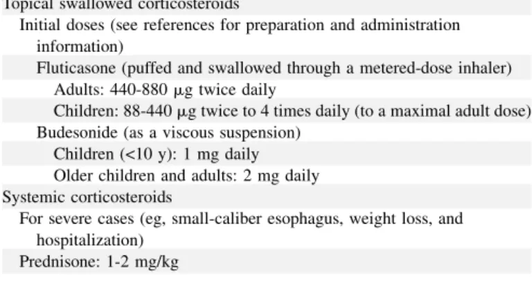
MEDICAL AND DIETARY THERAPY PPI therapy
Update of 2007 recommendations. As previously re-ported in the 2007 CR, acid suppression continues to be an effective tool in fulfilling the diagnostic guidelines for EoE. PPI therapy is useful in treating patients with esophageal eosinophilia secondary to GERD.6,132,133 Patients with isolated esophageal eosinophilia who are treated with PPIs and have a significant improvement of their symptoms and esophageal eosinophilia either have GERD or a yet undefined PPI-responsive esophageal eosinophilia3,134; the lack of a clinicopathologic response to PPI treatment in patients adherent to the treatment regimen with compatible symptoms of EoE and isolated esophageal eosinophilia is consistent with the diagnosis of EoE (see the ‘‘Diagnostic guide-lines’’ section).110However, apart from PPI-responsive esophageal eosinophilia, PPIs might be useful as a cotherapy in patients with diagnosed EoE because they might alleviate symptoms related to secondary GERD, which might be present with EoE.59PPI therapy alone is not effective as a primary treatment for patients with EoE.
Committee clinical recommendations.In many patients PPIs are useful to help eliminate GERD as a cause of esophageal eosinophilia. The recommended PPI dose that should be used to eliminate PPI-responsive esophageal eosinophilia is 20-40 mg, once or twice daily for 8 to 12 weeks in adults (depends on patient and chosen PPI), and 1 mg/kg per dose, twice daily for 8 to 12 weeks in children (for maximal dosing use adult recommenda-tions). PPIs are useful in treating patients with EoE in whom GERD is a comorbid disease. Finally, although the mechanism of PPIs is thought to primarily involve acid blockade, PPIs might also affect esophageal eosinophilia by means of other mecha-nisms and thus be helpful in a subset of patients described as having PPI-responsive esophageal eosinophilia.
Committee future recommendations.Patients with EoE might have an enhanced sensitivity to acid, even in the absence of pathologic reflux defined by conventional pH criteria. As a result, the presence of both GERD and EoE in a patient might not represent simple coexistence but instead a synergistic mecha-nism. Therefore the committee recommends additional studies clarifying the relationship among esophageal acid exposure and its capacity to increase eotaxin-3 production, esophageal eosin-ophilia, and clinical symptoms. In addition, future studies are
needed not only to clarify PPI-responsive esophageal eosinophilia but also the endoscopic and histologic features that might distinguish GERD from EoE. Emerging data suggest a potential role for endoscopic ultrasound, deep tissue biopsies to examine for lamina propria fibrosis, identification of activated mast cells, and evaluation of the esophageal transcriptome.
Dietary therapy
Update of 2007 recommendations. Dietary therapy continues to be effective in children given a diagnosis of EoE. The literature continues to demonstrate that the use of dietary therapy leads to near-complete resolution of both clinical and histologic abnormalities.110,135One study suggested that dietary restriction might reverse esophageal fibrosis.136 Three dietary regimens have been shown to be effective: (1) the strict use of an amino acid–based formula, (2) dietary restriction based on multimodality allergy testing, and (3) dietary restriction based on eliminating the most likely food antigens. Similar results (clin-ical and histologic response) have been documented when using either method of dietary restriction; however, when compared with the administration of a strict elemental formula in allergic patients, elemental formula continues to be the most effective dietary therapy.137Available data suggest that tolerance to foods associated with EoE is unlikely to develop spontaneously, even after prolonged elimination.110Furthermore, methods to induce tolerance in patients with EoE have not been evaluated.
Committee clinical recommendations. Dietary therapy should be considered in all children given a diagnosis of EoE. Preliminary observations suggest that dietary restriction should also be considered for motivated adult patients with EoE. When deciding on the use of a specific dietary therapy, the patient’s lifestyle, adher-ence to therapy, and family resources need to be considered. Consul-tation with a registered dietitian is strongly encouraged to ensure that proper calories, vitamins, and micronutrients are maintained. The committee suggests that foods proved to cause EoE continue to be restricted from the diet, whereas those foods not definitively proved to be antigenic can be reintroduced systematically, with careful observation for recurrence of EoE. Restriction of foods proved to trigger EoE might need to be continued indefinitely.
Committee future recommendations. The use of die-tary therapy in adults requires further study. In addition, further research needs to be performed with regard to the effect of dietary restriction on esophageal fibrosis, quality-of-life issues, adher-ence to therapy, development of food antigen tolerance, nutri-tional effects and possible consequences of prolonged dietary restriction, and best ways to identify foods (eg, skin prick, serum IgE, and patch testing) that cause EoE.
Corticosteroid therapy