34
Effect of different concentrations of Aloe vera leave's extract on the healing process of rat's second degree burn
Kholoud Ramz1, Naser Abbasi2,Mohammadreza Hafeziahmadi3, Monireh Azizi1,Azim Hedayatpour4, Hojjatallah Abbaszadeh5, Ardeshir Moayeri1*
1. Department of Anatomy, Faculty of Medicine, Ilam University of Medical Sciences, Ilam, Iran
2. Department of Pharmacology, Faculty of Medicine, Ilam University of Medical Sciences, Ilam, Iran
3. Department of Pathology, Faculty of Medicine, Ilam University of Medical Sciences, Ilam, Iran
4. Department of Anatomy, Faculty of Medicine, Tehran University of Medical Sciences, Tehran, Iran
5. Cellular and Molecular Research Center, Faculty of Medicine, Shahid Beheshti University of Medical Sciences,Tehran, Iran
Abstract
Introduction: The aim of this work was to evaluate the effects of four different concentrations of Aloe vera extract on healing of second degree burns in male Wistar rats. Materials and methods: By an experimental study 42 male Wistar rats were divided into 7 equal groups. The burn injury was made on the back of all rats, according to standard methods. The burned areas in the group1: were left without any intervention, group 2: were treated topically with eucerin, group 3: were treated with SSD and groups 4-7 were covered with 0.5%, 1%, 1.5%, 2% Aloe vera extract in eucerin (basal cream) twice a day, respectively. 21 days later the rats were sacrificed and samples of burnt skin tissue were collected for histological examinations. The parameters were evaluated to be considered for review included the number of hair follicles, sebaceous glands of angiogenesis, acute inflammation (severe infiltration of neutrophils) and the formation of epithelial layers.
Results: Diagram of healing levels and reduction rats of wounds' sizes in 3th week indicated that the former was reduced more significantly in groups 3, 6 and 7 than in groups 1 (P<0.005) and 2 (P<0.01). Histological findings showed that burn healing was significantly better in groups 6 and 7 than the groups 1 and 2 at the day 21.
Conclusion: It could be concluded that concentration of 1.5% and 2% of Aloe vera extract have an effective role in the treatment of burn wound healing.
Keywords: Aloe vera, SSD, Second degree burn Introduction
Millions of people suffer from different kinds of disability due to incidental injuries annually (1). Burns are among the most common factors associated with social, economical and mental disorders which are sometimes irreversible ) 2, 3).
Burns are classified by level of severity as the 1th, 2nd and 3rd grades of burn. The most common are the first-degree or superficial burns which are the least serious and cause tenderness that are similar to sunburn. The second-degree
*Corresponding author:Tel: +98 8432235713 Fax: +98 8432227136
Address: Department of Anatomy, Faculty of Medicine, Ilam University of Medical Sciences, Ilam, Iran E-mail: [email protected]
Received; 2015/11/19 revised; 2015/12/1accepted; 2016/02/18
35
burns, known as partial thickness burns, are deeper than the first-degree burns and are characterized by blotchy white, pink or red patches which cause blisters. The most severe type of burn, a third-degree, known as a full thickness burn, penetrates through all layers of the skin and may injure tissue beneath skin, so the skin is not capable of healing itself (4, 5).
Some antibiotics such as mafinide acetate and silver sulfadiazine (SSD) are used for healing and shortening the course of wound's repairment (6). Aloe vera is a naturally moisturizing plant containing some amino acids contributing to regrowth and repairment of damaged skin cells (7). During burn's healing phenomenon, fibroblasts and some other cells are called into the damaged area. Fibroblasts cause the edges of wound or damaged area to gain an adhesion (8). But sometimes they create some abnormal tissue called scar due to insufficient regulation of their activities (9).
Considering properties of Aloe vera plant, we decided to study the effects of four different concentrations of its extract on healing of second grade of burn wounds in male Wistar rats. Thus, this paper was aimed to answer the question that which concentrations of the Aloe vera extract can affect more efficiently on the wound healing of second degree burn?
Materials and methods
Animals: 42 male Wistar rats (300-350 gr) aged of nearly 2-3 months were utilized as experimental animals and were isolated into 7 groups of six rats. The animals were housed in standard environmental states of temperature (22 ±3°C), moistness (60 ± 5%), and 12:12 light: dark cycle. Accordingly, during the experimental period the rats were directed to a standard pellet diet (10). Aloe vera leaves extracts were provided according to standard methods (11). The extracts were filtered through Whatman filter paper (No. 1) and
the filtrate was evaporated in vacuum evaporator (brand: IKA-RV10digital) at 40°C. The percentage yield obtained from the extraction of coarse powder of Aloe vera leaves was 5.8. The extracts were stored at -40 °C in a desiccant until requirement. The extracts were dissolved in the cream basis containing 0.5%, 1%, 1.5% and 2% net extracts and cream was made by mixing of this concentration extracts in eucerin (12).
Design of experiments: All 42 animals were divided into 7 different groups, each containing 6 animals as follows: Group 1: control group with no intervention. Group 2: treated with only basal cream (eucerin: without effective agent). Group 3: treated by SSD (silver sulfadiazine 0.01, SOBHAN DAROU – Rasht industrial city -IRAN). Group 4: treated by eucerin containing Aloe vera extract 0.5%. Group 5: treated by eucerin containing Aloe vera extract1%. Group 6: treated by eucerin containing Aloe vera extract 1.5%. Group 7: treated by eucerin containing Aloe vera extract 2%. Burn wounds were created under sterile and standard conditions and then were treated twice daily. At that point the surfaces of wounds were secured by comparing salve and no dressing was connected (13). Considering the wound healing as a study's goal, the size of dead injuries was set to be clear well at the 7th, 14th, 21st days after burn injury. Some parameters that were considered to be evaluated for review included: the number of hair follicles, sebaceous glands, angiogenesis, acute inflammation (severe infiltration of neutrophils) and the formation of an epithelial layer (14). On the day 21, the healing tissue samples of rats (one rat from each groups) were examined using Hematoxylin-eosin. Stained tissues were histologicaly examined by a histologist, and the results were analyzed by SPSS, version 16. Mann-Whitney U test followed by Kruskal-Wallis test was used to evaluate the differences between groups. A P-value
36
of less than 0. 05 was considered as significant.
Results
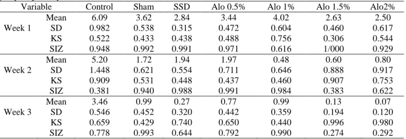
The healing process and wound size reduction rates of all 7 studied groups were compared and their results are shown in Table 1. At the end of week 3 significant differences were observed between groups
3-7 in comparison with group 1 (P<0.005) and 2 (P<0.01), respectively. In addition, in Table1, normality of values of these 7 groups in 3 successive weeks were examined using Kolmogorov- Smirnov (K-S) test and the significance level was determined at P< 0.05 for 3 successive weeks.
Table 1. Descriptive indices of means and standard deviations of wound sizes among control, sham, SSD and 4
groups separately within 3 successive weeks.
Variable Control Sham SSD Alo 0.5% Alo 1% Alo 1.5% Alo2%
Mean 6.09 3.62 2.84 3.44 4.02 2.63 2.50
Week 1 SD 0.982 0.538 0.315 0.472 0.604 0.460 0.617
KS 0.522 0.433 0.438 0.488 0.756 0.306 0.544
SIZ 0.948 0.992 0.991 0.971 0.616 1/000 0.929
Mean 5.20 1.72 1.94 1.97 0.48 0.60 0.80
Week 2 SD 1.448 0.621 0.554 0.711 0.646 0.888 0.917
KS 0.909 0.531 0.448 0.437 0.460 0.907 0.753
SIZ 0.381 0.940 0.988 0.991 0.984 0.383 0.622
Mean 3.46 0.99 0.27 0.77 0.99 0.13 0.07
Week 3 SD 0.546 0.452 0.320 0.442 0.359 0.194 0.120
KS 0.659 0.429 0.740 0.650 0.440 0.996 0.980
SIZ 0.778 0.993 0.644 0.792 0.990 0.274 0.292
The Figure 1 shows a comparison between either group 7 or SSD group and other groups, in terms of mean wound sizes at week 3, that indicated a significantly difference. The result indicated that group 1 and 2 (control and sham) had slightly
healing effects and Aloe vera with concentrations of 1.5% and 2% could shorten the recovery time of burns' wounds comparing to the control group (P<0.005) and group 2 (sham) (P<0.01).
Figure1. Mean wound sizes for treatment groups were shown separately during 3 successive weeks graphically.
** P<0.005 the groups compared control #P<0.05 the groups compared SSD ##P<0.01 the groups compared SSD ɸɸP<0.01 the groups compared sham
37
Table 2 indicates the histological recovery of different indices and the scores allocated to each group. Samples were scored as follows: 0= none and slight (+),
slight or mild (+/-), moderate (++), severe (+++) profound or advance, or very severe (Hematoxylin and eosin stained sections).
Table 2. Histological scoring criteria for burn wounds. Histological evaluation of 7 groups on wound's healing process in different groups of treatment in rats.
Groups Follicle
hair
Seabasea gland
Fibroblasts Macrophages Blood
vessels
Neutrophiles Layer of
epithelium
1 0 0 + ++ +++ +++ 0
2 0 0 + ++ ++++ +++ 0
3 ++ ++ ++ -/+ -/+ -/+ ++
4 0 0 + ++ +++ +++ 0
5 0 0 + ++ ++ ++ +
6 + + ++ - + / - + / + +++
7 + + ++ - + / - + / + +++
As Figure 2 indicates the wound size in the control group was considerably different from those in others and the descending
trend line of wound size in Aloe vera groups of 1.5%, and 2% lies below the others groups.
Figure 2. The diagram of mean size of wounds' margins for 7 study groups during 3 successive weeks.
Discussion
The present study evaluated the effects of a four different concentrations of Aloe vera, which were applied locally, in improvement of healing process of wound and inflammation caused by second grade
burns in Wistar rats. The healing indices in this study were considered as number of Neutrophiles, blood vessels, macrophages, fibroblasts, sebaceous glands and follicles.
38
Based on the international society of burn and injury reports, skin layers are damaged or destroyed, partially or completely, due to burns caused by direct contacts with an energy source such as hot liquid / solid materials (15). According to some studies, application of Aloe vera extract improves healing process of degenerated tissues due to having micronutrients and anti-inflammatory effects and stimulation of skin fibroblasts by this plant (16) .
Aloe vera gel contains Mannose-6-phosphat component which is existed in this plant as glucose and mannose and its chain is the most effective substance presented in Aloe vera extract for wound healing (2). Moreover, Aloe vera extract has other biological and recovery effects for other diseases including diabetes and skin diseases such as acne and also effects on immune system, neoplasms and possess an antioxidant feature (5).
Vascularization is the physiological process through which new blood vessels created from pre-existing vessels. This is a normal and vital process in growth and development, as well as in wound healing and in the formation of granulation tissue. During this process, a number of blood
vessels are destroyed due to apoptosis, and the wound contracts because of cells activities, extracellular matrix and cytokines. Some substances such as vitamins A, E and C, glucose, amino acids, fatty acids, proteins, H2O and zinc are important to the wound healing process (10).
In general, our findings showed a difference in the amount of blood vessels for all groups which included the highest amounts on day 21.
Aloe vera extract presented in our studied compound had some antibacterial properties and could facilitate the healing of all wound types with different origins and of course improves the healing process of burn wounds. Aloe vera extraction and salicylic acid (aspirin), revealed the best results for groups 6 and 7 among all studied groups.
Conclusion
In conclusion application of 1.5% and 2% concentrations of Aloe vera extract has a positive effect on the healing of burn wounds in the second degree of male Wistar rats significantly.
References
1. Kaufman T, Kalderon N, Ullmann Y, Berger J. Aloe vera gel hindered wound healing of experimental second-degree burns: a quantitative controlled study. J Burn Care Rehabil. 1988;9(2):156-9.
2. Moghbel A, Ghalambor A, Allipanah S. Wound healing and toxicity evaluation of Aloe vera cream on outpatients with second degree burns. IJPS. 2007; 3(3):157-60.
3. Bossini PR. Low-level laser therapy (670 nm) on viability of random skin flap in rats. Lasers Med Sci. 2009;2(24): 209-13.
4. Akhoondinasab M, Akhoondinasab M, Saberi M. Comparison of healing effect of Aloe Vera extract and silver
sulfadiazine in burn injuries in experimental rat model. Burns. 2014;3 (1):29-34.
5. Eyal Z, Rachel Z, Alon Y, Yacoby-ZO. Heparanase accelerates wound angiogenesis and wound. Faseb J. 2005; 19(2):211-21.
6. Oryan A, Naeini T. Effect of aqueous extract of Aloe vera on experimental cutaneous wound healing in rat. Veterinary. 2010; 80(4): 509-22. 7. Khaing TA. Evaluation of the
antifungal and antioxidant activities of the leaf extract of Aloe vera (Aloe barbadensis Miller). J Tradit Complement Med. 2011;5(3): 110-2. 8. Zcharia E, Zilka R, Yaar A,
Yacoby-Zeevi O, Zetser A, Metzger S, et al.
39
Heparanase accelerates wound angiogenesis and wound healing in mouse and rat models. FASEB J. 2005;19(2):211-21.
9. Durmus AS, Yaman M, Can HN. Effects of extractum cepae, heparin, allantoin gel and silver sulfadiazine on burn wound healing: an experimental study in a rat model. Veterinární Med.2012; 57(6):287-92.
10. Shahzad MN1, Ahmed N.
Effectiveness of Aloe Vera gel compared with 1% silver sulphadiazine cream as burn wound dressing in second degree burns. J Pak Med Assoc. 2013;63(2):225-30.
11. Somboonwong J, Kankaisre M, Tantisira B, Tantisira MH. Wound healing activities of different extracts of Centellaasiatica in incision and burn wound models: an experimental animal study. BMC Complement Altern Med. 2012; 12:103.
12. Hui-ying Z, Yan-hua S, Bao-ming L. The effects of Aloe gel juice on
second-degree burn wound of Taihu pig. Acta Academiae Medicinae Suzhou. 2010;6(4):1204-6.
13. Beya W, Davidson B, Erlwanger KH. The effects of crude aqueous and alcohol extracts of Aloe vera on growth and abdominal viscera of suckling rats. Afr J Tradit Complement Altern Med. 2012; 9(4): 553–60. 14. Maenthaisong R1, Chaiyakunapruk N,
Niruntraporn S, Kongkaew C. The efficacy of Aloe vera for burn wound healing: a systematic review. Burns. 2007;33(6):713-8.
15. Rathor N, Mehta AK, Sharma AK, Mediratta PK, Sharma KK. Acute effect of Aloe vera gel extract on experimental models of pain. Inflammation. 2012;35(6):1900-3. 16. Danhoff IE, McAnally BH. Stabilised
Aloe Vera, its effect on human skin cells. Drugs in the Cosmetics Industry. 1983; 133: 52-196.