The Use of Nanodroplets in the Extraction and Fragmentation of Chromatin from Formalin-Fixed Paraffin Embedded Samples
By Suud Ashur
Senior Honor Thesis Biology
University of North Carolina at Chapel Hill
14th April 2016
Approved:
____________________________________ Dr. Samantha Pattenden, Thesis advisor Greg Copenhaver, Reader
1 Abstract
DNA is compacted into the cell nucleus through the formation of chromatin, which consists of DNA wound around octamers of histone proteins into nucleosomes. Chromatin that is
accessible, or “open” is associated with active gene transcription, while inaccessible, or “closed” chromatin with intact nucleosomes is thought to be transcriptionally repressed. To identify sequence signatures in open chromatin, we use a technique called Formaldehyde Assisted Isolation of Regulatory Elements (FAIRE). Currently, FAIRE can only be performed on fresh tissues or cells. We wanted to extend our protocol to include formalin-fixed paraffin embedded (FFPE) human biopsy samples since these samples are readily available in tissue banks.
However, the only available protocol for extracting chromatin from FFPE samples requires an enzyme that degrades open chromatin, making it incompatible with FAIRE. As an alternative to chemical extraction, we used nanodroplets, in combination with acoustic sonication, to
mechanically disrupt FFPE tissue samples. This novel method sufficiently disrupted FFPE samples and fragmented chromatin to make it compatible with FAIRE analysis. Since fixation times in pathology labs range from a few hours to several days, we will next test whether FFPE tissue fixation times affect our ability to extract chromatin. We hypothesize that the addition of nanodroplets to the FAIRE protocol would normalize the quantity and quality of isolated chromatin independent of tissue fixation time. If successful, our protocol could be applied as a screening assay or diagnostic for diseases with aberrantly open or closed chromatin signatures.
Introduction
2 DNA sequence1. Such changes can alter the packaging of DNA, which in turn could affect the ability of transcription factors to access DNA1. Hence, epigenetic modifications can induce
aberrant activation or repression of genes, thereby promoting tumor formation and growth. Since chromatin is a complex of DNA and histones that packages DNA to fit into a cell nucleus, any changes in the chromatin structure can affect gene expression. One example of cancer linked to a specific change in chromatin signature is Ewing sarcoma, a rare type of pediatric cancer in bones and soft tissues2. This disease is characterized by a chromosomal translocation involving the EWSR1 and FLI1 genes, resulting in an oncoprotein called EWS-FLI1 that causes aberrant opening of chromatin at specific regions of DNA2. Dr. Ian Davis’s lab used a technique called Formaldehyde Assisted Isolation of Regulatory Elements (FAIRE) to isolate open chromatin and demonstrate that loss of EWS-FLI1 protein through mRNA knockdown results in closing of aberrantly open chromatin and suppression of tumor cell growth2. Therefore, these
disease-specific regions of open chromatin have the potential to be used as a diagnostic for Ewing sarcoma, further emphasizing the importance of chromatin signature in tumor formation. Hence, devising a standard method to extract high quality chromatin from all types of tissues that is compatible with all downstream chromatin-based assays is very desirable and likely to advance the field of oncological medicine.
3 machinery compared to regions of heterochromatin associated with tightly packed nucleosomes4. The nucleosome is the fundamental unit of chromatin composed of DNA wrapped around a set of eight proteins called histones5. It plays an important role in gene regulation since its presence reduces the ability of enzymes, such as RNA polymerase II, to access DNA for transcription. Therefore, nucleosome depletion is generally a marker for active gene expression6.
By isolating open chromatin, the FAIRE technique allows for the identification of
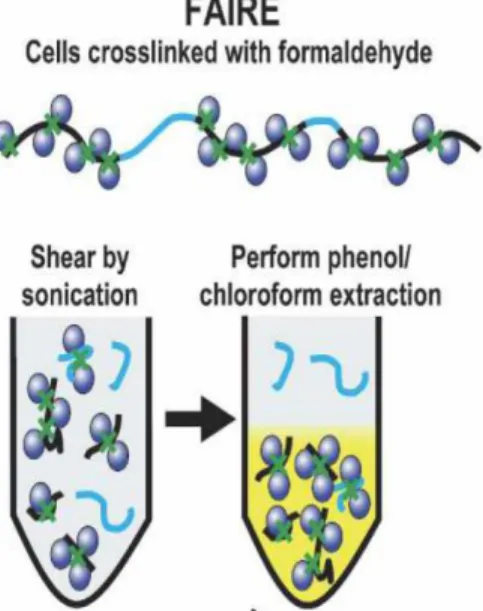
regulatory elements in eukaryotes, such as promoters. The original procedure of FAIRE involves three steps: crosslinking DNA to proteins using
formaldehyde, shearing the chromatin by sonication, and performing a phenol/chloroform extraction3 (Fig 1). Since open chromatin containing active regulatory elements is depleted of nucleosomes, the crosslinking phase only involves nucleosome-rich, or closed regions of chromatin. As a result, shearing by sonication essentially divides the chromatin into two regions: open and closed. The
phenol/chloroform solution is then used to separate the two regions by forming two phases: organic and aqueous. Due to its rich nature of proteins, closed chromatin segregates into the organic phase, while the open
chromatin of interest, containing mostly negatively charged DNA, gets segregated to the aqueous phase. Although this technique can work with a broad range of cell types, we have modified the third phase of the procedure by replacing the phenol/chloroform extraction with silica-containing columns7. These columns are simple to use and do not require toxic chemicals like chloroform.
Fig. 1: The FAIRE technique makes it
possible to isolate “open” or
transcriptionally active regions of chromatin that are associated with
4 By pouring the sonicated solution containing a mixture of open and closed chromatin through the column, the negatively charged DNA from the open chromatin binds to the positively charged silica, while the DNA from closed chromatin passes right through the column due to the presence of proteins that weaken the negatively charged DNA. In other words, columns work by taking advantage of the differential isoelectric point between DNA bound by proteins and free DNA. Hence, our modified FAIRE offers a simple method for identifying regions of open chromatin.
Our experiments involve tissue or cultured cell pellets that have been Formalin-Fixed (FF) and Paraffin Embedded (PE). Due to its inexpensive nature and long-term storage potential, FFPE samples offer the best choice for preserving and processing specimens in pathology
departments8. However, the use of FFPE samples limits us on the ways chromatin can be extracted. Currently, the only available method for the extraction of chromatin from FFPE samples requires an enzyme called micrococcal nuclease that degrades open chromatin, which in turn makes it incompatible with the FAIRE method9. Hence, a method that extracts chromatin without degrading it is very desirable.
5 bubble wall can move up to 700 meters per second10. At higher acoustic pressures, which can be achieved by the E110 Covaris Sonicator, microbubble cavitation becomes intense to the point that it violently collapses, releasing tremendous mechanical energy responsible for the extraction and fragmentation of chromatin10. We use nanodroplets instead of microbubbles because
nanodroplets require energy to not only vaporize but also to cavitate, thereby extending the cavitation enhancement effect compared to microbubbles alone10. By incorporating nanodroplets during sonication of FFPE samples, we showed that nanodroplets provide sufficient energy needed to extract chromatin from such samples, thereby obviating the use of micrococcal nuclease in chromatin extraction.
Before devising a standardized protocol that involves the use of chromatin from FFPE samples, there are certain factors that we need to consider, such as tissue type and duration of fixation. Fixation times in pathology labs tend to range from a few hours to couple of days, such as samples being left fixed over the weekend. We are interested in testing whether FFPE tissue fixation times affect our ability to extract chromatin using nanodroplets. We hypothesize that the addition of nanodroplets to the FAIRE protocol would normalize the quantity and quality of isolated chromatin independent of tissue fixation time. Here, I describe and test the optimization of the protocol for nanodroplet-mediated extraction of chromatin from FFPE samples including rodent xenograft tissue that has been fixed for various time points.
Method
1. FFPE FAIRE protocol
1.1 Deparaffinizing -
6 rodent xenograft tissue that was fixed for 4, 24, and 48 hours. A slide containing the FFPE sample was obtained and placed in a slide holder. The holder was then placed in a tub containing 150 mL xylene for 3 minutes. This was repeated three times in different tubs of xylene, before draining and blotting the excess solution from the holder.
1.2 Hydration -
The holder containing the FFPE sample was placed in a tub containing 150 mL 100% ethanol for 3 minutes, before moving it to another tub of 100% ethanol for the same period of time. The above procedure was repeated but in a tub of 85% ethanol instead (15% de-ionized water). The holder was then placed in a new tub of 150 mL 70% ethanol (30% de-ionized water) for 3 minutes, before moving the holder to a tub containing 150 mL de-ionized water for 3 minutes. The slides were then blotted by pressing it between paper towels. Using a razor blade, the sample was scrapped off the slide and deposited into a 8MM clear round bottom crimp vials (Thermo Scientific #C4008-632R)
1.3 Sonication -
FAIRE buffer (10mM Tris-HCl pH 8.0, 2% Triton-X-100, 1% SDS, 100mM NaCl, 1mM EDTA) was added to the vial containing the sample followed by 10 µL of nanodroplets (courtesy of Paul Dayton). The vial was then capped (Thermo Scientific #C4008-2A) and sonicated in a Covaris E110 Sonicator at 4°C for 4 minutes with the following settings: 20% duty cycle; intensity = 8; and 200 cycles per burst. The sonication cycle was then repeated for an additional 4 minutes for a total of 8 minutes sonication. There was a 5 minute limit for continuous
7
1.41 DNA Recovery –
This method was used to determine the cell lysis efficiency and calculate DNA recovery. After separating the supernatant from the pellet from the sonication step, the pellet was
resuspended in 100 µL FAIRE buffer. Two µL proteinase K was then added to both the pellet and the supernatant tubes. The tubes were mixed and briefly centrifuged, followed by incubation at 55⁰C overnight. On the following day, 2µL RNase was added to both the pellet and
supernatant tubes and incubated at 37⁰C for 10 minutes. Using the Zymo kit, the DNA was extracted and purified from both the pellet and supernatant tubes, followed by elution of DNA using a 25µL elution buffer. To calculate DNA recovery, the Qubit 2.0 flurometer was used to determine the amount of DNA in the pellet and the supernatant. The amount of DNA in the supernatant was divided by the amount of DNA left over in the pellet. Fragmentation efficiency was verified by running the DNA on a 1.5% agarose gel.
1.42 FAIRE -
This method was used to determine open chromatin recovery. After separating the
8 at 37⁰C for 10 minutes. The purified DNA was extracted from both the FAIRE DNA and the input solution using the Zymo kit and eluted to a final volume of 25 µL. The total amount of open chromatin was determined using a Qubit 2.0 fluorometer by dividing the quantity of FAIRE DNA by the input DNA.
1.5 Quantitative PCR (qPCR)
To assay individual loci associated with aberrantly open chromatin signatures, qPCR was utilized. We used four primer sets: a positive control (AURKAIP1), a negative control
(BC006361), and two primers associated with Ewing sarcoma (P1 and P2) (See Appendix A). For each primer set, there were wells designated for Input DNA, FAIRE DNA, and a no template control. The master mix was prepared according to the number of wells used +2 (See Appendix B). For example, if two wells would be used, then a master mix for four wells would be
9
2. Fixation time protocol
This protocol was used to determine the effect of different fixation times on the quantity and purity of isolated chromatin from MCF-7 cell-derived xenograft tissue. This method involves three phases: 1) To quantify the amount of DNA present in MCF-7 cell-derived xenograft tissue 2) To find the optimal sonication time for high ratios of supernatant to pellet and 3) To analyze the disease-specific regions of open chromatin using FAIRE and qPCR.
2.1Quantify total DNA per scroll
The MCF-7 cell-derived xenograft tissues were deparafinzed and hydrated as outlined in the FFPE FAIRE protocol. The DNA was purified using the Biostic FFPE Tissue DNA Isolation Kit according to manufacturer’s instructions. After treatment with 2µL RNase and subsequent purification using the Zymo Kit, the total DNA was quantified using the Qubit 2.0 flurometer by following the Qubit Manual.
2.2Calculate DNA recovery
After deparaffinization, hydration and sonication of MCF-7 scrolls, the DNA recovery part of the FFPE FAIRE protocol was followed.
2.3Perform FAIRE and qPCR
This part of the protocol is identical to the FFPE FAIRE protocol for FAIRE and qPCR. Results
10 The initial stages for the development of the protocol tested the possibility of chromatin
A
.
8 12 0 5 10 15 20 - Nanodroplets + Nanodroplets Time (minutes) DNA Re c o v e ry Ra ti o (S u p e rn a n ta n t/ P e ll e t) ND NDB
Posi tive Negative ES1 ES2
0.0 0.5 1.0 1.5 2.0 - Nanodroplets + Nanodroplets qPCR Amplicon P e rc e n t In p u t ND ND
Fig. 3. A) Comparison of DNA recovery with and without nanodroplets at different sonication times in FFPE cell pellets. Y-axis represents the ratio of the amount of DNA obtained from the supernatant compared to DNA obtained from the pellet. Error bars represent the standard deviation of three biological replicates. ND denotes Nanodroplets B) Comparison of FAIRE signal with and without nanodroplets at different chromatin regions in FFPE cell pellets. Positive represents chromatin regions that are always open, while Negative represent regions that are always closed. ES1 and ES2 represent regions associated with aberrantly open
chromatin. Y-axis represents the percent input, which is calculated by dividing the FAIRE qPCR signal by the
input (background) qPCR signal. Error bars represent the standard error of three biological replicates. ND denotes nanodroplets.
A
12 16 0 20 40 6080 - Nanodroplets
+ Nanodroplets Time (minutes) P e rc e n t DN A Re c o v e ry (S u p e rn a n ta n t/ P e ll e t) (n = 3 ) ND ND
B
Pos itiv e Negative ES1 ES2
0.0 0.5 1.0 1.5 2.0 2.5 - Nanodroplets + Nanodroplets qPCR Amplicon P e rc e n t F A IRE DNA /I n p u t DNA (n = 3 ) ND ND
Fig. 4. A) Comparison of DNA recovery with and without nanodroplets at different sonication times in FFPE rodent xenograft tissues. Y-axis represents the ratio of the amount of DNA obtained from the supernatant compared to DNA obtained from the pellet. Error bars represent the standard deviation of three biological replicates. ND denotes Nanodroplets B) Comparison of FAIRE signal with and without nanodroplets at different chromatin regions in FFPE rodent xenograft tissues. Positive represents chromatin regions that are always open, while Negative represent regions that are always closed. ES1 and ES2 represent regions
associated with aberrantly open chromatin. Y-axis represents the percent input which is calculated by dividing the FAIRE qPCR signal by the input (background) qPCR signal. Error bars represent the standard error of three biological replicates. ND denotes nanodroplets.
p=0.03
p=0.03
p=0.02
p=0.19
p=0.006 p=0.01
p=0.07
p=0.12
p=0.05
p=0.02
11 extraction using nanodroplets in Ewing sarcoma FFPE cell pellets (EWS894 cells). Previous work by Anna Kenan and I showed that the addition of nanodroplets during acoustic sonication provided sufficient mechanical energy to disrupt regular cell pellets and fragment chromatin. We started with cell pellets instead of xenograft tissues because cell pellets are easily available, while xenograft tissues are expensive to generate. Further, Ian Davis’s lab has FAIRE next-generation sequencing (FAIRE-seq) data for EWS894 cell lines, which makes FFPE cell pellets a logical place to start for the purposes of comparing data. To test the efficiency of chromatin extraction in FFPE cell pellets, we calculated DNA recovery in the presence and absence of nanodroplets. Our experiments reveal that the addition of nanodroplets significantly enhances DNA recovery compared to FFPE cell pellets without nanodroplets (Fig. 3A). This increase of DNA yield is also observed when FFPE cell pellets were sonicated for additional 4 minutes (Fig. 3A). We then tested if our nanodroplet-disrupted samples could be used for the FAIRE assay. Analysis using quantitative PCR revealed that samples disrupted by nanodroplets produced
increased FAIRE signals over background in chromatin regions that are always open as well as regions associated with Ewing sarcoma (Fig 3B). This result means that the use of nanodroplets increases
chromatin recovery, which allowed us to detect good FAIRE signal over
background. Moreover, as seen in Fig. 5, nanodroplets consistently produce around 500 base pair fragment size in the
Fig. 5: Gel electrophoresis of EWS894 cell pellets
sonicated for 4 and 8 minutes.. +/- ND indicates
presence or absence of nanodroplets respectively. The supernatant contains the DNA of interest while the pellet mostly contains debris.
Pellet Supernatant
8 8
4 4
–
+
–
+
–
+
–
+
1500
500
12 supernatant containing the DNA of interest compared to ones without nanodroplets. Further work by Anna Kenan revealed that nanodroplets also improve chromatin recovery and
fragmentation from EWS894 FFPE xenograft tissues (Fig. 4A and 4B), thereby broadening the utility of our protocol. However, qPCR only involves specific regions of DNA, which limits us on the comparisons we can make with FAIRE-sequencing data from fresh Ewing sarcoma cell lines. We have therefore sent the FFPE xenograft tissue for next-generation sequencing in order to look at genome-wide DNA sequence. Nevertheless, our qPCR results helped us create a reliable protocol that has the potential to be used as a screening assay for diseases that have aberrantly open and closed chromatin signatures (Fig. 2).
The effectiveness of nanodroplets is inversely proportional to formaldehyde fixation time
Due to a lack of time, we were unable to determine the effect of different fixation times on the quality of isolated chromatin using FAIRE by qPCR. Instead, we assessed the effect of different fixation times on chromatin recovery upon the addition of nanodroplets. This method involved obtaining three MCF-7 cell-derived xenograft tissues that were each fixed in
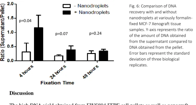
13 of the amount of DNA present overall (200-400ng) for comparisons with DNA obtained through nanodroplets. We then moved to the second phase of the protocol: to find the optimal sonication time for high ratios of supernatant containing the DNA of interest over pellet. We started with 12 minutes sonication with and without nanodroplets but we found that a significant amount of DNA remained in the pellet even after the use of nanodroplets. This lack of soluble DNA is partly due to the complexity of tissues, which require more energy by nanodroplets to extract chromatin as when compared to cell pellets. As a result, we performed a similar experiment with 16 minutes sonication (Fig. 6). The results reveal that, with the addition of nanodroplets, the highest increases in DNA recovery is seen in tissues fixed for 4 hours. The effectiveness of nanodroplets decreases when tissues are fixed for greater period of times, as seen in tissues fixed for 24 hours. This decrease continues until there is no signficiant difference between samples with nanodroplets and those without, as seen in tissues fixed for 48 hours. Therefore, not only does lengthy formaldehyde fixation impose a barrier on the extraction of DNA from FFPE samples, the results also reveal that the effectiveness of nanodroplets is inversely proportional to the duration of formalin-fixation. .
Fixation Time
DNA Extracted (ng/uL)
Total DNA Obtained (ng) 4 hours 3.54 ng/uL 177 ng
24 hours 7.84 ng/uL 392 ng 48 hours 4.82 ng/uL 241 ng
14 Discussion
The high DNA yield obtained from EWS894 FFPE cell pellets as well as xenograft tissues with the addition of nanodroplets compared to the ones without implies that nanodroplet cavitation has the ability to not only disrupt FFPE samples but also to extract and fragment chromatin (Fig. 3A and 4A). Since Ewing sarcoma is associated with aberrant opening of chromatin at specific DNA sequences2, we expect that nanodroplet cavitation will increase chromatin recovery on regions associated with the disease (ES1 and ES2). Indeed, the increase in DNA yield is concomitant with an increase in open chromatin associated with those regions (Fig. 3B and 4B). The increase in FAIRE signal over background was also observed for chromatin regions that are always open (positive control), thereby confirming the effectiveness of nanodroplets. There was no significant increase in chromatin regions that are always closed (negative control), which confirms that FAIRE only isolates open chromatin. In addition, nanodroplet cavitation produced consistent DNA fragment size distribution (Fig. 5), which is important for next-generation sequencing, since the primers used in sequencing have a limit to the size of fragments they can extend.
Fig. 6: Comparison of DNA recovery with and without nanodroplets at variously formalin-fixed MCF-7 Xenograft tissue samples. Y-axis represents the ratio of the amount of DNA obtained from the supernatant compared to DNA obtained from the pellet. Error bars represent the standard deviation of three biological replicates.
p=0.04
15 An alternative method available for the extraction of chromatin from FFPE samples requires the use of micrococcal nuclease, an enzyme with both endo- and exo-nuclease activity, to digest unprotected DNA9. However, the use of micrococcal nuclease increases the
susceptibility to open chromatin degradation, which in turn makes it an incompatible method for analyzing open chromatin signatures through FAIRE. By improving the recovery and
fragmentation of chromatin, mechanical disruption of FFPE samples using nanodroplets can replace the micrococcal nuclease digestion step for the isolation of chromatin from FFPE
samples. Therefore, for the first time, our protocol provides a tool for identifying regions of open chromatin from FFPE samples.
We are in the process of determining if FAIRE-seq data from EWS894 xenograft samples compares well to that obtained from fresh EWS894 cells and frozen patient samples. This comparison is especially important if the protocol is to be used as a screening assay for diseases that have aberrantly open or closed chromatin signatures. There is a possibility that the use of nanodroplets could strip proteins off the chromatin and generate artificial open sequences, thereby creating discrepancy between the two sequencing data. Other factors could include degradation of samples caused by the aging of FFPE samples, thereby causing the chromatin to be less intact12. In addition, recovery of chromatin from FFPE tissue might not be sufficient to
produce a strong signal over background with FAIRE compared to fresh or frozen tissues. Therefore, next-generation sequencing will be an integral part of our studies to analyze some of these variables by providing the genome-wide DNA sequence for the samples.
16 formaldehyde. Therefore, before devising a standardized protocol that involves the use of
chromatin from FFPE samples, we need to determine a sonication time range for recovery of high quality chromatin from any type of tissue. Our results show that a significant amount of DNA remained in the pellet in the absence of nanodroplets in all variously fixed MCF-7 tissues (Fig. 6). DNA recovery from all these tissues fell below 1, which indicates that there is more DNA in the pellet compared to the supernatant. After the addition of nanodroplets, the total DNA yield in the supernatant increased for samples fixed for 4 hours, where a less significant effect was seen for samples fixed for 24 hours (Fig. 6). There was no improvement in DNA yield for samples fixed for 48 hours (Fig. 6). This finding indicates that a longer fixation period for FFPE samples imposes a barrier for DNA extraction even in the presence of nanodroplets. Further work, such as FAIRE and qPCR, will be performed on these samples to confirm if a similar effect is observed on open chromatin recovery.
Before performing FAIRE and qPCR to assess the effect of fixation on chromatin recovery in the presence of nanodroplets, we intend to test on ways that can enhance the effect of
17 References
1. Sharma S, Kelly TK, Jones PA. Epigenetics in cancer. Carcinogenesis. 2010;31(1):27-36. doi:10.1093/carcin/bgp220.
2. Patel M, Simon JM, Iglesia MD, et al. Tumor-specific retargeting of an oncogenic transcription factor chimera results in dysregulation of chromatin and transcription.
Genome Res. 2012;22(2):259-270. doi:10.1101/gr.125666.111.
3. Giresi PG, Lieb JD. Isolation of active regulatory elements from eukaryotic chromatin using FAIRE (Formaldehyde Assisted Isolation of Regulatory Elements). Methods. 2009;48(3):233-239. doi:10.1016/j.ymeth.2009.03.003.
4. Workman JL. Nucleosome displacement in transcription. Genes Dev. 2006;20(15):2009-2017. doi:10.1101/gad.1435706.
5. Kireeva ML, Walter W, Tchernajenko V, Bondarenko V, Kashlev M, Studitsky VM. Nucleosome Remodeling Induced by RNA Polymerase II. Mol Cell. 2002;9(3):541-552. doi:10.1016/S1097-2765(02)00472-0.
6. Wallrath LL, Lu Q, Granok H, Elgin SC. Architectural variations of inducible eukaryotic promoters: preset and remodeling chromatin structures. Bioessays. 1994;16(3):165-170. doi:10.1002/bies.950160306.
7. Pattenden SG, Simon JM, Wali A, et al. High-throughput small molecule screen identifies inhibitors of aberrant chromatin accessibility. Proc Natl Acad Sci U S A. February 2016. doi:10.1073/pnas.1521827113.
8. Orina A Dela, Oil NE, Oghin ANL, Acariu VI V, Ogdan VA, Ala H. The storage period of the formalin-fixed paraffin-embedded tumor blocks does not influence the
concentration and purity of the isolated DNA in a series of 83 renal and thyroid carcinomas. 2015;56(Table 1):759-763.
9. Fanelli M, Amatori S, Barozzi I, Minucci S. Chromatin immunoprecipitation and high-throughput sequencing from paraffin-embedded pathology tissue. Nat Protoc.
2011;6(12):1905-1919. doi:10.1038/nprot.2011.406.
10. Kasoji SK, Pattenden SG, Malc EP, et al. Cavitation Enhancing Nanodroplets Mediate Efficient DNA Fragmentation in a Bench Top Ultrasonic Water Bath. Baskakov I V, ed.
PLoS One. 2015;10(7):e0133014. doi:10.1371/journal.pone.0133014.
11. Sheeran PS, Luois SH, Mullin LB, Matsunaga TO, Dayton PA. Design of ultrasonically-activatable nanoparticles using low boiling point perfluorocarbons. Biomaterials. 2012;33(11):3262-3269. doi:10.1016/j.biomaterials.2012.01.021.
18 of aging of formalin-fixed paraffin-embedded tissues on the in situ hybridization and immunohistochemistry signals in cervical lesions. Diagn Mol Pathol. 2013;22(3):164-173. doi:10.1097/PDM.0b013e3182823701.
Appendix A: Quantitative PCR Primers
Name Oligo Sequence
AURKAIP1 Forward TATACCCGCAGGTCCAGAATCGTT
AURKAIP1 Reverse AATAGCTCTAGACGCTTCCGCCTT
BC006361 Forward TTCTCCAACTTTGGAAGCCCAGGA
BC006361 Reverse TGTCTCCTTCTAGGCCCTCACAAT
P1 Forward AAGGAAGGAAGGGAGGGACACATAC
P1 Reverse CCTGTGAGTGTGACAGATTACTTGG
P7 Forward GGGTGACAGAGTAAGATCCTGTCAGA
P7 Reverse TGGGCGTGGTTCTCATGT
Appendix B: qPCR Master Mix
PCR Master Mix (for each primer set) o 2.8µl de-ionized water per well o 0.1µl of Primer 1 [50µM] per well o 0.1µl Primer 2 [50µM] per well
o 5µl of FastStart 2XSYBR Green Master Mix (Roche) per well Acknowledgements