345
CYTOCHEMICAL FEATURES OF OLFACTORY RECEPTOR CELLS IN BENTHIC AND
PELAGIC SCULPINS (COTTOIDEI) FROM LAKE BAIKAL
Igor V. Klimenkov1,2,*, Nikolay P. Sudakov2,3,4, Mikhail V. Pastukhov5 and Nikolay S. Kositsyn6
1Limnological Institute, Siberian Branch, Russian Academy of Sciences, 3 Ulan-Batorskaya St., Irkutsk, 664033 Russia
2Irkutsk State University, 1 Karl Marx St., Irkutsk, 664003 Russia
3 Scientific Center for Reconstructive and Restorative Surgery, Siberian Branch, Russian Academy of Medical Sciences,
1 Bortsov Revolyutsii St., Irkutsk, 664003 Russia
4Irkutsk Scientific Center, Siberian Branch of the Russian Academy of Sciences, Lermontov St. 134, Irkutsk, 664033, Russia
5Vinogradov Institute of Geochemistry, Siberian Branch, Russian Academy of Sciences, 1a Favorsky St., Irkutsk, 664033 Russia
6Institute of Higher Nervous Activity and Neurophysiology, Russian Academy of Sciences, 5a Butlerova St., Moscow 117485
*Corresponding author: [email protected]
Received: July 1, 2015; Revised: August 17, 2015; Accepted: October 6, 2015; Published online: March 21, 2016
Abstract: Electron and laser confocal microscopy were used to analyze the adaptive cytochemical features of the olfactory epithelium in three genetically close deep-water Cottoidei species endemic to Lake Baikal − golomyanka (Baikal oilfish) Comephorus baicalensis, longfin Baikal sculpin Cottocomephorus inermis and fat sculpin Batrachocottus nikolskii − whose foraging strategies are realized under different hydrostatic pressure regimes. Hypobaric hypoxia that developed in B. nikol-skii (a deep-water benthic species) upon delivery to the surface caused distinct destructive changes in cells of the olfactory epithelium. In C. baicalensis and C. inermis, whose foraging behavior involves daily vertical migrations between deep and shallow layers, these cells are characterized by a significantly higher structural and functional stability than in deep-water B. nikolskii. The results of morphological study and quantitative analysis of functionally active mitochondria in cells of the olfactory epithelium of closely related deep-water fish species with different modes of life provide evidence that tolerance of the olfactory apparatus to hypobaric hypoxia is different in pelagic and benthic species. These results help elucidate the mechanisms responsible for the consistent functioning of the olfactory system in animals evolutionarily adapted to extreme environmental factors, and provide theoretical and practical implications in different fields of biology, neurology and extreme medicine.
Key words: olfaction; neuron; hydrostatic pressure; adaptation; ultrastructural alterations; hypoxia
INTRODUCTION
Analysis of the processes involved in the perception of biologically significant olfactory signals is one of the key areas of interest in modern neurobiology and medicine [1,2]. In particular, this concerns the mechanisms of olfactory receptor cell functioning, not only in environments with different chemical char-acteristics but also under conditions of exposure to a wide range of hydrostatic pressures. Such studies are especially relevant in certain hydrobionts, which, in the course of evolution, have adapted to living under
This study is devoted to analysis of the peripheral olfactory apparatus in genetically close sculpins (Cot-toidei), endemic to Lake Baikal, which, in the course of evolution, have occupied separate ecological niches at different depths and developed distinct behavioral strategies. The evolutionary kinship of these species has been confirmed in comparative morphological and genetic studies [12,13]. In addition to electron microscopy, we have used cytochemical methods to analyze cellular elements of the sensory epithelium for the level of structural and functional integrity of mitochondria, which are known to be most vulner-able to hypoxia [14]. Studies on the olfactory system of animals living under extreme deep-water condi-tions will help to gain an insight into the cytological and molecular mechanisms allowing them to acquire tolerance to hypoxia and, consequently, to maintain the pattern of chemosensory behavior under variable hydrostatic pressure regimes.
Because of major technical difficulties in collecting biological material from large depths, we could analyze tissues of the olfactory apparatus only after the fish were delivered to the surface. Obviously, cells of the olfactory epithelium in these fish could be damaged due to hypobaric hypoxia developing upon the rapid transfer to a low-pressure environment [15]. Under such conditions, the adaptive properties of the olfacto-ry system in deep-water fish species could be evaluated from different aspects: we could compare the degrees of intracellular lesions resulting from depressurization in fishes with different modes of life and, on the other hand, reveal the most stable ultrastructural character-istics of olfactory cells that have adaptive significance for deep-water fishes in their natural environment. In our opinion, such characteristics should remain largely unchanged upon depressurization.
MATERIALS AND METHODS Animals
The study was performed on three species of deep-water Baikal sculpins: the golomyanka (Baikal oilfish)
Comephorus baicalensis (Pallas, 1776), longfin Baikal
sculpin Cottocomephorus inermis (Jakowlew, 1890),
and fat sculpin Batrachocottus nikolskii (Berg, 1900).
Comephorus baicalensis (Fig. 1a) is a bladderless pe-lagic species that does not form schools and prefers depths from 100 to 750 m but also occurs in the bot-tom water layer (to depths of about 1600 m). These fish usually ascend to the upper water layers only at night, following the movements of their preferred prey (pelagic amphipods Macrohectopus branickii and lar-vae of their own species and Comephorus dybowski), and descend again during the day. Thus, C. baicalensis
performs daily vertical migrations and is exposed to a wide range of hydrostatic pressures. Cottocomephorus inermis (Fig. 2a) is a bladderless benthopelagic species that occurs at depths of 10-15 to 1000 m and can also move between water layers with different hydrostatic pressures during the day. In summer, these fish live separately at depth of 300-1000 m; in winter they per-form foraging migrations, rising to a 50-m water layer. The diet of C. inermis is similar to that of C. baicalensis
and consists mainly of Macrohectopus amphipods and larvae of pelagic Cottoidei. Batrachocottus nikolskii
(Fig. 3a) is a deep-water benthic species inhabiting mud-stone bottom areas at depths of 100-200 to more than 1500 m in the open-water zone of Baikal. Due to neutral buoyancy, B. nikolskii can swim up from the bottom and feed on pelagic fish, but its main prey are benthic amphipods and sculpins [13]. All manipula-tions with fish were carried out in compliance with the Helsinki Declaration of the World Medical As-sociation (2000) and the EEC Directive 86/609 ЕЕС (1986) on the protection and welfare of experimental animals. All experimental procedures were approved by the Bioethics Committee at the Scientific Council of the Department of Biology at Irkutsk State Univer-sity 30.11.2007 year (Permit Number 3).
Sampling
Svyatoi Nos Peninsula (depth 1140 m) and delivered to the surface during the following 5 h. The olfactory epithelium of this fish was analyzed by transmission electron microscopy. Other specimens of all three spe-cies were caught in southern Baikal at depths of 220-260 m with nets set under the ice in March-April 2008-2014. Their delivery to the surface took 30 min. Ten specimens of each species were analyzed by methods of electron and laser confocal microscopy (Table 1).
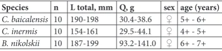
Table 1. Morphometric, weight and sexual features of studied sculpins (Cottoidei) from Lake Baikal.
Species n L total, mm Q, g sex age (years)
C. baicalensis 10 190-198 30.4-38.6 ♀ 5+ - 6+
C. inermis 10 154-161 29.5-44.1 ♀ 4+ - 5+
B. nikolskii 10 187-199 93.2-141.0 ♀ 6+ - 7+
Transmission electron microscopy
The olfactory rosettes were fixed in 2.5% glutaralde-hyde (Sigma-Aldrich, United States) solution in 0.1 M phosphate buffer, pH 7.3 at 4°C for 2 h, postfixed in 2% OsO4 (Merck, Germany) in the same buffer for 12 h, dehydrated in an ascending alcohol gradient with acetone and embedded in Araldite 502 resin with a DMP-30 accelerator (Araldite 502 Kit, SPI Supplies, United States). Ultrathin sections (70-80 nm) made with an Ulracut R microtome (Leica, Germany) were examined under a Leo 906E transmission electron microscope at an accelerating voltage of 80 kV. Mi-croscopic images made with a MegaView II digital camera were processed using the MegaVision program package (Soft Imaging System GmbH, Germany).
Laser confocal microscopy
To objectively assess the functional state of mito-chondria, we analyzed total preparations of olfactory rosettes. The rosettes were washed with medium 199 (Sigma-Aldrich) to remove blood and other contami-nation, stained for active mitochondria by incubating in 25 nM MitoTracker® Orange (Life Technologies, United States) in medium 199 for 25 min, and fixed in 2% paraformaldehyde solution for 15 min at 4°C. Cell nuclei were stained with 0.5 µg/mL DAPI solution
(Sigma-Aldrich) for 15 min. The preparations were then covered with the ProLong® Gold antifade reagent (Life Technologies) and analyzed under an LSM 710 laser confocal microscope (Carl Zeiss). The Z-stack images of the olfactory epithelium were processed with the Imaris® Bitplane 7.2.3 program package to calculate the number of mitochondria per 1 × 106 µm3 tissue volume. The results were processed statistically by non-parametric methods, and intergroup differences were assessed by the Kruskal-Wallis test, using Statistica 10 and Microsoft Excel 2010 program packages.
RESULTS
The structure of olfactory rosettes is similar in all three species of Baikal sculpins: they consist of 5-6 folds (lamellae) lined on both sides with the receptor (chemosensory) epithelium, except for the non-senso-ry epithelium at their margins. As in other vertebrates, the chemosensory epithelium consists of three basic cell types: receptor, supporting and basal. The olfac-tory epithelium of Baikal sculpins includes receptor neurons of ciliated and microvillar types, the former prevailing in number.
Comephorus baicalensis
numer-ous vacuole-like structures with irregular edges and electron-lucid contents. The size of these structures in different cells ranged from 0.2 to more than 2 µm. Apparently, they were not necessarily derived from the Golgi apparatus but often appeared due to fragmenta-tion and vacuolafragmenta-tion of nearby canals of the endoplas-mic reticulum (ER). Such vacuoles sometimes adjoined each other and occupied almost all free cytoplasmic space around the nucleus. In some neurons, such vacuoles occurred not only in the cell body but also throughout the dendrite, including the cell apex. How-ever, the vacuoles were not incorporated into the cell membrane but remained in the apical cytoplasm. The olfactory knob in such vacuolated receptor cells usu-ally had a low electron density and lacked structurusu-ally complete basal bodies and cilia or microvilli (Fig. 1d).
It should be noted that some receptor neurons in the olfactory epithelium showed similar degenerative changes but still retained well-defined chemosensory structures in the apical part (Fig. 1e).
Despite the poor development of intracellular organelles, receptor neurons contained well-defined cytoskeletal components, microtubules in particular. Dense microtubule bundles extended along the den-drite, amid vacuoles and mitochondria, to the olfac-tory knob (Fig. 1f). As could be seen in cross sections through the dendrites, there were also single neuro-filaments extending in different directions, which oc-curred throughout the dendrite length (Figs. 1f, 1g). The cell bodies and dendrites of olfactory neurons contained relatively low numbers of small (0.2-0.3 µm) round or elongated mitochondria with sparse cristae, but without any signs of swelling or degen-eration. Confocal microscopy of sections treated with MitoTracker®Orange, which stains only functionally
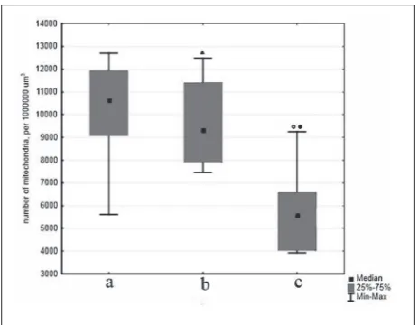
active mitochondria, provided evidence for a high de-gree of preservation of these organelles (Figs. 1h, 1i), which was further confirmed by statistical analysis of the results of these cytochemical experiments(Fig. 4a).
Cottocomephorus inermis
As in C. baicalensis, the cytoplasm of supporting cells in C. inermis usually had increased electron density and relatively electron-lucid cell bodies, and dendrites of the olfactory neurons clearly stood out against this background (Fig. 2b). The level of organelle develop-ment in sensory cells was also similar to that in C. baicalensis. In the electron-lucid cytoplasmic matrix, an inactive Golgi apparatus and dilated ER canals with signs of vacuolation could be seen (Fig. 2c). The dendrites contained numerous vacuoles of different diameters (0.1-0.7 µm) with electron-lucid contents. Numerous vesicles of similar diameter and electron density were also detected in supporting cells, main-ly those located in the upper layers of the olfactory epithelium (Fig. 2d). Analysis of sections made in different planes showed that the dendrites of recep-tor neurons contained well-developed microtubules and mitochondria (Figs. 2e, 2f). A noteworthy fact Fig. 1. Cytochemical features of the olfactory epithelium in the
golomyanka, Comephorus baicalensis. (a) The golomyanka, exterior view. Transmission electron microscopy: (b) section through the perinuclear zone of a receptor cell with vacuoles in the cytoplasm; (c) a fragment of receptor cell cytoplasm with vacuoles and a lipo-fuscin granule; (d) the olfactory knob of a ciliated receptor cell with signs of degenerative changes; (e) an “intact” apex of ciliated recep-tor neuron; (f) longitudinal and (g) transverse neurofilaments in a dendrite; arrows indicate microfilaments. Confocal microscopy: (h) a fragment of olfactory epithelium in transmitted light; (i) a fragment of the olfactory epithelium, Z-stack image. Cell nuclei were stained with DAPI (blue), active mitochondria with Mito-Tracker® Orange (red). Designations: rc − receptor cell, ga − Golgi
is that receptor cells with very large vacuoles occu-pying a major part of dendrite volume could often be detected amid electron-dense supporting cells in some areas of the olfactory epithelium. Moreover, it should be emphasized that, despite strong vacuolation, mitochondria located in the remaining cytoplasmic regions proved to retain their structure: they had a typical shape and contained sparse cristae and single glycogen granules (Fig. 2g). The results of confocal microscopy (Figs. 2h, 2i) and statistical analysis (Fig. 4b) confirmed the high functional stability of mito-chondria in large areas of the olfactory epithelium. Fig. 2. Cytochemical features of the olfactory epithelium in the longfin Baikal sculpin, Cottocomephorus inermis. (a) The longfin sculpin, exterior view. Transmission electron microscopy: (b) the cell body of a receptor neuron with dilated ER canals; (c) cytoplas-ic vacuolation in receptor and supporting cells; (d) receptor cell dendrite surrounded by supporting cells with numerous vesicles; (e) cross section through receptor cell dendrites (cytoskeletal ele-ments and mitochondria are indicated); (f) the apex of a microvil-lar receptor cell with preserved microvilli (cytoskeletal elements are indicated); (g) cross section through receptor cell dendrite with a large vacuole and well-preserved mitochondria. Confocal microscopy: (h) a fragment of olfactory epithelium in transmitted light; (i) a fragment of olfactory epithelium, Z-stack image. Cell nuclei were stained with DAPI (blue), active mitochondria with MitoTracker® Orange (red). Designations: rc − receptor cell, mrc
− microvillar receptor cell, sc − supporting cell, er − endoplasmic reticulum, v − vacuole, m − mitochondria, c − centrioles; arrows indicate microfilaments. Scale bars: (b) – 2 µm; (c) – 0.5 µm, (d) – 2 µm, (e-g) – 0.5 µm; (h) – 15 µm.
Batrachocottus nikolskii
Unlike those in pelagic sculpins, the receptor cells of the olfactory epithelium in B. nikolskii contained a well-developed Golgi apparatus and abundant ri-bosomes, both free and bound to ER, which was evidence for a high functional activity of these cells (Fig. 4b). The canals of the granular ER in both recep-tor and supporting cells were often fragmented and vacuolated, probably under the effect of acute hypoxia caused by the abrupt change in hydrostatic pressure when the deep-water fish were lifted to the surface. Fig. 3. Cytochemical features of olfactory epithelium in the fat sculpin, Batrachocottus nikolskii. (a) The fat sculpin, exterior view. Transmission electron microscopy: (b) section through the peri-nuclear zone of a vacuolated receptor cell with a high density of ribosomes; (c, d) receptor cells with dilated ER canals and large li-pofuscin granules; (e) the apex of a ciliated receptor cell with vacu-olated mitochondria; (f) a fragment of receptor cell dendrite with a lipofuscin granule and mitochondria with signs of vacuolation; (g) “intact” (arrows) and vacuolated (arrowheads) axons of olfac-tory neurons in basal regions of the olfacolfac-tory epithelium. Confocal microscopy: (h) a fragment of olfactory epithelium in transmitted light; (i) a fragment of olfactory epithelium, Z-stack image. Cell nuclei were stained with DAPI (blue); active mitochondria, with MitoTracker® Orange (red). Designations: rc − receptor cell, n −
A characteristic feature of cells in the olfactory epithelium of B. nikolskii was that they contained nu-merous lipofuscin granules. In the receptor neurons, they were usually located both in the cell body and in different dendrite segments. As a rule, these were electron-dense granules of irregular shape with an average size of 0.2-1 µm. Lipofuscin also aggregated into large cytoplasmic bodies comparable in size to the cell nucleus. Analysis of olfactory rosette sections in different planes showed that the total volume of lipo-fuscin in the organ was significant, with this pigment being equally detected in the cytoplasm of receptor and supporting cells (Figs. 3c, 3d).
It is noteworthy that mitochondria in cells of B. nikolskii olfactory epithelium were usually vacuolated to different degrees (Figs. 3e, 3f), which was indicative of their swelling, with a consequent loss of functional activity and eventual degradation. Confocal microsco-py of cytochemically stained preparations confirmed that the amount of positively stained (functionally active) mitochondria in the olfactory epithelium was reduced (Figs. 3h, 3i). Analysis of Z-stack images showed that their numbers in B. nikolskii were 2-fold smaller than in C. baicalensis (p = 0.047) and 1.7-fold smaller than in C. inermis (p=0.119) (Fig. 4).
Structural disturbances were detected, not only in dendrites and perinuclear cytoplasmic regions of receptor neurons, but also in their axons. The axons of such neurons were usually dilated to different ex-tents and could be easily distinguished from those of other, structurally normal olfactory cells. They were devoid of microfilaments and intact mitochondria, and the electron density of their axoplasm was lower than in the axons of viable neurons (Fig. 3g). Mor-phological analysis of cross sections of axon bundles showed that the proportion of degenerating olfactory neurons averaged 5-10%. This proportion apparent-ly included neurons undergoing natural cell death, which is known to occur in the olfactory epithelium throughout the animal life span [16].
DISCUSSION
Morphological studies on the olfactory rosettes of the three Baikal sculpin species were performed in order to reveal the cytological features characteristic of ol-factory cells in the norm and discriminate them from the results of rearrangements caused by depressuriza-tion upon delivery to the surface. To this end, we used not only published data but also the criteria for evalu-ating the structure and functional status of receptor cells that were determined in our previous studies on another Baikal sculpin, Cottocomephorus grewingkii
(Dybowski, 1874), at different stages of the life cycle [17,18], which appears to be methodologically valid. Thus, cytological features characteristic of the olfac-tory cells of C. grewingkii, which is not exposed to changes in hydrostatic pressure during the period of living in coastal waters, were taken as control.
The results of comparative morphological analysis showed that the olfactory receptor cells in B. nikol-skii have a higher level of ultrastructural development compared to those in the two pelagic species. They contain a well-developed Golgi apparatus, an exten-sive network of ER canals and abundant ribosomes, both free and ER-bound. A probable explanation is that B. nikolskii, which has adopted a benthic mode of life, is exposed to higher sensory load compared to the pelagic species, because the benthic environ-Fig. 4. Numbers of functionally active mitochondria per 1 × 10⁶
µm3 of olfactory epithelium in (a) C. baicalensis, (b) C. inermis,
and (c) B. nikolskii: ( ) p=1.000 compared to C. baicalensis; ()
ment is more saturated with organic substances of different origin. It should be noted that, according to measurements of chemical oxygen demand (COD) by dichromate method [19], the contents of organic mat-ter in Baikal wamat-ters are generally low. Thus, COD in the pelagic zone averages 3-4.5 mg О2/L [20]; however, this parameter markedly increases in the near-bottom water and especially in water from the upper layer of bottom sediments, reaching 8-10 and 20-30 mg О2/L, respectively. Moreover, calculations show that the rate of organic matter decomposition in the upper layer of bottom sediments reaches 2.5 g Corg/m2 per year [21], with this process being a constant source of olfactory signals.
It is noteworthy that both pelagic and benthic species included in the study had signs of destructive changes in cells of the olfactory epithelium, which in-cluded fragmentation and vacuolation of rough ER canals and structural disturbances in the axons and dendrites. No such ultrastructural alterations have been revealed in the Baikal sculpins that live in the coastal zone and are not exposed to abrupt changes in hydrostatic pressure [18]. Therefore, it appears that the above changes are the consequence of hypobaric hypoxia developing in deep-water fish upon delivery to the surface.
Another point of interest concerns the degree of preservation of mitochondria, which are especially vulnerable to oxygen deficiency [14]. Our data show that the morphological stability of mitochondria is higher in the pelagic species, C. baicalensis and C. in-ermis, and the sameis true of their functional activity: as can be seen in Fig. 4, the proportion of mitochon-dria with an intact level of oxidative phosphorylation in these species is, on average, two-fold higher than in the deep-water benthic B. nikolskii.
Are there special mechanisms in the peripheral ol-factory apparatus of the fish that can ensure the safety of cells and their organelles under extreme conditions of acute hypoxia? As noted above, foraging pelagic fish periodically ascend from depth to surface and then descend again, following the movements of their prey. In the course of evolution, such a regime of
alternat-ing exposure to high and low hydrostatic pressures could well provide for the development of relevant metabolic adaptations. Therefore, it appears that the good preservation of mitochondria in the olfactory receptor cells of pelagic fish could be due to the pro-tective effect of hypoxic preconditioning, which has been recently revealed in experiments on mammals. It has been shown that animal exposure to interval hypobaric hypoxia leads to the activation of adaptive genetic mechanisms improving the tolerance of the brain to oxygen deficiency [22]. These mechanisms in-clude the activation of early genes responsible for the production of antiapoptotic factors, cytoplasmic and mitochondrial antioxidants, metalloproteases, heat shock proteins and other agents protecting nerve cells from adverse effects under conditions of acute cerebral ischemia. These data concern terrestrial mammals, which have always lived in an atmospheric-pressure environment and could not develop any evolution-ary adaptations to hypobaric hypoxia. Therefore, it may well be that fish species adapted to a wide range of hydrostatic pressures have effective mechanisms providing for a high tolerance to oxygen deficiency, and analysis of these mechanisms is of major theoreti-cal and practitheoreti-cal importance. In particular, studies on these fish can open an opportunity to identify new neuroprotector molecules improving the resistance of the brain to ischemic damage, which is known to be responsible for the development of many socially significant neurodegenerative disorders [23].
Thus, the cytoskeleton of olfactory neurons acquires a highly structured pattern not only under high func-tional load in the spawning period but also in waters with low concentrations of dissolved substances. It also appears that the structural and functional char-acteristics of receptor neurons in the olfactory epithe-lium are not common but individual to each of them, because these characteristics depend not only on the initial, genetically determined sensitivity of receptors [25], but also on the effect of environmental factors on their expression [26,27] and the potential of these cells for subsequent stimulus-dependent differentia-tion [28,29].
CONCLUSIONS
The structural and functional status of olfactory recep-tor cells in closely related deep-water sculpin species (Cottoidei) from Lake Baikal is largely determined by the hydrostatic pressure regime (depending on their mode of life) and chemical composition of the water. In pelagic species, the receptor cells are characterized by a low level of organelle development, and their structural integrity is maintained due to a well-or-ganized system of cytoskeletal elements. The reduced functional status of receptor cells can be explained by the low contents of organic substances (potential olfactory stimuli) in the oligotrophic lake. In contrast, the olfactory neurons of benthic sculpins, which live in an environment enriched with various chemical substances, have a well-developed system of cell or-ganelles, including an extensive network of endoplas-mic reticulum canals and a high density of ribosomes. The results of cytochemical analysis of the peripheral olfactory apparatus in sculpins sampled from their natural environment provide evidence for a direct correlation between the level of receptor cell devel-opment and the overall intensity of olfactory signaling from the environment. Electron microscopic data and quantitative analysis for functionally active mitochon-dria in the olfactory epithelium of deep-water sculpins show that their exposure to hypobaric hypoxia during delivery to the surface leads to degenerative changes in the olfactory cells. Moreover, the extent of these changes depends on fish mode of life: it is markedly
greater in B. nikolskii (a typical Baikal low-mobile ben-thic fish) than in pelagic C. baicalensis and C. inermis, which are regularly exposed to changes in hydrostatic pressure during vertical foraging migrations. This is evidence that closely related fish species evolutionarily adapted to living under different hydrological condi-tions have different levels of tolerance to hypoxia. The cytological data obtained in this study are important for understanding basic mechanisms ensuring the reliable functioning of the olfactory apparatus under different environmental conditions. Moreover, they show that hydrobionts adapted to changes in hydro-static pressure are promising as models for the search of neuroprotective agents that improve the tolerance of nerve cells to hypoxia and other extreme environ-mental factors.
Acknowledgments: The authors are very grateful to Dr. Ye.V. Likhoshway and Chief Specialist A.P. Lopatin for their help with electron-microscopic procedures; to the captains and crews of the RVs G.Yu. Vereshchagin and Akademik V.A.Koptyug and the crews of manned submersibles Mir and Metropolia for provision and performance of underwater work; and to Dr. V.A. Fialkov, Director of the Baikal Museum, and V.V. Pastukhov, Head of the aquarium department, for their help in conducting field research. This study was performed using the facilities of the United Ul-tramicroanalysis Center, Limnological Institute, Siberian Branch, Russian Academy of Sciences (http://www.lin.irk.ru/copp/rus/) and supported by the Russian Foundation for Basic Research, project nos. 11-04-01231-a and № 12-04-10007-k
Authors’ contributions: Equal contribution of authors Conflict of interest disclosure: No conflicts of interest
REFERENCES
1. Coppola DM. Studies of olfactory system neural plasticity: the contribution of the unilateral naris occlusion technique. Neural Plast. 2012;2012:351752.
2. Ruan Y, Zheng, XY, Zhang HL, Zhu W, Zhu J. Olfactory dysfunctions in neurodegenerative disorders. Neurosci Res. 2012;90:1693-700.
3. Kasumyan AO. The Olfactory System in Fish: Structure, function, and role in behavior. Ichthyol. 2004;44(2):180-223. 4. Reuter K, Kapoor BG. Fish Chemosenses. Science
Publish-ers, Inc. USA; 2005.
6. Fishelson L, Golani D, Galil B, Goren M. Comparison of the nasal olfactory organs of various species of lizardfishes (Tel-eostei: Aulopiformes: Synodontidae) with additional remarks on the brain. Int J Zool. 2010;2010:807913.
7. Popper AN. Scanning electron microscopic studies of the sacculus and lagena in several deep-sea fishes. Am J Anat. 1980;157:115-36.
8. Marshall NJ. The lateral line system of three deep-sea fish. J Fish Biol. 1996;49:239-58.
9. Raymond EH Widder EA. Behavioral responses of two deep-sea fish species to red, far-red, and white light. Mar Ecol Prog Ser. 2007;350:291-8.
10. Wagner HL. Sensory brain areas in three families of deep-sea fish (slickheads, eels and grenadiers): comparison of mes-opelagic and demersal species. Mar Biol. 2002;141(5):807-17. 11. Herring PJ. The Biology of the Deep Ocean. Oxford: Oxford
University Press; 2002.
12. Kirylchyk SV, Slobodyanyuk SI, Belikov SI, Pavlova ME. Phy-logenetic relationships among 16 species of sculpins Lake Baikal by analyzing the nucleotide sequence of the gene fragment in the mitochondrial DNA cytochrome. Mol Biol. 1995;29(4):817-25.
13. Sideleva VG. Endemic fishes of Lake Baikal. Leiden: Back-huys Pub; 2003.
14. Vettier A, Labbe C, Amerand A, Da Costa G, Le Rumeur E, Moisan C, Sebert P. Hydrostatic pressure effects on eel mitochondrial functioning and membrane fluidity. Undersea Hyperb Med. 2006;33(3):149-56.
15. Casillas E, Smith L, D’Aoust BG. The response of fish blood cells, particularly thrombocytes, to decompression. Undersea Biomed Res. 1976;3(3):273-81.
16. Graziadei GA, Graziadei PP. Neurogenesis and neuron regeneration in the olfactory system of mammals. II. Degen-eration and reconstitution of the olfactory sensory neurons after axotomy. J Neurocytol. 1979;8(2):197-213.
17. Klimenkov IV. Ultrastructural rearrangement in receptor cells of olfactic analyzer in fishes at different stages of life cycle. [dissertation]. [Irkutsk]: Irkutsk State University.1990. 155 p. 18. Kositsyn NS, Klimenkov IV. Transformation of elements of cytoskeleton in receptor cells of olfactic analyzer in fishes
at different stages of life cycle. Dokl Ross Akad Nauk. 1994;336(2):261-3.
19. Skopintsev BA. Organic matter in natural waters. Trudy GOIN. In: Proceedings of GOIN. 1950;17(29):5.
20. Votincev KK, Meshceryakova GI, Popovskaya GI. Organic matter turnover in the Lake Baikal. Novosibirsk: Nauka; 1975.
21. Votintsev KK. On the question to the modern sedimenta-tion in Lake Baikal. Reports of the Academy of Sciences. 1967;174(2):419-22.
22. Rybnikova E, Glushchenko T, Tyulkova E, Baranova K, Samoilov M. Mild hypobaric hypoxia preconditioning up-regulates expression of transcription factors c-Fos and NGFI-A in rat neocortex and hippocampus. Neurosci Res. 2009;65:360-6.
23. Pluta R. From brain ischemia-reperfusion injury to pos-sible sporadic Alzheimer’s disease. Curr Neurovasc Res. 2004;1(5):44-153
24. Klimenkov IV, Kositsyn NS. Morphological reorganizations olfactory neurons at fishes after their periodic stimulation by sucrose. The bulletin of Irkutsk State University. 2008;2:33-6. 25. Buck L, Axel R. A novel multigene family may encode
odor-ant receptors: a molecular basis for odor recognition. Cell. 1991;65(1):175-87.
26. Wang HW, Wysocki CJ, Gold GH. Induction of olfactory receptor sensitivity in mice. Science. 1993;260:998-1000. 27. Harden MV, Newton LA, Lloyd RC, Whitlock KE.
Olfac-tory imprinting is correlated with changes in gene expres-sion in the olfactory epithelia of the zebrafish. J Neurobiol. 2006;66:1452-66.
28. Klimenkov IV, Kositsyn NS, Svinov MM. Common features of stimulus-dependent differentiation of olfactory receptor neurons and b cells of the immune system. Dokl Ross Akad Nauk. 2011;436(2):273-5.