ARTIGO ORIGINAL
Fine-needle Aspiration of Thyroid Nodules: Is it Worth
Repeating?
Punção Aspirativa de Nódulos da Tiroide: Vale a Pena
Repetir?
1. Serviço de Endocrinologia. Hospital de Braga. Braga. Portugal.
2. ICVS - Instituto de Investigação em Ciências da Vida e Saúde. Escola de Ciências da Saúde. Universidade do Minho. Braga. Portugal. 3. ICVS/3Bs - PT Government Associate Laboratory. Braga/Guimarães. Portugal.
4. IPATIMUP Diagnósticos. Instituto de Patologia e Imunologia Molecular. Universidade do Porto. Porto. Portugal. 5. Departamento de Patologia. Faculdade de Medicina. Universidade do Porto. Porto. Portugal.
Autor correspondente: Vera Fernandes. [email protected]
Recebido: 02 de março de 2016 - Aceite: 18 de novembro de 2016 | Copyright © Ordem dos Médicos 2017
Vera FERNANDES1,2,3, Tânia PEREIRA4, Catarina ELOY4,5
Acta Med Port 2017 Jun;30(6):472-478 ▪ https://doi.org/10.20344/amp.8215
RESUMO
Introdução: A punção aspirativa por agulha fina tem um papel relevante na avaliação do risco de malignidade de nódulos da tiroide. Persistem dúvidas quanto ao valor da repetição da punção aspirativa por agulha fina em nódulos com resultado benigno. Este trabalho pretende avaliar a concordância de resultados em punções aspirativas por agulha fina consecutivas e estudar a pertinência da repetição nos resultados benignos.
Material e Métodos: Estudo retrospetivo dos resultados das 4800 punções aspirativas por agulha fina da tiroide realizadas no Instituto de Patologia e Imunologia Molecular da Universidade do Porto entre 1 de janeiro de 2014 e 2 de maio de 2016. Da amostra inicial, selecionaram-se as punções aspirativas por agulha fina repetidas no mesmo nódulo.
Resultados: O resultado da primeira punção aspirativa por agulha fina dos 309 nódulos submetidos a reavaliação foi não diagnóstico em 103 (33,3%), benigno em 120 (38,8%) e atipia/lesão folicular de significado indeterminado em 86 (27,8%). A concordância entre a primeira e segunda punção aspirativa por agulha fina foi significativamente mais elevada nos casos com resultado inicial benigno (benigno: 85,8%, não diagnóstico: 27,2% e atipia/lesão folicular de significado indeterminado: 17,4%, p < 0,005). Os motivos de repetição da punção aspirativa por agulha fina em resultados inicialmente benignos (n = 78) foram a sugestão de repetição em 58, crescimento do nódulo em 17 e características ecográficas suspeitas de malignidade em três.
Discussão: A repetição da punção aspirativa por agulha fina em nódulos com resultado inicial não diagnóstico e atipia/lesão folicular de significado indeterminado alterou o diagnóstico inicial numa proporção significativa de doentes, modificando a sua orientação terapêutica. A elevada concordância de resultados em casos inicialmente benignos torna a repetição da punção aspirativa por agulha fina não custo-efetiva na maioria dos casos.
Conclusão: A repetição da punção aspirativa por agulha fina deve ser realizada quando o resultado citológico inicial é não diagnóstico ou atipia/lesão folicular de significado indeterminado.
Palavras-chave: Biópsia por Agulha Fina; Nódulo da Tiroide
ABSTRACT
Introduction: The fine-needle aspiration has a significant role in assessing the malignancy risk of thyroid nodules. There is uncertainty regarding the value of repeat fine-needle aspiration in benign nodules. This study aims to evaluate the concordance of results in consecutive fine-needle aspiration and to study the relevance of repetition in benign results.
Material and Methods: Retrospective study of the 4800 thyroid nodules fine-needle aspiration held in Instituto de Patologia e Imunologia Molecular da Universidade do Porto between January 1, 2014 and May 2, 2016. Of the initial sample, we selected the repeated fine-needle aspiration on the same nodule.
Results: The first fine-needle aspiration result of the 309 nodules underwent revaluation was non-diagnostic in 103 (33.3%), benign in 120 (38.8%) and atypia/follicular lesion of undetermined significance in 86 (27.8%). The agreement between the first and second fine-needle aspiration was significantly higher in cases with an initial benign result (benign: 85.8%, non-diagnostic: 27.2% and atypia/ follicular lesion of undetermined significance: 17.4%, p < 0.005). The fine-needle aspiration repeating motifs in initially benign nodules (n = 78) were repetition suggestion in 58, nodule growth in 17 and suspicious ultrasonographic features in 3.
Discussion: The fine-needle aspiration repetition in nodules with initial non-diagnostic and atypia/follicular lesion of undetermined significance result changed the initial diagnosis in a significant proportion of patients, modifying their therapeutic approach. The high concordance of results in initially benign nodules makes fine-needle aspiration repetition not cost-effective in most cases.
Conclusion: The fine-needle aspiration should be repeated when the initial cytology result is non-diagnostic or atypia/follicular lesion of undetermined significance.
Keywords: Biopsy, Fine-Needle; Thyroid Nodule
INTRODUCTION
Thyroid gland nodular hyperplasia is a common thyroid pathology, with a 50-60% prevalence in general population.1 The selection of malignant nodules for surgery is challenging and fine-needle aspiration cytology (FNAC) has a very important role in the identification of nodules with
an increased risk for malignancy.2,3
ARTIGO ORIGINAL According with the international recommendations,
the indication for FNAC depends on ultrasound nodule characteristics and size.1,6 When FNAC results are associated with an increased risk of malignancy, there is a clear indication for surgery; however, in the presence of any other result, the usefulness of repeat FNAC is questionable. Repeat FNAC is recommended following a cytology report as non-diagnostic or as an atypia of undetermined significance or follicular lesion of undetermined significance (AUS/FLUS) according with the Bethesda Classification.3,7 Repeat FNAC in patients with initial benign cytology result is questionable. Repeat FNAC has been recommended by some authors, based on a decreased rate of false negative results and therefore leading to an early surgical approach in the presence of any malignancy8-10 while other authors recommend not to repeat the FNAC based on the low rate of false negative results in the initial cytology and on the absence of impact on patient’s outcome of a potential delayed diagnosis.11
Our study aimed mainly at the assessment of the degree of agreement between consecutive FNACs as well as the relevance of a repeat FNAC upon an initial benign cytology result.
MATERIAL AND METHODS
This was a retrospective study of thyroid ultrasound-guided FNAC carried out between 1 Jan 2014 and 2 May 2016 at the Instituto de Patologia e Imunologia Molecular da Universidade do Porto (IPATIMUP).
Thyroid FNAC reporting was carried out by the same pathologist, with a few exceptions. The Bethesda System has been used for reporting all thyroid FNACs.3 According with this system, non-diagnostic biopsies are those not compliant with the quantitative or qualitative criteria that were established for a thyroid specimen to be satisfactory for evaluation; at least six groups of benign follicular cells are required, each group composed of at least 10 well-preserved epithelial cells.3 Thyroid aspirates with borderline cellularity and quantity were classified as benign and repeat FNAC was recommended in a period of no less than three months; these were included into a benign subtype and were hereinafter referred to as ‘benign – R’.
Repeat FNAC from the same patient were selected from an initial sample of 4,800 carried out over the study period and cytopathology reports were analysed. A second FNAC of a different nodule from the first one and unavailable data regarding either or both cytologies were the study exclusion criteria. Only eight nodules (2.6%) were assessed three times and as these were considered as a non-representative sample, no further data regarding any third cytology will be described.
Data were collected by using SISPAT® software and were exported to Microsoft Office Excel® 2011 software. The statistical analysis was carried out by using IBM® SPSS® Statistics v. 22 software. Data regarding the categorical variables were summarized into frequency charts. Continuous variable normal distribution has been
assessed and was described as mean ± standard deviation or median and interquartile range (IQR). Pearson’s chi-square test has been used in inferential statistical analysis for the assessment of the association between qualitative variables. Student’s t-test and one-way ANOVA were used to compare means among normally distributed variables and non-parametric Mann-Whitney and Kruskall-Wallis tests were used for the analysis of non-normally distributed variables.
Statistical significance has been considered for p-values < 0.05 and a statistically significant trend for 0.05 ≤ p < 0.1. Data collection and data statistical analysis complied with the ethical and deontological principles regarding the good practice during all the stages of the study.
RESULTS
Reporting result distribution of the 4,800 thyroid FNACs carried out at the IPATIMUP over the study period is shown in Fig. 1. From the initial sample, 638 corresponded to repeat FNACs of the same thyroid nodule. A final sample of 309 cases has been obtained upon the application of the exclusion criteria, corresponding to 291 patients.
The final sample of 309 cases corresponded to patients aged on average 54 ± 14 years (range 18-87; mostly female [84.5%]). Six months was the median time interval between both FNACs (IQR: 25th percentile: 4 – 75th percentile: 9) and 3.6% were located in the isthmus, 52.1% in the right lobe and 44.3% in the left. Median 15-mm nodule size was found at the time of the initial FNAC (IQR: 12 - 19), ranging between 5.8 and 50 mm while median 16-mm size was found at the time of the repeat FNAC (IQR: 12 – 21.8), ranging between 7 and 85 mm.
The group of 309 nodules that were submitted to re-assessment in a repeat FNAC were initially reported as one from three categories according with the classification of Bethesda: 103 (33.3%) reported as non-diagnostic, 120 (38.8%) as benign and 86 (27.8%) as AUS/FLUS (Fig. 2). The two cases initially reported as benign and subsequently as showing a follicular tumour corresponded to nodules located in the isthmus, a 30-mm nodule showing follicular cell hyperplasia and a 43-mm nodule with a 7-mm nodule growth in 11 months. The case initially reported as
Figure 1 – Distribution of the 4,800 FNACs as per the cytology report, according with the Bethesda classification
AUS/FLUS: Atypia of Undetermined Significance or Follicular Lesion of Undetermined Significance
I Não diagnóstico II Benign III AUS/FLUS IV Follicular neoplasm V Suspicious for malignancy VI Malignant
81.2% 7.3% 1.6% 1.3%
ARTIGO ORIGINAL
benign and subsequently as suspicious for malignancy showed borderline cellularity with a recommendation for repeat FNAC, which was carried out six months later. Those nodules initially reported as AUS/FLUS were subsequently re-classified in repeat FNAC according with the system of Bethesda and these were the only cases that were re-classified as malignant.
The analysis of the demographic variables, nodule characteristics and the degree of agreement between both FNACs by the diagnostic category reported in the initial FNAC is shown in Table 1. No statistically significant differences have been found between the groups with different reporting results as regards the demographic characteristics and the location of the nodule. A significantly higher degree of agreement between consecutive FNACs has been found regarding initially reported benign cases (85.8%) when compared to the degree of agreement between consecutive FNACs initially reported as non-diagnostic or as AUS/ FLUS (27.2% and 17.4%, respectively) (p < 0.005). This
already high degree of agreement was increased to 95.4% when cases reported as benign – R were removed from the analysis (74.5% agreement among benign – R cases) (Table 2).
Rationale for repeat FNAC in initially reported benign cases was available in 78 FNACs (32.1% clearly described in clinical information and 67.9% implied by patient’s clinical presentation) and included the following: recommended repeat FNAC in previous reporting regarding 58 cases (reported as benign – R or as follicular cell hyperplasia), nodule growth regarding 17 cases (6 with clear information and 11 without such information, even though with an increased size in consecutive FNACs) and suspicious ultrasound characteristics for malignancy in three cases. Follow-up clinical data were available in 12 (80%) from the 15 cases consistently reported as AUS/FLUS in both consecutive FNACs. From these, five (41.7%) were referred for surgery, two with a malignant result found in the histologic examination (40% risk for malignancy), Figure 2 – Distribution of the 309 cases with repeat FNAC as per the cytology report, according with the classification of Bethesda
AUS/FLUS: Atypia of Undetermined Significance or Follicular Lesion of Undetermined Significance Initial FNAC
309
120 (38.8%) 103 (33.3%)
28 (27.2%) 67 (65.0%) 4 (3.9%) 4 (3.9%) 14 (11.7%) 103 (85.8%) 2 (1.7%) 1 (0.8%)
3 (3.5%) 2 (2.3%)
8 (9.3%) 15 (17.4%)
47 (54.7%) 11 (12.8%)
86 (27.8%)
Repeat FNAC
Repeat FNAC Repeat FNAC
Non-diagnostic
Non--diagnostic
Non--diagnostic
Non--diagnostic Follicular
neoplasm
Follicular neoplasm Follicular
neoplasm Suspicious for malignancy
Suspicious for
malignancy Malignant Benign
Benign
Benign AUS/FLUS
AUS/FLUS
Benign AUS/FLUS
Table 1 - Demographic variables, nodule characteristics and results of repeat FNAC by diagnostic category reported in the initial FNAC
Variable Non-diagnosticn (%) Benignn (%)# AUS/FLUSn (%) p
Gender
Male
Female 20 (19.4)83 (80.6) 102 (85.0)18 (15.0) 13 (15.1)73 (84.9) 0.621
Age (years), mean ± SD* 55 ± 14 53 ± 13 54 ± 15 0.349
Nodule location
Right lobe Left lobe Isthmus
61 (59.2) 39 (37.9) 3 (2.9)
53 (44.2) 63 (52.5) 4 (3.3)
47 (54.7) 35 (40.7) 4 (4.7)
0.198
Thyroid nodule size described in the initial FNAC (mm),
Median (IQR)* 14 (12 - 19) 16 (13 - 22) 14,5 (11 - 18) 0.083 Agreement between cytologies
Yes
No 28 (27.2)75 (72.8) 103 (85.8)17 (14.2) 15 (17.4)71 (82.6) < 0.005
ARTIGO ORIGINAL
two reported as benign and one reported as benign and diagnosed with an incidental papillary micro-carcinoma not found in FNAC.
Thyroid nodule size
The distribution of thyroid nodule sizes reported in two consecutive FNACs is shown in Fig. 3 and 4, respectively. The information regarding thyroid nodule size was only available in 79 out of the 103 cases initially reported as non-diagnostic. Five from these (6.3%) were less than 10 mm and 41 (51.9%) less than 15 mm in size. One (5%) from the 20 cases subsequently reported as non-diagnostic was less than 10 mm and 15 (75%) were less than 15 mm.
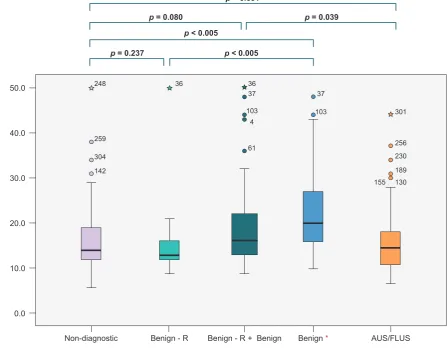
The nodules initially reported as AUS/FLUS and
non-diagnostic were significantly (p = 0.039) and tendentially (p = 0.08) smaller than those reported as benign (including those reported as benign - R), respectively (Fig. 3). Nodules initially reported as benign - R had a similar size as those reported as non-diagnostic (p = 0.237).
Nodules reported as non-diagnostic in the second FNAC were smaller than the remaining (non-diagnostic: 12 mm vs. benign: 17 mm vs. AUS/FLUS: 16 mm vs. follicular neoplasm: 25 mm vs. suspicious for malignancy: 24 mm, p < 0,005) (Fig. 4).
Time interval between FNACs
Time interval between both FNACs was significantly longer in the group of cases initially reported as benign. The Table 2 - Analysis of the degree of agreement between cytologies, reported as non-diagnostic, benign - R or benign (benign - R not included)
Initial FNAC
Repeat FNAC Non-diagnosticn (%) Benign - R n (%) Benignn (%) p
Result
Non-diagnostic Benign AUS/FLUS Follicular neoplasm Suspicious for malignancy
28 (27.2) 67 (65) 4 (3.9) 4 (3.9)
0
13 (23.6) 41 (74.5)
0 0 1 (1,8)
1 (1.5) 62 (95.4)
0 2 (3.1)
0
UNDET
UNDET: Undetermined, as the required conditions for the use of chi-square test were not met
Figure 3 – Thyroid nodule size in the initial cytology, according with the classification of Bethesda
AUS/FLUS: Atypia of Undetermined Significance or Follicular Lesion of Undetermined Significance.* Benign – R results not included.
Non-diagnostic Benign - R Benign - R + Benign Benign * AUS/FLUS
p = 0.237
p = 0.080 p = 0.039
p = 0.651
p < 0.005 p < 0.005
Cytological classification (system of Bethesda) in the initial FNAC
Nodule size in initial FNAC (mm)
0.0 10.0 20.0 30.0 40.0
50.0 248 36 36
37 37
103 301
256 230 189 130
155
103
4
61 259
304
ARTIGO ORIGINAL group of cases reported as benign – R underwent a second FNAC significantly later than the group of cases initially reported as non-diagnostic (non-diagnostic: five months vs. benign - R: seven months, p < 0.005) and significantly earlier than the remaining benign cases (seven months vs. 14 months, respectively, p < 0.005). No statistically significant differences were found between the group of cases initially reported as non-diagnostic and AUS/FLUS (non-diagnostic: five months vs. benign: 10 months vs. AUS/FLUS: four months, p < 0.005).
DISCUSSION
Our group of FNACs of the thyroid gland included cases with an adequate result distribution according with the classification of Bethesda3 and only repeat FNACs carried out in nodules reported as non-diagnostic, benign and AUS/ FLUS were included.
The data obtained in our study allowed for the conclusion that there is a benefit in repeating the FNAC in thyroid nodules initially reported as non-diagnostic and that an adequate sampling in repeat cytology will be obtained in 73% of the nodules. Repeat FNAC of a nodule previously reported as non-diagnostic was re-assessed as benign in 65% of the cases, allowing these patients to be clinically monitored and surgical over-treatment to be avoided. Thyroid nodule size seems to be one of the possible reasons underlying initially reported non-diagnostic cytologies. In fact, nodules initially reported as non-diagnostic were significantly smaller than those reported as benign (except those reported as benign
- R) in both cytologies.12 Therefore, ultrasound monitoring of these small nodules should be considered before any nodule growth would emerge and the chances of a better sampling would be increased.
Despite the current controversy involving repeat FNAC of nodules initially reported as benign, our study’s results support the recommendation not to repeat the FNAC in most nodules. Approximately 86% of the nodules initially reported as benign remained as such in repeat FNAC and a 2.5% (three cases) rate of false negative results (absence of any histological results) has been found. Therefore, according with our study, repeat FNAC of benign nodules is not a cost-effective procedure and should only be carried out in large or growing nodules, as well as in the presence of follicular cell hyperplasia. The results showed that nodules reported as benign - R are benign nodules with scarce cellularity, probably due to their small size. These nodules were significantly smaller than the remaining benign nodules and the result of the second cytology was closer to those in the group of non-diagnostic than to the benign group. This was the main reason for repeat FNAC of nodules initially reported as benign and suggested that compliance with cytological criteria of sampling adequacy recommended by the system of Bethesda should be met3 and benign - R nodules should comply with the same recommendation as the remaining nodules reported as non-diagnostic, supporting the recommendation for repeat FNAC in this group whenever thyroid size criteria are met.
The results of FNAC re-assessment of nodules reported
Figure 4 – Thyroid nodule size in repeat cytology, according with the classification of Bethesda
AUS/FLUS: Atypia of Undetermined Significance or Follicular Lesion of Undetermined Significance.* Benign - R included
Non-diagnostic Benign Follicular
neoplasm Suspicious for malignancy AUS/FLUS
Cytological classification (system of Bethesda) in the repeat FNAC
Nodule size in repeat cytology (mm)
0.0 20.0 40.0 60.0 80.0 100.0
220 197
69
36
61 259
ARTIGO ORIGINAL as AUS/FLUS showed that this recommendation should be
adopted, as repeat FNAC was adequate in most nodules (87.2%) and allowed for a more adequate and definitive treatment approach to a significant percentage of patients. In fact, re-assessment of these nodules led to 13 cases (15.1%) reported as with a high risk for malignancy (eight follicular neoplasms, two suspicious for malignancy and three malignant), allowing for the surgical approach to these patients. In addition, more than half (54.7%) of repeat FNAC of nodules initially reported as AUS/FLUS led to a benign result and surgery has been avoided. Benign nodules initially reported as AUS/FLUS seem to have been due to the presence of cytological artifacts related to sampling procedure (air drying of smears and entrapment of cells in clot material), lymphocytic thyroiditis, previous treatment with radioactive iodine or with synthetic anti-thyroid medications and cell repair in the presence of cystic degeneration and/or haemorrhage.3 The morphological features leading to an AUS/FLUS diagnosis must be re-assessed by an experienced pathologist for a second opinion, as this measure can avoid the need for any repeat FNAC. Malignancy has been found in 40% of the cases as regards nodules persistently reported as AUS/FLUS, therefore supporting surgical approach to these patients. A significant percentage of small nodules has been found (half of these were initially reported with no more than 15 mm in diameter). The recommendations for FNAC of thyroid nodules are increasingly becoming more conservative and the classical 1-cm threshold has been replaced by a 1.5-cm threshold in the latest recommendations of the American Thyroid Association. Nodules 1-1.4 cm in diameter have an indication for cytology assessment only in the presence of imaging characteristics of suspicious for malignancy1 and nodules <1 cm have no routine indication for this assessment as papillary thyroid microcarcinoma (< 1 cm) usually has an indolent outcome.1 Therefore, with the increased use of thyroid ultrasound imaging in general population, the relevant role of complying with the recommendations for the assessment by FNAC of the nodules of the thyroid gland should be mentioned and the risk of non-diagnostic results and the risk of over-treatment (and its complications) should be considered.
The results of this study showed that time interval between aspirations was significantly longer in nodules initially reported as benign, as previously described2 and
probably showing physician’s lower concern with results associated with lower risk for malignancy.
Even though a significant number of FNACs carried out and reported by the same pathologist has been included, showing the reality of a national reference centre, some limitations should be mentioned. This was a retrospective study with some gaps in clinical information regarding most cases, which has induced some limitations regarding the rationale for repeat FNAC of nodules initially reported as benign, the cyto-histological correlation as well as the correlation with imaging data. The analysis of thyroid nodule imaging characteristics must be considered in further studies, as these are the first determinants not only of the decision for the initial cytology assessment as also of the decision to repeat the FNAC.1
CONCLUSION
This study draws attention to the relevance of repeat fine-needle aspiration cytology (FNAC) of the thyroid gland in patients initially reported as non-diagnostic and AUS/ FLUS, leading to changes in treatment approach to a significant percentage of patients. As regards the patients initially reported as benign, repeat cytology does not seem cost-effective, except in the presence of large nodules, those showing an increased growth or with follicular cell hyperplasia.
HUMAN AND ANIMAL PROTECTION
The authors declare that the followed procedures were according to regulations established by the Ethics and Clinical Research Committee and according to the Helsinki Declaration of the World Medical Association.
DATA CONFIDENTIALITY
The authors declare that they have followed the protocols of their work centre on the publication of patient data.
CONFLICTS OF INTEREST
The authors declare that there were no conflicts of interest in writing this manuscript.
FINANCIAL SUPPORT
The authors declare that there was no financial support in writing this manuscript.
REFERENCES
1. Haugen BR, Alexander EK, Bible KC, Doherty GM, Mandel SJ, Nikiforov YE, et al. 2015 American Thyroid Association Management Guidelines for Adult Patients with Thyroid Nodules and Differentiated Thyroid Cancer: The American Thyroid Association Guidelines Task Force on Thyroid Nodules and Differentiated Thyroid Cancer. Thyroid. 2016;26:1-133.
2. Graciano AJ, Chone CT, Fisher CA, Bublitz GS, Peixoto AJ. Repeated fine-needle aspiration cytology for the diagnosis and follow-up of thyroid nodules. Braz J Otorhinolaryngol. 2014;805:422-7.
3. Cibas ES, Ali SZ. The Bethesda System for reporting thyroid cytopathology. Am J Clin Pathol. 2009;132:658-65.
4. Schlumberger MJ, Filetti S, Alexander EK, Hay ID. Nontoxic diffuse goiter, nodular thyroid disorders, and thyroid malignancies. In: Melmed
S, Polonsky KS, Larsen PR, Kronenberg HM, editors. Williams Textbook of Endocrinology. 13th ed. Philadelphia: Elsevier Saunders; 2016. p.
449–88.
5. Olson MT, Zelger MA. Thyroid fine-needle aspiration and cytological diagnosis. In: Jameson JL, De Groot LJ, editors. Endocrinology adult and pediatric. 7th edition Philadelphia: Elsevier Saunders; 2016. p.
1417–22.
ARTIGO ORIGINAL
C. Cost-effectiveness analysis of repeat fine-needle aspiration for thyroid biopsies read as atypia of undetermined significance. Surgery. 2012;152:423-30.
8. Gabalec F, Cáp J, Ryska A, Vasátko T, Ceeová V. Benign fine-needle aspiration cytology of thyroid nodule: to repeat or not to repeat? Eur J Endocrinol. 2009; 61:933–7.
9. llouz F, Rodien P, Saint-Andre JP, Triau S, Laboureau-Soares S, Dubois S, et al. Usefulness of repeated fine-needle cytology in the follow-up of non-operated thyroid nodules. Eur J Endocrinol. 2007;156:303–8. 10. Orlandi A, Puscar A, Capriata E, Fideleff H. Repeated fine-needle
aspiration of the thyroid in benign nodular thyroid disease: critical evaluation of long-term follow-up. Thyroid. 2005;15:274–8.
11. Merchant SH, Izquierdo R, Khurana KK. Is repeated fine-needle aspiration cytology useful in the management of patients with benign nodular thyroid disease? Thyroid. 2000;10:489–92.