http://www.ijmedicine.com pISSN 2349-3925 | eISSN 2349-3933
Original Research Article
Study of role of pulse oxymetry during nonsedated upper
gastrointestinal endoscopic procedure
Sukanya Dasgupta, Supriya Barsode*
INTRODUCTION
Although upper gastrointestinal endoscopy is considered safe even in critically ill patients, it is known to be associated with arterial oxygen desaturation, resulting in rare serious cardiopulmonary events. Hypoxemia seems to be the key factor in causing cardiopulmonary complications.1,2 Decrease in partial pressure of oxygen
has been known to occur during upper gastrointestinal endoscopy.3 Hypoxia episodes have been observed in
association with endoscopy and various studies have been
performed to identify the risk factors. The effect of smoking, ascites sedations during endoscopy and duration of procedure have been studied.
With the advent of pulse oximetry, it is possible to obtain an accurate measurement of hemoglobin oxygen desaturation. There are many reports concerning arterial oxygenation during upper gastrointestinal endoscopywith sedation, little information is available concerning oxygenation in non-sedated endoscopies.4 The present
study was carried out to determine the incidence and ABSTRACT
Background: Hypoxia episodes have been observed in association with endoscopy and various studies have been performed to identify the risk factors .The effect of smoking, deoxygenation during endoscopy and duration of procedure have been studied. With the advent of pulse oximetry, it is possible to obtain an accurate measurement of hemoglobin oxygen desaturation. There are many reports concerning arterial oxygenation during upper gastrointestinal endoscopy with sedation, little information is available concerning oxygenation in non-sedated endoscopies. The results of this study have extensive relevance because of increasing incidence of tobacco users, smokers and sedations during upper gastrointestinal endoscopies.
Methods : This study including 300 patients was carried out from July 2014 to September 2016 in gastroenterology clinic of medicine department at a large teaching hospital in Western Maharashtra. 250 patients underwent diagnostic procedures and 50 underwent therapeutic procedures.
Result: A significant correlation was found between oxygen desaturation and patients above 60 years of age (P< 0.01)in our study. There were 40.67% non-smoker patients with hypoxia and 21.33% non-smoker patients without hypoxia. In this study, 92 patients underwent UGI endoscopy for a duration of less than 5 minutes out of which only 24 (26.08 %) developed O2 desaturation.
Conclusion: It was concluded from the study patients with age >60 years, severe anaemia, presence of ascites, patients showing adverse events during the endoscopy procedure can be considered at the risk for developing oxygen desaturation during nonsedated UGI endoscopy.
Key words: Ascites, Hypoxia, Non-sedation, Pulse oximetry, Smoking, UGI Endoscopy
Department of Medicine, Bharati Vidyapeeth, Deemed to be University and Bharati Hospital, Pune, Maharashtra, India
Received: 29 May 2019
Revised: 10 June 2019
Accepted: 03 July 2019
*Correspondence:
Dr. Supriya Barsode,
E-mail: supriyabarsode@gmail.com
Copyright: © the author(s), publisher and licensee Medip Academy. This is an open-access article distributed under the terms of the Creative Commons Attribution Non-Commercial License, which permits unrestricted non-commercial use, distribution, and reproduction in any medium, provided the original work is properly cited.
severity of oxygen desaturation and to study the effects of various variables on oxygen saturation.
METHODS
This study was carried out from July 2014 to September 2016 in gastroenterology clinic of medicine department at a large teaching hospital in Western Maharashtra. 250 patients underwent diagnostic procedures and 50 underwent therapeutic procedures. After inclusion of the patients in the study history regarding personal habits (especially smoking ) was recorded. Similarly a history of previous ischemic heart disease, hypertension, COPD, Chronic lung diseases or tuberculosis was noted.
A general examination was done with special emphasis on the presence of pallor and cyanosis. A systemic examination was done and presence of crepitations or rhonchi were specifically recorded. If a patient had ascitis, the degree of ascitis was noted as mild, moderate and severe. Patients were then investigated which included Hemogram, Liver function test, HIV antibodies and HBsAg. ECG, X-ray chest and USG abdomen were done to detect presence of liver disease and signs of portal hypertension. Pre procedural monitoring of basal O2 saturation and pulse rate for 1 minute was done.
Simultaneous observations of O2 saturation and pulse rate
and phase of endoscopy where O2 desaturation occurred
was noted. Reading was taken every 15 seconds and also any adverse events were recorded. Post procedure monitoring for two minutes was done. Correlation of O2
desaturation with statistical analysis 𝜒2 test was
calculated.
RESULTS
There were 16.33% Female patients and 83.67% Male patients in the present study. There are 11% Female patients with hypoxia and 5.33% Female patients without hypoxia. There are 55% Male patients with hypoxia and 28.67% Male patients without hypoxia. In age group 15-30 16% patients had hypoxia and 9.67% patients did not have hypoxia. In age group 30-45 there were 22% patients with hypoxia and 11.33% patients without hypoxia. In age group 45-60, 13.67% patients had hypoxia and 8% patients did not have hypoxia. In age group 60-75 there were 9.67% patients with hypoxia and 4% patients without hypoxia.
In age group 75-90 4.67% patients had hypoxia and 1% patients did not hypoxia. There were 62% nonsmokers and 38% patients who were smokers. There were 40.67% nonsmoker patients with hypoxia and 21.33% nonsmoker patients without hypoxia. 66.66% smokers had hypoxia and 12.67% did not. There are 2.00% patients with Chronic Obstructive Pulmonary Disease. No significant correlation was found between O2 desaturation and a
history of respiratory disease or COPD as compared to other patients (P >0.05). In haemoglobin ranges from less than 5 gm there were 4.6% patients, 5 to 7 gm 22.67%
patients and more than 10 gm 17% patients who had hypoxia.
There were 25.67% Alcoholic patients and 74.33% Nonalcoholic patients. 71.42% alcoholic patients had hypoxia and 28.57% did not have hypoxia. Hypoxia was seen in 2.33% patients with gross ascitis and 1.00% patients did not have hypoxia in moderate ascitis, there were 4.67% patients with hypoxia and 3.33% patients without hypoxia. In mild ascitis, there are 8.00% patients with hypoxia and 2.00% patients without (Figure 1).
Figure 1: Presence of ascitis and SPO2.
About 97.67% patients were without supplemental oxygen while only 2.33% patients were given supplemental oxygen. In 63.67% patients desaturation occurred at antrum level and 14.67% patients whose desaturation occurred at duodenum level. In 12.33% patients desaturation occurred at the lower end of oesophagus level, in 8.67% patients desaturation occurred at oesophagus level and in 0.667% patients at stomach level (Figure 2).
Figure 2: Levels at which desaturation levels occurred.
In 75.67% patients tachycardia occurred at oesophagus (O) level and in 24.33% patients at pharyngeal (P) level. There were 0.7% patients whose systolic blood pressure (BP) was low before and 2.7% patients whose BP was
0.00% 10.00% 20.00% 30.00% 40.00% 50.00% 60.00%
gross ascites mild acites moderate ascites
normal
hypoxia non hypoxia
63.67% 14.67%
12.33% 8.67%
0.66%
antrum duodenum
low after GI endoscopy. Systolic BP was high in 10.7% patients before and in 6.3% patients after GI endoscopy. 6.7% patients had a low diastolic blood pressure after GI endoscopy. Minimum duration of endoscopy at which oxygen desaturation occurred was 5 minutes and maximum duration was 10 minutes. In our study 92
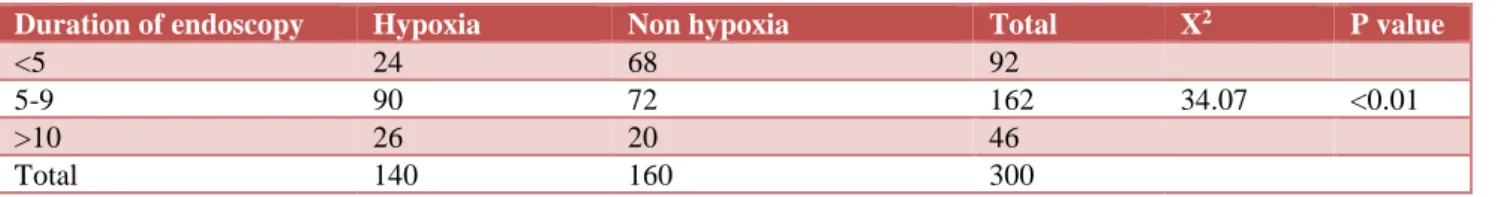
patients underwent UGI endoscopy for a duration of less than 5 minutes out of which only 24 (26.08 %) developed O2 desaturation. As against these 46 patients who required more than 10 minutes for the entire study 26 patients (56.52%) developed oxygen desaturation (Table 1).
Table 1: Duration of endoscopy and oxygen desaturation.
Duration of endoscopy Hypoxia Non hypoxia Total X2 P value
<5 24 68 92
5-9 90 72 162 34.07 <0.01
>10 26 20 46
Total 140 160 300
DISCUSSION
Gastrointestinal endoscopy procedures are reported as safe procedures with little morbidity and mortality and the risk associated with upper GI endoscopy is low.5,6
Decrease in partial pressure of oxygen has been shown to occur in UGI endoscopy .In a study conducted by Wang et al. Hypoxia (SaO2 92% or less of at least 15 s duration)
occurred in 17% and 6% of sedated patients and nonsedated patients, respectively (p<0.03).7
During the present study, all procedures were performed by single experienced endoscopist as oxygen desaturation is found to have significant correlation with the operator’s experience and skill, as studied by Laevis et al 8. We studied changes in oxygen desaturation, pulse rate, in 300 patients using “CONTEC” pulse oximeter.
Our study demonstrated arterial oxygen desaturation occured commonly with the use of UGI (Upper gastrointestinal) endoscopy. 198 patients out of 300 (i.e. 66%) had oxygen desaturation (SaO2 < 95%) during UGI
endoscopy. Unsedated endoscopic procedures have several potential advantages, including decreased rates of hypoxemia, respiratory depression, and hypotension during the procedure.9 Six unsedated patients (2%)
desaturated to 90% or less during endoscopy compared to 32 sedated patients (21%) (P andlt; 0.0001). SpO2 levels
in unsedated patients were not related to patient sex, age, cigarette smoking, endoscope diameter, basal SpO2 levels
or duration of endoscopy. In contrast, examination of the pharyngeal area and epiglottis (P=0.0002) and a longer intubation time (P=0.0002) were associated with lower SpO2 levels.10 In patients with 15-30 years of age 16%
had oxygen desaturation. Similarly in 30-45 years of age patients 22 % , 45-60 years 13.67% , 60-75 9.67% and 75-90% 4.67% had hypoxia. A significant correlation was found between oxygen desaturation and patients above 60 years of age (P <0.01) in our study.
Javed G et al, observed that desaturation occurred more in patients aged 60 and above 60 years of age, this is
suggested as an important factor in determining patients at high risk of morbidity. Dhariwal et al, found that in patients aged >65 years the baseline saturation was significantly lower and a reduced SaO2 was seen
throughout the procedure.11
In this study, there were 251 (83.67%) male and 49 (16.33) female patients however authors did not find any correlation between oxygen desaturation and either sex (p>0.05). Similar observation were made by Berg et al who observed no correlation between gender and oxygen desaturation.12
Patients who had reduced haemoglobin levels also had episodes of hypoxia during upper GI endoscopy procedure. In patients with severe anaemia i.e. <5gm % Hb out of 79 patients 51 (64.55%) had hypoxia. In patients with moderate anaemia i.e. 5-7gm % Hb out of 117 patients 111(94.8%) had hypoxia, and in those with mild Hb level i.e. >10 gm% out of 50 patients 36 (72%) had hypoxia. A significant correlation was found between O2 desaturation and patients with severe anaemia i.e. (Hb
value<5.0gm%) . (P<0.05).Similar observation was made earlier by Javed G et al, they found that there was significant drop in oxygen saturation in patients with Hb <10 gm% as compared to others.
In patients who were alcoholic 55 (71.42%) out of 77 patients had hypoxia during upper GI endoscopy procedure. Patients suffering from liver diseases and those patients with ascites were categorised according to the USG findings with gross, moderate and mild ascites. In gross ascites there were 2.33% patients who had hypoxia during the procedure. In moderate ascites there are 4.67% who patients had hypoxia during the procedure. 8% patients with mild ascites had hypoxia during the procedure A significant correlation was found between clinically detectable ascites and O2 desaturation
(P <0.01).
by Reed et al.13 In a study by Javid G et al, 39.6%
patients were found to have oxygen desaturation the average duration was 30 seconds (range 18-42) sec. Authors monitored patients for 2 minutes after the procedure for post procedure oxygen desaturation. Inspite of not using premedication the procedure was well tolerated by our patients. In this study, only 2.33% patients required supplemental oxygen during recovery.
In a similar study by Vittorio et al it was found that only two patients out of 273 who underwent UGI endoscopy without sedation had unsatisfactory results mostly because of patient’s intolerance and hence said that sedation is generally not required for endoscopy and the procedure is well tolerated by patients.14 Mild
desaturation (SaO2 between 90% and 94%) was found in
23.7% of the patients, and severe desaturation (SaO2
<90%) was found in 6.4%15 in a study conducted by Guillermo et al. Mild to moderate hypoxia was noted more in sedated patient than in non-sedated patient.15
Severe hypoxia was noted in 3.3% of sedated patient by Sharma et al.16 Laevis et al, who tried to assess the effect
of sedation and operator experience on oxygen saturation during UGI endoscopy, found that there was similar O2
desaturation in patients who did not receive sedation. Whorwell et al, proposed that endoscopy itself produced oxygen desaturation.17
In this study, 114 patients were smokers out which 76 (66.66%) patients had oxygen desaturation. However, no significant correlation was found between smokers and oxygen desaturation (P >0.05). Pecora et al noted a significant drop in Pao2 was noted in each group(smokers
and nonsmokers) however sedation, length of procedure, or smoking did not affect the Pao2 levels significantly.
Liebermann et al, studied the effect of UGI endoscopy on arterial oxygen tension in smokers and nonsmokers with and without premedication and found that a significant drop in PaO2 occurred in each group.18
Similarly Mistry et al, found no significant basal oxygen saturation or change in these parameters during UGI endoscopy and recovery in smokers as compared to nonsmokers. However, Javed G et al, found a significant correlation between oxygen desaturation and smokers. The difference in observation made in our study and the study stated above may be because the number of patients who were smokers (i.e. 20%) was smaller in our study than in theirs (i.e. 44%).No significant correlation was found between O2 desaturation and a history of
respiratory disease or COPD as compared to other patient (p >0.05).
COPD is proposed as a risk factor for developing O2
desaturation as studied by Javed G et al. Rostikus et al, in their study in patients with COPD who developed O2
desaturation during UGI endoscopy demonstrated that patients with moderate to severe air flow obstruction commonly develop hypoxia during the procedure.19 An
observation similar to our study was made by Dark et al,
who found that there was no significant correlation between O2 desaturation and severity of obstructive lung
disease.
In this study, 92 patients underwent UGI endoscopy for a duration of less than 5 minutes out of which only 24 (26.08 %) developed O2 desaturation. As against these 46
patients who required more than 10 minutes for the entire study 26 patients (56.52%) developed oxygen desaturation. We tried to assess the significance of duration of the entire procedure with oxygen desaturation and found that there was a highly significant correlation between O2 desaturation and patients requiring more than
10minutes for the UGI endoscopy (p <0.01) as compared to patients requiring less than 5mins for the procedure. Laevis et al, studied the significance of operator experience with oxygen desaturation during UGI endoscopy and found that the experienced endoscopist produced less desaturation in his patients than the inexperienced one, as he required about half the time to perform endoscopy. Also the patients who underwent endoscopies done by experienced consultant had better tolerance for the procedure. Dark et al, also found that in some instances the longer the procedure greater was the risk for significant arterial oxygen desaturation. 63.67 % had oxygen desaturation only at the level of antrum and saturation returned to normal as the UGI endoscopy progressed further.
The remaining patients had oxygen desaturation at some further phase of endoscopy also. Other levels at which O2
desaturation occurred were at the oesophagus 8.6%, lower end of oesophagus 12.33%, 0.6% and duodenum 14.67%. A strong correlation was found between the patients who continued to have O2 desaturation at a later
phase of endoscopy suggests that physical presence of endoscope could also be responsible for oxygen desaturation. Similar observation in patients were made by Whorwell et al. Similar results to our study were shown by Mistry et al who observed that out of 24 patients who had significant oxygen desaturation 16 patients had this in the first minute of endoscopy at the time of introduction of endoscope (PE) junction in our study (which recovered later). They proposed that gagging and coughing at the time of introduction of the endoscope could account for this O2 desaturation in the
early phase.
CONCLUSION
study were that it was a retrospective study and some adverse events might have occurred and not been noted. Our study did not assess pre-procedure anxiety and history of previous UGI endoscopy. The endoscopic procedures were performed by many endoscopists including fellows in training. Therefore, the varied experience may have influenced the results.
Funding: No funding sources Conflict of interest: None declared
Ethical approval: The study was approved by the Institutional Ethics Committee
REFERENCES
1. Dark DS, Campbell DR, Wesselius LJ, arterial oxygen desaturation during gastrointestinal endoscopy American J Gastrointestinal. 1990;85(10):1317-21.
2. Javid G, Khan B, Wani MM, Shah A, Gulzar GM. Role of pulse oximetry during nonsedated upper gastrointestinal endoscopic procedures. Indian J Gastroenterol Off J Indian Soc Gastroenterol. 1999;18(1):15-7.
3. Pecora AA, Chiesa JC, Alloy AM, Santoro J, Lazarus B. The effect of upper gastrointestinal endoscopy on arterial O2 tension in smokers and
non-smokers with and without premedication. Gastrointestinal Endosc. 1984;30(5):284-8.
4. Mistry FP, Abraham P, Bhatia SJ. Oxygen desaturation and tachycardia during upper gastrointestinal endoscopy are transient and benign. J Association Physicians India. 1992;40(8):524-7. 5. Keeffe EB, Schrock TR. Complications of
gastrointestinal endoscopy. In: Sleisenger MH, Fordtran JS, eds. Gastrointestinal disease. 5th ed.
Philadelphia: Saunders, 1993: 301-308.
6. Silvis DE, Nebel O, Rogers G, Sugwa C, Stam PM. Endoscopic complications, results of the 1974. Am Soc GI Endosc Survey. JAMA. 1976;235(9):928-30. 7. Wang CY, Ling LC, Cardosa MS, Wong AK, Wong NW. Hypoxia during upper gastrointestinal endoscopy with and without sedation and the effect of pre-oxygenation on oxygen saturation. Anaesthesia. 2000;55(7):654-8.
8. Laevis NG, Crisy T, Harris K, Haning CD. Arterial Oxygen saturation during UGI endoscopy. Influence of sedation and operator experience. Am J Gasterol. 1988;83(6):618-22.
9. Huang R, Eisen GM. Efficacy, safety, and limitations in current practice of sedation and
analgesia. Gastrointestinal Endosc Clin North Am. 2004;14(2):269-88.
10. Banks MR, Kumar PJ, Mulcahy HE. Pulse oximetry saturation levels during routine unsedated diagnostic upper gastrointestinal endoscopy. Scandinavian J Gastroenterol. 2001;36(1):105-9.
11. Dhariwal A, Plevris JN, Lo NT, Finlayson ND, Heading RC, Hayes PC. Age anaemia and obesity- associated O2 desaturation during UGI endoscopy Gastrointestinal Endosc. 1992-38(6):684-8.
12. Berg JC, Millar R, Burkhalter E. Clinical value of pulse oximetry during routine diagnostic and therapeutic endoscopic procedures. Endosc. 1991;23(6):328-30.
13. Reed MW, O'leary DP, Duncan JL, Majeed AW, Wright B, Reilly CS. Effects of sedation and supplemental oxygen during upper alimentary tract endoscopy. Scandinavian J Gastroenterol. 1993;28(4):319-22.
14. Peri V, Amuso M, Gatto G, Traina M. Monitoring during endoscopy. Italian data support upper gastrointestinal endoscopy without sedation. BMJ. Brit Med J. 1995;311(7002):453.
15. Alcaín G, Guillén P, Escolar A, Moreno M, Martín L. Predictive factors of oxygen desaturation during upper gastrointestinal endoscopy in nonsedated patients. Gastrointestinal Endosc. 1998;48(2):143-7. 16. Sharma SK, Maharjan DK, Thapa PB, Adhikari SB, Byenjankar B, Khadka S, et al. The role of sedation and pulse oximetry during upper gastrointestinal endoscopy. JNMA. 2009;48(174):92-8.
17. Whorwell PJ, Smith CL, Foster KJ. Arterial blood gas tensions during upper gastrointestinal endoscopy.Gut1976;17(10):797-800.
18. Lieberman DA, Wuerker CK, Katon RM. Cardiopulmonary risk of esophago gastroduodenoscopy: role of endoscope diameter and systemic sedation. Gastroenterol. 1985;88(2):468-72.
19. Rostykus PS, McDonald GB, Albert RK. Upper intestinal endoscopy induces hypoxemia in patients with obstructive pulmonary disease. Gastroenterol. 1980;78(3):488-91.